The Channel - Cook Medical
The Channel - Cook Medical
The Channel - Cook Medical

Create successful ePaper yourself
Turn your PDF publications into a flip-book with our unique Google optimized e-Paper software.
<strong>The</strong> <strong>Channel</strong><br />
A COOK NeWs PuBLiCATiON issue 2, 2011<br />
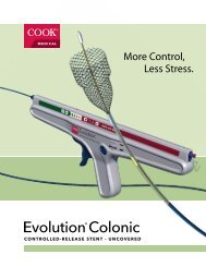
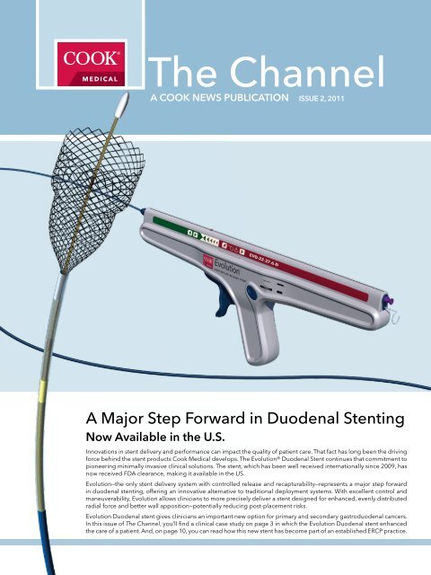
A Major Step Forward in Duodenal Stenting<br />
Now Available in the u.s.<br />
Innovations in stent delivery and performance can impact the quality of patient care. That fact has long been the driving<br />
force behind the stent products <strong>Cook</strong> <strong>Medical</strong> develops. <strong>The</strong> Evolution® Duodenal Stent continues that commitment to<br />
pioneering minimally invasive clinical solutions. <strong>The</strong> stent, which has been well received internationally since 2009, has<br />
now received FDA clearance, making it available in the US.<br />
Evolution—the only stent delivery system with controlled release and recapturability—represents a major step forward<br />
in duodenal stenting, offering an innovative alternative to traditional deployment systems. With excellent control and<br />
maneuverability, Evolution allows clinicians to more precisely deliver a stent designed for enhanced, evenly distributed<br />
radial force and better wall apposition—potentially reducing post-placement risks.<br />
Evolution Duodenal stent gives clinicians an important new option for primary and secondary gastroduodenal cancers.<br />
In this issue of <strong>The</strong> <strong>Channel</strong>, you’ll find a clinical case study on page 3 in which the Evolution Duodenal stent enhanced<br />
the care of a patient. And, on page 10, you can read how this new stent has become part of an established ERCP practice.
2<br />
inside this issue<br />
Histology in EcHoEndoscopy 2<br />
REsolving gastRic outlEt<br />
obstRuction<br />
asgE ambassadoR pRogRam 4<br />
immunostaining: making a<br />
dEfinitivE diagnosis<br />
diagnosing low-gRadE<br />
nEuRoEndocRinE nEoplasm<br />
wHat’s up doc? 7<br />
yalE univERsity sEction<br />
of digEstivE disEasEs<br />
yalE nEw HavEn Hospital<br />
fEatuREs<br />
smilow Hospital fEatuREs 9<br />
about tHE yalE ambulatoRy<br />
sERvicE division<br />
montREal’s ERcp and<br />
duodEnal-stEnting innovatoR<br />
www.cookmedical.com<br />
3<br />
6<br />
6<br />
8<br />
9<br />
9<br />
10<br />
tHE nuRsEs' station 12<br />
ovER 2200 paRticipants<br />
attEnd asia pacific<br />
digEstivE wEEk<br />
nEws fRom signEa - Endoscopy<br />
and infEction contRol<br />
tRiton bRt additional<br />
sizEs availablE<br />
13<br />
14<br />
15<br />
gi360 16<br />
An official publication of <strong>Cook</strong> <strong>Medical</strong>.<br />
4900 Bethania Station Road<br />
Winston-Salem, NC 27105<br />
If you would like to submit material<br />
for <strong>The</strong> <strong>Channel</strong>, please email us at<br />
thechannel@cookmedical.com.<br />
We welcome your comments<br />
and suggestions.<br />
Histology in Echoendoscopy<br />
Two points of view, one common goal<br />
<strong>The</strong> 1st Spanish Symposium on “Histology in<br />
Echoendoscopy” was held June in Santiago<br />
de Compostela, Galicia, Spain. <strong>The</strong> event,<br />
which attracted 12 echoendoscopists and 13<br />
pathologists from the surrounding region, was<br />
aimed at exploring the potential of histology<br />
from two different points of view: the pathologist<br />
and the echoendoscopist.<br />
Echoendoscopist Dr. Julio Iglesias and pathologist<br />
Dr. Ihad Abdulkader—both with the University<br />
Hospital of Santiago de Compostela—were course<br />
directors for the symposium, which highlighted a<br />
number of key issues, including: the implications<br />
of sample size, the importance of robust<br />
collaboration between echoendoscopists and<br />
pathologists, the value of sample manipulation<br />
and the use of the EchoTip® ProCore HD<br />
Ultrasound Biopsy Needle.<br />
Symposium attendees reviewed 12 clinical cases<br />
in which histology was integral to diagnose<br />
and treat patients. “<strong>The</strong> bigger the sample,”<br />
says Dr. Iglesias, “the easier it is to get a real<br />
core, providing more information about the<br />
characteristics of the lesions being studied,<br />
including architecture, etc. But sample size<br />
is also important because it can allow you to<br />
perform many different studies with the sample,<br />
including immunohistochemistry, molecular<br />
analysis and more.”<br />
Reviewing the ProCore needle, Dr. Iglesias<br />
adds: “It is an excellent needle, as easy to use<br />
as the standard cytology needles. It is a real<br />
step forward.”<br />
<strong>The</strong> symposium concluded with an enthusiastic<br />
open discussion on the reviewed cases, followed<br />
by a delicious traditional Galician meal. “All<br />
attendees were very appreciative,” says Dr.<br />
Iglesias. “<strong>The</strong>y enjoyed the symposium and<br />
found the format was very interesting. In fact,<br />
we are considering organizing at least two more<br />
meetings in the next months.”
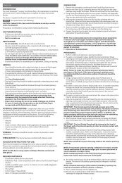
Resolving<br />
Gastric outlet<br />
obstruction<br />
Figure 1 Figure 2<br />
Dr. Douglas Howell<br />
Director, Pancreaticobiliary Center<br />
Director, Advanced Interventional Endoscopy Fellowship<br />
Maine <strong>Medical</strong> Center<br />
Portland, Maine<br />
Duodenal obstruction complicates the late course of pancreatic head cancer in at least 10<br />
percent of patients. <strong>The</strong> following case illustrates placement of the new flexible Evolution®<br />
duodenal stent to resolve gastric outlet obstruction and permit restoration of oral intake.<br />
A 72-year-old patient initially presented in the fall of 2010 with obstructive jaundice. CT<br />
imaging revealed a large pancreatic head mass, producing an obvious bile duct stricture<br />
and deemed unresectable due to vascular encasement. ERCP was successful in accessing<br />
a short, malignant, periampullary stricture and a positive intraprocedural forceps SMASH<br />
protocol tissue diagnosis of adenocarcinoma was made. A Zilver® (10 x 40 mm) uncoated<br />
SEMS was placed at the close of the ERCP. <strong>The</strong> patient subsequently underwent palliative<br />
chemoradiation therapy with an excellent response.<br />
Ten months later, the patient presented with bloating, vomiting, recurrent jaundice and rapid<br />
weight loss. Repeat CT confirmed duodenal obstruction at the level of the periampullary<br />
mass. Repeat ERCP revealed high-grade duodenal obstruction with frank invasion of the<br />
duodenal lumen. <strong>The</strong> previous stent was enveloped by the surrounding mass. (Figure 1)<br />
To reestablish biliary drainage, the biliary lumen was "hooked" from above using a DASH<br />
25 and the preloaded Metro .025-inch wire guide was advanced through the previously<br />
placed stent. A new very flexible Zilver 635® (10 x 60 mm) was then advanced over the wire<br />
guide, positioned precisely with the bottom of the stent flush with the duodenal wall, and<br />
deployed. Excellent biliary drainage was noted.<br />
Without requiring duodenal dilation, the wire guide was then advanced well down the<br />
duodenum into the jejunum. A new through-the-endoscope Evolution (22 x 120 mm)<br />
duodenal stent was positioned using both direct vision and fluoroscopic guidance. (Figure<br />
2) Due to the low position of the stricture, the upper aspect of the stent was left in the<br />
duodenum and rapid expansion confirmed complete expansion in the distal bulb again<br />
under direct vision. (Figure 3) Spot radiography also documented good positioning.<br />
(Figure 4) <strong>The</strong> patient experienced no complications and no significant pain. Vomiting<br />
rapidly resolved, a diet was resumed, and jaundice cleared. He was discharged to home<br />
24 hours after placement.<br />
Two weeks later, significant weight gain was noted and upper GI barium study revealed full<br />
expansion, optimal positioning and complete patency. (Figure 5)<br />
This case demonstrates that rapid resolution of combined biliary and duodenal obstruction<br />
due to late stage pancreatic cancer following a single outpatient ERCP procedure can be<br />
well tolerated. Further experience is anticipated.<br />
<strong>The</strong> <strong>Channel</strong><br />
Figure 3<br />
Figure 4<br />
Figure 5<br />
www.cookmedical.com<br />
3
ASGE Ambassador Program<br />
Treatment for those who might otherwise do without<br />
“You have worked hard and it shows. You are a leader with a thriving practice or academic career….<br />
You are at a point where you can see the fruits of your labors and you are ready to do more. It’s<br />
time to give back. Time to share your knowledge. Time to treat those who might not otherwise get<br />
treatment. Time to reach beyond your comfort zone. It's time to become an ASGE Ambassador.”<br />
How to become an<br />
ASGE Ambassador<br />
and future<br />
Ambassador<br />
opportunitites<br />
ASGE Ambassador physicians<br />
must be active or international<br />
members of ASGE for a<br />
minimum of five years.<br />
<strong>The</strong> first ASGE Ambassador<br />
Program for 2012 is scheduled<br />
for February 4-11, 2012 in Taipei,<br />
Taiwan. If you would like to apply<br />
to be an ASGE Ambassador,<br />
visit: http://www.asge.org/<br />
ambassador.aspx<br />
4 www.cookmedical.com<br />
That is the inspiring challenge to members of the American Society for Gastrointestinal<br />
Endoscopy (ASGE) to participate in a philanthropic initiative designed to export two of<br />
the Society’s greatest assets—endoscopic medical care and training expertise—to centers<br />
around the world in need of such care. <strong>The</strong> call-to-action also says: “Educating and training<br />
physicians in underserved countries provides an opportunity for ASGE and its Ambassadors<br />
to have an enduring effect in these areas.”<br />
Prof. Ian Gralnek (Chief of Ambulatory Services at Rambam <strong>Medical</strong> Center, Haifa, Israel,<br />
and Chair of the ASGE International Committee) says: “This is an outstanding philanthropic<br />
program that has had an overwhelming response from gastroenterologists who offer to<br />
train overseas. We have had many applications from our ASGE members. <strong>The</strong> selections<br />
are highly competitive. Members who have attended find the experience very worthwhile.<br />
“Also, we have had many excellent grassroots proposals from institutions abroad who want<br />
the endoscopy training program,” Prof. Gralnek continues. “<strong>The</strong> selection has become very<br />
competitive. Many of the physicians who are being trained may not have had the means to<br />
attend an international conference.”<br />
To date, eleven ASGE professionals (all physicians) have participated in the Ambassador<br />
Program. Five participated in a pilot program in Cairo, Egypt; six participated in a Vietnam<br />
program. More than 60 physicians in those countries have been trained in endoscopic<br />
techniques. In addition, those who have been trained can now train others who were unable<br />
to participate in the program. A third program has been scheduled for Cuenca, Ecuador<br />
this November. In December, a fourth Ambassador Program will be held in the Solomon<br />
Islands in conjunction with the Soloman Islands Living Memorial Project.<br />
Cairo, egypt<br />
February 19-26, 2010<br />
In Cairo, the one-week program covered topics related to upper GI bleeding—one of the most<br />
common gastrointestinal afflictions facing developing nations in Africa today. <strong>The</strong> extensive<br />
curriculum incorporated didactic sessions as well as hands-on training as physicians and<br />
trainees treated patients.<br />
John M. DeWitt, MD, FASGE (Indiana University <strong>Medical</strong> Center) participated in the Cairo,<br />
program. DeWitt was joined by David H. Robbins, MD (Center for Advanced Endoscopy,<br />
Lenox Hill Hospital, New York) and Virendra Singh, MD (Postgraduate Institute of <strong>Medical</strong><br />
Education and Research, Chandigarh, India). <strong>The</strong> three endoscopists helped train not only<br />
Egyptian gastroenterologists but also GIs and surgeons from Kenya, Sudan and Nigeria.<br />
“I was struck by the passion of the training center director, Dr. Ibrahim Mostafa of the <strong>The</strong>odor<br />
Bilharz Research Institute, Cairo for teaching physicians,” says Dr. DeWitt. “It was contagious<br />
and clearly something he enjoyed. Also clearly evident was the gratitude the trainees had<br />
for the chance to learn and improve their skills.<br />
“Some physicians coming to train had very little experience prior to the sessions,” Dr. DeWitt<br />
continues. “<strong>The</strong> trainees were not the only ones to benefit from the program. For example,<br />
I gained further experience with cyanoacrylate injection which was routinely done there<br />
for gastric varices.”<br />
One trainee, Dr. Aderemi Omololu Oluyemi (Lagos, Nigeria), says, “I want to thank the<br />
Ambassadors for making themselves available to us. <strong>The</strong> program was a huge benefit. In<br />
fact, you have re-launched my endoscopy career in an interventional sense.”
Dr. DeWitt adds: “Overall, this program was incredibly valuable to serving the ASGE<br />
Ambassador mission. To my fellow gastroenterologists, I would say, ‘We are blessed in this<br />
country with technology and training not available elsewhere. Share that knowledge and<br />
experience with others to benefit patients in other parts of the world.’”<br />
Vietnam<br />
October 30-November 11, 2010<br />
In Vietnam, the program focused on diagnostic and therapeutic ERCP for the treatment of<br />
bile duct stones and pancreatic cancer, both prevalent conditions in Vietnam. A second focus<br />
was endoscopic ultrasonography for the management of gastric and esophageal cancers,<br />
also common in the region.<br />
In addition, the doctors in Vietnam were successfully trained in percutaneous endoscopic<br />
gastrostomy (PEG) placement to manage patients who are unable to eat due to head trauma.<br />
This is a common injury seen in Vietnam because of the widespread use of small motorbikes.<br />
Dr. Andrew Q. Giap, MD, FASGE (Lakeview <strong>Medical</strong> Offices, Anaheim, California), a member<br />
of the team training in Vietnam, says, “All procedures—ERCP, EUS and PEG placement—were<br />
done in the OR, which was well equipped with C-arm fluoroscopy and well staffed with<br />
nursing staff and OR techs.<br />
“In addition to the need for PEG placement training, we realized that there is a need for<br />
additional endoscopic training in Vietnam—especially pertaining to advanced endoscopic<br />
skills such as ERCP and EUS.<br />
“<strong>The</strong> ASGE Ambassador Program in Vietnam was a great success. During our visit, we trained<br />
over 50 physicians in ERCP and PEG placement along with instruction in EUS. I continue to<br />
receive compliments and gratitude from these physicians for our work in Vietnam. Since<br />
then, these trainees have also passed on their knowledge and skills to their colleagues in<br />
local hospitals and universities.”<br />
Ambassadors joining Dr. Giap were Franklin Kasmin, MD, FASGE, and Jerry Siegel, MD, FASGE<br />
(both of Beth Israel <strong>Medical</strong> Center, New York); Raj Shah, MD (University of Colorado, Denver);<br />
and Kai Matthes, MD, PhD (Beth Israel Deaconess <strong>Medical</strong> Center, New York). Physicians<br />
trained came from North, Central and South Vietnam, Indonesia and Laos.<br />
“Having good English interpreters who are well-trained in medical terminology is paramount,<br />
especially when teaching in a country with physicians who are not proficient in the English<br />
language," says Dr Giap.<br />
Patient updates continue<br />
During both programs, approximately 70 patients were treated. Many more continue to<br />
be treated as a result of the programs. ASGE has been receiving updates on patients who<br />
were treated.<br />
Dr. Kasmin sums up his experience: “We have taught Vietnamese GIs how to do PEG<br />
placement, and those we have taught will teach others who could not attend.”<br />
Grant funding for these programs was provided by Pentax <strong>Medical</strong> Company, US Endoscopy,<br />
Olympus Corporation of the Americas and the AstraZeneca Foundation. Equipment<br />
and supplies were provided by <strong>Cook</strong> <strong>Medical</strong>, Pentax <strong>Medical</strong> Company and Olympus<br />
Corporation of the Americas.<br />
<strong>The</strong> <strong>Channel</strong><br />
www.cookmedical.com<br />
5
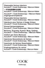
Ductal anatomy relative to the mass<br />
<strong>The</strong> bevel of the ProCore is clearly visible<br />
during FNA of the patient's mass.<br />
New (Lt) old (Rt): Cellularity<br />
at low power appears more robust<br />
with the ProCore.<br />
New (Lt) old (Rt): NETs are<br />
examples of "small blue cell tumors,"<br />
showing uniform cells which have<br />
a round to oval stippled nucleus<br />
and scant, pink granular cytoplasm.<br />
6 www.cookmedical.com<br />
immunostaining<br />
Making a definitive diagnosis<br />
In a recent case (summarized below), Bedford, Texas gastroenterologist Dr. Jay Yepuri,<br />
working closely with a pathologist, relied on immunostaining to obtain a definitive diagnosis.<br />
Immunostaining requires an adequately sized cytologic sample, and for that, Dr. Yepuri<br />
relied on the EchoTip® ProCore HD Ultrasound Biopsy Needle.<br />
“Cytology sample size is essential for an unequivocal diagnosis,” says Dr. Yepuri. “Pathologists<br />
need as much representative material as possible to make a definitive diagnosis, and<br />
often utilize sophisticated immunostaining techniques in this endeavor. Immunostaining<br />
techniques require a robust cytologic yield.”<br />
Dr. Yepuri refers to the ProCore needle as a “true asset in the endosonographer’s arsenal.<br />
It is becoming my ‘go to’ needle for solid lesions. Its design allows for maximal yield with<br />
minimal trauma, as it minimizes the number of sticks required to obtain diagnostic cytology.”<br />
Diagnosing lowgrade<br />
neuroendocrine<br />
neoplasm<br />
Jay Yepuri, MD<br />
Digestive Health Associates of Texas, PA<br />
Bedford, TX<br />
An 86-year-old otherwise healthy patient presented with complaints of mid-epigastric pain<br />
in the fall of 2009. A CT scan of the abdomen at that time was revealing for a mass in the<br />
head of the pancreas with associated pancreatic ductal dilation, but no significant biliary<br />
ductal dilation. EUS/FNA of this lesion in November 2009 was unrevealing for definitive<br />
malignancy. Tumor markers at that time were negative as well. As the patient’s symptoms<br />
improved, additional evaluation was deferred.<br />
In April 2011, the patient presented with recurrent pain and nausea. A CT scan of the<br />
abdomen was again revealing for a mass in the head of the pancreas, unchanged from<br />
the prior CT. After a thorough discussion regarding management options, the patient<br />
elected to proceed with a repeat EUS/FNA. A EUS in May 2011 was revealing for a 2.6-cm<br />
heterogeneous, hypoechoic, well-circumscribed mass with scattered calcifications in the<br />
head of the pancreas with associated pancreatic ductal dilation. <strong>The</strong> common bile duct was<br />
non-dilated. <strong>The</strong>se findings were unchanged from those noted at the time of the patient's<br />
initial EUS in November 2009. FNA of this lesion was performed using a <strong>Cook</strong> 22-gauge<br />
ProCore needle. While preliminary review of the aspirate by Pathology in the procedure<br />
suite was revealing for atypia, final cytology in conjunction with immunostains was revealing<br />
for a low-grade neuroendocrine neoplasm. Per discussions with Pathology, immunostains<br />
were possible only because of the greater volume of material obtained during the patient's<br />
second FNA, and immunostains were the key to making a definitive diagnosis.
Michel Kahaleh, MD,<br />
AGAF, FACG, FASGE<br />
Chief, Advanced<br />
Endoscopy<br />
Division of<br />
Gastroenterology<br />
& Hepatology<br />
Department of<br />
Medicine<br />
Weill Cornell<br />
<strong>Medical</strong> College<br />
Savreet Sarkaria, MD<br />
Assistant Professor<br />
of Clinical Medicine<br />
Weill Cornell<br />
<strong>Medical</strong> College<br />
We i l l Cornell M e d i c a l<br />
College (WCMC) has recently<br />
seen an influx of patients with<br />
altered anatomy requiring ERCP.<br />
Typical patients in this subgroup have<br />
a history of gastric bypass surgery for obesity or liver<br />
transplantation with roux en Y anastomosis. Techniques<br />
used at WCMC by Drs. Sarkaria and Kahaleh include single<br />
balloon enteroscopy and cholangiography using long<br />
instruments, such as long retrieval balloon, sphincterotome<br />
or needle knife. <strong>The</strong> ability to do combined single balloon<br />
enteroscopy with therapeutic ERCP offers patient a new<br />
alternative to percutaneous drainage or repeat surgery.<br />
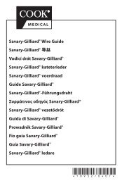
<strong>The</strong> first patient was a liver transplant patient whose air<br />
cholangiogram appeared as seen in Figure 1.<br />
What type of altered<br />
anatomy is depicted?<br />
We required the use of a needle knife to gain access into the biliary tree<br />
followed by balloon dilation and stent placement (Figure2).<br />
<strong>The</strong> second case was also a liver transplant patient (Figure 3).<br />
What does this<br />
image show?<br />
<strong>The</strong> cholangiogram appeared as seen in Figure 4. This was extracted<br />
using a biopsy forceps (Figure 5) and after gaining access and<br />
performing balloon dilation, stents were placed.<br />
To confirm your diagnosis, click on newsletter button on endoscopy<br />
homepage of www.cookmedical.com <br />
We are looking for more submissions and welcome your participation.<br />
if you want to submit an image with a written case history and<br />
clinical explanation, please contact Kevin Chmura at kevin.chmura@<br />
cookmedical.com.<br />
<strong>The</strong> <strong>Channel</strong><br />
Figure 2<br />
Figure 1<br />
Figure 3<br />
Figure 4<br />
Figure 5<br />
www.cookmedical.com<br />
7
“An ideal endoscopic<br />
environment”<br />
<strong>The</strong> Smilow Ambulatory Procedure Center of<br />
Yale New Haven Hospital at Yale University<br />
School of Medicine is a patient-centered<br />
tertiary referral center for advanced<br />
endoscopy with highly committed physicians<br />
and staff.<br />
Priya Jamidar, MD, FACG (Professor of<br />
Medicine and Director of Endoscopy,<br />
Section of Digestive Diseases, Yale School<br />
of Medicine) is quick to describe the center’s<br />
priority: “We try very hard to be responsive<br />
to our patients and to be highly accessible<br />
to the physicians who refer to us. We realize<br />
we are not in an ivory tower; our objective is<br />
to make life better for patients and to make<br />
a difference in people’s lives with everything<br />
we offer and do.”<br />
<strong>The</strong> Yale Interventional Endoscopy team<br />
includes: Dr. Jamidar, Harry Aslanian,<br />
MD, FASGE, AGAF (Director of Advanced<br />
Endoscopic Fellowship and Associate<br />
Director of Endoscopy, Associate<br />
Professor of Medicine, Yale University School<br />
of Medicine) and Uzma Siddiqui, MD (Director<br />
of EUS and Associate Professor of Medicine,<br />
Yale University School of Medicine). New<br />
additions to the interventional team include<br />
Nurse Practitioner Hillary Drumm and Clinical<br />
Referral Specialist Natasha Williams, who<br />
assist in streamlining patient care.<br />
New, state-of-the-art<br />
endoscopy facilities<br />
<strong>The</strong> physician team and staff recently moved<br />
to their new headquarters. <strong>The</strong> Ambulatory<br />
Procedure Center, which opened in February<br />
2010, is on the fourth floor of the new Smilow<br />
Cancer Hospital. During the first ten months<br />
of operation, physicians there performed<br />
more than 7,500 procedures.<br />
“It is a beautiful facility,” says Cindy<br />
Dabbraccio, RN, Manager of the Center.<br />
“It is filled with color and artwork. On the<br />
seventh floor is a healing garden with a water<br />
fountain, trees and benches. It overlooks<br />
New Haven. Patient satisfaction, as measured<br />
8 www.cookmedical.com<br />
Yale University Section<br />
of Digestive Diseases<br />
by Press Ganey numbers, is 93.5%. It takes<br />
everyone working together to meet and<br />
exceed our patient needs.”<br />
“We have a wonderful place to practice,”<br />
agrees Dr. Jamidar. “<strong>The</strong> environment is<br />
nice and bright, and we are blessed with<br />
wonderful nurses, techs and secretaries. In<br />
addition, the rooms are fully integrated for<br />
observation and teaching purposes.”<br />
<strong>The</strong>re are two fluoroscopy rooms, both<br />
equipped with fixed C-arms. “It is much more<br />
convenient for patients and physicians,”<br />
he says. “In our previous facility, we had to<br />
take patients to the radiology department<br />
in another area of the hospital. Now it is all<br />
done here.”<br />
Three of the five procedure rooms also are<br />
equipped for general anesthesia. “This is an<br />
important upgrade because, in the past, we<br />
called the Anesthesia Department and they<br />
brought their equipment and supplies to the<br />
old facility,” says Dr. Jamidar.<br />
“We have an excellent, state-of-the-art<br />
facility where patients find the environment<br />
comfortable and supportive,” says Dr.<br />
Aslanian. “<strong>The</strong> physical environment, excellent<br />
nursing care and anesthesia all make this an<br />
ideal endoscopic environment to achieve<br />
the best results. Even parking is convenient.”<br />
Dr. Siddiqui agrees: “I find our new endoscopy<br />
suite a significant improvement from both our<br />
viewpoint and from the patients’ perspective.<br />
It represents a major commitment to patient<br />
care by Yale-New Haven Hospital. In addition<br />
to being equipped for anesthesia, all the<br />
equipment is on booms, which allow for<br />
flexibility in how the rooms are arranged.<br />
We can adjust monitors and patients in any<br />
configuration that is needed and achieve<br />
the best ergonomic positions for a variety<br />
of procedures. This feature provides optimal<br />
conditions for the patient and endoscopist.”<br />
A wide range of expertise<br />
“ We enjoy the collaboration with<br />
our colleagues in managing complex<br />
cases including many gastrointestinal<br />
malignancies,” says Dr. Aslanian. We have<br />
an excellent group of radiologists, surgeons,<br />
oncologists and a pathologist, which is<br />
an important part of an interventional<br />
endoscopy practice. Furthermore, for the<br />
past two years, there has also been an<br />
expert cytology attending and reviewing<br />
our FNA samples bedside during an EUS<br />
and are often able to provide an immediate<br />
diagnosis of malignancy.”<br />
Endoscopic research is an important priority<br />
for the Yale Interventional Endoscopy Team,<br />
which has performed and are involved<br />
in multiple clinical research studies.<br />
Dr. Jamidar's current interests include:<br />
comparisons of metal and plastic stents<br />
for preoperative biliary decompression,<br />
confocal endomicroscopy of the bile ducts<br />
and treatment of cholangiocarcinoma and<br />
Sphincter of Oddi dysfunction. In addition,<br />
he has led a double-blind study to determine<br />
if intraduodenal indomethacin can decrease<br />
the incidence of post-ERCP pancreatitis.<br />
Drs. Aslanian’s and Siddiqui's current<br />
interests include: confocal endomicroscopy<br />
of pancreatic cysts and Barrett's esophagus,<br />
endoscopic mucosal resection, and EUS-<br />
FNA, and RFA for Barrett's with dysplasia.<br />
Furthermore, for EUS-FNA of pancreatic<br />
tumors, the team's published data has<br />
shown they can obtain a diagnosis in greater<br />
than 90% of patients. <strong>The</strong>y favor smaller, 25<br />
gauge needles and benefit from having an<br />
attending cytologist at the bedside during<br />
EUS-FNA that can often provide an on-site<br />
diagnosis, which means the patient can be<br />
informed of their diagnosis before leaving<br />
the endoscopy suite. Dr. Siddiqui states, “This<br />
gives us a unique opportunity to expedite<br />
the next steps in a patient's oncologic care.”<br />
<strong>The</strong> physicians perform approximately 1,000<br />
EUS and 600 ERCPs in addition to other<br />
advanced endoscopy procedures (RFA, EMR<br />
and palliative stenting of GI malignancies).<br />
Yale Advanced<br />
endoscopy Fellowship<br />
Drs. Jamidar, Aslanian and Siddiqui have<br />
trained one advanced fellow per year since<br />
2002 in advanced endoscopy. "It has been
Yale New Haven<br />
Hospital features<br />
966 beds<br />
3,600 university and community<br />
physicians have privileges<br />
8,580 employees<br />
Patients in 2010:<br />
Inpatient discharges: 56,620<br />
Outpatient visits: 638,411<br />
ER visits: 137,627<br />
a very rewarding experience to see our<br />
fellows' skills and careers develop, perform<br />
endoscopic research and develop long<br />
lasting friendships,” reports Aslanian.<br />
Live endoscopic workshops<br />
Twice a year, the center conducts live<br />
endoscopic workshops for up to 100 GIs<br />
and registered nurses from the Northeast<br />
region. Workshops typically feature national<br />
and international experts in EUS and ERCP.<br />
All sessions are CME-accredited.<br />
“Workshops in our new facilities are light<br />
years ahead of our former rooms,” says Dr.<br />
Jamidar. “In the recent past, we had only<br />
one room with wires everywhere. Now we<br />
have three state-of-the-art wireless rooms<br />
and multiple screens for EUS, fluoroscopy,<br />
confocal, cholangioscopy and EUS-FNA<br />
cytology views. “We perform up to ten<br />
cases in three rooms,” he continues. “We<br />
are wired so we can send images and sound<br />
to a conference room across the street as<br />
well as to remote locations worldwide. <strong>The</strong><br />
technology also allows us to interact with the<br />
audience in different clinical scenarios via<br />
closed-circuit interactive television monitors,<br />
while doing EUS and ERCP procedures.<br />
<strong>The</strong> audience is able to ask questions and<br />
hear our responses as we perform the<br />
procedures. We held our twelfth session in<br />
Smilow Hospital<br />
features<br />
<strong>The</strong> Smilow Cancer Hospital is 14 stories<br />
tall, has 168 inpatient beds, 12 operating<br />
rooms and five procedure rooms. Twelve<br />
physician specialties are represented<br />
within the center.<br />
For more information on<br />
CMe courses at Yale:<br />
CME@Yale.edu<br />
(203) 785-4578 FAX: (203) 785-3083<br />
September 2011 and have received excellent<br />
feedback.” <strong>The</strong> conference recently received<br />
a Best Practice award from the Association of<br />
American <strong>Medical</strong> Colleges.<br />
In addition, the center is striving for future<br />
collaborations with other academic medical<br />
centers. “In the spring of 2012,” says Jamidar,<br />
“Dr. John Saltzman of Brigham and Women’s<br />
Hospital in Boston and I are planning a<br />
Harvard-Yale collaborative course to be held<br />
in Bermuda. It will focus on controversies,<br />
debates and challenges in endoscopy. We<br />
have nicknamed it, ‘<strong>The</strong> Course’ after ‘<strong>The</strong><br />
(Yale- Harvard football) Game.’”<br />
On the horizon at Yale<br />
“We want to continue to grow as a resource<br />
for gastroenterolgists and oncologists<br />
in our area, providing tertiary care in<br />
advanced endoscopy,” says Dr. Jamidar.<br />
“We want to remain on the cutting edge<br />
of early diagnoses, and be at the forefront<br />
of pancreatic and biliary endoscopy. We<br />
foresee better methods of imaging, tissue<br />
acquisition and stenting. We also foresee<br />
better treatments for reflux and obesity.<br />
Ultimately, we hope to earn national and<br />
international recognition for our practice and<br />
for our training of other physicians who will<br />
then make a difference in their communities.”<br />
<strong>The</strong> <strong>Channel</strong><br />
About the Yale<br />
Ambulatory Service<br />
Division<br />
<strong>The</strong> Center is located within the new<br />
Smilow Cancer Hospital. It opened in<br />
2010 as an addition to the Yale New<br />
Haven Hospital and features:<br />
40 MDs<br />
20 RNs<br />
3 Patient Care Associates<br />
5 GI Technicians<br />
2 Scope Technicians<br />
2 Secretaries<br />
7 MD fellows (one advanced endoscopy<br />
Fellow each year since 2002)<br />
5 Procedure rooms<br />
12 Prep/recovery rooms<br />
Procedures:<br />
1,000 EUS/year<br />
600 ERCP/year<br />
Endoscopic mucosal resection<br />
Enteral stents<br />
Balloon dilations<br />
Barrx<br />
Confocal microscopy<br />
Direct choledocoscopy<br />
EUS-guided access procedures<br />
Colonoscopy/EGD<br />
<strong>The</strong>re are also satellite facilities for routine<br />
colonoscopies within the Ambulatory<br />
Service Division.<br />
Service Area Includes:<br />
Connecticut and other areas in the<br />
Northeast (including New York,<br />
Rhode Island and Massachusetts). Patients<br />
come from other states and countries,<br />
as well.<br />
Press Ganey score<br />
93.5% high satisfaction rate.<br />
www.cookmedical.com<br />
9
10 www.cookmedical.com<br />
Montreal’s<br />
ERCP and<br />
Duodenal-<br />
Stenting<br />
innovator<br />
At first glance, the clinical objectives seem to be in conflict: A renowned Canadian liverpancreatic<br />
surgeon, with expertise in liver transplants, is pushing the envelope to find nonsurgical,<br />
endoscopic solutions to complex medical problems.<br />
Meet Dr. Andre Roy, Director of the Liver Transplantation Program at Hopital Saint-Luc<br />
de Centre Hospitalier de l’Universite de Montreal, known as CHUM. He also heads one<br />
of the busiest endoscopic retrograde cholangiopancreatography (ERCP) practices in the<br />
Quebec Province.<br />
A surgeon who received training in Lyon, France, Dr. Roy began to specialize in liver surgery<br />
in London, Ontario, in the early 1990s, where he performed transplants and, simultaneously,<br />
ERCP procedures. He began his present surgical practice and endoscopic practice at<br />
St. Luc in 1993.<br />
During his first year at St. Luc, in conjunction with a radiologist, he introduced duodenal<br />
stenting. “It went smoother than expected,” recalls Dr. Roy. “It required only two hours<br />
versus a full day. Also, we had good results. <strong>The</strong> alternative was open surgery, with<br />
dangers of complications. But with the duodenal stent, the patient was able to return<br />
home the same evening.<br />
“That same year, we did 100 ERCPs,” continues Dr. Roy. “Since then, our practice has<br />
grown in size and volume, with increasing referrals and challenges. We continually<br />
see more complicated cases and we are being introduced to new, more advanced<br />
endoscopic devices.”<br />
Growing potential for biliary and duodenal stenting<br />
“Today, we perform more than 1,000 ERCPs a year, with up to 15 cases a day,” continues<br />
Dr. Roy. “Our procedure room has a good set up. Two nurses in the room assist me, along<br />
with one or two radiology techs, and a transportation aide to move the patients in and out.”<br />
Dr. Roy’s patients come from all over the Quebec and Maritime Provinces. “Many are<br />
transfers from other hospitals,” says Dr. Roy, “because ERCP is not available outside<br />
large cities in Canada. In some cases, we receive ERCP patients from other large<br />
medical centers, when the procedure was unsuccessful. Thus, we see the more complicated<br />
cases: pregnant women, patients with drainage from cysts, patients requiring<br />
duodenal and enteral stenting.<br />
“I also use stenting for gastric cancer and proximal small bowels. Guided fluoroscopy<br />
allows us to reach difficult areas to insert metal stents. We try to push the envelope, when<br />
it is do-able and logical.”
Research and innovations<br />
Dr. Roy is involved in several registries and is about to start another one this fall. In a registry<br />
for the <strong>Cook</strong> Evolution® duodenal stent, he found that “these are amazing stents. <strong>The</strong><br />
enlarged cuffs prevent migration and they decrease the risk of perforation. <strong>The</strong> big handle<br />
allows clinicians to control the release. It is very reliable and very steady.<br />
“In addition, we can recapture and reposition the stent. This is very handy. I do my part, the<br />
nurse does hers; it is much easier for the nurse to release. Further, the meshing of the stent<br />
is tighter and more durable.“<br />
In addition to registries, Dr. Roy conducts research on endoscopic and liver surgery, and he<br />
has written or contributed to more than 100 journal articles. An example is “Treatment of<br />
post liver transplantation bile duct stricture with self-expandable metallic stent.” This article<br />
describes a study of 21 patients diagnosed with anastomotic biliary stricture who were<br />
treated by ERCP. <strong>The</strong> conclusion: “Treatment of post-transplant biliary stenosis using a stent<br />
is a valuable option for delaying or avoiding surgery in up to 70 percent of patients. Proximal<br />
stenosis can be treated in the same manner in selected patients with major co-morbidities.”<br />
In addition, Dr. Roy shares his knowledge through seminars in the Province of Quebec, such<br />
as a recent presentation to the Americas Hepato Pancreato Biliary Association (AHBPA).<br />
Pushing the envelope<br />
Dr. Roy has already introduced new uses for ERCP. He recalls a 44-year-old patient who had<br />
previously undergone surgery for cancer of the stomach. <strong>The</strong> patient returned eight months<br />
after surgery with cancer blocking the small bowel.<br />
Dr. Roy explains: “<strong>The</strong> surgeon tried a bypass but was unsuccessful. <strong>The</strong> blockage was<br />
far down in the small bowel. We jointly concluded that the patient’s only chance was an<br />
endoscopic bowel stent. So we worked hard to apply a stent to relieve the obstruction. It<br />
was unorthodox but successful. <strong>The</strong> patient returned home and was able to eat, and enjoyed<br />
the time he had. <strong>The</strong> stent made a big difference in his life.”<br />
In the future, Dr. Roy foresees more sophisticated stents for draining pseudocysts and<br />
duodenal cysts, and inserting more enteral stents in the proximal small bowel, which he<br />
recently performed.<br />
“Overall, I believe we will see even more innovative equipment and that we will able to do<br />
more and more procedures that prevent the need for surgery,” says Dr. Roy. “I say this because<br />
we can already insert stents in places previously inaccessible, such as deep into the liver.<br />
“<strong>The</strong> keys to pushing the envelope are to perform procedures that are logical and do-able,<br />
and that produce good results. <strong>The</strong>re are a large number of very sick patients who need<br />
these services,” he concludes.<br />
<strong>The</strong> <strong>Channel</strong><br />
www.cookmedical.com<br />
11
the<br />
nurses’ station<br />
Annual Education<br />
Day Focuses on<br />
Latest innovations<br />
in Gi<br />
One hundred and forty GI nurses<br />
and technicians recently gathered<br />
at Chicago’s Westin O’Hare for the<br />
3rd Annual Advocate Health Care<br />
“Innovations in GI: Advancing Nursing<br />
Practice” Education Day, where key<br />
opinion leaders delivered lectures, led<br />
discussions and presented cases.<br />
Programs and presenters at this year’s<br />
Education Day included: “Capnography<br />
in Colonoscopy” by Cecelia Pezdek,<br />
ADN,BS, MSHA, RN, CGRN, Julie<br />
Flamm, ADN,BS, RN, CGRN, and Karyn<br />
Pechinski, BSN, RN, CGRN; “Advanced<br />
Diagnostic Bronchoscopy: EBUS &<br />
Electromagnetic Navigation” by Sara R.<br />
Greenhill MD, FCCP; “Stop the Bleeding!<br />
Burn, Band, Bite and Other Methods for<br />
Endoscopic Hemostasis” by Dean Silas,<br />
MD; “Impact of EUS & Innovations in<br />
<strong>The</strong>rapeutic Endoscopy: A Community<br />
Perspective” by Kenneth Chi, MD; and<br />
“EUS & ERCP: A Partnership in Care” by<br />
<strong>The</strong>resa Vos MS, RN, CGRN.<br />
Attendees who completed the course<br />
received five contact hours of continuing<br />
education. For more information<br />
on Advocate Health Care’s Annual<br />
Education Day, contact Steven Werner<br />
at Steven.Werner@advocatehealth.com<br />
or 773-296-5562.<br />
12 www.cookmedical.com<br />
Capnography<br />
Helping to keep our patients safe<br />
Cecelia Pezdek, MSHA, CGRN<br />
Manager, Endoscopy and Ambulatory Center<br />
Advocate Good Samaritan Hospital<br />
Downers Grove, IL<br />
<strong>The</strong> impact of healthcare reform and determining what this reform will look like, developing<br />
metrics for performance outcomes, and most importantly, providing safe affordable<br />
healthcare is a monumental task in these economically challenging times. <strong>The</strong> recent change<br />
in guidelines from the American Society of Anesthesiologists (ASA) regarding monitoring<br />
standards for ventilation has resulted in providers scrambling for resources. <strong>The</strong> “Standards<br />
for Basic Anesthetic Monitoring” are as follows:<br />
“3.2.4 During regional anesthesia (with no sedation) or local anesthesia<br />
(with no sedation), the adequacy of ventilation shall be evaluated by<br />
continual observation of qualitative clinical signs. During moderate or<br />
deep sedation the adequacy of ventilation shall be evaluated by continual<br />
observation of qualitative clinical signs and monitoring for the presence of<br />
exhaled carbon dioxide unless precluded or invalidated by the nature of<br />
the patient, procedure, or equipment.” (ASA Standards, 2010).<br />
This standard of care from the ASA is for anesthesiologists and would suggest that monitoring<br />
ventilation by non-anesthesiologists is reasonable (Kodali B, 2011). Many institutional<br />
structures have sedating areas as well as their operating rooms under the direction of an<br />
anesthesiologist. <strong>The</strong>refore, provision of resources for capnography monitoring has taken<br />
on top priority for many facilities. Pulse oximetry monitoring for oxygenation levels has long<br />
been a standard of care for patients undergoing moderate sedation. <strong>The</strong> literature cautions<br />
providers about interchanging the terms “oxygenation” and “ventilation”; they are not the<br />
same. Research has repeatedly shown a delay of apnea detection with pulse oximetry as<br />
compared to capnography. <strong>The</strong> challenge of monitoring ventilation in endoscopy suites is<br />
compounded when patients receive supplemental oxygen, lighting is limited for clinical<br />
assessment, positioning of the patient is not optimal for air exchange and the use of<br />
benzodiazepines reduces lung capacity. Earlier detection results in earlier intervention and<br />
improved safety for patients. Capnography is a welcome adjunct to keeping our patients safe.<br />
For more information, visit:<br />
www.asahq.org/publicationsAndServices/sgstoc.htm<br />
http://www.capnography.com
over 2200 Participants<br />
attend Asia Pacific<br />
Digestive Week<br />
<strong>The</strong> Asia Pacific Digestive Week (APDW) held at SUNTEC Singapore<br />
International Convention and Exhibition Centre from 1st to 4th October<br />
2011, attracted over 2200 participants over the whole Asia Pacific Region.<br />
Supporting the event as a Bronze sponsor, <strong>Cook</strong> <strong>Medical</strong> exhibited a wide<br />
range of GI products in three work stations, targeting different procedures,<br />
namely ERCP, non-ERCP and EUS.<br />
In addition to the important products such as Evolution Controlled-Release<br />
Stent, EchoTip® ProCore HD Ultrasound Biopsy Needle, Fusion® Dual<br />
Platform ERCP, Hercules® 3 Stage Wire Guided Balloon and Zilver 635®<br />
Biliary Self-Expanding Stent, two new hemostasis products – Hemospray<br />
Endoscopic Hemostat and Instinct Endoscopic Hemoclip – had been<br />
introduced in the Innovation Room. By touring and watching product<br />
demonstrations, participants were excited about the products and were<br />
looking forward to the launch in their own countries.<br />
To provide a deeper understanding towards the hemostasis devices, <strong>Cook</strong><br />
<strong>Medical</strong> conducted an “Advances in Hemostasis” dinner symposium on<br />
October 2nd and invited Prof. Joseph Sung who is the Vice-Chancellor<br />
and President of the Chinese University of Hong Kong as the guest<br />
speaker. With over 60 participants, Prof. Sung presented the Hemospray<br />
Endoscopic Hemostat and Instinct Endoscopic Hemoclip with clinical<br />
evidence where audiences enthusiastically exchanging ideas and asking<br />
questions throughout the event.<br />
<strong>The</strong> <strong>Cook</strong> EUS Roundtable Meeting directed by both honorary guests:<br />
Dr. Jong H. Moon from Korea and Dr. Takao Itoi from Japan was held on<br />
3rd October. Within this one hour session, latest information related to<br />
EUS were shared. Three lectures as well as a special case review of Pro<br />
Core by Dr. Sandeep Lakhtakia from India were given. <strong>The</strong> panel discussion<br />
at the end with the topic of EUS guided sampling technique (for better<br />
diagnosis) was also successfully involved active participants from all the<br />
attendees that concluded the event with a wider horizon of understandings<br />
on EUS technology.<br />
<strong>The</strong> <strong>Channel</strong><br />
(Top Left) Prof. Joseph Sung is pleased to have<br />
active response during the questions and answer<br />
sessions from the participants<br />
(Top Right) <strong>Cook</strong> <strong>Medical</strong> booth<br />
(bottom Right) Participants are actively<br />
involving in the roundtable discussion<br />
www.cookmedical.com<br />
13
N E W S F R O M<br />
SIGNEA Society of International Gastroenterological Nurses and Endoscopy Associates<br />
introduction<br />
<strong>The</strong> method of diagnosis and treatment of<br />
digestive diseases known as gastrointestinal<br />
endoscopy has existed for about 120 years.<br />
As the science and art of endoscopy has<br />
evolved worldwide there has been enormous<br />
beneficial interventions for patients. <strong>The</strong>re<br />
has been rapid advancement of endoscopic<br />
technology with innovative instrumentation<br />
and new advanced techniques (Sivak, 2000).<br />
My own personal experience commenced<br />
just some 16 years ago when I entered<br />
endoscopy as a Registered Nurse as a parttimer<br />
while completing my Masters in Health<br />
Science. Very quickly I was drawn to this<br />
specialty and focused on the patient journey<br />
and the advancing technology in scopes,<br />
equipment and techniques and changes in<br />
cleaning and decontamination processes.<br />
My passion for the “endoscopy” specialty<br />
grew into a genuine interest in endoscopy<br />
management and infection control.<br />
endoscopy Developments<br />
Over the two last decades research data<br />
has supported significant changes in best<br />
practice within endoscopy and specifically<br />
with cleaning and decontamination<br />
processes. International research studies<br />
have identified infections transmitted by<br />
endoscopy procedures (Favero, 1996;<br />
Seone-Vazquez, Rodriguez-Monguio,<br />
Visaria, Carlson, 2007; Morris, Duckworth,<br />
Ridgway, 2006). Key medical and nursing<br />
leaders have driven significant changes<br />
and improvements to prevent these<br />
transmissions.<br />
<strong>The</strong> specialty has become even more<br />
complex and to the beginner there is little<br />
appreciation of the extensive infrastructure<br />
that is now necessary for efficient and safe<br />
endoscopy procedures. According to<br />
Cotton & Williams (2006) endoscopy has<br />
become a sophisticated industry, setting<br />
up and running an endoscopy unit is a<br />
complex topic where endoscopy is a team<br />
activity that requires the collaborative<br />
talents of many people to ensure an<br />
appropriate environment and professional<br />
staff to maintain patient comfort and safety<br />
and to optimize clinical outcomes. With<br />
phenomenal increases in technical capability<br />
and scientifically derived knowledge come<br />
the challenges to ensure patient safety at<br />
all times.<br />
Endoscopic equipment presents particularly<br />
unsatisfactory surfaces for disinfection. As<br />
we are aware, endoscopes are fragile and<br />
14 www.cookmedical.com<br />
Endoscopy and infection Control<br />
Raewyn Paviour, NZNO Gastroenterology Nurses Section Treasurer, SIGNEA Director<br />
heat labile, channels within the endoscope<br />
are made of plastic material and are subject<br />
to trauma of biopsy forceps and therapeutic<br />
equipment, all of which may cause surface<br />
irregularity. New endoscopic equipment<br />
and accessories are a frequent occurrence<br />
within the endoscopy departments. Hence<br />
the impact on everyday practice poses<br />
a constant challenge to ensure that all<br />
endoscopic equipment is cleaned and<br />
disinfected according to best practice and<br />
that a clean safe endoscope and accessories<br />
are always presented for every patient’s<br />
episode of care.<br />
Some specific infection control challenges<br />
in everyday endoscopy practice:<br />
• Selecting best practice national and /<br />
or international standards, protocols,<br />
policies and guidelines<br />
• Establishing in-depth knowledge and<br />
understanding on the various types<br />
and models of endoscopes<br />
• Ensuring sound knowledge and<br />
understanding of substantial range of<br />
endoscopic accessories and ancillary<br />
equipment from multiple companies<br />
worldwide<br />
• Selecting national and/or international<br />
best practice standards for infection<br />
control in cleaning and disinfection,<br />
mechanical cleaning and disinfectants,<br />
drying and storing<br />
• Ensuring ability to safely use and<br />
apply multiple types of energy sources<br />
• Establishing appropriate staff<br />
education and training, on-going<br />
monitoring and evaluation<br />
• Ensuring quality control measures<br />
• Applying bacteriological surveillance<br />
as a quality marker<br />
• Ensuring best practice standards for<br />
detailed and accurate records<br />
• Establishing appropriate and regular<br />
equipment maintenance and records<br />
• Ensuring staff protection, staff training,<br />
monitoring and on-going evaluation<br />
International leaders have driven significant<br />
changes in endoscopy standards and<br />
policies, educational courses, publications<br />
and exposed current issues worldwide with<br />
the intent of ensuring best practice and<br />
patient safety. Access to communication<br />
tools of the 21st century means that<br />
international links to multiple reference sites<br />
for endoscopy best practice guidelines,<br />
standards and information are quick<br />
and easy.<br />
Listed below are a few examples:<br />
w w w. i n f e c t i o n c o n t r o l . s g n a . o r g<br />
SGNA (Society of Gastroenterology<br />
Nurses & Associates, Inc)– GI/Endoscopy<br />
Infection Control Resource Center<br />
w w w. i n f e c t i o n c o n t ro l t o d a y. c o m<br />
ASGE (<strong>The</strong> American Society for<br />
Gastrointestinal Endoscopy)–ASGE issues<br />
Updated Infection Control Guidelines for<br />
Gastrointestinal Endoscopy<br />
www.genca.org (Gastroenterological<br />
Nurses College of Australia) and<br />
www.gesa.org (GESA-Gastroenterologic<br />
Society of Australia)–Infection Control in<br />
Endoscopy 2010<br />
www.bsg.org.uk (BSG-British Society<br />
of Gastroenterology)–BSG Clinical<br />
Guidelines<br />
www.signea.org (SIGNEA-Society of<br />
International Gastroenterological Nurses<br />
& Endoscopy Associates–Newsletter<br />
Archives/Future technical & publication<br />
developments planned<br />
Over the years I have had the privilege to<br />
network professionally with many of the<br />
GENCA Committee members, who have<br />
actively developed the GENCA/GESA<br />
“Infection Control Guidelines” with the<br />
newest update being the 3rd edition in<br />
September 2010. As Australia is one of our<br />
immediate neighbors we are incredibly<br />
fortunate to have excellent professional<br />
working relationships where we benefit from<br />
the outstanding work that the Australians<br />
have invested in Infection Control Standards<br />
in Endoscopy. In reality, our practices are<br />
fairly similar and the country’s close proximity<br />
facilitates very good attendance of nurses<br />
and doctors at Australian Gastroenterology<br />
Conferences.<br />
Most Endoscopy Units/Centres in New<br />
Zealand work and apply the best practice<br />
guidelines of the GENCA/GESA Infection<br />
Control in Endoscopy Guidelines. New<br />
Zealand has maintained an innovative and<br />
up to date presence in endoscopy infection<br />
control and best practice standards.<br />
In 2001 Standards New Zealand developed<br />
the New Zealand Handbook – Microbiological<br />
Surveillance of Flexible Hollow Endoscopes
(SNZ HB 8149:2001). Surveillance cultures<br />
had become increasingly popular in NZ<br />
as one of the quality control markers of<br />
effective cleaning and disinfection of flexible<br />
hollow endoscopes. As a member of the<br />
working party, the Handbook for Endoscopy<br />
Users was developed in order to further<br />
rationalize and standardize the method for<br />
testing and validate results interpretation<br />
with relevant patient outcomes. <strong>The</strong> ultimate<br />
goal was to reduce the risk of transmission<br />
of infection during endoscopic procedures<br />
in New Zealand. Hence a formal process was<br />
established with the intent for prospective<br />
evaluation of microbiological cultures<br />
in New Zealand. We are now 10 years<br />
on and we are hoping there will be an<br />
opportunity in the near future to complete<br />
this retrospective review.<br />
Looking into the future there are further<br />
opportunities to evaluate endoscopy practice<br />
in New Zealand and make improvements in<br />
patient care delivery. Recently the Ministry<br />
of Health has announced that New Zealand<br />
will have a bowel cancer screening pilot<br />
study commencing in October 2011. This<br />
is wonderful news as bowel cancer is the<br />
most frequently diagnosed cancer and<br />
the second most common cause of cancer<br />
death in New Zealand. <strong>The</strong>re have been new<br />
Available in<br />
the US only.<br />
GPN<br />
roles established with a Doctor and Nurse<br />
National Endoscopy Service Improvement<br />
Lead Bowel Cancer positions. <strong>The</strong>se roles<br />
have the potential for New Zealand to<br />
have further endoscopic developments<br />
and improvements based on international<br />
experiences where patient care and safety<br />
are based on proven and tested best<br />
practice systems.<br />
Over the last decade I have held various<br />
Committee roles within our New Zealand<br />
Nurses Organization, Gastroenterology<br />
Nurses Section, ranging from Chair to<br />
Editor of our Nursing Journal called the<br />
“TUBE” to, currently, the Treasurer. Since<br />
2009 I have experienced working with the<br />
SIGNEA Directors, which again offers the<br />
most amazing collegial networking and<br />
opportunities for learning and development.<br />
summary<br />
<strong>The</strong> significant advances we have seen<br />
in the endoscopy field are outstanding<br />
and to have been a small part of the<br />
endoscopy specialty in New Zealand has<br />
been a privilege. Management and infection<br />
control in New Zealand have taken quantum<br />
leaps for better patient care and outcomes,<br />
with more advancements to take place<br />
in the near future. Networking, nationally<br />
A D D i T i o N A L S i z E S A v A i L A b L E<br />
Order<br />
Number<br />
<strong>The</strong> <strong>Channel</strong><br />
New Triton Balloon Replacement Tube helps deliver patient care with added efficiency.<br />
<strong>Cook</strong> <strong>Medical</strong> recently introduced the new Triton Balloon Replacement Tube. With separate<br />
lumens for inflation, feeding and delivering medications, the Triton adds convenience and<br />
efficiency to enteral feeding procedures. <strong>The</strong> dedicated medication tube accepts smaller<br />
syringes and the clear feeding tube, made of flexible, high-grade silicone, allows caregivers<br />
to easily visualize nutrient flow. <strong>The</strong> external bolster has been newly designed for patient<br />
comfort. For more information on the Triton BRT, contact your <strong>Cook</strong> representative.<br />
Feeding Tube<br />
Fr<br />
Balloon Capacity<br />
cc<br />
G56111 PEG-14-BRT-TRI 14 5<br />
G56112 PEG-16-BRT-TRI 16 10<br />
G56113 PEG-18-BRT-TRI 18 15<br />
G53883 PEG-20-BRT-TRI 20 20<br />
G56114 PEG-22-BRT-TRI 22 20<br />
G53885 PEG-24-BRT-TRI 24 20<br />
Components include: 1 bolster and 1 water soluble lubricant pack.<br />
and internationally, has offered the most<br />
amazing connections and friendships along<br />
with significant learning and development<br />
opportunities.<br />
References<br />
Sivak, M.V., (2000). Gastroenterologic Endoscopy<br />
(2nd ED.). W.B.Saunders: Philadelphia.<br />
Cotton, P.B & Williams, C.B. (2006). Practical<br />
Gastrointestinal Endoscopy <strong>The</strong> Fundamentals<br />
(5th ED.). Blackwell Science Ltd: Massachusetts.<br />
GENCA/GESA Clinical Update - Infection Control<br />
in Endoscopy (2010) (3rd Ed.). Gastroenterological<br />
Society of Australia. Victoria, Australia.<br />
Favero, M.S., & Pugliese, G. Infections transmitted<br />
by endoscopy: an international problem<br />
(editorial). AM J Inf Control 1996; 24 (5):343-5.<br />
Seoane-Vazquez, E., Rodriguez-Monguio, R.,<br />
Visara, J., Carlson, A. Endoscopy-related infections<br />
and toxic reactions: an international comparison.<br />
Endoscopy 2007 Aug; 39 (8): 742-78.<br />
Morris, J., Duckworth, G.J., Ridgway, G.L.<br />
Gastrointestinal endoscopy decontamination<br />
failure and the risk of blood-borne viruses: a<br />
review. J Hosp Infect. 2006, May 63 (1): 1-13.<br />
New Zealand Ministry of Health Standards,<br />
New Zealand. New Zealand Handbook –<br />
Microbiological Surveillance of Flexible Hollow<br />
endoscopes. SNZ HB 8149:2001. 2001.<br />
www.cookmedical.com<br />
15
<strong>Cook</strong> <strong>Medical</strong> has long understood that<br />
optimal patient care is your focus, and it<br />
continues to be our focus as well. That’s why<br />
for more than twenty years we have assisted<br />
healthcare professionals in learning the latest<br />
in endoscopic GI technology and related<br />
disease information.<br />
That tradition continues as <strong>Cook</strong> <strong>Medical</strong>, in<br />
partnership with HealthStream (an accredited<br />
provider of continuing nursing education),<br />
offers three new educational activities:<br />
Business Management<br />
of the Endoscopy Unit<br />
Malignant Biliary<br />
Disease Management<br />
Updates in Colorectal Cancer<br />
Barrett’s Esophagus<br />
<strong>The</strong>se activities are presented without<br />
charge by your <strong>Cook</strong> <strong>Medical</strong> district<br />
manager. Educational activity descriptions,<br />
objectives and the related accreditation<br />
information can be found at http://www.<br />
cookmedical.com/esc/educationResource.<br />
do?id=Educational_Activity.<br />
Contact your <strong>Cook</strong> representative for more<br />
information or to arrange a presentation<br />
opportunity.<br />
A continuing nursing education activity sponsored by<br />
HealthStream. Grant funds provided by <strong>Cook</strong> <strong>Medical</strong>.<br />
16 www.cookmedical.com<br />
upcoming events<br />
XXXIII Endoscopy Society Spanish Congress Madrid Nov. 11<br />
Interventional Endoscopy: New Frontiers San Francisco, CA Nov. 11-12<br />
UCI Hands-On ERCP for GI Nurses & Techs Orange, CA Nov. 15-16<br />
2nd Annual Jefferson GI Live Conference Philadelphia, PA Nov. 18<br />
Frontiers in Gastroenterology Farmington, PA Nov. 18-19<br />
National Endoscopy Week Leon, Mexico Nov. 19<br />
Brazilian Digestive Week Porto Alegre, Brazil Nov. 20<br />
XXXVIII Chilean Congress of Gastroenterology Vina del Mar, Chile Nov. 23<br />
Gastroenterology National Congress Medellin, Colombia Dec. 1<br />
IU ERCP Workshop for Nurses Indianapolis, IN Dec. 1-2<br />
UCI Hands-On EUS for GI Nurses & Techs Orange, CA Dec. 6-7<br />
26th International Workshop on <strong>The</strong>rapeutic Endoscopy Hong Kong Dec. 13<br />
UCI Hands-On ERCP for GI Nurses & Techs Orange, CA Dec. 13-14<br />
35th Annual NYSGE<br />
(New York Society Gastrointestinal Endo)<br />
Club Francophone d’Echoendoscopie<br />
(French Endoscopy Meeting)<br />
New York, NY Dec. 14-17<br />
Paris, France<br />
Jan. 27-28<br />
Pancreatic & Biliary Endoscopy - Simon Lo Los Angeles, CA Jan. 27-29,12<br />
Düsseldorf International Endoscopy Symposium<br />
(Prof. Neuhaus)<br />
Düsseldorf, Germany Feb. 3-4<br />
XXIVth Belgian Week of Gastroenterology 2012 Ostende, Belgium Feb. 9-10<br />
11th. GI Live Endoscopy Demonstration at the<br />
Chulalongkorn University Hospital in Bangkok<br />
Bangkok, Thailand Feb. 13-15<br />
CDDW-Canadian Digestive Disease Week Montreal, Quebec Feb. 24-27<br />
Westmead International Endoscopy Workshop Sydney, Australia Mar. 7-9<br />
Journées Francophones d’Hépato-gastroentérologie et<br />
d’Oncologie Digestive (JFHOD)<br />
Paris, France Mar. 15-18<br />
42. Kongress der DGE-BV Munich, Germany Mar. 22-23<br />
Annual International Course in Gastroenterology and<br />
Advanced Endoscopy, WGO Training Center<br />
La Paz, Bolivia Mar. 26-Apr. 1<br />
18° Congresso Nazionale delle Malattie Digestive (FISMAD) Naples, Italy Mar. 28-31<br />
2nd Prague an 13th Endoscopic Day IKEM Prague Mar. 29<br />
GI Roundtable Knoxville, TN Mar. 30-31<br />
University Hospital Workshop Kuala Lumpur, Malaysia Mar. 30-Apr. 1<br />
DDW San Diego, CA May 20-22<br />
SGNA Phoenix, AZ May 20-22<br />
ESC-WNL-50033-EN-201110