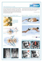
Transforaminal Endoscopic Stenosis Surgery - joimax GmbH
Transforaminal Endoscopic Stenosis Surgery - joimax GmbH
Transforaminal Endoscopic Stenosis Surgery - joimax GmbH

Create successful ePaper yourself
Turn your PDF publications into a flip-book with our unique Google optimized e-Paper software.
www.touchbriefings.com<br />
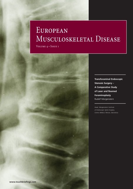
European<br />
Musculoskeletal Disease<br />
Volume 4 • Issue 1<br />
<strong>Transforaminal</strong> <strong>Endoscopic</strong><br />
<strong>Stenosis</strong> <strong>Surgery</strong> –<br />
A Comparative Study<br />
of Laser and Reamed<br />
Foraminoplasty<br />
Rudolf Morgenstern<br />
Head, Morgenstern Institute<br />
of <strong>Endoscopic</strong> Spine <strong>Surgery</strong>,<br />
Centro Médico Teknon, Barcelona
Orthopaedic <strong>Surgery</strong> Spine<br />
Abstract<br />
This article presents a new endoscopic procedure: transforaminal endoscopic stenosis surgery (TESS). This technique is performed through<br />
a posterolateral transforaminal approach and allows the foramen in a collapsed lumbar disc to be widened by undercutting the superior<br />
facet under direct endoscopic control. A new endoscopic small reamer is used for this purpose, which allows the aggression to the<br />
surrounding tissues to be minimised. This study of 216 cases of lumbar foraminal stenosis compares the results of one group in which the<br />
new endoscopic bone reamers were used for the foraminoplasty with the results of another group in which only classic foraminoplasty was<br />
performed with a standard holmium: yttrium–aluminium–garnet (Ho:YAG) laser. The methods were as follows: 216 patients with lumbar<br />
foraminal stenosis underwent endoscopic spine surgery from 2003 to 2008 at Centro Médico Teknon in Barcelona. One hundred and twentyfive<br />
patients underwent classic endoscopic surgery, i.e. only a Ho:YAG laser was used for the foraminoplasty (Group A); 91 patients<br />
underwent TESS, i.e. the new endoscopic bone reamers were used for the foraminoplasty (Group B). The inclusion criteria were unilateral or<br />
bilateral radicular leg pain associated with imaging evidence of foraminal or lateral stenosis, and inadequate response to conservative<br />
treatment for >6 months. All 216 procedures were performed in the prone position and under local anaesthesia. Pain was scored for every<br />
patient both pre- and post-operatively using a visual analogue scale (VAS); disability was scored using the Oswestry Disability Index (ODI).<br />
The post-operative scores were updated every three months. The mean follow-up period was 2.8 years (range: six to 61 months). The results<br />
were as follows: 216 patients who met the inclusion criteria underwent TESS. These 216 patients comprised 143 men and 73 women with<br />
ages ranging from 17 to 82 years (mean age 45.8 years). The overall results, evaluated according to Macnab criteria, for the 216 cases were:<br />
151 excellent (69.9%), 45 good (20.8 %), 16 fair (7.4 %) and four poor (1.9 %). The results for group A (125 cases) were: 90 excellent (72 %), 20<br />
good (16 %), 14 fair (11.2 %) and one poor (1.9 %). The results for group B (91 cases) were: 61 excellent (67 %), 25 good (27.5 %), two fair (2.2<br />
%) and three poor (3.3 %). The average surgical time was approximately 50 minutes for group A and approximately 30 minutes for group B.<br />
Keywords<br />
The first posterolateral discectomy was a percutaneous central<br />
nuclectomy and was reported by Hijikata1 in 1975, followed by<br />
Kambin and Gellman’s2 report of nine cases in 1983. In 1983, Forst<br />
and Hausmann3 described the direct visualisation of intervertebral<br />
disc space with a modified arthroscope. Schreiber et al. 4 used a<br />
biportal endoscopic technique. In 1996, Mathews5 wrote about the<br />
transforaminal approach, but it is the same portal utilised by Kambin6 and Yeung. 7<br />
<strong>Transforaminal</strong> <strong>Endoscopic</strong> <strong>Stenosis</strong> <strong>Surgery</strong> –<br />
A Comparative Study of Laser and Reamed Foraminoplasty<br />
Spinal endoscopy, minimally invasive spine surgery, transforaminal endoscopic surgery, endoscopic stenosis surgery, endoscopic laser<br />
foraminoplasty, endoscopic reamed foraminoplasty<br />
Disclosure: The author receives payments from <strong>joimax</strong> <strong>GmbH</strong> for consulting activities, as well as royalty fees.<br />
Received: 11 October 2008 Accepted: 8 December 2008<br />
To aid visualisation, Yeung7–9 used a laser as an adjunct to<br />
endoscopic discectomy. Yeung8 and Knight10 later used the holmium:<br />
yttrium–aluminium–garnet (Ho:YAG) laser for foraminoplasty and<br />
decompression of stenosis (endoscopic laser foraminoplasty [ELF]).<br />
In 1997, Yeung7–9 introduced a rigid rod-lens, flow-integrated and<br />
multichannel operating spinal endoscope with slotted and bevelended<br />
cannulas that allowed for same-field viewing of the epidural<br />
space, annular wall and intradiscal space. Hijikata1 in 1989, Kambin et<br />
al., 6 Mathews5 in 1996, Yeung et al. 7,8 and Tsou et al. 9 in 2002 reported<br />
Rudolf Morgenstern<br />
Head, Morgenstern Institute of <strong>Endoscopic</strong> Spine <strong>Surgery</strong>, Centro Médico Teknon, Barcelona<br />
Correspondence: Rudolf Morgenstern, Orthopaedic Spine <strong>Surgery</strong>, Centro Médico Teknon, E-08034 Barcelona, Spain. E: rudolf@morgenstern.es<br />
on the posterolateral approach for intradiscal access employing the<br />
transforaminal technique.<br />
Schreiber et al. 11 in 1989, Mayer and Brock 12 in 1993, Hermantin et al. 13<br />
in 1999 and Ruetten14 in 2008 compared the percutaneous<br />
endoscopic discectomy with microsurgical discectomy and open<br />
conventional discectomy. All of them concluded that the outcome of<br />
endoscopic discectomy was similar to the conventional discectomy,<br />
but the endoscopic procedure was less invasive and less aggressive<br />
to the surrounding tissue than the open surgery.<br />
In 2003 Ahn et al. 15 and Hoogland16 introduced new techniques to<br />
perform foraminoplasty using a transforaminal posterolateral<br />
approach that allowed reaming out the capsule and the foraminal<br />
ligament by undercutting the cranial part of the superior facet.<br />
In this article, we will present a new endoscopic surgical<br />
procedure, which we call transforaminal endoscopic stenosis<br />
2 © TOUCH BRIEFINGS 2009
surgery (TESS). This technique is used to widen the foramen<br />
through a posterolateral transforaminal approach, reaming out the<br />
capsule and the foraminal ligament by undercutting the cranial<br />
part of the superior facet under direct endoscopic control using a<br />
new endoscopic small reamer (endoreamer) with minimal<br />
aggression to the surrounding tissues. The endoscopic surgical<br />
procedure itself is similar to the surgical technique previously<br />
described by Yeung et al., 8 Lee et al. 17 and others, 18,19 but with the<br />
additional use of bone reamers, as described by Ahn et al. 15 and<br />
Hoogland, 16 to perform foraminoplasty, especially at L5–S1. This<br />
study of transforaminal endoscopic foraminoplasty, comprising a<br />
total of 216 cases of lumbar lateral or foraminal stenosis,<br />
compared the results of one group of patients on which bone<br />
reamers were used for the foraminoplasty with the results of<br />
another group of patients on which only a Ho:YAG laser was used<br />
to widen the bony foramen.<br />
Materials and Methods<br />
Patient Selection<br />
The current author performed surgery on a total of 216 patients<br />
between January 2003 and December 2007 at Centro Médico<br />
Teknon in Barcelona, Spain. Of these patients, 125 underwent<br />
posterolateral transforaminal endoscopic surgery using Ho:YAG<br />
laser for foraminoplasty (group A), and 91 underwent posterolateral<br />
TESS with bone reamers and endoreamers for foraminoplasty<br />
(group B).<br />
The inclusion criteria for endoscopic surgery were unilateral or<br />
bilateral radicular leg pain associated with foraminal or lateral<br />
stenosis (see Figure 1) and an inadequate response to conservative<br />
treatment for >6 months. The exclusion criteria were intervertebral<br />
instability, central stenosis, bone infection, drug abuse and<br />
tumours. Scoliosis was not an exclusion criterion. The general<br />
inclusion criteria required clinical evidence of lumbar foraminal or<br />
lateral stenosis (associated or not associated with a disc herniation)<br />
and findings from a physical examination consistent with the<br />
magnetic resonance imaging (MRI) findings. Every patient had had<br />
at least six months of failed non-surgical treatment and clinical<br />
signs of radiculopathy, which included intractable leg or buttock<br />
pain with or without back pain. Lumbar sagittal and frontal X-rays<br />
and MRI were the standard minimal images used to correlate<br />
symptoms of back and neuropathic pain.<br />
To perform an endoscopic transforaminal approach, it is necessary<br />
to first insert a needle into the disc. The addition of discography as<br />
a complementary step provides additional information to confirm<br />
that the disc is painful under increased internal pressure. It also<br />
helps to verify the herniation shape, if present, and stains the<br />
degenerative nucleus pulposus blue with a vital dye (indigo<br />
carmine) for targeted disc extraction. The entire procedure is<br />
performed under local anaesthesia and light patient sedation so<br />
that the patient is able to respond to simple verbal orders and react<br />
to pain stimuli.<br />
Routine pre-operative electrocardiogram (ECG), thorax X-ray and<br />
blood analysis were systematically performed for every patient. All<br />
medical records were carefully reviewed and neurological<br />
examinations were performed pre- and post-operatively. All<br />
patients underwent pre-operative MRI and anterior–posterior (A/P)<br />
and lateral lumbar spine X-ray.<br />
<strong>Transforaminal</strong> <strong>Endoscopic</strong> <strong>Stenosis</strong> <strong>Surgery</strong> (TESS)<br />
Figure 1: Magnetic Resonance Imaging Saggital<br />
View of L4–L5 in a Patient with Unilateral Right<br />
Foraminal <strong>Stenosis</strong><br />
Surgical Technique<br />
Left normal foramen Right stenotic foramen<br />
All 216 procedures were performed in a prone position by<br />
posterolateral transforaminal approach under endoscopic control.<br />
The patient was placed in a prone position with slight hip flexion on<br />
a radiolucent table. The skin was anaesthetised with lidocaine 1%<br />
and the patient was kept conscious under light sedation during the<br />
surgical procedure. The skin-entry point was chosen and an 18G<br />
needle was located following Yeung’s9 technique. A contrast<br />
discography with indigo carmine (Taylor Pharmaceuticals, Decatur,<br />
Illinois) diluted with iopamidol 300 1:10 was performed to blue-stain<br />
abnormal nucleus pulpous and annular fissures.<br />
Surgical Procedure for Group A<br />
Under the endoscopic procedure, the foraminoplasty was<br />
performed as described by Yeung1–3 using a 20º rigid endoscope<br />
with a working channel of 2.8mm (YESS; Richard Wolf <strong>GmbH</strong>,<br />
Knittlingen, Germany), laser Ho:YAG 80W with 90º side-firing<br />
electrodes (Trimedyne Inc., Irvine, California), radiofrequency<br />
coagulation system (Ellman International Inc., Hewlett, New York)<br />
and indigo carmine (Taylor Pharmaceuticals, Decatur, Illinois)<br />
diluted with iopamidol 300 1:10 to blue-stain abnormal nucleus<br />
pulpous and annular fissures. 8<br />
In accordance with the procedure, after the needle insertion and the<br />
discogram, a dilator is passed using the needle central guide. This<br />
central guide is then extracted and a 30º bevelled cannula is passed<br />
over the dilator and the dilator extracted. The fluoroscopy X-ray arch<br />
is used to control in A/P and lateral view the proper position of the<br />
dilator and cannula into the disc through the foraminal approach;<br />
the endoscope is then passed through the cannula and, under saline<br />
irrigation, the disc and foraminal structures are visible on the<br />
camera monitor.<br />
The careful dissection of the ligament and disc tissues with laser<br />
energy and single-action basquets allows the surgeon to see the<br />
blue-stained nucleus pulposus and the herniation, if present, and,<br />
with careful identification, the neural structures. A foraminoplasty<br />
was performed with the Ho:YAG laser to ablate the cranial part of<br />
the superior facet and the articular capsule (see Figure 2). The<br />
laser was set to an output of 80W and limited to a maximum<br />
duration of 120 seconds (9,600J per session). The area of exposure<br />
was the caudal foraminal region, as the laser was used to ablate all<br />
soft tissues (ligament and capsula) and 2 or 3mm of the bony facet.<br />
After the herniation removal (if present) and a disc curettage of the<br />
remaining nuclear loose or degenerated fragments, the endoscope<br />
can be removed and the skin sutured (5mm).<br />
EUROPEAN MUSCULOSKELETAL REVIEW 3
Orthopaedic <strong>Surgery</strong> Spine<br />
Figure 2: <strong>Endoscopic</strong> View of Side-firing Laser Probe<br />
Figure 3: Anterior–Posterior X-ray View of<br />
<strong>Endoscopic</strong> Reamer<br />
4<br />
LP<br />
NS<br />
<strong>Endoscopic</strong> view of the 90º side-firing laser probe (LP) (upper left side) undercutting the bony<br />
facet (BF) by direct visual control. This allows the decompression of the neural structure (NS)<br />
located on the bottom of the image without the possibility of harmful side effects.<br />
Anterior–posterior fluoroscopic view of the 3.5mm-diameter endoscopic reamer<br />
undercutting the facet at L5–S1 in a patient with unilateral foraminal stenosis. The<br />
endoscope’s tip is reaching out of the guiding cannula and the reamer is passing through the<br />
endoscope’s working channel into the neuroforamen.<br />
Figure 4: <strong>Endoscopic</strong> View of <strong>Endoscopic</strong> Reamer<br />
BF<br />
<strong>Endoscopic</strong> view of the 3.5mm-diameter endoscopic reamer (ER) (upper right side)<br />
undercutting the bony facet (BF) by direct visual control. This allows the decompression of<br />
the neural structure (NS) located on the right side of the image (see arrow) without the<br />
possibility of harmful side effects.<br />
Figure 5: 5mm-diameter Reamer Cutting the Facet<br />
Under Cannula Protection<br />
ER<br />
NS<br />
BF<br />
Surgical Procedure for Group B<br />
The reamed foraminoplasty under endoscopic procedure was<br />
performed using a 30º endoscope with an outer diameter of 6.3mm<br />
and a working channel of 3.7mm (<strong>joimax</strong> <strong>GmbH</strong>, Karlsruhe, Germany)<br />
that was inserted through the foramen to visually identify the<br />
superior facet. Progressive tissue dilatation was achieved by placing<br />
a bevelled cannula with 7.5mm of outer diameter (<strong>joimax</strong> <strong>GmbH</strong>,<br />
Karlsruhe, Germany) in immediate contact with the foraminal border<br />
of the annulus.<br />
A new endoscopic bone reamer with a 3.5mm outer diameter<br />
(Morgenstern <strong>Stenosis</strong> System, <strong>joimax</strong> <strong>GmbH</strong>, Karlsruhe, Germany)<br />
was introduced through the endoscope’s working channel (3.7mm<br />
diameter) to undercut the superior facet (see Figures 3 and 4). This<br />
new reamer (outer diameter 3.5mm) allowed sculpturing of the facet<br />
through the endoscope’s working channel under direct endoscopic<br />
visual control despite the narrow space available in the collapsed<br />
foramen. In doing so, it was possible to enlarge the foramen without<br />
touching or harming the neural structures. This new endoscopic<br />
reaming technique was developed by <strong>joimax</strong> in co-operation with<br />
the current author so that foraminoplasty could also be performed<br />
in narrow neuroforamens.<br />
After protecting the exiting root by rotating the bevelled cannula (see<br />
Tsou et al. 9 and Ahn et al. 15 ), the endoscope was withdrawn. This<br />
allowed the surgeon to pass a 5mm bone reamer (<strong>joimax</strong> <strong>GmbH</strong>,<br />
Karlsruhe, Germany) through the bevelled cannula to ream out the<br />
bony facet. The reamer is advanced under fluoroscopic control until it<br />
reaches the medial pedicular line (A/P projection) (see Figure 5). After<br />
removing the reamer, the endoscope was used again to remove<br />
remnant bone fragments with an endoscopic basket (<strong>joimax</strong> <strong>GmbH</strong>,<br />
Karlsruhe, Germany), while ligament and soft tissue was removed with<br />
a radiofrequency electrode (Ellman Innovations LLC, Oceanside, New<br />
York), also used to coagulate blood vessels or inflammatory tissue.<br />
After clearing the way to the intervertebral space, degenerated<br />
nuclear material, if present, was removed.<br />
Finally, in both procedures (group A and group B) the endoscope was<br />
used to visually confirm the decompression of the exiting nerve root.<br />
Additionally, the exiting root can be mobilised under direct<br />
endoscopic vision with a flexible probe (<strong>joimax</strong> <strong>GmbH</strong>, Karlsruhe,<br />
Germany and Ellman Innovations LLC, Oceanside, New York) to<br />
confirm the decompression. Interestingly, it has also been observed,<br />
directly through the endoscope, that the exiting nerve root usually<br />
pulses at heart rate when decompressed. After retrieving the<br />
endoscope, the skin was sutured and A/P and lateral fluoroscopic<br />
controls were printed for the case’s records. A corticoid such as<br />
Depomedrol 125mg is locally injected before the skin suture.<br />
In 125 cases (patient group A) the surgical procedure was performed<br />
as described, using the laser for the bony foraminoplasty (see Figure<br />
2). For 91 cases (patient group B), the surgical procedure was<br />
performed as described, but using the reamers for the bony<br />
foraminoplasty (see Figure 4).<br />
Patient selection criteria did not vary between group A and group<br />
B. For both groups, approximately four hours after surgery most of<br />
the patients had started walking and approximately 12 hours after<br />
surgery the patients had been discharged. Additional A/P and<br />
lateral X-ray control on the lumbar spine was performed for each<br />
EUROPEAN MUSCULOSKELETAL REVIEW
patient five to six months after surgery. The surgical time evolution<br />
was recorded for both procedures. The surgical time is the time<br />
elapsed between the first needle puncture and the final skin suture.<br />
To minimise the influence of the number of operated discs and the<br />
difficulty of every single case, the surgical time was averaged every<br />
20 cases. 18<br />
Outcome Evaluation<br />
Pain was scored with a visual analogue scale (VAS) and the<br />
disability was evaluated with the Oswestry Disability Index (ODI) for<br />
every patient. The scoring was performed pre- and post-operatively<br />
for the VAS and the ODI score. The post-operative scores were<br />
updated every three months to achieve a minimal follow-up of 18<br />
months for every case.<br />
The scoring was performed anonymously by independent<br />
professional physiotherapists who routinely participate in the<br />
rehabilitation of the surgeon’s patients. 20 Standard pre- and postoperative<br />
neurological and clinical examinations were performed by<br />
the authors for every case immediately (maximum one hour) after the<br />
operation and were repeated after 24 hours and again 30 days after<br />
the surgical procedure. If neurological symptomatology (paresia,<br />
dysesthesia, hyposthesia, etc.) was detected, electromyography<br />
(EMG) controls were additionally performed in every evaluation. The<br />
mean follow-up period was 33.8 months (2.8 years) (range six to 61<br />
months), as can be seen in Table 1. Finally, follow-up Macnab21 criteria<br />
were applied to classify the outcome of the cases.<br />
Results<br />
Two hundred and sixteen patients who met the inclusion criteria<br />
underwent TES surgery, comprising 143 men and 73 women with ages<br />
ranging from 17 to 82 years (mean age 45.8 years) (see Table 1). The 216<br />
operated patients were divided into two groups: 125 patients with laser<br />
foraminoplasty (group A) and 91 patients with bone-reamed<br />
foraminoplasty (group B). The age distribution of both groups was<br />
similar, as the mean age of group A was 45.78 years and the mean age<br />
of group B was 45.92 years. The operated disc distribution can be seen<br />
in Table 2.<br />
The overall outcome results, as well as the separate outcome results<br />
for both groups evaluated according to Macnab21 criteria, are shown<br />
in Table 3. The overall outcome (216 cases) was: 151 excellent (69.9%),<br />
45 good (20.8%), 16 fair (7.4%) and four poor (1.9%). The outcome of<br />
group A (125 cases) was: 90 excellent (72%), 20 good (16%), 14 fair<br />
(11.2%) and one poor (0.8%) (see Table 3). The outcome of group B (91<br />
cases) was: 61 excellent (67%), 25 good (27.5%), two fair (2.2%) and<br />
three poor (3.3%) (see Table 3). The overall mean values of the VAS<br />
and ODI scores, as well as the scores for both groups, can be found<br />
in Table 4. In both groups, some cases of transitory dysaesthesia were<br />
reported (50% of total disc height), the dorsal root<br />
ganglion, the exiting nerve root and, in many cases, a herniated<br />
portion of the nucleus and/or disc tissue with excess foraminal<br />
ligament and capsula may fill the foramen and increase the difficulty<br />
of introducing an endoscope. For these narrow spaces, it is<br />
imperative to perform the reamed foraminoplasty under direct<br />
endoscopic vision in order not to harm the neural structures. This is<br />
why 91 patients in this study (group B) were operated on using a<br />
new endoscopic 3.5mm bone reamer for undercutting the superior<br />
facet under direct endoscopic vision. It is worth noting that 36<br />
patients were operated at level L5–S1. The remaining 125 patients<br />
(group A) underwent surgery with the classic and supposedly less<br />
efficient laser foraminoplasty technique. 7–9<br />
EUROPEAN MUSCULOSKELETAL REVIEW 5
Orthopaedic <strong>Surgery</strong> Spine<br />
The control variables of this study were the VAS and ODI scores and<br />
the clinical outcome. Failures were determined in terms of recurrent<br />
leg pain and/or low-back pain incidence. Poor or fair results had<br />
clinical relevance only when the post-operative VAS scores reached<br />
values ≥5 and/or the post-operative ODI scores were >25. Therefore,<br />
the evolution of the 216 patients was clinically controlled every three<br />
months for a total of 18 months for every individual case.<br />
The final surgery outcome was similar for both techniques: laser<br />
foraminoplasty achieved excellent and good results in 88%, while the<br />
reamed foraminoplasty achieved excellent and good results in 94.5%.<br />
Therefore, the new surgical technique presented in this article seems<br />
to be promising and useful for narrow foramens in which access is<br />
generally difficult. Only after using the 3.5mm reamer under direct<br />
endoscopic vision, the 5mm bone reamer (without endoscopic vision)<br />
can be used in the already widened foramen, as described by Ahn et<br />
al. 15 The endoscopic-controlled reaming of the foramen, as previously<br />
described, should therefore be considered an important first step for<br />
a successful nerve-root decompression.<br />
Additionally, the surgical time for group A was similar to the time<br />
previously reported by the author18 and also by others. 8 However, laser<br />
foraminoplasty as performed for group A is, in comparison with the<br />
surgical time achieved with the reamed foraminoplasty of group B, a<br />
more time-consuming technique due to the difficulty of ablating bone<br />
only with the laser beam (maximum 0.4mm of penetration). Therefore,<br />
the reamed technique used for group B is more effective and time-<br />
1. Hijikata S, Percutaneous nucleoctomy. A new concept<br />
technique and 12 years’ experience, Clin Orthop, 1989;238:<br />
9–23.<br />
2. Kambin P, Gellman H, Percutaneous lateral discectomy of<br />
the lumbar spine: a preliminary report, Clin Orthop Relat Res,<br />
1983;174:127–32.<br />
3. Forst R, Hausmann G, Nucleoscopy: a new examination<br />
technique, Arch Orthop Traum Surg, 1983;101:219–21.<br />
4. Schreiber A., Suezawa Y, Leu H, Does percutaneous<br />
nucleoctomy with discoscopy replace conventional<br />
discectomy? Eight years of experience and results in<br />
treatment of herniated lumbar disc, Clin Orthop, 1989;<br />
238:35–42.<br />
5. Mathews HH, <strong>Transforaminal</strong> endoscopic<br />
microdiscectomy, Neurosurg Clin N Am, 1996;7:59–63.<br />
6. Kambin P, Casey K, O’Brien E, et al., <strong>Transforaminal</strong><br />
arthroscopic decompression of lateral recess stenosis,<br />
J Neurosurg, 1996;84:462–7.<br />
7. Yeung AT, The evolution of percutaneous spinal<br />
endoscopy and discectomy, Mount Sinai J Med, 2000;67:<br />
327–32.<br />
8. Yeung AT, Tsou PM, Posterolateral <strong>Endoscopic</strong> Excision<br />
for Lumbar Disc Herniation: Surgical Technique, Outcome,<br />
and Complications in 307 Consecutive Cases, Spine,<br />
2002;27(7): 722–31.<br />
9. Tsou PM, Yeung AT, <strong>Transforaminal</strong> endoscopic<br />
decompression for radiculopathy secondary to intracanal<br />
noncontained lumbar disc herniations: outcome and<br />
6<br />
In this article, we present a new<br />
endoscopic surgical procedure,<br />
which we call transforaminal<br />
endoscopic stenosis surgery.<br />
technique, Spine J, 2002;2:1.<br />
10. Knight MTN, Goswami AKD, <strong>Endoscopic</strong> laser<br />
foraminoplasty. In: Savitz MH, Chiu JC, Yeung AT (eds), The<br />
Practice of Minimally Invasive Spinal Technique. First ed.,<br />
Richmond, VA: AAMISMS Education, LLC, 2000;42:337–40.<br />
11. Schreiber A, Suezawa Y, Leu H, Does percutaneous<br />
nucleotomy with discoscopy replace conventional<br />
discectomy? Eight years of experience and results in<br />
treatment of herniated lumbar disc, Clin Orthop Relat Res,<br />
1989;(238):35–42.<br />
12. Mayer H, Brock M, Percutaneous endoscopic discectomy.<br />
Surgical technique and preliminary results compared to<br />
microsurgical discectomy, J Neurosurg, 1993;78:216–25.<br />
13. Hermantin F, Peters T, Quartararo L, Kambin P, A<br />
prospective, randomized study comparing the results of<br />
open disectomy with those of video-assisted<br />
arthroscopic microdiscectomy, J Bone Joint Surg,<br />
1999;81(7):958–65.<br />
14. Ruetten S, Komp M, Merk H, Godolias G, Full-<strong>Endoscopic</strong><br />
Interlaminar and <strong>Transforaminal</strong> Lumbar Discectomy<br />
Versus Conventional Microsurgical Technique, Spine,<br />
2008;33(9):931–9.<br />
15. Ahn Y, Lee SH, Park WM, et al., Posterolateral<br />
percutaneous endoscopic lumbar foraminotomy for L5-S1<br />
foraminal or lateral exit zone stenosis. Technical note,<br />
J Neurosurg, 2003;99(Suppl. 3):320–23.<br />
16. Hoogland T, <strong>Transforaminal</strong> endoscopic discectomy with<br />
foraminoplasty for lumbar disc hernation, Surgical<br />
saving than the laser foraminoplasty techniques7–10 thanks to the<br />
radical action of the bone reamer for foraminoplasty.<br />
Conclusions<br />
This study has demonstrated the efficacy and efficiency of a new<br />
surgical technique (TESS) for foraminal stenosis that uses bone<br />
reaming under direct endoscopic control to widen the foramen in<br />
cases of foraminal or lateral stenosis. This endoscopic technique<br />
appears to be more accurate than other reaming techniques that<br />
are used only with X-ray C-arm control and not under direct<br />
endoscopic vision. Similar outcome and scoring results were<br />
achieved for the reamed and laser bone foraminoplasty, but the<br />
latter was less efficient as it presented longer average surgical time<br />
(an average of approximately 20 minutes or more) and higher<br />
material costs.<br />
This new endoscopic-reamed technique opens the way for surgeons to<br />
primarily avoid more aggressive methods of decompression and minimise<br />
the surgical costs. ■<br />
Reprint Citation: Morgenstern R, <strong>Transforaminal</strong> <strong>Endoscopic</strong> <strong>Stenosis</strong><br />
<strong>Surgery</strong> – A Comparative Study of Laser and Reamed Foraminoplasty,<br />
European Musculoskeletal Review, 2009;4(1) in press.<br />
Rudolf Morgenstern is Head of the Morgenstern<br />
Institute of <strong>Endoscopic</strong> Spine <strong>Surgery</strong> at Centro<br />
Médico Teknon in Barcelona and an orthopaedic<br />
surgeon in private practice. He specialises in<br />
percutaneous endoscopic implants for degenerative<br />
spine problems and has 28 years of experience in<br />
orthopaedic surgery. Dr Morgenstern is an active<br />
member of several scientific societies dedicated to<br />
spine surgery in Europe and the US. He has published<br />
over 15 international papers and has made more than<br />
150 contributions at international conferences related to spine surgery and<br />
biomechanics. He has also received several international and national scientific<br />
awards for his extensive scientific work. He is a former Associate Professor of<br />
Anatomy and Biomechanics at the School of Medicine of the University of Barcelona<br />
(UB). He holds an MD from UB and a PhD in mechanical engineering from the<br />
Polytechnic University of Catalonia.<br />
Techniques in Orthopaedics and Traumatology, 2003;C40:<br />
55–120.<br />
17. Lee S, Kim SK, Lee SH, Shin SW, New surgical techniques<br />
of percutaneous endoscopic lumbar discectomy for<br />
migrated disc herniation, Joint Disease and Related <strong>Surgery</strong>,<br />
2005;16(2):102–10.<br />
18. Morgenstern R, Morgenstern C, Yeung AT, The Learning<br />
Curve in Foraminal <strong>Endoscopic</strong> Discectomy: Experience<br />
Needed to Achieve a 90% Success Rate, Journal of the Spine<br />
Arthroplasty Society, 2007;1(3):1–8.<br />
19. Morgenstern R, Lavignolle B, Chirurgie endoscopique du<br />
rachis lombaire par voie postero-laterale transforaminale,<br />
Le Rachis, 2007;3:5–6.<br />
20. Morgenstern R, Morgenstern C, Abelló A, et al., Eine<br />
Studie von 144 Fällen nach unterzogener endoskopischer<br />
Lendenwirbelsäulenchirurgie. Klassische Rehabilitation im<br />
Vergleich zur FPZ Methode, Orthopädische Praxis, 2005;12:<br />
674–81.<br />
21. Macnab I, Negative disc exploration, An analysis of the<br />
causes of nerve-root involvement in sixty-eight patients,<br />
J Bone Joint Surg Am, 1971;53:891–903.<br />
22. Maher CO, Henderson FC, Lateral exit zone stenosis and<br />
lumbar radiculopathy, J Neurosurg (Spine 1), 1999;90:52–8.<br />
23. Rauschning W, Normal and pathologic anatomy of the<br />
lumbar root canals, Spine, 1987;12:1008–19.<br />
24. Jenis LG, An HS, Gordin R, Foraminal stenosis of the<br />
lumbar spine; a review of 65 surgical cases, Am J Orthop,<br />
2001;30:205–11.<br />
EUROPEAN MUSCULOSKELETAL REVIEW
Fragmentectomy<br />
> Foraminoplasty<br />
> Decomression<br />
> Nucleotomy<br />
> Annuloplasty<br />
> Discography<br />
TES ® - <strong>Transforaminal</strong> <strong>Endoscopic</strong> <strong>Surgery</strong> with TESSYS ® – the Unique All-in-One System<br />
+<br />
360°<br />
+<br />
+ New MISS Implants<br />
+ Neuro Monitoring<br />
<strong>joimax</strong> ® <strong>GmbH</strong><br />
An der RaumFabrik 33a,<br />
Amalienbadstraße 36<br />
76227 Karlsruhe - Germany<br />
PHONE +49 (0) 721 255 14-0<br />
FAX +49 (0) 721 255 14-920<br />
MAIL info@<strong>joimax</strong>.com<br />
NET www.<strong>joimax</strong>.com<br />
<strong>joimax</strong> ® Digital <strong>Endoscopic</strong> System<br />
<strong>joimax</strong> ® provides the latest digital technology for endoscopic surgery,<br />
particularly for innovative „joined minimal access“ procedures.<br />
<strong>joimax</strong> ® , Inc.<br />
275 E. Hacienda Avenue<br />
Campbell, CA 95008 USA<br />
TESSYS ® Instrument Sets TESSYS ® Disposable Access Kits<br />
TESSYS ® Foraminoscope<br />
Surgi-Max TM Trigger-Flex TM RF-Probes TESSYS ® Spinal <strong>Stenosis</strong> Set acc. to Dr. R. Morgenstern<br />
PHONE<br />
FAX<br />
+1 408 370 3005<br />
+1 408 370 3015<br />
MAIL usa@<strong>joimax</strong>.com<br />
NET www.<strong>joimax</strong>.com Copyright © 2009 <strong>joimax</strong> ® iLESSYS<br />
<strong>GmbH</strong>. All rights reserved.<br />
TM Patents pending<br />
interLaminar <strong>Endoscopic</strong> Surgical System
Cardinal Tower<br />
12 Farringdon Road<br />
London<br />
EC1M 3NN<br />
EDITORIAL<br />
Tel: +44 (0) 20 7452 5133<br />
Fax: +44 (0) 20 7452 5050<br />
SALES<br />
Tel: +44 (0) 20 7452 5101<br />
Fax: +44 (0) 20 7452 5601<br />
E-mail: info@touchbriefings.com<br />
www.touchbriefings.com