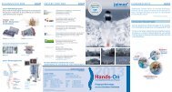
Developments in the Area of Endoscopic Spine ... - joimax GmbH
Developments in the Area of Endoscopic Spine ... - joimax GmbH
Developments in the Area of Endoscopic Spine ... - joimax GmbH

You also want an ePaper? Increase the reach of your titles
YUMPU automatically turns print PDFs into web optimized ePapers that Google loves.
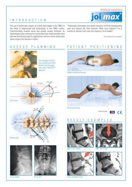
I N T R O D U C T I O N<br />
The use <strong>of</strong> endoscopic surgery on a daily basis began <strong>in</strong> <strong>the</strong> 1980’s <strong>in</strong><br />
<strong>the</strong> fields <strong>of</strong> laparoscopy and arthroscopy. In <strong>the</strong> 1990´s endoscopic/m<strong>in</strong>imally<br />
<strong>in</strong>vasive neuro and cardiac surgery followed. As<br />
technologies have cont<strong>in</strong>ued to evolve <strong>the</strong>y have made possible more<br />
delicate and disease specific applications, and as a result endoscopic<br />
sp<strong>in</strong>e surgery has become a reality.<br />
`<strong>Endoscopic</strong> techniques may speed recovery, m<strong>in</strong>imize postoperative<br />
pa<strong>in</strong> and improve <strong>the</strong> f<strong>in</strong>al outcome. What once required 3 to 6<br />
months to recover from now only requires 3 to 6 weeks!´<br />
The Cleveland Cl<strong>in</strong>ic Foundation<br />
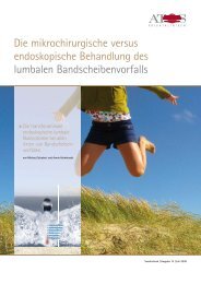
A C C E S S P L A N N I N G P A T I E N T P O S I T I O N I N G<br />
Axial view<br />
Dorsal view<br />
40°- 50°<br />
30°- 40°<br />
The triangular work<strong>in</strong>g<br />
zone accord<strong>in</strong>g to Parviz<br />
Kamb<strong>in</strong> and <strong>the</strong> target<br />
area <strong>of</strong> <strong>the</strong> THESSYS<br />
approach.<br />
10 -14 cm<br />
25°- 35°<br />
X-ray: lateral X-ray: A-P<br />
35°<br />
45° 30°<br />
Entry po<strong>in</strong>t<br />
10°- 40°<br />
25°<br />
Stable lateral position<strong>in</strong>g<br />
Prone position<strong>in</strong>g<br />
MRI: L5-S1 lateral<br />
L4-L5, bi-lateral view<br />
Patents pend<strong>in</strong>g<br />
R E S U L T E X A M P L E S<br />
MRI: L5-S1 axial
a report by<br />
Florian Maria Alfen, Beate Lauerbach and Wolfgang Ries<br />
Orthopaedic Surgeon, Private Practice, Würzburg<br />
Pioneers <strong>in</strong> <strong>Endoscopic</strong><br />
Sp<strong>in</strong>e Surgery<br />
M<strong>in</strong>imally <strong>in</strong>vasive sp<strong>in</strong>al surgery is emerg<strong>in</strong>g as an<br />
alternative, reliable method <strong>of</strong> treatment for a variety<br />
<strong>of</strong> sp<strong>in</strong>al disorders. The operative techniques be<strong>in</strong>g<br />
used for discectomy, retrieval <strong>of</strong> herniated disc<br />
fragments and stabilisation <strong>of</strong> unstable sp<strong>in</strong>al motion<br />
segments are be<strong>in</strong>g utilised more widely on a daily<br />
basis. 1 Mixter, Barr and Dandy are credited with <strong>the</strong><br />
diagnosis and treatment <strong>of</strong> herniated lumbar discs via<br />
lam<strong>in</strong>ectomy and <strong>the</strong> exposure <strong>of</strong> <strong>the</strong> sp<strong>in</strong>al canal. 2,3<br />
With<strong>in</strong> <strong>the</strong> last 40 years many <strong>in</strong>vestigators have<br />
attempted to f<strong>in</strong>d alternatives to lam<strong>in</strong>ectomy and<br />
discectomy, <strong>in</strong>clud<strong>in</strong>g evacuation <strong>of</strong> <strong>the</strong> nucleus via<br />
an anterior retro-peritoneal approach, 4 automated<br />
percutaneous nucleotomy, 5 suction-excision <strong>of</strong><br />
herniated lumbar discs, and chemonucleolysis and<br />
laser nuclear ablation. 6-13<br />
In <strong>the</strong> 1970s, Parviz Kamb<strong>in</strong> and Hijikata started to<br />
use specifically designed cannulas for perform<strong>in</strong>g a<br />
percutaneous dorso-lateral nucleotomy, with a<br />
reported satisfactory outcome <strong>of</strong> 75% <strong>in</strong> <strong>the</strong>ir<br />
patients. 14-16 The pr<strong>in</strong>ciple <strong>of</strong> mechanical nucleotomy<br />
was subsequently pursued by o<strong>the</strong>r <strong>in</strong>vestigators <strong>in</strong><br />
<strong>the</strong> 1980s. 17,18 Increased understand<strong>in</strong>g <strong>of</strong> <strong>the</strong><br />
endoscopic anatomy <strong>of</strong> <strong>the</strong> foram<strong>in</strong>al and extraforam<strong>in</strong>al<br />
region, 19,20 <strong>the</strong> description <strong>of</strong> radiographic<br />
landmarks <strong>of</strong> <strong>the</strong> work<strong>in</strong>g zone on <strong>the</strong> dorsolateral<br />
annulus by Parviz Kamb<strong>in</strong> comb<strong>in</strong>ed with <strong>the</strong><br />
availability <strong>of</strong> smaller calibre rod-lens fibre-optics<br />
have permitted fur<strong>the</strong>r lateralisation <strong>of</strong> <strong>the</strong> sk<strong>in</strong> entry<br />
po<strong>in</strong>ts. 15,21,22 Specifically <strong>the</strong> later approaches had been<br />
pursued by Anthony Yeung, 23 Mart<strong>in</strong> Knight, 13 Sang<br />
Ho Lee, 21 Thomas Hoogland and o<strong>the</strong>rs. 12,25-33 Over<br />
<strong>the</strong> last decade, all <strong>of</strong> <strong>the</strong>m have performed some<br />
thousands <strong>of</strong> endoscopic sp<strong>in</strong>al procedures us<strong>in</strong>g a<br />
very similar approach, but vary<strong>in</strong>g <strong>the</strong>ir specific<br />
methods and technologies.<br />
The THESSYS Concept<br />
For <strong>the</strong> removal <strong>of</strong> herniated <strong>in</strong>ter-vertebral disc<br />
material, THESSYS (Transforam<strong>in</strong>al <strong>Endoscopic</strong><br />
Sp<strong>in</strong>e System) utilises a special lateral, trans-foram<strong>in</strong>al<br />
Orthopaedic Surgery SPINE<br />
<strong>Developments</strong> <strong>in</strong> <strong>the</strong> <strong>Area</strong> <strong>of</strong> <strong>Endoscopic</strong> Sp<strong>in</strong>e Surgery<br />
endoscopic approach. This represents a less traumatic<br />
approach for <strong>the</strong> patient than <strong>the</strong> typically used dorsal<br />
approach. With <strong>the</strong> use <strong>of</strong> dorsal lam<strong>in</strong>ectomy<br />
procedures for <strong>the</strong> removal <strong>of</strong> <strong>in</strong>tra- and<br />
transforam<strong>in</strong>al disc fragments, extensive sacrific<strong>in</strong>g <strong>of</strong><br />
vital sp<strong>in</strong>al stability structures can be required <strong>in</strong> order<br />
to reach <strong>the</strong> target po<strong>in</strong>t, <strong>of</strong>ten lead<strong>in</strong>g to immediate<br />
sp<strong>in</strong>al fusion. In contrast, THESSYS allows access to<br />
every herniated disc fragment or protrusion except<br />
those located fully dorsally. The documented<br />
recurrence rate with <strong>the</strong> THESSYS method is very<br />
low. 11,25,30-34 With this method, sequestered disc<br />
material is completely removed directly through <strong>the</strong><br />
foramen, which is gradually widened <strong>in</strong> a step-wise<br />
fashion with specially designed reamers and<br />
accompany<strong>in</strong>g <strong>in</strong>strumentation. The patient can be<br />
placed <strong>in</strong> both <strong>the</strong> prone position and <strong>the</strong> lateral<br />
position. He/she is awake dur<strong>in</strong>g <strong>the</strong> entire<br />
operation, which is carried out under local<br />
anaes<strong>the</strong>sia. This allows communication with <strong>the</strong><br />
patient dur<strong>in</strong>g <strong>the</strong> entire procedure. The whole<br />
operation can be performed effectively <strong>in</strong> both a<br />
hospital and an out-patient surgery centre.<br />
Operat<strong>in</strong>g Technique<br />
Proper patient position<strong>in</strong>g and thorough plann<strong>in</strong>g<br />
<strong>of</strong> <strong>the</strong> approach to <strong>the</strong> herniated <strong>in</strong>tervertebral disc<br />
via <strong>the</strong> sk<strong>in</strong> entry po<strong>in</strong>t, as shown <strong>in</strong> <strong>the</strong> figures, is<br />
crucial <strong>in</strong> obta<strong>in</strong><strong>in</strong>g a good surgical outcome.<br />
Confirmations <strong>of</strong> <strong>the</strong> exact position <strong>of</strong> an annular<br />
tear protrusion and/or sequestered <strong>in</strong>tervertebral<br />
disc material can be obta<strong>in</strong>ed us<strong>in</strong>g <strong>the</strong> <strong>in</strong>traoperative<br />
discography.<br />
With <strong>the</strong> comb<strong>in</strong>ation <strong>of</strong> methodology and<br />
technology, <strong>the</strong> THESSYS total system makes access<br />
possible at all lumbar levels, <strong>in</strong>clud<strong>in</strong>g L5-S1. Any<br />
sequestered disc fragment or protrusions can be<br />
removed with <strong>the</strong> system immediately (see Figure 1).<br />
The specific approach for herniated <strong>in</strong>tervertebral<br />
discs us<strong>in</strong>g <strong>the</strong> method is through <strong>the</strong> <strong>in</strong>tervertebral<br />
foramen, which most <strong>of</strong> <strong>the</strong> time is very narrow due<br />
to <strong>the</strong> disease. The affected nerve roots exit cranially<br />
Florian Alfen is an orthopaedic<br />
surgeon <strong>in</strong> private practice <strong>in</strong><br />
Würzburg, Germany. He also<br />
provides <strong>Endoscopic</strong> Transforam<strong>in</strong>al<br />
Neucleotomy treatments at <strong>the</strong><br />
ARKADE Private Cl<strong>in</strong>ic,<br />
Niederschmalkalden, as well as at<br />
<strong>the</strong> HELIOS Private Cl<strong>in</strong>ic, Volkach,<br />
Germany. Previous to this he had<br />
been work<strong>in</strong>g at <strong>the</strong> ALPHA-Cl<strong>in</strong>ic<br />
Munich as an assistant medical<br />
director <strong>in</strong> <strong>the</strong> sp<strong>in</strong>e surgery<br />
section. A graduate <strong>of</strong> J-W-von<br />
Goe<strong>the</strong> University <strong>in</strong> 1993, Dr Alfen<br />
was a visit<strong>in</strong>g doctor at Al Shatti<br />
Hospital, Oman, <strong>in</strong> 2003, and at<br />
Armed Forces Hospital, Oman, <strong>in</strong><br />
2005. Dr Alfen is a member <strong>of</strong><br />
numerous societies and pr<strong>of</strong>essional<br />
bodies, <strong>in</strong>clud<strong>in</strong>g <strong>the</strong> Orthopaedic<br />
Surgery Society, German Society for<br />
Sp<strong>in</strong>e Surgery, German<br />
Orthopaedists Association, and<br />
International Society for Medical<br />
Streng<strong>the</strong>n<strong>in</strong>g Therapy.<br />
BOOK TITLE 2006 3
Orthopaedic Surgery SPINE<br />
Figure 1: MRI Pre-operatively and Three Months Post-operatively<br />
and are <strong>of</strong>ten encapsulated by fibrous tissue and/or<br />
bony structures. In order not to irritate any nerve<br />
close to <strong>the</strong> foramen and to ensure a safe access to <strong>the</strong><br />
sp<strong>in</strong>al canal, <strong>the</strong> caudal part (safety zone) <strong>of</strong> <strong>the</strong><br />
foramen is widened millimetre by millimetre with<br />
specially designed reamers.<br />
A stepwise, three-staged guide wire pr<strong>in</strong>ciple is<br />
used, <strong>in</strong>sert<strong>in</strong>g <strong>in</strong>to <strong>the</strong> foramen, under X-ray<br />
control from <strong>the</strong> THESSYS manual <strong>in</strong>strumentation<br />
tray, which consists <strong>of</strong> a variety <strong>of</strong> guid<strong>in</strong>g rods,<br />
guid<strong>in</strong>g tubes, work<strong>in</strong>g cannulas and <strong>the</strong> previously<br />
mentioned crown reamers. The foramen is gradually<br />
widened by ream<strong>in</strong>g bone material away. Because <strong>of</strong><br />
this procedure, safe access to <strong>the</strong> sp<strong>in</strong>al canal is<br />
enabled. Through this access channel and <strong>the</strong><br />
specifically developed sp<strong>in</strong>al foram<strong>in</strong>oscopes, which<br />
allow full endoscopic visualisation, <strong>the</strong> prolapsed<br />
disc material caus<strong>in</strong>g serious radicular symptoms can<br />
be removed us<strong>in</strong>g <strong>the</strong> specially developed forceps,<br />
graspers and punches.<br />
Indication<br />
The THESSYS operat<strong>in</strong>g method can be used <strong>in</strong><br />
any m<strong>in</strong>imally <strong>in</strong>vasive surgical procedure on a<br />
herniated <strong>in</strong>tervertebral disc. All sequestered disc<br />
material and protrusions <strong>of</strong> <strong>the</strong> lumbar sp<strong>in</strong>e<br />
(<strong>in</strong>clud<strong>in</strong>g level L5-S1,) can be removed with <strong>the</strong><br />
complete system through <strong>the</strong> lateral transforam<strong>in</strong>al<br />
approach under local anaes<strong>the</strong>sia.<br />
The use <strong>of</strong> <strong>the</strong> complete system is <strong>in</strong>dicated <strong>in</strong> any<br />
radicular syndrome caused by a herniated<br />
<strong>in</strong>tervertebral disc or major protrusion and longterm<br />
pa<strong>in</strong>, <strong>in</strong> which conservative <strong>the</strong>rapy failed to<br />
achieve sufficient improvement. A cauda equ<strong>in</strong>a<br />
syndrome represents an <strong>in</strong>dication for immediate<br />
<strong>in</strong>tervention. As with any herniated disc operation,<br />
a THESSYS operation also requires pre-operative<br />
magnetic resonance tomography (MRT) and<br />
regular X-ray imag<strong>in</strong>g.<br />
Statistics<br />
Published <strong>in</strong>ternational literature report a 75–85%<br />
success rate for percutaneous nucleotomies<br />
performed by experienced sp<strong>in</strong>e specialists. 15,35<br />
Herniated <strong>in</strong>tervertebral disc operations performed<br />
with <strong>the</strong> help <strong>of</strong> microscopic technologies, used <strong>in</strong><br />
most centres, demonstrate an average success rate<br />
<strong>of</strong> 87%. 19,35,36 In one-year and two-year follow-up<br />
studies, <strong>the</strong> application <strong>of</strong> <strong>the</strong> THESSYS method<br />
for <strong>the</strong> removal <strong>of</strong> sequestered <strong>in</strong>tervertebral disc<br />
material, by referenced THESSYS users, has<br />
achieved a success rate <strong>of</strong> more than 90%. 25,26,30-33,34<br />
Overall, <strong>the</strong> early recurrence rate is under five per<br />
cent. In patients with a recurrent herniation, <strong>the</strong><br />
success rate is more than 84%. 25,26,30,31<br />
Statistics – Private practice Florian<br />
Maria Alfen (MD)<br />
Evaluation<br />
• 4/2004–5/2005, N=189<br />
• Retrospective non-randomised cl<strong>in</strong>ical study<br />
• Data compiled for s<strong>in</strong>gle/multi-level endoscopic<br />
transforam<strong>in</strong>al nucleotomies (ETNs) from<br />
4/2004–5/2005<br />
• N=189 (48 female/141 male)<br />
• Average age 50 years (range 24–79 years)<br />
ETN – disc level (frequency <strong>of</strong> occurrence; N=189)<br />
ETN – complication rate: 5.5% (N=147, FU 6w)<br />
ETN – patient’s satisfaction on <strong>the</strong> visual analog scale<br />
(VAS)<br />
Conclusion<br />
In our op<strong>in</strong>ion, <strong>the</strong> ETN with THESSYS is a fully<br />
developed technique to remove lumbar sp<strong>in</strong>e disc<br />
herniations. The disadvantages are: a long learn<strong>in</strong>g<br />
curve, a two-dimensional view, <strong>the</strong> dist<strong>in</strong>ction <strong>of</strong><br />
tissue and <strong>in</strong>itial expense. The advantages are: local<br />
anaes<strong>the</strong>sia only, a reduced risk <strong>of</strong> <strong>in</strong>fection, reduced<br />
risk <strong>of</strong> <strong>in</strong>stability, less subsequent scars, open-door<br />
surgery and a shorter rehabilitation time. ■<br />
4 EUROPEAN MUSCULOSKELETAL REVIEW 2006
References<br />
<strong>Developments</strong> <strong>in</strong> <strong>the</strong> <strong>Area</strong> <strong>of</strong> <strong>Endoscopic</strong> Sp<strong>in</strong>e Surgery<br />
1. Darzi A, Mackay S, “Recent advances <strong>in</strong> m<strong>in</strong>imal access surgery”, BMJ (2002);324: pp. 31–34.<br />
2. Mixter W J, Barr J S, “Rupture <strong>of</strong> <strong>the</strong> <strong>in</strong>tervertebral disc with <strong>in</strong>volvement <strong>of</strong> <strong>the</strong> sp<strong>in</strong>al canal”, New England Journal<br />
Med (1934); 211: pp. 205–210.<br />
3. Dandy W E, “Loose cartilage from <strong>in</strong>ter-vertebral disc simulat<strong>in</strong>g tumour <strong>of</strong> <strong>the</strong> sp<strong>in</strong>al cord”, Arch Surg (1929);19: pp.<br />
660–672.<br />
4. Hult L, “Retroperitoneal disc fenestration <strong>in</strong> low back pa<strong>in</strong> and sciatica”, Acta Orthop Scand (1956);20: pp. 342–348.<br />
5. Cour<strong>the</strong>oux F, Theron J, “Automated percutaneous nucleotomy <strong>in</strong> <strong>the</strong> treatment <strong>of</strong> cervicobrachial neuralgia due to disc<br />
herniation”, J Neuroradiol (1992);19: pp. 211–216.<br />
6. Kamb<strong>in</strong> P, Sampson S, “Posterolateral percutaneous suction-excision <strong>of</strong> herniated lumbar <strong>in</strong>ter-vertebral discs; Report <strong>of</strong><br />
<strong>in</strong>terim results”, Cl<strong>in</strong> Orthop (1986); 207: pp. 37–43.<br />
7. Hoogland T, Scheckenbach C, “Low-Dose Chemonucleolysis Comb<strong>in</strong>ed with Percutaneous Nucleotomy <strong>in</strong> Herniated<br />
Cervical Discs”, Journal <strong>of</strong> Sp<strong>in</strong>al Disorders (1995); 8(3): pp. 228–232.<br />
8. Schubert M, Hoogland T, “The endoscopic transforam<strong>in</strong>al nucleotomy <strong>in</strong> comb<strong>in</strong>ation <strong>of</strong> a low-dose chemonucleolysis:<br />
Results <strong>of</strong> a prospective study with 2-year follow-up”, Program Abstract at <strong>the</strong> 18th Annual Meet<strong>in</strong>g <strong>of</strong> <strong>the</strong> International<br />
Intradiscal Therapy Society (2005); San Diego.<br />
9. Chiu J, Clifford T, “Micro-decompressive percutaneous discectomy: Sp<strong>in</strong>al discectomy with new laser <strong>the</strong>rmodiscoplasty for<br />
non extruded herniated nucleus pulposus”, Surg Technol Int (1999);3: pp. 343–351.<br />
10. Choy D S J, Case R E, Field<strong>in</strong>g W, “Percutaneous laser nucleolysis <strong>of</strong> lumbar discs”, New England Journal Med<br />
(1987);317: pp. 770–771.<br />
11. Gastambide D, “<strong>Endoscopic</strong> posterolateral foram<strong>in</strong>otomy with <strong>in</strong>struments or laser for lateral lumbar stenosis”, Program<br />
Abstract at <strong>the</strong> 17th Annual Meet<strong>in</strong>g <strong>of</strong> <strong>the</strong> International Intra-discal Therapy Society (2004) Munich.<br />
12. Hell<strong>in</strong>ger J, “Technical aspects <strong>of</strong> <strong>the</strong> percutaneous cervical and lumbar laser-disc-decompression and laser-nucleotomy”,<br />
Neurol Res (1999);21: pp. 99–102.<br />
13. Knight M et al (eds), “<strong>Endoscopic</strong> Laser Foramn<strong>in</strong>oplasty. A two year follow-up <strong>of</strong> a prospective study on 200 consecutive<br />
patients”, Lumbar Sp<strong>in</strong>al Stenosis, Lipp<strong>in</strong>cott Williams and Wilk<strong>in</strong>s, Ed. Gunzberg and Spalski (1999): pp.<br />
244–254.<br />
14. Kamb<strong>in</strong> P, “Arthroscopic microdiscectomy: lumbar and thoracic”, Sp<strong>in</strong>e Care St. Louis, Mosby White A H, Sch<strong>of</strong>fermann<br />
J A (eds)(1955);2: pp. 1002–1016.<br />
15. Kamb<strong>in</strong> P, Gellman H, “Percutaneous lateral discectomy <strong>of</strong> lumbar sp<strong>in</strong>e, a prelim<strong>in</strong>ary report”, Cl<strong>in</strong> Orthop<br />
(1983);174: pp. 127–132.<br />
16. Hijikata S, Yamagishi M, Nakayama T, et al, “Percutaneous nucleotomy. A new treatment method for lumbar disc<br />
herniation”, J Toden Hosp (1975);5: pp. 5–13.<br />
17. Onik G, Helms C, G<strong>in</strong>sburg L, et al, “Percutaneous lumbar discectomy us<strong>in</strong>g a new aspiration probe”, AJR (1985);144:<br />
pp. 1137–1140.<br />
18. Schreiber A, Suezawa Y, Leu H J, “Does percutaneous nucleotomy with discoscopy replace conventional discectomy? Eight<br />
years <strong>of</strong> experience and results <strong>in</strong> treatment <strong>of</strong> herniated lumbar disc”, Cl<strong>in</strong> Orthop (1989);238: pp. 35–42.<br />
19. Hermant<strong>in</strong> F, Peters T, Quartararo L, “A prospective, randomized study compar<strong>in</strong>g <strong>the</strong> results <strong>of</strong> open discectomy with<br />
those <strong>of</strong> video-assisted arthroscopic microdiscectomy” J Bone Jo<strong>in</strong>t Surg (A)(1999);81: pp. 958–965.<br />
20. Kamb<strong>in</strong> P, “Arthroscopic techniques for sp<strong>in</strong>al surgery”, McG<strong>in</strong>ty J B, Caspari R B, Jackson R W, et al., (eds) Operative<br />
Arthroscopy Philadelphia, Lipp<strong>in</strong>cott-Raven (1996);2: pp.1207–1235<br />
21. Kamb<strong>in</strong> P, “Posterolateral percutaneous lumbar discectomy and decompression” Arthroscopic Microdiscectomy,<br />
M<strong>in</strong>imal Intervention <strong>in</strong> Sp<strong>in</strong>al Surgery, Kamb<strong>in</strong> P,(ed) Baltimore,Urban & Schwarzenberg(1991): pp. 67–121.<br />
22. Kamb<strong>in</strong> P, Casey K, O’Brien E, et al., “Trans-foram<strong>in</strong>al arthroscopic decompression <strong>of</strong> lateral recess stenosis.”, J<br />
Neurosurg (1996); 84: pp. 462–467.<br />
23. Yeung A, Tsou P, “Posterolateral <strong>Endoscopic</strong> Excision for Lumbar Disc Herniation”, Sp<strong>in</strong>e (2001); 27(7): pp.<br />
722–731.<br />
24. Ahn Y, Lee SH, Park W M, et al., “Posterolateral percutaneous endoscopic lumbar foram<strong>in</strong>otomy for L5-S1 foram<strong>in</strong>al or<br />
lateral exit zone stenosis”, J Neurosurg (2003);99(3): pp. 320–323.<br />
25. Hoogland T, “Transforam<strong>in</strong>al endoscopic discectomy with foram<strong>in</strong>oplasty for lumbar disc herniation”, Surg Techn <strong>in</strong><br />
Orthopaedics and Traumatology (2003);40; pp. 55–120.<br />
26. Hoogland T, “Percutaneous nucleotomy <strong>of</strong> thoracic discs”, Program Abstract at <strong>the</strong> 17th Annual Meet<strong>in</strong>g <strong>of</strong> <strong>the</strong><br />
International Intra-discal Therapy Society (2004); Munich.<br />
27. Hoogland T, Godon T, Wagner C, “Die endoskopische transforam<strong>in</strong>ale Diskoplastik bei überwiegend lumbalen<br />
Rückenschmerzen”, Program Abstract at <strong>the</strong> 52. Jahrestagung der Vere<strong>in</strong>igung Süddeutscher Orthoäden(2004);Baden-Baden.<br />
28. Alfen F M , “<strong>Endoscopic</strong> Transforam<strong>in</strong>al Nocleotomy (ETN)”, Program Abstract at <strong>the</strong> 3rd Dubai Sp<strong>in</strong>e Conference<br />
(2005); Dubai.<br />
EUROPEAN MUSCULOSKELETAL REVIEW 2006 5
Orthopaedic Surgery SPINE<br />
29. Gibson A, “Surgery for lumbar disc prolapse: Evidence from <strong>the</strong> 2004 Cochrane review”, Program Abstract at <strong>the</strong> 17th<br />
Annual Meet<strong>in</strong>g <strong>of</strong> <strong>the</strong> International Intradiscal Therapy Society (2004) Munich.<br />
30. Iprenburg M, “Percutaneous Transforam<strong>in</strong>al <strong>Endoscopic</strong> Discectomy; <strong>the</strong> learn<strong>in</strong>g curve to achieve a more than 90% success<br />
rate”, Program Abstract at <strong>the</strong> 19th Annual Meet<strong>in</strong>g <strong>of</strong> <strong>the</strong> International Intradiscal Therapy Society (2006);Phoenix.<br />
31. Krzok G, “Early results after posterolateral endoscopic discectomy with <strong>the</strong>rmal annuloplasty”, Program Abstract at <strong>the</strong> 17th<br />
Annual Meet<strong>in</strong>g <strong>of</strong> <strong>the</strong> International Intradiscal Therapy Society (2004) Munich.<br />
32. Lev<strong>in</strong>kopf M, Caspi I et al., “Posterolateral <strong>Endoscopic</strong> Discectomy”, Program Abstract at <strong>the</strong> 18th Annual Meet<strong>in</strong>g <strong>of</strong> <strong>the</strong><br />
International Intra-discal Therapy Society (2005);San Diego.<br />
33. Schubert M, Hoogland T, “<strong>Endoscopic</strong> Transforam<strong>in</strong>al Nucleotomy with Foram<strong>in</strong>oplasty for Lumbar Disc Herniation”,<br />
Oper Orthop Traumatol (2005);17: pp. 641–661.<br />
34. Alfen FM, Lauerbach B, “Technique and Results <strong>of</strong> <strong>Endoscopic</strong> Transforam<strong>in</strong>al Nucleotomy (ETN)”, Program Abstract<br />
at <strong>the</strong> 13th Congress <strong>of</strong> <strong>the</strong> International Musculoskeletal Laser Society (2006):Barcelona.<br />
35. Leu H J, Schreiber A, “ Percutaneous fusion <strong>of</strong> <strong>the</strong> lumbar sp<strong>in</strong>e, a promis<strong>in</strong>g technique”, Sp<strong>in</strong>e State Art Rev (1992);6:<br />
pp. 593–604.<br />
36. Lühmann D, Burkhardt-Hammer T, Borowski C, et al., “M<strong>in</strong>imally <strong>in</strong>vasive surgical procedures for <strong>the</strong> treatment <strong>of</strong><br />
lumbar disc herniation” , DIMDI, German Agency for Health Technology Assessment at <strong>the</strong> German Institute <strong>of</strong> Medical<br />
Documentation and Information (2005): DAHTA@DIMDI.<br />
37. Mayer H, Brock M, “Percutaneous endoscopic discectomy: Surgical technique and prelim<strong>in</strong>ary results compared to<br />
microsurgical discectomy”, J Neurosurg (1993);78: pp. 216–225.<br />
38. Casey K F, Chang M K, O’Brien E D, et al., “Arthroscopic microdiscectomy, comparison <strong>of</strong> preoperative and postoperative<br />
imag<strong>in</strong>g studies”, Arthroscopy (1997);13: pp. 438–445.<br />
39. Hochschuler S H, “Posterior lateral arthroscopic microdiscectomy”, Sem<strong>in</strong> Orthop (1991);6: pp. 113–114.<br />
40. Kamb<strong>in</strong> P, O’Brien E, Zhou L, “Arthroscopic microdiscectomy and fragmentectomy”, Cl<strong>in</strong> Orthop (1998);347: pp.<br />
150–167.<br />
41. LaRocca H, McNab I, “The lam<strong>in</strong>ectomy membrane. Studies <strong>in</strong> its evolution, characteristics, effects and prophylaxis <strong>in</strong><br />
dogs”, J Bone Surg (Br)(1974); 56: pp. 545–550.<br />
42. Lowery GL, Bernauer D, Casper D, et al., “<strong>Endoscopic</strong> posterolateral lumbar fusion and pedicle screw fixation: Early<br />
results from a multi-center prospective study”, Program Abstract at <strong>the</strong> World Sp<strong>in</strong>e II Meet<strong>in</strong>g(2003):Chicago.<br />
43. Ma<strong>the</strong>ws H, Long B, “M<strong>in</strong>imally Invasive Techniques for <strong>the</strong> Treatment <strong>of</strong> Intervertebral Disc Herniation”, Journal <strong>of</strong><br />
<strong>the</strong> American Academy <strong>of</strong> Orthopaedic Surgeons (2002);10(2): pp. 80–85.<br />
44. McAfee P C , “Thoracolumbar sp<strong>in</strong>al corpectomy”, Atlas <strong>of</strong> <strong>Endoscopic</strong> Sp<strong>in</strong>e Surgery, St. Louis, Regan J J, McAfee<br />
P C, Mark M J (eds) Quality Medical Publish<strong>in</strong>g (1995): pp. 189–197.<br />
45. Mochida J, Nishimura K, Okuma M, et al., “Percutaneous Nucleotomy <strong>in</strong> Elite Athletes”, In: Journal <strong>of</strong> Sp<strong>in</strong>al<br />
Disorders (2001);14(2): pp. 159–164.<br />
46. Osman S, Nibu K, Panjabi M, et al., “Transforam<strong>in</strong>al and Posterior Decompressions <strong>of</strong> <strong>the</strong> Lumbar Sp<strong>in</strong>e” Sp<strong>in</strong>e<br />
(1997);22: pp. 1690–1693.<br />
47. Parke W W, “Cl<strong>in</strong>ical anatomy <strong>of</strong> <strong>the</strong> lower lumbar sp<strong>in</strong>e”, Arthroscopic Microdiscectomy, M<strong>in</strong>imal Intervention <strong>in</strong> Sp<strong>in</strong>al<br />
Surgery, Baltimore, Kamb<strong>in</strong> P (ed) Urban & Schwarzenberg (1991): pp. 11–29.<br />
48. Schaffer J L, Kamb<strong>in</strong> P, “Percutaneous posterolateral lumbar discectomy and decompression with a 6.9 millimeter cannula.<br />
Analysis <strong>of</strong> operative failures and complications”, J Bone Jo<strong>in</strong>t Surg (Am) (1991);73: pp. 822–831.<br />
49. Scheckenbach C, Hoogland T, “Endoskopische transforam<strong>in</strong>ale Diskektomie (EDT) – Ergebnisse nach 2 Jahren”,<br />
Orthopädische Praxis (1999);35(2): pp. 104–105.<br />
6 EUROPEAN MUSCULOSKELETAL REVIEW 2006
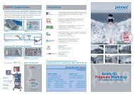
THESSYS <br />
Transforam<strong>in</strong>al <strong>Endoscopic</strong> Sp<strong>in</strong>e System<br />
The THESSYS Procedure Kit System<br />
THESSYS TM Disposable Access Kits<br />
THESSYS TM Instrument Set<br />
THESSYS TM Foram<strong>in</strong>oscopes<br />
Surgi-Max TM TriggerFlex TM RF-Probes<br />
Pre-surgical: L3-L4,<br />
lateral<br />
Colour coded reamers <strong>in</strong><br />
different specifications<br />
Patented LOPS<br />
forceps system<br />
Herniated fragment Freed nerve root<br />
Colour coded<br />
<strong>in</strong>struments<br />
Post-surgical: L3-L4,<br />
lateral<br />
> Discography<br />
> Foram<strong>in</strong>oplasty<br />
> Fragmentectomy<br />
> Deherniation<br />
> Nucleotomy<br />
> Annuloplasty<br />
Transforam<strong>in</strong>al <strong>Endoscopic</strong> Sp<strong>in</strong>e System<br />
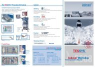
<strong>joimax</strong> provides <strong>the</strong> latest digital technologies for endoscopic<br />
surgery, specifically for <strong>the</strong> performance <strong>of</strong> <strong>the</strong> <strong>in</strong>novat<strong>in</strong>g<br />
(jo<strong>in</strong>ed) m<strong>in</strong>imal access THESSYSTM sp<strong>in</strong>e procedure.<br />
1<br />
1<br />
2<br />
3<br />
4<br />
5<br />
6<br />
Fully <strong>in</strong>tegrated image and<br />
video data record<strong>in</strong>g with<br />
multi-task command<strong>in</strong>g<br />
S<strong>in</strong>gle Cable Technology for Endoscopy<br />
High resolution images for any signal source<br />
For Arthroscopy and m<strong>in</strong>imally <strong>in</strong>vasive Sp<strong>in</strong>e Surgery<br />
A new era <strong>of</strong> Radiowave Technology <strong>of</strong>fer<strong>in</strong>g<br />
maximum surgical precision, control, versatility<br />
and safety<br />
ICPP xxx<br />
<strong>joimax</strong> Photo Pr<strong>in</strong>ter<br />
<strong>joimax</strong>, Inc.<br />
718 University Avenue, Suite 116<br />
Los Gatos, CA 95032 USA<br />
PHONE +1 408 399 40 20<br />
FAX +1 408 399 40 29<br />
MAIL <strong>in</strong>fo@<strong>joimax</strong>.com<br />
NET www.<strong>joimax</strong>.com<br />
jo<strong>in</strong>ed m<strong>in</strong>imal acess technologies<br />
<strong>joimax</strong> <strong>GmbH</strong><br />
RaumFabrik 33a, Amalienbadstraße<br />
76227 Karlsruhe - Germany<br />
PHONE +49 (0) 721 255 14-0<br />
FAX +49 (0) 721 255 14-920<br />
MAIL <strong>in</strong>fo@<strong>joimax</strong>.com<br />
NET www.<strong>joimax</strong>.com<br />
Patents pend<strong>in</strong>g<br />
3<br />
c<br />
2<br />
4<br />
5<br />
6
www.touchbrief<strong>in</strong>gs.com<br />
Card<strong>in</strong>al Tower<br />
12 Farr<strong>in</strong>gdon Road<br />
London EC1M 3NN<br />
EDITORIAL<br />
Tel: +44 (0) 20 7526 2384<br />
Fax: +44 (0) 20 7452 5050<br />
SALES<br />
Tel: +44 (0) 20 7452 5164<br />
Fax: +44 (0) 20 7452 5606<br />
E-mail: <strong>in</strong>fo@touchbrief<strong>in</strong>gs.com<br />
www.touchbrief<strong>in</strong>gs.com
European<br />
Musculoskeletal<br />
Review 2006<br />
EXTRACT<br />
<strong>Developments</strong> <strong>in</strong> <strong>the</strong> <strong>Area</strong> <strong>of</strong><br />
<strong>Endoscopic</strong> Sp<strong>in</strong>e Surgery<br />
a report by<br />
Florian Maria Alfen, Beate Lauerbach<br />
and Wolfgang Ries<br />
Orthopaedic Surgeon, Private Practice, Würzburg<br />
www.touchbrief<strong>in</strong>gs.com