Endoscopic sinus surgery for the odontogenic maxillary cysts ...
Endoscopic sinus surgery for the odontogenic maxillary cysts ...
Endoscopic sinus surgery for the odontogenic maxillary cysts ...

You also want an ePaper? Increase the reach of your titles
YUMPU automatically turns print PDFs into web optimized ePapers that Google loves.
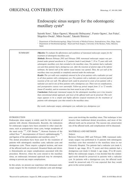
ORIGINAL CONTRIBUTION Rhinology, 47, 305-309, 2009<br />
SUMMARY<br />
INTRODUCTION<br />
<strong>Endoscopic</strong> <strong>sinus</strong> <strong>surgery</strong> is widely used <strong>for</strong> <strong>the</strong> treatment of<br />
patients with chronic rhino<strong>sinus</strong>itis. Recently, <strong>the</strong> indications<br />
<strong>for</strong> endoscopic <strong>surgery</strong> have been extended to o<strong>the</strong>r nasal diseases,<br />
such as postoperative <strong>sinus</strong> <strong>cysts</strong>, benign neoplasms of<br />
<strong>the</strong> nasal cavity (1,2) , CSF fistulas (3) , blowout fractures of <strong>the</strong><br />
orbital floor (4) , decompression of Grave’s ophthalmopathy (5) ,<br />
and ligation of <strong>the</strong> sphenopalatine and <strong>maxillary</strong> artery (6) .<br />
The dental procedures have been used <strong>for</strong> <strong>the</strong> treatment of<br />
patients with <strong>odontogenic</strong> <strong>maxillary</strong> <strong>cysts</strong>; radicular <strong>cysts</strong> and<br />
dentigerous <strong>cysts</strong>. These require a gingival incision, and most<br />
of <strong>the</strong> affected teeth are extracted. Oroantral fistula and chronic<br />
rhino<strong>sinus</strong>itis are major complications associated with this<br />
procedure. However, when <strong>the</strong> <strong>cysts</strong> extend to <strong>the</strong> <strong>maxillary</strong><br />
<strong>sinus</strong>, an endoscopic intranasal approach may be attempted,<br />
aiming to prevent any major complications.<br />
In this article, we present our experience with intranasal endoscopic<br />
<strong>surgery</strong> <strong>for</strong> <strong>the</strong> treatment of radicular <strong>cysts</strong> and dentige-<br />
<strong>Endoscopic</strong> <strong>sinus</strong> <strong>surgery</strong> <strong>for</strong> <strong>the</strong> <strong>odontogenic</strong><br />
<strong>maxillary</strong> <strong>cysts</strong>*<br />
Satoshi Seno 1 , Takao Ogawa1, Masayuki Shibayama 1 , Fumio Ogawa 1 , Jun Fukui 1 ,<br />
Shigehiro Owaki 1 , Mikio Suzuki 2 , Takeshi Shimizu 1<br />
1<br />
Department of Otorhinolaryngology, Shiga University of Medical Science, Setatsukinowa, Otsu, Shiga, Japan<br />
2<br />
Department of Otorhinolaryngology - Head and Neck Surgery, University of <strong>the</strong> Ryukyu, Naha, Okinawa,<br />
Japan<br />
Objective: To evaluate <strong>the</strong> effectiveness and usefulness of transnasal endoscopic <strong>surgery</strong> <strong>for</strong> <strong>the</strong><br />
treatment of <strong>odontogenic</strong> <strong>maxillary</strong> <strong>cysts</strong>.<br />
Methods: Between February 2003 and February 2008, transnasal endoscopic <strong>surgery</strong> was per<strong>for</strong>med<br />
under general anes<strong>the</strong>sia in 13 patients (male 6 and female 7, 19 to 75 years old) with<br />
<strong>odontogenic</strong> <strong>maxillary</strong> <strong>cysts</strong> that extended to <strong>the</strong> <strong>maxillary</strong> <strong>sinus</strong>. Ten patients had a radicular<br />
cyst and three patients had a dentigerous cyst. After <strong>the</strong> resection of anterior edge of <strong>the</strong> inferior<br />
turbinate, <strong>the</strong> lateral wall of <strong>the</strong> inferior nasal meatus was opened. Then, <strong>the</strong> cyst wall of <strong>the</strong><br />
<strong>maxillary</strong> <strong>sinus</strong> was partially or completely removed under <strong>the</strong> endoscope.<br />
Results: The cyst walls were completely removed in five of ten patients with a radicular cyst and<br />
in all three patients with a dentigerous cyst. Five patients with a radicular cyst received partial<br />
resection of <strong>the</strong> cyst wall. The affected teeth could be preserved in seven of ten patients with a<br />
radicular cyst and in one of three patients with a dentigerous cyst. There were no complications,<br />
and postoperative courses were uneventful. Follow-up period ranged from 11 to 72 months<br />
(mean 42 months), and no recurrence has been noted in any of <strong>the</strong> cases.<br />
Conclusions: <strong>Endoscopic</strong> transnasal <strong>surgery</strong> <strong>for</strong> <strong>the</strong> <strong>odontogenic</strong> <strong>maxillary</strong> cyst is less invasive<br />
than conventional dental approach, and most of <strong>the</strong> affected teeth can be preserved. This technique<br />
appears to be a simple and highly effective surgical treatment <strong>for</strong> <strong>the</strong> treatment of<br />
patients with <strong>odontogenic</strong> <strong>cysts</strong> that extend to <strong>the</strong> <strong>maxillary</strong> <strong>sinus</strong>.<br />
Key words: endoscopic <strong>surgery</strong>, <strong>odontogenic</strong> cyst, radicular cyst, dentigerous cyst<br />
nous <strong>cysts</strong> involving <strong>the</strong> <strong>maxillary</strong> <strong>sinus</strong>. This technique is less<br />
invasive than traditional dental procedures, and most of <strong>the</strong><br />
affected teeth can be preserved. The success rate, preservation<br />
rate of <strong>the</strong> affected teeth, and complications will be discussed.<br />
MATERIALS AND METHODS<br />
Subjects<br />
Between February 2003 and February 2008, transnasal endoscopic<br />
<strong>surgery</strong> was per<strong>for</strong>med in 13 patients with <strong>odontogenic</strong><br />
<strong>maxillary</strong> <strong>cysts</strong> that extended to <strong>the</strong> <strong>maxillary</strong> <strong>sinus</strong> in our<br />
University Hospital. Ten patients had a radicular cyst (male 4,<br />
female 6; age range, 20 to 75 years) and three patients had a<br />
dentigerous cyst (male 2, female 1; age range, 19 to 60 years).<br />
Be<strong>for</strong>e <strong>the</strong> endoscopic <strong>surgery</strong>, it was decided that <strong>the</strong> undamaged<br />
teeth should be preserved in patients with a radicular<br />
cyst. In patients with a dentigerous cyst, <strong>the</strong> affected teeth<br />
would be preserved only if it was expected that <strong>the</strong>y would<br />
grow in <strong>the</strong> correct position.<br />
*Received <strong>for</strong> publication: August 22, 2008; accepted: February 2, 2009 DOI: 10.4193/Rhin 08.162
306 Seno et al.<br />
Operation<br />
All operations were per<strong>for</strong>med under general anes<strong>the</strong>sia with<br />
controlled hypotension. The following endoscopic procedure<br />
was per<strong>for</strong>med with a 4-mm rigid endoscope (0° and 70°). The<br />
anterior edge of <strong>the</strong> inferior turbinate was resected to visualize<br />
<strong>the</strong> lateral wall of <strong>the</strong> inferior meatus. A small mucoperiosteal<br />
flap from <strong>the</strong> lateral wall was made, and <strong>the</strong> bony wall of <strong>the</strong><br />
inferior meatus was opened. The cyst wall was observed in <strong>the</strong><br />
<strong>maxillary</strong> <strong>sinus</strong>, and it was partially or completely removed<br />
endoscopically from <strong>the</strong> <strong>maxillary</strong> <strong>sinus</strong> using <strong>for</strong>ceps or<br />
curettes. After removing <strong>the</strong> cyst wall, <strong>the</strong> mucoperiosteal flap<br />
was layered on <strong>the</strong> floor of <strong>maxillary</strong> <strong>sinus</strong>.<br />
RESULTS<br />
Table 1 summarizes <strong>the</strong> patients and outcomes of <strong>the</strong> endoscopic<br />
<strong>surgery</strong> <strong>for</strong> <strong>the</strong> treatment of <strong>odontogenic</strong> <strong>maxillary</strong><br />
<strong>cysts</strong>. The cyst walls were completely removed in five of ten<br />
patients with a radicular cyst and in all 3 patients with a<br />
dentigerous cyst. Five patients with a radicular cyst underwent<br />
partial resection of <strong>the</strong> cyst wall. The affected teeth could be<br />
preserved in seven of ten patients with a radicular cyst, and in<br />
one of three patients with a dentigerous cyst. The postoperative<br />
courses were uneventful. There were no complications<br />
such as oroantral fistula and chronic rhino<strong>sinus</strong>itis, following<br />
<strong>the</strong> <strong>surgery</strong> in this series. The follow-up period ranged from 11<br />
to 72 months (mean 42 months), and no recurrence has been<br />
noted in any of <strong>the</strong> cases.<br />
CASE REPORT (Number 10)<br />
A 48-year-old woman was referred to <strong>the</strong> Department of<br />
Otorhinolaryngology, Shiga University hospital, complaining of<br />
right cheek pain and upper gingival swelling of three months’<br />
duration. Computed tomography (CT) showed a large cystic<br />
mass with a thin bony layer that extended into <strong>the</strong> right <strong>maxillary</strong><br />
<strong>sinus</strong> (Figure 1). Dental examination revealed a radicular<br />
cyst arising from <strong>the</strong> right upper second molar. The affected<br />
tooth received <strong>the</strong> dental treatment prior to <strong>the</strong> endoscopic<br />
<strong>surgery</strong>. After <strong>the</strong> fenestration of <strong>the</strong> lateral wall of inferior<br />
Table 1. Patients and outcome of <strong>the</strong> endoscopic transnasal <strong>surgery</strong> <strong>for</strong><br />
<strong>odontogenic</strong> <strong>maxillary</strong> <strong>cysts</strong>. The post operative courses were<br />
uneventful and <strong>the</strong>re were no complications during or after <strong>the</strong> <strong>surgery</strong><br />
in this series. No recurrence has been noted after 11 to 72 months<br />
follow-up.<br />
No. Type of Age Cyst wall Affected Follow Recurcyst<br />
(years) removal tooth -up rence<br />
/Gender (months) Duration<br />
1 Radicular 55/Male Complete Preserved 72 No<br />
2 Radicular 61/Male Partial Extracted 67 No<br />
3 Radicular 38/Male Complete Extracted 57 No<br />
4 Dentigerous 27/Male Complete Extracted 56 No<br />
5 Dentigerous 19/Male Complete Extracted 54 No<br />
6 Radicular 75/Female Complete Preserved 47 No<br />
7 Dentigerous 60/Female Complete Preserved 37 No<br />
8 Radicular 42/Male Partial Preserved 35 No<br />
9 Radicular 20/Female Partial Preserved 34 No<br />
10 Radicular 48/Female Complete Preserved 34 No<br />
11 Radicular 44/Female Complete Preserved 30 No<br />
12 Radicular 28/Female Partial Extracted 15 No<br />
13 Radicular 63/Female Partial Preserved 11 No<br />
meatus, <strong>the</strong> cyst wall could be observed in <strong>the</strong> right <strong>maxillary</strong><br />
<strong>sinus</strong> (Figure 2). On opening of <strong>the</strong> radicular cyst, a purulent<br />
discharge was noted (Figure 3), and <strong>the</strong> cyst wall was completely<br />
removed (Figure 4). The affected tooth was preserved,<br />
and a gingival incision was not needed. The patient’s postoperative<br />
course was uneventful. The floor of <strong>the</strong> <strong>maxillary</strong> <strong>sinus</strong><br />
was well epi<strong>the</strong>lized at 3 months after <strong>the</strong> <strong>surgery</strong> (Figure 5).<br />
The complete removal of <strong>the</strong> radicular cyst was confirmed by<br />
<strong>the</strong> postoperative CT at 6 months after <strong>the</strong> <strong>surgery</strong> (Figure 6).<br />
No recurrence has been noted at more than 34 months of follow-up.<br />
DISCUSSION<br />
Odontogenic <strong>cysts</strong> are lesions that rise in <strong>the</strong> maxilla and/or<br />
mandible, originating from epi<strong>the</strong>lial remains associated with<br />
odontogenesis. The most frequent type is a radicular cyst<br />
(65%), followed by dentigerous <strong>cysts</strong> (24%) and <strong>odontogenic</strong><br />
a b<br />
Figure 1. Axial (a) and coronal (b) CT scan showing a large cystic mass with a thin bony layer that extends into <strong>the</strong> right <strong>maxillary</strong> <strong>sinus</strong> (arrows).
<strong>Endoscopic</strong> treatment of <strong>odontogenic</strong> <strong>cysts</strong> 307<br />
Figure 2. <strong>Endoscopic</strong> photograph showing <strong>the</strong> radicular cyst in <strong>the</strong> right<br />
<strong>maxillary</strong> <strong>sinus</strong>. After <strong>the</strong> fenestration of <strong>the</strong> lateral wall of inferior<br />
meatus, <strong>the</strong> cyst wall was observed in <strong>the</strong> <strong>maxillary</strong> <strong>sinus</strong> (arrows).<br />
Figure 4. <strong>Endoscopic</strong> photograph showing <strong>the</strong> right <strong>maxillary</strong> <strong>sinus</strong><br />
after <strong>the</strong> complete resection of <strong>the</strong> cyst wall of radicular cyst (arrows).<br />
kerato<strong>cysts</strong> (5%) (7) . The majority of <strong>the</strong>se <strong>cysts</strong> have been<br />
treated by dentists using surgical approaches such as enucleation,<br />
curettage, marsupialization, and tooth extraction (8) .<br />
However, most of <strong>the</strong> affected teeth were extracted along with<br />
<strong>the</strong> cyst walls. The complications, such as oroantral fistulas and<br />
chronic rhino<strong>sinus</strong>itis, may be caused by dental extractions in<br />
<strong>odontogenic</strong> <strong>cysts</strong> which involve <strong>the</strong> <strong>maxillary</strong> <strong>sinus</strong>.<br />
In <strong>the</strong> last decade, endoscopic <strong>sinus</strong> <strong>surgery</strong> has been widely<br />
used, and <strong>the</strong> indications <strong>for</strong> endoscopic procedures were<br />
extended to many nasal diseases. For <strong>the</strong> treatment of patients<br />
with <strong>odontogenic</strong> <strong>maxillary</strong> <strong>cysts</strong>, endoscopic treatment has<br />
been used in addition to <strong>the</strong> dental procedure. Suarez-<br />
Conqueiro et al. (9) reported <strong>the</strong> endoscopically assisted dental<br />
approach <strong>for</strong> <strong>the</strong> removal of an ectopic lower third molar tooth<br />
associated with a dentigerous cyst. Costa et al. (10) per<strong>for</strong>med<br />
Figure 3. <strong>Endoscopic</strong> photograph showing <strong>the</strong> radicular cyst in <strong>the</strong><br />
right <strong>maxillary</strong> <strong>sinus</strong>. At <strong>the</strong> opening of radicular cyst, a purulent<br />
discharge was noted (arrow).<br />
<strong>the</strong> endoscopic <strong>sinus</strong> <strong>surgery</strong> <strong>for</strong> <strong>the</strong> treatment of chronic <strong>maxillary</strong><br />
<strong>sinus</strong>itis of dental origin after <strong>the</strong> dental treatment of<br />
<strong>odontogenic</strong> lesions. Cedin et al. (8) first removed <strong>the</strong> cyst walls<br />
from <strong>the</strong> <strong>maxillary</strong> <strong>sinus</strong> using small clamps with an endoscope<br />
through a 10 mm circumferential hole of <strong>the</strong> canine fossa.<br />
Recently, transnasal removal of large dentigerous <strong>cysts</strong> of <strong>the</strong><br />
<strong>maxillary</strong> <strong>sinus</strong> has been also reported using <strong>the</strong> endoscopic<br />
approach through <strong>the</strong> middle meatus (11,12) .<br />
In <strong>the</strong> present study, a transnasal endoscopic approach was<br />
used to remove <strong>the</strong> cyst walls of odonotogenic <strong>cysts</strong> through<br />
<strong>the</strong> <strong>maxillary</strong> <strong>sinus</strong> and <strong>the</strong> opening of inferior meatus. This is<br />
<strong>the</strong> first report to remove <strong>the</strong> cyst walls of <strong>the</strong> radicular cyst<br />
with no incision of oral mucosa. This technique can be applied<br />
in cases with <strong>odontogenic</strong> <strong>maxillary</strong> <strong>cysts</strong>. It is less invasive<br />
than conventional dental procedures, and it prevents oroantral<br />
fistula <strong>for</strong>mation and chronic <strong>maxillary</strong> <strong>sinus</strong>itis. There were<br />
no complications during and after <strong>the</strong> <strong>surgery</strong> in this series.<br />
We removed <strong>the</strong> cyst walls completely from <strong>the</strong> <strong>maxillary</strong><br />
<strong>sinus</strong> in five of ten patients with a radicular cyst. Ano<strong>the</strong>r five<br />
patients undwernt <strong>the</strong> partial resection of <strong>the</strong> cyst walls of <strong>the</strong><br />
radicular cyst. However, no recurrence has been noted after 11<br />
to 72 months of follow-up. These results indicate that <strong>the</strong> partial<br />
resection of <strong>the</strong> cyst wall may be sufficient <strong>for</strong> <strong>the</strong> endoscopic<br />
treatment of <strong>the</strong> radicular <strong>cysts</strong>. In patients with a<br />
dentigerous cyst, we completely removed <strong>the</strong> cyst walls in all<br />
three cases. Because malignant change in <strong>the</strong> dentigerous walls<br />
<strong>cysts</strong> have been reported (10,13) , <strong>the</strong> walls of <strong>the</strong> dentigerous<br />
<strong>cysts</strong> should be completely removed.<br />
Teeth of <strong>the</strong> damaged affected radicular cyst should be extracted,<br />
as inadequate dental treatment may cause <strong>the</strong> recurrence<br />
of <strong>the</strong> cyst (14) . Most of <strong>the</strong> affected teeth have usually been<br />
extracted by <strong>the</strong> traditional dental procedure. In <strong>the</strong> present
308 Seno et al.<br />
study, we could preserve <strong>the</strong> undamaged affected teeth in<br />
seven of ten patients with a radicular cyst. Although <strong>the</strong> dental<br />
treatment of <strong>the</strong> affected teeth is important prior to <strong>the</strong> endoscopic<br />
procedure, this approach can preserve <strong>the</strong> affected teeth<br />
in cases of radicular <strong>cysts</strong>. We also preserved <strong>the</strong> affected tooth<br />
in one of three patients with a dentigerous cyst, in <strong>the</strong> condiotn<br />
that <strong>the</strong> tooth was expected to grow in <strong>the</strong> correct position.<br />
REFERENCES<br />
1. Jameson MJ, Kountakis SE. <strong>Endoscopic</strong> management of extensive<br />
inverted papilloma. Am J Rhinol 2005; 19: 446-451.<br />
2. Hofmann T, Bernal-Sprekelsen M, Koele W, Reittner P, Klein E,<br />
Stammberger H. <strong>Endoscopic</strong> resection of juvenile angiofibromas—<br />
long term results. Rhinology 2005; 43: 282-289.<br />
3. Silva LR, Santos RP, Zymberg ST. <strong>Endoscopic</strong> endonasal<br />
approach <strong>for</strong> cerebrospinal fluid fistulae. Minim Invasive<br />
Neurosurg 2006; 49: 88-92.<br />
4. Hinohira Y, Yumoto E, Shimamura I. <strong>Endoscopic</strong> endonasal<br />
reduction of blowout fractures of <strong>the</strong> orbital floor. Otolaryngology<br />
Head Neck Surg 2005; 133: 741-747.<br />
a b<br />
Figure 5. <strong>Endoscopic</strong> photograph of <strong>the</strong> inferior meatus (a) and right <strong>maxillary</strong> <strong>sinus</strong> (b) at 3 months after <strong>the</strong> <strong>surgery</strong>. The floor of <strong>the</strong><br />
<strong>maxillary</strong> <strong>sinus</strong> was well epi<strong>the</strong>lialized (arrows).<br />
a b<br />
Figure 6. Axial (a) and coronal (b) CT scan at 6 months after <strong>the</strong> <strong>surgery</strong>. The complete removal of <strong>the</strong> radicular cyst was confirmed (arrows).<br />
5. Suzuki M, Sakurai H, Seno S, Kitanishi T, Shimizu T. Use of realtime<br />
magnetic resonance image guidance in endoscopic <strong>sinus</strong><br />
<strong>surgery</strong>. Minim Invasive Ther Allied Technol 2005; 14: 376-384.<br />
6. Seno S, Arikata M, Sakurai H, Owaki S, Fukui J, Suzuki M, and<br />
Shimizu T. <strong>Endoscopic</strong> ligation of <strong>the</strong> sphenopalatine artery and<br />
<strong>the</strong> <strong>maxillary</strong> artery <strong>for</strong> <strong>the</strong> treatment of intractable posterior epistaxis.<br />
Am J Rhinol, in press.<br />
7. Daley TD, Wisoccki GP, Pringle GA. Relative incidence of <strong>odontogenic</strong><br />
tumors and oral jaw <strong>cysts</strong> in Canadian population. Oral<br />
Surg 1994; 77: 276-280.<br />
8. Cedin AC, Paula Junior FA, Landim ER, Silva FL, Oliveira LF,<br />
Sotter AC. <strong>Endoscopic</strong> treatment of <strong>odontogenic</strong> cyst with intra<strong>sinus</strong>al<br />
extension. Rev Bras Otorrinolaringol (Engl Ed) 2005; 71:<br />
392-395.<br />
9. Suarez-Conqueiro MM, Schoen R, Schramm A, Gellrich NC,<br />
Schmelzeisen R. <strong>Endoscopic</strong> approach to removal of an ectopic<br />
mandibular third molar. Br J Oral Maxillofac Surg 2003; 41: 340-<br />
342.<br />
10. Costa F, Emanuelli E, Robiony M, Zerman N, Polini F, Politi M.<br />
<strong>Endoscopic</strong> surgical treatment of chronic <strong>maxillary</strong> <strong>sinus</strong>itis of<br />
dental origin. J Oral Maxillofac Surg 2007; 65: 223-228.<br />
11. Bradfield WJ, Broadway ES. Malignant change in a dentigerous<br />
cyst. Br J Surg 1958; 45: 657-659.
<strong>Endoscopic</strong> treatment of <strong>odontogenic</strong> <strong>cysts</strong> 309<br />
12. Di Pasquale P and Shermetaro C. <strong>Endoscopic</strong> removal of a<br />
dentigerous cyst producing unilateral <strong>maxillary</strong> <strong>sinus</strong> opacification<br />
on computed tomography. Ear Nose Throat J 2006; 85: 747-748.<br />
13. Micozkadioglu SD, Erkan AN. <strong>Endoscopic</strong> removal of a <strong>maxillary</strong><br />
dentigerous cyst. B-ENT. 2007; 3: 213-216.<br />
14. Gulbranson SH, Wolfrey JD, Raines JM, McNally BP. Squamous<br />
cell carcinoma arising in a dentigerous cyst in a 16-month-old girl.<br />
Otolaryngol Head Neck Surg 2002; 127: 463-464.<br />
15. Kulacz R, Fishman G, Levine H. An unsuccessful <strong>sinus</strong> <strong>surgery</strong><br />
caused by dental involvement within <strong>the</strong> floor of <strong>the</strong> <strong>maxillary</strong><br />
<strong>sinus</strong>. Oper Tech Otolaryngol – Head Neck Surg 2004; 15: 2-3.<br />
ANNOUNCEMENT<br />
Satoshi Seno, M.D.<br />
Department of Otorhinolaryngology<br />
Shiga University of Medical Science<br />
Tsukinowa Seta<br />
Otsu<br />
Shiga 520-2143<br />
Japan<br />
Tel: +81-77-548-2264<br />
Fax: 81-77-548-2783<br />
E-mail: senosato@belle.shiga-med.ac.jp<br />
_______________________________________________________________________________________________________________<br />
International<br />
MASTERCOURSE ON ENDOSCOPIC SINUS SURGERY<br />
5-6-7-8 May 2010 - Brussels, Belgium<br />
__________________________________________________________________________________________<br />
International faculty: P. CLEMENT (Brussels), L. CLOSE (New York), O. MICHEL<br />
(Brussels), S. SCHAEFER (New York), W. THUMFART (Innsbruck), J. ZINREICH<br />
(Baltimore) Specials guests: G. BACHMANN-HARILDSTAD (Oslo), J. D'HAENS<br />
(Brussels), E. PASQUINI (Bologna), WANG D.Y. (Singapore) Anatomy: J. CLARYS<br />
Guest lectures: S. Halewyck, Van Rompaey K.<br />
All in English<br />
Lectures, video sessions, live <strong>surgery</strong>, cadaver head demonstration, extensive<br />
hands-on cadaver dissection with CT from each head, intensive interactive<br />
discussion with <strong>the</strong> faculty members.<br />
Advanced techniques: HRCT/MR, fusing, erasing, navigation, frontal <strong>sinus</strong>,<br />
anterior skull base, CSF-leakage, DCR, sphenoid, benign and malign paranasal<br />
<strong>sinus</strong> tumors, pituary gland <strong>surgery</strong>, pterygo-palatine fossa and beyond…<br />
Registration fee ENT specialist 1450 , ENT resident in training (certificate<br />
requested) 1250 , Lectures only 1100 , Accompanying person 450 <br />
Registrations/info/course secretariat<br />
UZ BRUSSEL (Hospital Vrije Universiteit Brussels) Dept. of ENT, H&N Surgery, Prof.<br />
Dr. O. Michel c/o Mrs. K. Nuyts, Laarbeeklaan 101, B-1090 Brussels, Belgium,<br />
karine.nuyts@uzbrussel.be or fax+32/2-4776880<br />
Website: www.everyoneweb.com/iMESS2010