Evaluation of the Interaction between Sodium Hypochlorite and ...
Evaluation of the Interaction between Sodium Hypochlorite and ...
Evaluation of the Interaction between Sodium Hypochlorite and ...

Create successful ePaper yourself
Turn your PDF publications into a flip-book with our unique Google optimized e-Paper software.
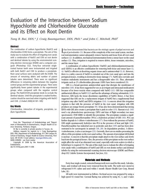
<strong>Evaluation</strong> <strong>of</strong> <strong>the</strong> <strong>Interaction</strong> <strong>between</strong> <strong>Sodium</strong><br />
<strong>Hypochlorite</strong> <strong>and</strong> Chlorhexidine Gluconate<br />
<strong>and</strong> its Effect on Root Dentin<br />
Tung B. Bui, DDS,* J. Craig Baumgartner, DDS, PhD,* <strong>and</strong> John C. Mitchell, PhD †<br />
Abstract<br />
The combination <strong>of</strong> sodium hypochlorite (NaOCl) <strong>and</strong><br />
chlorhexidine (CHX) forms a precipitate. The aim <strong>of</strong> this<br />
study was to evaluate <strong>the</strong> effect <strong>of</strong> irrigating root canals<br />
with a combination <strong>of</strong> NaOCl <strong>and</strong> CHX on root dentin<br />
<strong>and</strong> dentinal tubules by using <strong>the</strong> environmental scanning<br />
electron microscope (ESEM) <strong>and</strong> a computer program<br />
(Photoshop CS2). Forty-four extracted singlerooted<br />
human teeth were instrumented <strong>and</strong> irrigated<br />
with both NaOCl <strong>and</strong> CHX to produce a precipitate.<br />
Root canal surfaces were analyzed with <strong>the</strong> ESEM. The<br />
amount <strong>of</strong> remaining debris <strong>and</strong> number <strong>of</strong> patent<br />
tubules were determined. There were no significant<br />
differences in remaining debris <strong>between</strong> <strong>the</strong> negative<br />
control group <strong>and</strong> <strong>the</strong> experimental groups. There were<br />
significantly fewer patent tubules in <strong>the</strong> experimental<br />
groups when compared with <strong>the</strong> negative control<br />
group. The NaOCl/CHX precipitate tends to occlude <strong>the</strong><br />
dentinal tubules. Until this precipitate is studied fur<strong>the</strong>r,<br />
caution should be exercised when irrigating with NaOCl<br />
<strong>and</strong> CHX. (J Endod 2008;34:181–185)<br />
Key Words<br />
Chlorhexidine, interaction <strong>of</strong> irrigants, precipitate, sodium<br />
hypochlorite<br />
From <strong>the</strong> *Department <strong>of</strong> Endodontology <strong>and</strong> † Department<br />
<strong>of</strong> Biomaterials & Biomechanics, Oregon Health & Science<br />
University, Portl<strong>and</strong>, Oregon.<br />
Dr Bui is currently in private practice in Tucson, Az.<br />
Address requests for reprints to J. Craig Baumgartner,<br />
DDS, PhD, Department <strong>of</strong> Endodontology, 611 SW Campus Dr,<br />
School <strong>of</strong> Dentistry, Oregon Health & Science University, Portl<strong>and</strong>,<br />
OR 97239. E-mail address: baumgarc@ohsu.edu.<br />
0099-2399/$0 - see front matter<br />
Copyright © 2008 by <strong>the</strong> American Association <strong>of</strong><br />
Endodontists.<br />
doi:10.1016/j.joen.2007.11.006<br />
Basic Research—Technology<br />
It has been demonstrated that bacteria are <strong>the</strong> etiologic agents <strong>of</strong> pulpal necrosis <strong>and</strong><br />
apical periodontitis (1). Because <strong>of</strong> <strong>the</strong> complexity <strong>of</strong> <strong>the</strong> root canal system, mechanical<br />
instrumentation cannot adequately remove bacteria <strong>and</strong> tissue from all root canal<br />
surfaces (2). In addition, mechanical instrumentation forms a smear layer on <strong>the</strong> canal<br />
surface (3). Thus, irrigation is required to remove debris, tissue remnants, microbes,<br />
<strong>and</strong> <strong>the</strong> smear layer.<br />
A combination <strong>of</strong> sodium hypochlorite (NaOCl) <strong>and</strong> ethylenediaminetetraacetic<br />
acid (EDTA) is an effective combination for removing both tissue <strong>and</strong> smear layer (3,<br />
4). NaOCl is effective at dissolving organic tissue <strong>and</strong> is antimicrobial (5, 6). However,<br />
<strong>the</strong>re is a safety concern if NaOCl is extruded out <strong>of</strong> <strong>the</strong> root canal apex <strong>and</strong> into <strong>the</strong><br />
periapical tissues, resulting in destructive tissue damage (7). NaOCl also corrodes <strong>and</strong><br />
weakens endodontic instruments <strong>and</strong> has a disagreeable odor (8). Thus, alternative<br />
irrigants such as 2% chlorhexidine gluconate (CHX) have been considered (9).<br />
CHX is a broad-spectrum antimicrobial agent that disrupts <strong>the</strong> membranes <strong>of</strong><br />
microbes (10). It has been suggested for use as an irrigant <strong>and</strong> intracanal medicament<br />
because <strong>of</strong> its lower toxicity when compared with NaOCl (11). CHX has comparable<br />
antibacterial efficacy to NaOCl (12) <strong>and</strong> has <strong>the</strong> advantage <strong>of</strong> having substantivity (13).<br />
However, CHX lacks <strong>the</strong> tissue dissolution capabilities <strong>of</strong> NaOCl. Hence, it has been<br />
suggested that CHX not be a replacement irrigant <strong>of</strong> NaOCl but a supplemental final<br />
irrigation step after NaOCl <strong>and</strong> EDTA irrigation (14). A concern about this irrigation<br />
regimen is that with <strong>the</strong> presence <strong>of</strong> NaOCl in <strong>the</strong> root canal, irrigation with CHX<br />
produces an orange-brown precipitate (9). It has also been demonstrated that mixing<br />
EDTA with CHX also creates a precipitate that is pink in color (15). Basrani et al. (16)<br />
used x-ray photoelectron spectroscopy (XPS) <strong>and</strong> time <strong>of</strong> flight secondary ion mass<br />
spectrometry (TOF-SIMS) to identify this precipitate. The precipitate contains a significant<br />
amount <strong>of</strong> parachloroaniline (PCA); a hydrolysis product <strong>of</strong> CHX (16). PCA can<br />
fur<strong>the</strong>r degrade to 1-chloro-4-nitrobenzene (17). Even without <strong>the</strong> presence <strong>of</strong> NaOCl,<br />
CHX might spontaneously hydrolyze into PCA in <strong>the</strong> presence <strong>of</strong> heat <strong>and</strong> light (18).<br />
Parachloroaniline has industrial uses in pesticides <strong>and</strong> dyes (19) <strong>and</strong> has been demonstrated<br />
to be carcinogenic in animals (18, 20). Its degradation product, 1-chloro-<br />
4-nitrobenzene, is also a carcinogen (21). Currently, <strong>the</strong>re are no studies presenting <strong>the</strong><br />
effects <strong>of</strong> <strong>the</strong> precipitate on <strong>the</strong> root canal surface. The amount <strong>of</strong> precipitate left behind<br />
is unclear. A concern is that this precipitate might attach to <strong>the</strong> root surface <strong>and</strong> slowly<br />
leach into <strong>the</strong> periapical tissues. Also, <strong>the</strong> presence <strong>of</strong> this precipitate on <strong>the</strong> root surface<br />
might affect <strong>the</strong> seal <strong>of</strong> an obturated root canal, especially with resin sealers in which a<br />
hybrid layer is required (9). The aim <strong>of</strong> this study was to evaluate <strong>the</strong> effect <strong>of</strong> irrigating<br />
root canals with a combination <strong>of</strong> NaOCl <strong>and</strong> CHX on root dentin surface <strong>and</strong> dentinal<br />
tubules by using <strong>the</strong> environmental scanning electron microscope (ESEM) <strong>and</strong> Photoshop<br />
CS2 (Adobe Systems Incorporated, San Jose, CA).<br />
Materials <strong>and</strong> Methods<br />
Forty-four single-rooted, extracted human teeth were used for this study. Calculus,<br />
bone, <strong>and</strong> residual s<strong>of</strong>t tissue were removed from <strong>the</strong> teeth. The teeth were stored in<br />
refrigerated 0.9% saline <strong>and</strong> used within 1 month (Baxter Health Care Corp, Round<br />
Lake, IL).<br />
All teeth were instrumented as follows. Occlusal access was prepared by using a<br />
high-speed #2 round bur. Coronal flaring was achieved by using #2, 3, <strong>and</strong> 4 Gates<br />
JOE — Volume 34, Number 2, February 2008 Effect <strong>of</strong> <strong>Interaction</strong> <strong>between</strong> NaOCl <strong>and</strong> CHX on Root Dentin 181
Basic Research—Technology<br />
Glidden drills. With <strong>the</strong> aid <strong>of</strong> a surgical operating microscope, working<br />
length was determined with a #10 K-file introduced into <strong>the</strong> canal until<br />
<strong>the</strong> tip <strong>of</strong> <strong>the</strong> file was visible at <strong>the</strong> apical foramen. A glide path was<br />
established by using #15 <strong>and</strong> #20 K-files.<br />
A fresh mix <strong>of</strong> Aquasil vinyl polysiloxane impression material<br />
(Dentsply/Caulk, Milford, DE) was used to fill a small glass vial (VWR<br />
Scientific, West Chester, PA). The root end <strong>of</strong> <strong>the</strong> prepared tooth was<br />
inserted into <strong>the</strong> impression material <strong>and</strong> allowed to set. This prevented<br />
extrusion <strong>of</strong> irrigant out <strong>of</strong> <strong>the</strong> apex <strong>and</strong> allowed ease <strong>of</strong> h<strong>and</strong>ling during<br />
instrumentation. A rotary engine (Aseptico Inc, Dentsply, Woodinville,<br />
WA) with a 1:8 gear reduction h<strong>and</strong>piece was used for instrumentation<br />
<strong>of</strong> <strong>the</strong> root canals. The h<strong>and</strong>piece was set at 300 rpm <strong>and</strong> torque level<br />
two, which is within <strong>the</strong> range recommended by <strong>the</strong> rotary file manufacturer<br />
(Tulsa Dental, Dentsply, Tulsa, OK). Canal enlargement was<br />
performed with Pr<strong>of</strong>ile NiTi rotary files (Tulsa Dental, Dentsply) to a size<br />
40/.06 in a crown-down manner. The apical matrix was fur<strong>the</strong>r enlarged<br />
to a 60/.04 by using Pr<strong>of</strong>ile NiTi h<strong>and</strong> files (Tulsa Dental,<br />
Dentsply). Needle irrigation 1 mm short <strong>of</strong> <strong>the</strong> working length was<br />
delivered with a Monojet syringe (Sherwood Medical Co, St Louis, MO)<br />
<strong>and</strong> a 27-gauge needle. Irrigation with a total <strong>of</strong> 5 mL <strong>of</strong> 5.25% NaOCl<br />
was performed <strong>between</strong> instrument changes. A #15 K-file was intermittently<br />
used to maintain working length. After instrumentation, <strong>the</strong> teeth<br />
were irrigated with 5 mL <strong>of</strong> buffered (pH 7.4) 15% EDTA (Sigma-<br />
Aldrich Inc, St Louis, MO) <strong>and</strong> <strong>the</strong>n r<strong>and</strong>omly divided into 2 experimental<br />
groups (A <strong>and</strong> B) <strong>of</strong> 15 teeth <strong>and</strong> 2 control groups (C <strong>and</strong> D) <strong>of</strong> 7<br />
teeth each.<br />
Group A<br />
From our pilot study, to clinically simulate <strong>the</strong> maximum amount<br />
<strong>of</strong> precipitate formation, 5 mL <strong>of</strong> 5.25% NaOCl was used to irrigate, <strong>and</strong><br />
<strong>the</strong> canal was left filled with NaOCl. Then 5 mL 2% CHX was used as a<br />
final irrigant followed by immediately drying <strong>of</strong> <strong>the</strong> canal with paper<br />
points.<br />
TABLE 1. Results <strong>of</strong> Percentage Debris Remaining<br />
Root location<br />
Group A (maximum<br />
precipitate produced)<br />
Group B<br />
Similarly from our pilot study, to clinically simulate a minimum<br />
amount <strong>of</strong> precipitate, 5 mL <strong>of</strong> 5.25% NaOCl was used to irrigate <strong>and</strong><br />
<strong>the</strong>n aspirated from canal. The canal was immediately dried with paper<br />
points to remove excess NaOCl. A final irrigation with 5 mL 2% CHX was<br />
followed by immediate drying with paper points. Using additional irrigants<br />
to fur<strong>the</strong>r remove NaOCl was not done to avoid o<strong>the</strong>r possible<br />
interactions with CHX.<br />
Group C (Negative Control)<br />
A final irrigation with 5 mL 5.25% NaOCl was performed. The canal<br />
was aspirated <strong>and</strong> dried with paper points. From our pilot study, this<br />
st<strong>and</strong>ard root canal irrigation regimen left <strong>the</strong> tooth with minimal debris<br />
<strong>and</strong> patent tubules.<br />
Group D (Positive Control)<br />
A final irrigation <strong>of</strong> 5 mL 5.25% NaOCl was performed. The canal<br />
was left flooded <strong>and</strong> allowed to air dry under cover at room temperature.<br />
From our pilot study, crystalline debris was found under <strong>the</strong> SEM<br />
when a canal was flooded with NaOCl <strong>and</strong> left to dry.<br />
A group with only CHX as an irrigant was not included because it<br />
does not have <strong>the</strong> ability to dissolve tissue remnants.<br />
To prepare <strong>the</strong> samples for imaging, <strong>the</strong>y were placed in individual<br />
Petri dishes <strong>and</strong> allowed to air dry at room temperature. A small piece<br />
<strong>of</strong> sponge was placed deep into <strong>the</strong> access opening to avoid introduction<br />
<strong>of</strong> debris into <strong>the</strong> canal during processing. A #257 high-speed bur was<br />
used to decoronate <strong>the</strong> teeth. A #168-L high-speed bur was used to cut<br />
a thin longitudinal slot along <strong>the</strong> buccal <strong>and</strong> lingual aspect <strong>of</strong> <strong>the</strong> root,<br />
avoiding perforating into <strong>the</strong> canal. By using a wire cutter with gentle<br />
pressure, <strong>the</strong> roots were split longitudinally. One half <strong>of</strong> <strong>the</strong> split root<br />
was r<strong>and</strong>omly selected for imaging. To keep <strong>the</strong> imaging procedure<br />
blinded, samples were r<strong>and</strong>omized, <strong>and</strong> a key was constructed to decode<br />
<strong>the</strong> samples for analysis.<br />
Mean SD<br />
Group B (minimum<br />
precipitate produced)<br />
P < 0.0001<br />
P
TABLE 2. Results <strong>of</strong> Number <strong>of</strong> Patent Tubules per 4843 m 2 (SEM image area at 4000)<br />
Root location<br />
Group A (maximum<br />
precipitate produced)<br />
Root samples were imaged at <strong>the</strong> coronal, middle, <strong>and</strong> apical root<br />
third levels by using an ESEM (FEI Quanta 200, Hillsboro, OR) at a stage<br />
height approximately 10 mm, 15 kV, <strong>and</strong> magnification <strong>of</strong> 4000. The<br />
images were saved in TIFF format <strong>and</strong> were used for subsequent analysis.<br />
The ESEM does not require <strong>the</strong> samples to be sputter-coated. This<br />
reduces <strong>the</strong> possibility <strong>of</strong> artifacts. At 4000 magnification, representative<br />
areas for each third <strong>of</strong> <strong>the</strong> root canal were chosen for analysis.<br />
To study effects <strong>of</strong> <strong>the</strong> treatments on <strong>the</strong> root canal surface, we<br />
used <strong>the</strong> TIFF format images to quantify gross surface debris by using a<br />
computer program (Adobe Photoshop CS2; Adobe Systems) to calculate<br />
<strong>the</strong> number <strong>of</strong> debris pixels in each image. The magnetic lasso tool was<br />
used to outline <strong>and</strong> select areas <strong>of</strong> debris. A pixel count <strong>of</strong> <strong>the</strong> outlined<br />
debris was determined <strong>and</strong> was divided by <strong>the</strong> total pixel count for each<br />
image to determine <strong>the</strong> percentage <strong>of</strong> debris remaining. For <strong>the</strong> analysis<br />
<strong>of</strong> patent dentinal tubules, <strong>the</strong> total number <strong>of</strong> visually patent tubules in<br />
each image was manually counted. The total area <strong>of</strong> <strong>the</strong> image was<br />
4843 m 2 .<br />
Statistical analysis was performed with analysis <strong>of</strong> variance<br />
(ANOVA) followed by Fisher probable least-squares difference test<br />
(PLSD) at a significance level <strong>of</strong> P .05.<br />
Results<br />
Table 1 summarizes <strong>the</strong> results <strong>of</strong> <strong>the</strong> percentage debris remaining,<br />
<strong>and</strong> Table 2 summarizes <strong>the</strong> results <strong>of</strong> <strong>the</strong> number <strong>of</strong> patent tubules<br />
per 4843 m 2 . Fig. 1 presents representative SEM images <strong>of</strong> <strong>the</strong> experiment.<br />
The positive control group had more remaining debris in all thirds<br />
<strong>of</strong> root canals when compared with <strong>the</strong> negative control group, <strong>and</strong> this<br />
was statistically significant. There was no significant difference <strong>between</strong><br />
<strong>the</strong> experimental groups <strong>and</strong> <strong>the</strong> negative control group at all thirds <strong>of</strong><br />
<strong>the</strong> root canal. Fur<strong>the</strong>rmore, <strong>the</strong> experimental groups had statistically<br />
less debris when compared with <strong>the</strong> positive control group. When experimental<br />
groups A <strong>and</strong> B were compared with each o<strong>the</strong>r, no statistically<br />
significant differences were found.<br />
The number <strong>of</strong> patent tubules per 4843 m 2 was lower in <strong>the</strong><br />
positive control group when compared with <strong>the</strong> negative control group<br />
at each root third level. This was statistically significant except at <strong>the</strong><br />
apical level. At <strong>the</strong> coronal third level, experimental groups were not<br />
Mean SD<br />
Group B (minimum<br />
precipitate produced)<br />
Group C<br />
(negative control)<br />
Group D<br />
(positive control)<br />
Coronal third 102.33 41.40 87.93 54.14 151.00 29.16 44.29 45.58<br />
P < 0.05<br />
P < 0.05<br />
P < 0.05<br />
Middle third 15.93 19.40 39.60 38.83 65.71 17.71 24.57 20.85<br />
P < 0.05<br />
P < 0.05<br />
P < 0.05<br />
Apical third 16.80 21.82 11.20 16.47 21.29 16.79 16.57 15.99<br />
SD, st<strong>and</strong>ard deviation.<br />
Statistically significant differences are indicated with P values.<br />
P < 0.05<br />
P < 0.05<br />
P < 0.05<br />
Basic Research—Technology<br />
statistically different. At this same level, experimental groups had significantly<br />
more patent tubules than <strong>the</strong> positive control group <strong>and</strong> significantly<br />
less patent tubules than <strong>the</strong> negative control group. At <strong>the</strong> middle<br />
third, <strong>the</strong> negative control group had significantly more patent tubules<br />
when compared with both experimental groups. At this same level, <strong>the</strong>re<br />
were no significant differences <strong>between</strong> <strong>the</strong> experimental groups <strong>and</strong><br />
<strong>the</strong> positive control group. However, group B had statistically more<br />
patent tubules when compared with group A. At <strong>the</strong> apical third level,<br />
more patent tubules were found in <strong>the</strong> negative control group; however,<br />
no groups were statistically different.<br />
Discussion<br />
From this study, we found that when NaOCl was aspirated <strong>and</strong> dried<br />
with paper points, <strong>and</strong> even when it was left flooded in <strong>the</strong> root canal, its<br />
interaction with CHX did not leave behind a significant amount <strong>of</strong> gross<br />
precipitate on <strong>the</strong> root canal surface. There were no significant differences<br />
in <strong>the</strong> percentage <strong>of</strong> remaining debris <strong>between</strong> <strong>the</strong> groups. However,<br />
<strong>the</strong> interaction <strong>between</strong> CHX <strong>and</strong> NaOCl significantly affected <strong>the</strong><br />
patency <strong>of</strong> <strong>the</strong> dentinal tubules. There was a statistically significant reduction<br />
in <strong>the</strong> number <strong>of</strong> patent dentinal tubules in <strong>the</strong> 2 experimental<br />
groups when compared with <strong>the</strong> negative control group. Removing<br />
NaOCl by aspiration <strong>and</strong> paper points showed no significant reduction to<br />
this affect. Apparently, <strong>the</strong> dentin <strong>and</strong> its tubules harbor enough residual<br />
NaOCl that it reacts with <strong>the</strong> CHX in <strong>the</strong> canal. This indicates that a small<br />
amount <strong>of</strong> <strong>the</strong> precipitate is left behind <strong>and</strong> raises potential concerns<br />
with respect to leaching <strong>of</strong> <strong>the</strong> precipitate into <strong>the</strong> surrounding tissues<br />
<strong>and</strong> <strong>the</strong> seal <strong>of</strong> <strong>the</strong> root canal.<br />
The obliteration <strong>of</strong> dentinal tubules was not found to be significant<br />
at <strong>the</strong> apical third. There were no significant differences <strong>between</strong> all<br />
experimental <strong>and</strong> control groups. This might be due to <strong>the</strong> fact that <strong>the</strong><br />
apical third is more difficult to irrigate (5). Results at <strong>the</strong> apex might<br />
have been different if irrigation was supplemented with sonic, ultrasonic,<br />
or negative pressure irrigation. Because <strong>the</strong> coronal <strong>and</strong> middle<br />
thirds are significantly affected, <strong>the</strong>se results remain a concern.<br />
Examination <strong>of</strong> <strong>the</strong> ESEM micrographs revealed a subjective<br />
change in <strong>the</strong> morphology <strong>of</strong> <strong>the</strong> root surface (Fig. 1). The use <strong>of</strong> NaOCl<br />
<strong>and</strong> CHX appears to coat <strong>the</strong> root surface. The substance coating <strong>the</strong><br />
root surface <strong>and</strong> obliterating <strong>the</strong> dentinal tubules was not identified. We<br />
JOE — Volume 34, Number 2, February 2008 Effect <strong>of</strong> <strong>Interaction</strong> <strong>between</strong> NaOCl <strong>and</strong> CHX on Root Dentin 183
Basic Research—Technology<br />
Figure 1. Representative SEM micrographs <strong>of</strong> root surfaces at 4000. Negative control group shows no obvious debris <strong>and</strong> complete removal <strong>of</strong> smear layer. Presence<br />
<strong>and</strong> diameter <strong>of</strong> patent dentinal tubules decline in number from <strong>the</strong> coronal to <strong>the</strong> apical third. Positive control shows gross amounts <strong>of</strong> debris that obscure <strong>the</strong> dentinal<br />
tubules in all root thirds. Experimental groups do not show any obvious debris. However, <strong>the</strong> dentinal tubules appear obliterated especially in <strong>the</strong> middle third.<br />
Subjectively, <strong>the</strong> experimental groups’ root surfaces appear to be coated with unidentified material.<br />
speculate that it was likely <strong>the</strong> precipitate because <strong>the</strong> root surface was<br />
faintly stained on visual inspection. A future study would be to characterize<br />
<strong>the</strong> substance on <strong>the</strong> root surface by using a back-scatter sensor<br />
on <strong>the</strong> ESEM. These findings correlate to ano<strong>the</strong>r study in which a visible<br />
color change on <strong>the</strong> root surface was found along with increased dye<br />
leakage results (9). This might affect obturation systems such as Resilon<br />
(Pentron Clinical Technologies, Wallingford, CT) in which a dentin<br />
hybrid layer is required for proper sealing. However, one Resilon study<br />
demonstrated no significant differences in fluid filtration leakage when<br />
CHX was used as an irrigant (22).<br />
It is known that PCA <strong>and</strong> its degradation product are toxic <strong>and</strong><br />
carcinogenic (18, 21). It has been demonstrated that PCA is produced<br />
when NaOCl interacts with CHX (16). Our study demonstrated that such<br />
a precipitate on <strong>the</strong> root canal surfaces <strong>and</strong> in <strong>the</strong> dentinal tubules is<br />
likely PCA. This raises concerns about potential carcinogens leaching<br />
into surrounding tissues.<br />
A final irrigation with water or alcohol to remove residual NaOCl<br />
could be used. Using EDTA alone would be convenient; however, it also<br />
produces a precipitate in <strong>the</strong> presence <strong>of</strong> CHX (15). A future study<br />
would be to characterize <strong>the</strong> interaction <strong>between</strong> EDTA <strong>and</strong> CHX. A<br />
lower concentration <strong>of</strong> NaOCl produces proportionately less precipitate,<br />
but this strategy is <strong>of</strong> concern because it reduces its <strong>the</strong>rapeutic<br />
effectiveness (16).<br />
In summary, <strong>the</strong> presence <strong>of</strong> NaOCl in <strong>the</strong> root canal before CHX<br />
irrigation does not leave behind statistically significant precipitate on<br />
<strong>the</strong> canal walls. However, this reaction coats <strong>the</strong> canal surface <strong>and</strong><br />
significantly occludes <strong>the</strong> dentinal tubules in <strong>the</strong> coronal <strong>and</strong> middle<br />
thirds <strong>of</strong> <strong>the</strong> canal. Because <strong>the</strong> reaction <strong>between</strong> NaOCl <strong>and</strong> CHX produces<br />
a carcinogenic product, potential leaching <strong>of</strong> PCA into <strong>the</strong> surrounding<br />
tissues is a concern. An additional interface <strong>between</strong> <strong>the</strong> sealer<br />
<strong>and</strong> <strong>the</strong> dentin can also affect <strong>the</strong> seal <strong>of</strong> <strong>the</strong> root canal. Fur<strong>the</strong>r research<br />
is needed to determine <strong>the</strong> nature <strong>of</strong> <strong>the</strong> substance occluding <strong>the</strong> dentinal<br />
tubules.<br />
Acknowledgments<br />
The authors thank Ai Leen Chong, Dave Phillips, Mary Waller,<br />
<strong>and</strong> Harry Davis for <strong>the</strong>ir valuable technical support. This research<br />
project was funded by <strong>the</strong> Leslie A. Morgan Endowment Fund.<br />
References<br />
1. Kakehashi S, Stanley HR, Fitzgerald RJ. The effects <strong>of</strong> surgical exposures <strong>of</strong> dental<br />
pulps in germ-free <strong>and</strong> conventional laboratory rats. Oral Surg Oral Med Oral Pathol<br />
1965;20:340–9.<br />
2. Byström A, Sundqvist G. Bacteriologic evaluation <strong>of</strong> <strong>the</strong> efficacy <strong>of</strong> mechanical root<br />
canal instrumentation in endodontic <strong>the</strong>rapy. Sc<strong>and</strong> J Dent Res 1981;89:321–8.<br />
3. Baumgartner JC, Mader CL. A scanning electron microscopic evaluation <strong>of</strong> four root<br />
canal irrigation regimens. J Endod 1987;13:147–57.<br />
4. Yamada RS, Armas A, Goldman M, Lin PS. A scanning electron microscopic comparison<br />
<strong>of</strong> a high volume final flush with several irrigating solutions: part 3. J Endod<br />
1983;9:137–42.<br />
5. Senia ES, Marshall FJ, Rosen S. The solvent action <strong>of</strong> sodium hypochlorite on pulp<br />
tissue <strong>of</strong> extracted teeth. Oral Surg Oral Med Oral Pathol Oral Radiol Endod<br />
1971;1:96–103.<br />
6. Shih M, Marshall FJ, Rosen S. The bactericidal efficiency <strong>of</strong> sodium hypochlorite as<br />
an endodontic irrigant. Oral Surg Oral Med Oral Pathol 1970;29:613–9.<br />
7. Ehrich DG, Brian JD Jr, Walker WA. <strong>Sodium</strong> hypochlorite accident: inadvertent injection<br />
into <strong>the</strong> maxillary sinus. J Endod 1993;19:180–2.<br />
8. Peters OA, Roehlike JO, Baumann MA. Effect <strong>of</strong> immersion in sodium hypochlorite on<br />
torque <strong>and</strong> fatigue resistance <strong>of</strong> nickel-titanium instruments. J Endod 2007;33:<br />
589–93.<br />
9. Vivacqua-Gomes N, Ferraz CC, Gomes BP, Zaia AA, Teixeira FB, Souza-Filho FJ.<br />
Influence <strong>of</strong> irrigants on <strong>the</strong> coronal microleakage <strong>of</strong> laterally condensed guttapercha<br />
root fillings. Int Endod J 2002;35:791–5.<br />
184 Bui et al. JOE — Volume 34, Number 2, February 2008
10. Leonardo MR, Tanomaru Filho M, Silva LAB, Nelson Filho P, Bonifacio KC, Ito IY. In<br />
vivo antimicrobial activity <strong>of</strong> 2% chlorhexidine used as a root canal irrigation solution.<br />
J Endod 1999;25:167–71.<br />
11. Yesilsoy C, Whitaker E, Clevel<strong>and</strong> D, Phillips E, Trope M. Antimicrobial <strong>and</strong> toxic<br />
effects <strong>of</strong> established <strong>and</strong> potential root canal irrigants. J Endod 1995;21:513–5.<br />
12. Jeansonne MJ, White RR. A comparison <strong>of</strong> 2.0% chlorhexidine gluconate <strong>and</strong><br />
5.25% sodium hypochlorite as antimicrobial endodontic irrigants. J Endod<br />
1994;20:276 – 8.<br />
13. Dametto FR, Ferraz CC, de Almeida Gomes BP, Zaia AA, Teixeira FB, de Souza-Filho<br />
FJ. In vitro assessment <strong>of</strong> <strong>the</strong> immediate <strong>and</strong> prolonged antimicrobial action <strong>of</strong><br />
chlorhexidine gel as an endodontic irrigant against Enterococcus faecalis. Oral Surg<br />
Oral Med Oral Pathol Oral Radiol Endod 2005;99:768–72.<br />
14. Kuruvilla JR, Kamath MP. Antimicrobial activity <strong>of</strong> 2.5% sodium hypochlorite <strong>and</strong><br />
0.2% chlorhexidine gluconate separately <strong>and</strong> combined, as endodontic irrigants.<br />
J Endod 1998;24:472–6.<br />
15. Gonzalex-Lopez S, Camejo-Aguilar D, Sanchez-Sanchez P, Polanos-Carmona V. Effect<br />
<strong>of</strong> CHX on <strong>the</strong> decalcifying effect <strong>of</strong> 10% citric acid, 20% citric acid, or 17% EDTA.<br />
J Endod 2006;32:781–4.<br />
Basic Research—Technology<br />
16. Basrani BR, Manek S, Sodhi RNS, Fillery E, Manzur A. <strong>Interaction</strong> <strong>between</strong> sodium<br />
hypochlorite <strong>and</strong> chlorhexidine gluconate. J Endod 2007;33:966–9.<br />
17. Below H, Lehan N, Kramer A. HPLC determination <strong>of</strong> <strong>the</strong> antiseptic agent chlorhexidine<br />
<strong>and</strong> its degradation products 4-chloroaniline <strong>and</strong> 1-chloro-4-nitrobenzene in<br />
serum <strong>and</strong> urine. Microchimica Acta 2004;146:129–35.<br />
18. Van der Bijl P, Gelderblom WC, Thiel PG. On <strong>the</strong> mutagenicity <strong>of</strong> parachloroaniline,<br />
a breakdown product <strong>of</strong> chlorhexidine. J Dent Assoc S Afr<br />
1984;39:535–7.<br />
19. Available at: http://www.who.int/ipcs/publications/cicad/en/cicad48.pdf. Accessed<br />
July 2007.<br />
20. Chhabra RS, Huff JE, Haseman JK, Elwell MR, Peters AC. Carcinogenicity <strong>of</strong> p-chloroaniline<br />
in rats <strong>and</strong> mice. Food Chem Toxicol 1991;29:119–24.<br />
21. Matsumoto M, Aiso S, Senoh H, et al. Carcinogenicity <strong>and</strong> chronic toxicity <strong>of</strong> parachloronitrobenzene<br />
in rats <strong>and</strong> mice by two-year feeding. J Environ Pathol Toxicol<br />
Oncol 2006;25:571–84.<br />
22. Stratton R, Apicelle M, Mines P. A fluid filtration comparison <strong>of</strong> gutta-percha<br />
versus Resilon, a new s<strong>of</strong>t resin endodontic obturation system. J Endod<br />
2006;32:642–5.<br />
JOE — Volume 34, Number 2, February 2008 Effect <strong>of</strong> <strong>Interaction</strong> <strong>between</strong> NaOCl <strong>and</strong> CHX on Root Dentin 185