248 Diffuse leprosy of Lucio and Latapi - LEPRA Health in Action
248 Diffuse leprosy of Lucio and Latapi - LEPRA Health in Action
248 Diffuse leprosy of Lucio and Latapi - LEPRA Health in Action

You also want an ePaper? Increase the reach of your titles
YUMPU automatically turns print PDFs into web optimized ePapers that Google loves.
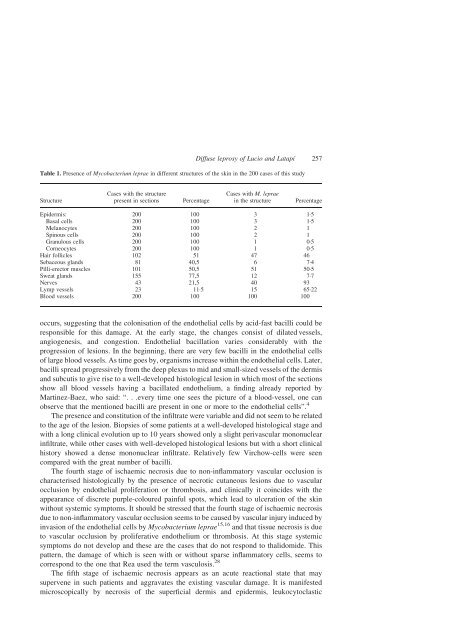
Table 1. Presence <strong>of</strong> Mycobacterium leprae <strong>in</strong> different structures <strong>of</strong> the sk<strong>in</strong> <strong>in</strong> the 200 cases <strong>of</strong> this study<br />
Structure<br />
Cases with the structure<br />
present <strong>in</strong> sections Percentage<br />
<strong>Diffuse</strong> <strong>leprosy</strong> <strong>of</strong> <strong>Lucio</strong> <strong>and</strong> Latapí 257<br />
Cases with M. leprae<br />
<strong>in</strong> the structure Percentage<br />
Epidermis: 200 100 3 1·5<br />
Basal cells 200 100 3 1·5<br />
Melanocytes 200 100 2 1<br />
Sp<strong>in</strong>ous cells 200 100 2 1<br />
Granulous cells 200 100 1 0·5<br />
Corneocytes 200 100 1 0·5<br />
Hair follicles 102 51 47 46<br />
Sebaceous gl<strong>and</strong>s 81 40,5 6 7·4<br />
Pilli-erector muscles 101 50,5 51 50·5<br />
Sweat gl<strong>and</strong>s 155 77,5 12 7·7<br />
Nerves 43 21,5 40 93<br />
Lymp vessels 23 11·5 15 65·22<br />
Blood vessels 200 100 100 100<br />
occurs, suggest<strong>in</strong>g that the colonisation <strong>of</strong> the endothelial cells by acid-fast bacilli could be<br />
responsible for this damage. At the early stage, the changes consist <strong>of</strong> dilated vessels,<br />
angiogenesis, <strong>and</strong> congestion. Endothelial bacillation varies considerably with the<br />
progression <strong>of</strong> lesions. In the beg<strong>in</strong>n<strong>in</strong>g, there are very few bacilli <strong>in</strong> the endothelial cells<br />
<strong>of</strong> large blood vessels. As time goes by, organisms <strong>in</strong>crease with<strong>in</strong> the endothelial cells. Later,<br />
bacilli spread progressively from the deep plexus to mid <strong>and</strong> small-sized vessels <strong>of</strong> the dermis<br />
<strong>and</strong> subcutis to give rise to a well-developed histological lesion <strong>in</strong> which most <strong>of</strong> the sections<br />
show all blood vessels hav<strong>in</strong>g a bacillated endothelium, a f<strong>in</strong>d<strong>in</strong>g already reported by<br />
Mart<strong>in</strong>ez-Baez, who said: “: ::every time one sees the picture <strong>of</strong> a blood-vessel, one can<br />
observe that the mentioned bacilli are present <strong>in</strong> one or more to the endothelial cells”. 4<br />
The presence <strong>and</strong> constitution <strong>of</strong> the <strong>in</strong>filtrate were variable <strong>and</strong> did not seem to be related<br />
to the age <strong>of</strong> the lesion. Biopsies <strong>of</strong> some patients at a well-developed histological stage <strong>and</strong><br />
with a long cl<strong>in</strong>ical evolution up to 10 years showed only a slight perivascular mononuclear<br />
<strong>in</strong>filtrate, while other cases with well-developed histological lesions but with a short cl<strong>in</strong>ical<br />
history showed a dense mononuclear <strong>in</strong>filtrate. Relatively few Virchow-cells were seen<br />
compared with the great number <strong>of</strong> bacilli.<br />
The fourth stage <strong>of</strong> ischaemic necrosis due to non-<strong>in</strong>flammatory vascular occlusion is<br />
characterised histologically by the presence <strong>of</strong> necrotic cutaneous lesions due to vascular<br />
occlusion by endothelial proliferation or thrombosis, <strong>and</strong> cl<strong>in</strong>ically it co<strong>in</strong>cides with the<br />
appearance <strong>of</strong> discrete purple-coloured pa<strong>in</strong>ful spots, which lead to ulceration <strong>of</strong> the sk<strong>in</strong><br />
without systemic symptoms. It should be stressed that the fourth stage <strong>of</strong> ischaemic necrosis<br />
due to non-<strong>in</strong>flammatory vascular occlusion seems to be caused by vascular <strong>in</strong>jury <strong>in</strong>duced by<br />
<strong>in</strong>vasion <strong>of</strong> the endothelial cells by Mycobacterium leprae 15,16 <strong>and</strong> that tissue necrosis is due<br />
to vascular occlusion by proliferative endothelium or thrombosis. At this stage systemic<br />
symptoms do not develop <strong>and</strong> these are the cases that do not respond to thalidomide. This<br />
pattern, the damage <strong>of</strong> which is seen with or without sparse <strong>in</strong>flammatory cells, seems to<br />
correspond to the one that Rea used the term vasculosis. 28<br />
The fifth stage <strong>of</strong> ischaemic necrosis appears as an acute reactional state that may<br />
supervene <strong>in</strong> such patients <strong>and</strong> aggravates the exist<strong>in</strong>g vascular damage. It is manifested<br />
microscopically by necrosis <strong>of</strong> the superficial dermis <strong>and</strong> epidermis, leukocytoclastic