THE IMMUNE SYSTEM OF SHRIMP Introduction Penaeid ... - Nicovita
THE IMMUNE SYSTEM OF SHRIMP Introduction Penaeid ... - Nicovita
THE IMMUNE SYSTEM OF SHRIMP Introduction Penaeid ... - Nicovita

Create successful ePaper yourself
Turn your PDF publications into a flip-book with our unique Google optimized e-Paper software.
By : Franklin S. Martínez (fmartinezt@alicorp.com.pe)<br />
<strong>Nicovita</strong>-ALICORP SAA Technical Service<br />
<strong>THE</strong> <strong>IMMUNE</strong> <strong>SYSTEM</strong> <strong>OF</strong> <strong>SHRIMP</strong><br />
<strong>Introduction</strong><br />
<strong>Penaeid</strong> shrimp aquaculture is an important economic activity worldwide. Nevertheless,<br />
shrimp production has been seriously affected by diseases, mostly those caused by viruses<br />
(Flegel, 2006) and Vibrio bacteria (Bachère, 2000). Shrimp resistance to invading<br />
organisms is strongly influenced by its immune status. The defense mechanisms of<br />
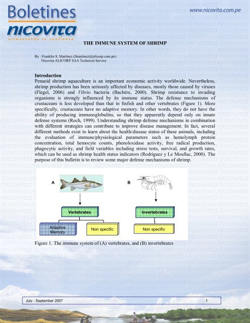
crustaceans is less developed than that in finfish and other vertebrates (Figure 1). More<br />
specifically, crustaceans have no adaptive memory. In other words, they do not have the<br />
ability of producing immunoglobulins, so that they apparently depend only on innate<br />
defense systems (Roch, 1999). Understanding shrimp defense mechanisms in combination<br />
with different strategies can contribute to improve disease management. In fact, several<br />
different methods exist to learn about the health/disease status of these animals, including<br />
the evaluation of immune/physiological parameters such as hemolymph protein<br />
concentration, total hemocyte counts, phenoloxidase activity, free radical production,<br />
phagocytic activity, and field variables including stress tests, survival, and growth rates,<br />
which can be used as shrimp health status indicators (Rodríguez y Le Moullac, 2000). The<br />
purpose of this bulletin is to review some major defense mechanisms of shrimp.<br />
A<br />
Adaptive<br />
Memory<br />
Vertebrates<br />
Non specific<br />
Invertebrates<br />
Non specific<br />
Figure 1. The immune system of (A) vertebrates, and (B) invertebrates<br />
July - September 2007 1<br />
B
Shrimp defense mechanisms<br />
The innate defense system – also known as natural or non-specific defense system –<br />
includes both cellular and humoral components (Figure 2) which work in jointly<br />
coordination for the detection/elimination of all foreign organisms potentially hazardous for<br />
the host (Jiravanichpaisal et al., 2006). Cellular defense components include all those<br />
reactions performed directly by hemocytes (phagocytosis, encapsulation, nodule<br />
formation). On the other hand, humoral components include the activation and release of<br />
molecules stored within hemocytes, such as anticoagulant proteins, agglutinins,<br />
phenoloxidase enzyme, antimicrobial peptides, protease inhibitors, etc. (Jiravanichpaisal et<br />
al., 2006; Holmblad and Söderhäll, 1999.)<br />
Function of crustacean immune system<br />
The cuticle works as the first physical barrier, and it contains antimicrobial substances<br />
(Söderhäll and Cerenius, 1992). Once the pathogen has crossed the outer defense barriers,<br />
hemocytes play an important role in the crustacean immune response. In addition to<br />
participating in the inactivation of invading organisms, hemocytes are also involved in the<br />
regulation of different physiological functions i.e., exoskeleton hardening, cuticle damage<br />
healing, coagulation, carbohydrate metabolism, and protein/amino acid transportation and<br />
storage (Jiravanichpaisal et al., 2006.)<br />
Cellular<br />
Components<br />
- Phagocytosis<br />
- Encapsulation<br />
- Formation of nodules<br />
Innate Immunity<br />
(Natural or Non-specific)<br />
Humoral<br />
Components<br />
- Anticoagulant proteins<br />
- Agglutinins<br />
- Phenoloxidase enzyme<br />
- Antimicrobial peptides<br />
- Free radicals<br />
Figure 2. Cellular and humoral components of crustacean immune system<br />
July - September 2007 2
Hemocyte classification is based on the presence and size of 3 types of cytoplasmic<br />
granules: hyaline, semi-granular, and granular hemocytes (Figure 3). Even though the<br />
proportion and function of hemocytes can vary among species, it is generally considered<br />
that granular and semi-granular hemocytes have the ability of producing melanin by the<br />
pro-phenoloxidase system (Johansson y Söderhäll, 1989). On the other hand, hyaline<br />
hemocytes and – in a lesser extent – semi-granular hemocytes are responsible for the<br />
phagocytosis process (Giulianini et al., 2007.)<br />
Pathogen recognition<br />
The first step in the immune process is the recognition of microorganisms. This process is<br />
carried out by hemocytes through molecules that have the ability of recognizing structures<br />
in the cell walls of invading organisms, such as attachment proteins, and by the recognition<br />
of β-1,3-glucans, lipopolysaccharides, and peptidoglycans (Lin et al., 2006; Vargas-Albores<br />
and Yepiz-Plascencia, 2000). Once invading organisms are detected, hemocytes get<br />
activated then a whole series of mechanisms is triggered to control or remove the intruders.<br />
Phenoloxidase activity<br />
The phenoloxidase system has been recognized as an efficient defense mechanism against<br />
the non-self. This system is stored and produced by semi-granular and granular hemocytes,<br />
and it can be activated by a minimum presence of microbes. Activation of the prophenoloxidase<br />
system results in the production of melanin, a dark-brown pigment<br />
responsible – among other processes – for inactivating foreign particles, and preventing<br />
their spread throughout the host body, as well as for healing cuticle damages<br />
(Sritunyalucksana and Söderhäll, 2000).<br />
A B<br />
C<br />
12.4 × 7.8 14.8 × 8.3 13.6 × 9.5<br />
Figure 3. Classification of hemocytes: (A) Hyaline, (B) Semi-granular, (C) Granular<br />
(Modified photos from Giulianini et al., 2007)<br />
July - September 2007 3
Free radicals and antioxidant mechanisms<br />
Destroying the phagocytized materials involves the intracellular production of free radicals.<br />
During contact with and recognition of the pathogen, host enzymes like NADPH-oxidase<br />
are activated, which in turn increase oxygen consumption, resulting in the production of<br />
free radicals such as superoxide anions (O2 - ) and hydrogen peroxide (H2O2), among others<br />
(Muñoz et al., 2000; Rodríguez and Le Moullac, 2000). These free radicals can directly kill<br />
the invading organism or work in combination with nitrogen compounds (nitric oxide), or<br />
exert a synergistic effect with lysozymes (Roch, 1999).<br />
Nevertheless, free radicals do not discriminate host-self cells from microbes, so that they<br />
can be deleterious in the event of acting in the extra-cellular spaces. Under normal<br />
conditions, the potential damage caused by free radicals is regulated by mechanisms such<br />
as antioxidant molecules i.e., ascorbic acid, poly-unsaturated fatty acids, and antioxidant<br />
enzymes (superoxide dismutase, various peroxidases) (Dandapat et al., 2003; Campa-<br />
Córdova et al., 2002.)<br />
Phagocytosis, encapsulation and nodule formation<br />
Phagocytosis is the most common reaction of defense cell mechanisms. By this process,<br />
cells (hemocytes) ingest and destroy invading pathogens, foreign particles or modified<br />
(aged) cells of the body itself (Secombes, 1996.)<br />
Encapsulation and nodule formation (Figure 4) are processes by which several hemocytes<br />
cooperate with each other aiming to stop the action of invading organisms, when the host is<br />
attacked by either extremely-large particles or numerous tiny particles, to be ingested then<br />
destroyed by individual cells (Söderhäll y Cerenius, 1992).<br />
A<br />
B C<br />
Figure 4. Defense cell processes include: (A) Phagocytosis, (B) Encapsulation, and (C)<br />
Nodule formation. Hemocytes are represented in green, while the invading<br />
organisms appear in red<br />
July - September 2007 4
References<br />
Bachère, E. 2000. Shrimp immunity and disease control. Aquaculture, 191:3-11.<br />
Campa-Córdova, A. I., N. Y. Hernández-Saavedra, R. De Philippis and F. Ascencio. 2002.<br />
Generation of superoxide anion and SOD activity in haemocytes and muscle of<br />
American white shrimp (Litopenaeus vannamei) as a response to β-glucan and<br />
sulphated polysaccharide. Fish & Shellfish Immunology, 12:353-366.<br />
Dandapat, J., G. B. N. Chainy and K. J. Rao. 2003. Lipid peroxidation and antioxidant<br />
defense status during larval development and metamorphosis of giant prawn,<br />
Macrobrachium rosenbergii. Comparative Biochemestry and Physiology Part C,<br />
135:221-233.<br />
Flegel, T. W. 2006. Detection of major penaeid shrimp viruses in Asia, a historical<br />
perspective with emphasis on Thailand. Aquaculture, 258:1-33.<br />
Giulianini, P. G., M. Bierti, S. Lorenzon, S. Battistella and E. A. Ferrero. 2007.<br />
Ultrastructural and functional characterization of circulating hemocytes from the<br />
freshwater crayfish Astacus leptodactylus: Cell types and their role after in vivo<br />
artificial non-self challenge. Micron, 38:49-57.<br />
Holmblad, T. and Söderhäll, K. 1999. Cell adhesion molecules and antioxidative enzymes<br />
in a crustaceans, possible role in immunity. Aquaculture, 172:111-123.<br />
Jiravanichpaisal, P., B. L. Lee and K. Söderhäll. 2006. Cell-mediated immunity in<br />
arthropods: Hematopoiesis, coagulation, melanization and opsonization. Immunobiology,<br />
211:213-236.<br />
Johansson, M. W. and Söderhäll, K. 1989. Cellular immunity in crustaceans and the proPO<br />
system. Parasitology Today, 5:171-176.<br />
Lin, C. Y., K. Y. Hu, S. H. Ho and Y. L. Song. 2006. Cloning and characterization of a<br />
shrimp clip domain serine protease homolog (c-SPH) as a cell adhesion molecule.<br />
Developmental and Comparative Immunology, 30:1132-1144.<br />
Muñoz, M., R. Cedeño, J. Rodríguez, W. P. W. van der Knaap, E. Mialhe and E. Bachère.<br />
2000. Measurement of reactive oxygen intermediate production in haemocytes of the<br />
penaeid shrimp, Penaeus vannamei. Aquaculture, 191:89-107.<br />
July - September 2007 5
Roch, P. 1999. Defense mechanisms and disease prevention in farmed marine invertebrates.<br />
Aquaculture, 172:125-145.<br />
Rodríguez, J. and Le Moullac, G. 2000. State of the art of immunological tools and health<br />
control of penaeid shrimp. Aquaculture, 191:109-119.<br />
Secombes, C. J. 1996. The Nonspecific Immune System: Cellular Defenses, In The Fish<br />
Immune System: Organism, Pathogen, and Environment, Iwama, G. and Nakanishi,<br />
T., Academic Press, San Diego, USA, pp. 63-103.<br />
Söderhäll, K. and Cerenius, L. 1992. Crustacean immunity. Annual Review of Fish<br />
Diseases, 3-23.<br />
Sritunyalucksana, K. and Söderhäll, K. 2000. The proPo and clotting system in crustaceans.<br />
Aquaculture, 191:53-69.<br />
Vargas-Albores, F. and Yepiz-Plascencia, G. 2000. Beta glucan binding protein and its<br />
role in shrimp immune response. Aquaculture, 191:13-21.<br />
Tumpis Edition<br />
Editors: Dagoberto Sánchez dsanchezc@alicorp.com.pe Carlos Ching cchingm@alicorp.com.pe<br />
Máximo Quispe mquispec@alicorp.com.pe<br />
July - September 2007 6