View - Lietuvos mokslų akademijos leidybos skyrius
View - Lietuvos mokslų akademijos leidybos skyrius
View - Lietuvos mokslų akademijos leidybos skyrius

You also want an ePaper? Increase the reach of your titles
YUMPU automatically turns print PDFs into web optimized ePapers that Google loves.
chemija. 2009. vol. 20. No. 3. P. 141–153<br />
© lietuvos <strong>mokslų</strong> akademija, 2009<br />
© lietuvos <strong>mokslų</strong> <strong>akademijos</strong> leidykla, 2009<br />
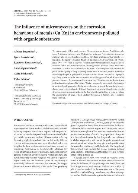
The influence of micromycetes on the corrosion<br />
behaviour of metals (Cu, Zn) in environments polluted<br />
with organic substances<br />
Albinas Lugauskas 1 *,<br />
Igoris Prosyčevas 2 ,<br />
Rimantas Ramanauskas 1 ,<br />
Asta Grigucevičienė 1 ,<br />
Aušra Selskienė 1 ,<br />
Vidas Pakštas 1<br />
1 Institute of Chemistry,<br />
A. Goštauto 9,<br />
LT-01108 Vilnius, Lithuania<br />
2 Institute of Physical Electronics,<br />
Kaunas University of Technology,<br />
Savanorių pr. 271,<br />
LT-50131 Kaunas, Lithuania<br />
INTRODUCTION<br />
Biocorrosion processes at metal surface are associated with<br />
microorganisms or the products of their metabolic activities<br />
including enzymes, exopolymers, organic and inorganic acids,<br />
as well as volatile compounds such as ammonia or hydrogen<br />
sulfide. Various mechanisms of biocorrosion, reflecting<br />
the variety of physiological activities carried out by different<br />
types of microorganisms have been identified and recent<br />
insights into these mechanisms reviewed. Many modern investigations<br />
have focused on the microbially influenced corrosion<br />
of ferrous and copper alloys, and particular microorganisms<br />
of interest have been sulfate-reducing bacteria and<br />
metal-depositing bacteria. Fungi are well known to produce<br />
organic acids, and are therefore capable of contributing to<br />
microbially influenced corrosion. For example, the fungus<br />
Hormoconis resinae (Lindau) v. Arx et de Vries, previously<br />
* corresponding author. e-mail: lugauskas@chi.lt<br />
The micromycetes of five species such as Chrysosporium merdarium, Penicillium cyclopium,<br />
Arthrinium phaeospermum, Cladosporium herbarum, Aspergillus niger, grown on<br />
metallic plates exposed to natural conditions have been investigated. Their specific ecological<br />
and biological peculiarities have been determined. Cu (>99.5%) and Zn (98.5%)<br />
plates 100 × 150 × 3 mm in size were contaminated with the mentioned fungi and placed<br />
into Petri dishes on a nutrient medium imitating organic pollution. It has been determined<br />
that Cu and Zn react differently to the impact of micromycetes. The influence depends<br />
on the capacity of fungi to develop on the metal surface and produce metabolites<br />
stimulating changes in polarization resistance and to destruct the surface. Aspergillus<br />
niger fungi proved to be the most active destructors of copper surface, while Arthrinium<br />
phaeospermum was the most active destructor of zinc. Chrysosporium merdarium is able<br />
to diminish the roughness of Zn surface. This fact is especially important in the first steps<br />
of metal and coating corrosion. The influence of various strains of the same fungi on metals<br />
was noted to be significantly different; therefore, it is important to determine specific<br />
strains or mycocommunities and describe their physiological abilities in order to evaluate<br />
the aggressiveness of fungi or their capability to produce metabolites able to suppress<br />
metal corrosion.<br />
Key words: copper, zinc, micromycetes, metabolites, corrosion, changes of surface<br />
classified as Amorphotheca resinae, Hormodendron resinae,<br />
Cladosporium avellaneum, C. resinae, some species from the<br />
genera Aspergillus, Penicillium, Fusarium utilize the hydrocarbons<br />
of fuel to produce organic acids. Surfaces in contact<br />
with the aqueous phase of fuel water mixtures and sediments<br />
are the common sites of attack. Large quantities of organic<br />
acid by-products induced by this fungus selectively dissolve<br />
or chelate copper, zinc and iron at the grain boundaries of<br />
aircraft aluminum alloys, forming pits which persist under<br />
the anaerobic conditions established under the fungal mat.<br />
The growth of fungi in diesel fuel storage tanks can produce<br />
large quantities of biomass, and this may provoke a crevissel<br />
attack on the metal [1–3].<br />
A grease-coated wire rope wound on wooden spools<br />
stored in a humid environment has been reported to corrode<br />
under the influence of Aspergilus niger and Penicillium spp.<br />
Both fungal species are known to produce citric acid [4, 5]<br />
which may be involved in the corrosion attack. The yeast,<br />
identified as a Candida spp., was also recovered from the copper<br />
surface. Biofilms containing these isolates were shown to
142<br />
Albinas Lugauskas, Igoris Prosyčevas, Rimantas Ramanauskas, Asta Grigucevičienė, Aušra Selskienė, Vidas Pakštas<br />
promote release of copper-by products in subsequent laboratory<br />
reactor experiments [6].<br />
How microorganisms influence corrosion can be in principle<br />
easily understood, as the kinetics of corrosion depends<br />
upon the physicochemical conditions at the interface, such as<br />
pH, oxygen concentration, redox potential, water content and<br />
ionic strength. As the microorganisms adhere to the corroding<br />
surface by means of their physiological activity, they will<br />
be able to change all of these parameters in the most corrosion-relevant<br />
way, i. e. directly at the interface [7–10]. Many<br />
metals are essential for fungal growth and metabolism. Metals<br />
exert toxic effects in many ways: they can inhibit enzymes,<br />
displace or substitute essential metal ions, cause disruption of<br />
membranes and interact with the systems that normally protect<br />
against the harmful effects of free radicals. Toxic metals<br />
can inhibit the growth and spore germination of fungi, affect<br />
reproduction and metabolism. In fungi, metal resistance is<br />
genetically inherited as species- or strain-specific characteristics<br />
and adaptation to metal stress. Resistance may rely on<br />
intrinsic biochemical and structural properties of the host.<br />
Metal can qualitatively and quantitatively affect fungal population<br />
in the environment, it may be different to distinguish<br />
metal effects from those of environmental components, environmental<br />
influence on metal speciation and toxicity and<br />
the nature of any microbial resistance [11, 12]; on the other<br />
hand, microbially influenced corrosion and biodeterioration<br />
of structural metals take place [13, 14].<br />
The most frequent soprotrophic microfungi isolated from<br />
heavy metals are species from the genera Aspergillus, Penicillium,<br />
Scopulariopsis, Paecilomyces, Trichoderma, Fusarium,<br />
Rhizomucor, Rhizopus, Mucor. Alternaria, Aureobasidium,<br />
Phoma, Cladosporium.<br />
Penicillium species are often reported to be dominant<br />
in copper-contaminated environments. Trichoderma viride<br />
grown on copper and zinc where the radial extension rate<br />
was commensurate with the availability of carbon, revealing<br />
a decrease in metal toxicity with increasing levels of glucose.<br />
Metals can be potent inhibitors of enzymatic reaction; for<br />
example copper decreased cellulose and amylase production<br />
by several fungi with a reduced enzyme activity correlating<br />
with an increasing metal concentration. Several metals can<br />
induce or accelerate melanin production in fungi, leading<br />
to blackening of colonies and chlamydospore development<br />
[11, 12]. Changes in mycelial morphology were observed in<br />
fungi during growth in metal-(Cu, Zn)-containing media.<br />
In the case of a toxic metal-containing domain, aggregated<br />
mycelia could produce high local concentrations of extracellular<br />
products such as complexing agents (e. g. organic acid<br />
siderophores, polyphenolic compounds), metal precipitating<br />
agents (e. g. oxalate), and polysaccharides and pigments with<br />
metal-binding abilities [15–17].<br />
Metals and their compounds can interact with fungi in<br />
various ways depending on the metal species, organism and<br />
environment, while metabolic activity can also influence<br />
speciality and mobility. Many metals are essential for fungal<br />
growth and metabolism (Cu, Zn, Fe). Fungi are able to restrict<br />
entry of toxic metal species into cells by:<br />
– reduced metal uptake and / or increased metal efflux,<br />
– metal immobilization, e. g. cell wall adsorption, extracellular<br />
precipitation of secondary neoformed minerals (e g.<br />
oxalates),<br />
– extracellular metal sequestration by e. g. exopolysaccharides<br />
and other extracellular metabolites.<br />
Fungi play an important biogeochemical role in the biosphere<br />
and are intimately involved in the cycling of elements<br />
and transformation of both organic and inorganic substrates.<br />
Their ability of oligotrophic growth, their explorative<br />
filamentous growth habit, flexible growth strategies and resistance<br />
to extreme environmental factors, including metal<br />
toxicity, irradiation and desiccation, make them successful<br />
colonizers of rock surfaces of metals and other metal-rich<br />
habitats [11, 12, 18–22].<br />
The aim of this study was to determine the influence of<br />
five species of micromycetes grown on metals in conditions<br />
identical to natural for a period of up to 2 years, on copper<br />
and zinc which are used as construction components of coatings<br />
diminishing corrosion in the environment polluted with<br />
organics.<br />
EXPERIMENTAL<br />
Five species of mitosporic fungi were selected for the studies:<br />
1. Chrysosporium merdarium (Link ex Grev.) J. W. Carmich.<br />
(Syn. Sporotrichum merdarium Link ex Grev. and many<br />
others) are detected in soils on vegetal remains, technical<br />
substances, epoxy glues, aromatic polyamides, polymetafenyleneisophthalamides,<br />
SiO 2 with bakelite tars, polyamides,<br />
fluorine compounds and other substrates. Fungi of this species<br />
actively destroy pectine, chitine and proteins. The optimal<br />
growth temperature is +20 °C, minimal +7 °C, and maximal<br />
approximately +37 °C. The fungi often dye the substrate<br />
yellow; growing on some substrates they abundantly produce<br />
citrinine (= antimycin) C 13 H 14 O 5 which belongs to the benzopyran-3-carboxylic<br />
acid group of substances. Citrinin is a<br />
mycotoxin of nephrotoxic and carcinogenic effect.<br />
2. Penicillium cyclopium Westling (many synonims are<br />
known) micromycetes grow rapidly: after 10 days colonies<br />
may be as great as 4.5–5 cm in diameter, they are common<br />
in soils of all the continents on vegetal remains, trees, timber,<br />
in the rhizosphere of plants. The fungi actively destroy<br />
substrates rich in lignine and cellulose, they are detected on<br />
various polymeric materials. They are able to destroy tanines<br />
and actively produce organic acids (cyclopiazonic, penicillic,<br />
citric, puberulonic, humic, fulvic) which allow them to develop<br />
on metals and affect their corrosion behaviour during<br />
exploitation. They develop in substrates of various acidity<br />
(pH 2.0–10.0), osmotrophyles, most rapidly grow at a humidity<br />
of 81–84%. The fungi release into the environment a<br />
variety of secondary metabolites which are often toxic and<br />
allergic.
The influence of micromycetes on the corrosion behaviour of metals (Cu, Zn) in environments polluted with organic substances<br />
3. Arthrinium phaeospermum (Corda) M. B. Ellis / Syn. Papularia<br />
sphaerosperma (Pers. ex Gray) Höhn / fungi are<br />
widely spread in the environment and are detected on vegetal<br />
remains, soils, often on other substrates of natural and<br />
sinthetic origin. The optimal growth temperature is about<br />
+20 °C, minimal about +8 °C, and maximal about +30 °C.<br />
They produce some specific substances, for example, the<br />
tetrahydroantraquinonic pygment bostrycine, which is regarded<br />
as a secondary metabolite close to altersolonales, succinic<br />
acid, ergosterol, phenolic compounds and α-threo-βhydroxyaspartic<br />
acid distinguished for its antibiotic activity.<br />
4. Cladosporium herbarum (Pers.) Link ex. Gray fungi<br />
are spread in all the continents on various substrates; being<br />
highly resistant to external factors, they can grow in extremal<br />
conditions lacking light, nutrition, under the action of<br />
chemical substances and other unfriendly factors. The optimal<br />
growth temperature is about +22 °C, minimal about 0 °C,<br />
and maximal about +35 °C. Conidia remain vital even at temperatures<br />
higher than +60 °C. The fungi adapt to a salty environment<br />
without difficulty, actively utilize pectine, lignine,<br />
cellulose, tars, resins and a variety of other compounds. The<br />
fungi of this species are very different from both the morphologic<br />
and functional points of view. The main metabolic<br />
products synthesized by this fungus are proteins, organic acids,<br />
polysaccharides. The fungi are used in biotechnology to<br />
produce steroid preparations (pregnenolon and progesteron)<br />
and allergens.<br />
5. Aspergillus niger Tiegh. colonies are formed rapidly.<br />
They synthesize and release into the environment a great<br />
variety of different metabolites. Their functional activity is<br />
peculiar and allows assimilating on various substrates and<br />
spreading in the environment. The fungi actively produce<br />
various acids: kojic, citric, oxalic, phenylacetic, 3-indolylacetic,<br />
dihydroxyphenylacetatic, glutaconic, 4-hydroxymandolic<br />
and many others. It should be noted that these fungi are distingnished<br />
for their ability to detox aflatoxins synthesized by<br />
other fungi belonging to this genus. The majority of strains of<br />
these fungi are toxic to warm-blooded organisms; they often<br />
cause mycoses in humans, animals and birds. They actively<br />
grow and develop at a temperature of +35 °C and higher;<br />
however, they can also be found even at a low temperature<br />
of +2 °C.<br />
Plates (100 × 150 × 3 mm) of the two principal metals<br />
– Cu (Cu > 99.5%) and Zn (Zn > 98.5%) – were used in<br />
the study. The exposed metal plates were cleaned with a fine<br />
suspension of Mg(OH) 2 and high purity acetone to minimize<br />
the initial contamination of the surface by nutritives from<br />
the environment. With the purpose to determine the capacity<br />
of micromycetes to adapt to metals, experiments were performed<br />
under laboratory conditions using a malt agar extract<br />
poor in nutritive matters [23–27].<br />
The contact of the metal with fungi was investigated.<br />
Metal plates were placed in Petri dishes filled with a sterile<br />
agar medium of malt extract supplied with chloramphenicol<br />
(50 mg/l). Then the medium with a metal was sown up with<br />
143<br />
fungi of the five species mentioned above: 1. Chrysosporium<br />
merdarium (Ch 1 ), 2. Penicillium cyclopium (P 2 ), 3. Arthrinium<br />
phaeospermum (Arth 3 ), 4. Cladosporium herbarum (C 4 ),<br />
5. Aspergillus niger (A. n. 5 ), 6. K 2 – the medium with metal<br />
plates was not contaminated with fungi; K 1 – metal plates<br />
were kept under common conditions at room temperature<br />
and usual humidity; the plates were not contaminated. The<br />
medium with metals was cultivated in a thermostat at a temperature<br />
of 26 ± 2 °C. The intensity of fungal growth and<br />
metal oxidation, as well as surface changes were evaluated after<br />
15 and 30 days. The extent of fungal growth was assessed<br />
with the naked eye and by light microscopy in accordance<br />
with the scheme:<br />
– no fungal growth observed on specimens under the<br />
light microscope – 1 point<br />
– mycelium which branched hyphae and possibly sporulation,<br />
visible under the light microscope – 2 points<br />
– growth of fungi, sparce but visible with the naked<br />
eye under the light microscope, sporulation clearly visible<br />
– 3 points<br />
– growth of fungi clearly visible but covering 25% of the surface – 5 points [27].<br />
For spectroscopical investigations of corrosion products<br />
on metal plates, a Nicolet model 5700 Fourier transformation<br />
infrared spectrometer (FTIR) in conjunction with a 10 Spec<br />
(10 Degree Specular Reflectance Accessory) was used. Samples<br />
were placed on the reflectance accessory, and spectra<br />
were obtained in the reflectance mode. In all cases, the spectral<br />
range was 4000–400 cm –1 (reciprocal wave length), with<br />
a 4 cm –1 resolution and 64 scans. The spectral manipulation<br />
included baseline correction, removal of carbon dioxide absorption<br />
bands, and subtraction of water vapour interferences.<br />
The samples of corroded metal plates were cleaned with<br />
dry filter paper. A metal plate stored for up to 30 days without<br />
biological interventions was used as a reference. In this way,<br />
the atmospheric corrosion products from a metal plate were<br />
eliminated, and the IR spectra of products formed by biological<br />
agents were registered [28].<br />
The morphology of the substrate surface of Cu and Zn<br />
was studied by SEM and AFM. The SEM method was employed<br />
to analyse the general appearance of the surface and its<br />
changes as a result of biocorrosion in the micrometry range<br />
of roughness, and the AFM was used to assess changes in the<br />
nanometer range of surface. Cu and Zn plates were evaluated<br />
after excerpts for 30 days under normal conditions and high<br />
humidity without a fungus and then with the fungi: Ch 1 , P 2 ,<br />
Arth 3 , C 4 , A. n. 5 . The images were analysed with Image J.<br />
One of the most promising techniques for the characterization<br />
of microbial adhesion and metal surface interaction<br />
is AFM. This method uses atomic force microscopy (AFM)<br />
operating in the force mode which offers both imaging capabilities<br />
and quantitative measurements of forces between the<br />
AFM tip and the sample. Topographical surface studies after
144<br />
Albinas Lugauskas, Igoris Prosyčevas, Rimantas Ramanauskas, Asta Grigucevičienė, Aušra Selskienė, Vidas Pakštas<br />
30 days of exposure of metals (Cu, Zn) to five different fungal<br />
species were performed by using an Exlorer scanning probe<br />
microscope (VECO Topometrix, USA).<br />
Polarization resistance changes of the metal plates were<br />
determined from EIS. The EIS measurements were carried<br />
out using an electrochemical glass cell equipped with holes<br />
for a metal specimen as well as Ag / AgCl reference and Pt<br />
counter electrodes. A metal sample was mounted in a special<br />
holder placed in a cell filled with 3.5% NaCl, and EIS measurements<br />
were carried out after 5–10 min. The measurements<br />
were performed at an open circuit potential applying a<br />
signal of the amplitude of ∆E = ± 5 mV. A P / G / FRA Autolab<br />
302 apparatus (The Netherlands) was used.<br />
RESULTS AND DISCUSSION<br />
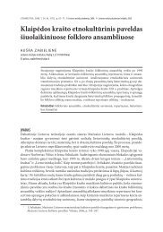
Fungal interaction with metal surface. The data presented<br />
in Table 1 and Fig. 1 show that the micromycetes studied<br />
are able to grow in a malt extract agar medium in contact<br />
with different metals. The Penicillium cyclopium fungi growing<br />
in a medium sporulated intensively and covered the surface<br />
of Cu plates with conidia almost completely (4 points);<br />
the Chrysosporium merdarium (Ch 1 ) and Arthrinium phaeospermum<br />
(Arth 3 ) fungi intensively grew on the interface of<br />
a metal with the medium from where their mycelium spread<br />
over the surface of a Cu plate (3 points). Chrysosporium<br />
merdarium (Ch 1 ) developed most intensively on a Zn plate<br />
(4 points). The Arthrinium phaeospermum (Arth 3 ) fungi<br />
covered the Zn plate surface with a sparse veil (3 points).<br />
In places of a Zn plate contact with Aspergillus niger fungi,<br />
Table 1. Reaction of Cu and Zn metals to the impact of micromycetes<br />
Fungal species Changes on metal surface<br />
colonies were formed in which conidia were seattered over<br />
the whole surface (3 points).<br />
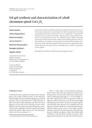
Some more prominent changes in metal plates (Cu, Zn) after<br />
30 days of contact with the developing different fungal species<br />
are illustrated in Fig. 2. These changes are less noticeable<br />
on copper plates, whereas on zinc they are more pronounced.<br />
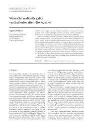
Products of metal surface and fungus interaction. The<br />
data obtained studying the surface reflection spectra of copper<br />
and zinc plates recorded after 30 days of their exposure to<br />
micromycetes of five fungi species are shown in Figs. 3–7. As<br />
shown by data presented in Fig. 3, ester –COOH groups were<br />
detected on the zinc surface after its exposure to the Chrysosporium<br />
merdarium fungus, while O–H–O bending of bound<br />
water vibrated on the zinc surface. The C–C deformation vibrations<br />
were similar on both surfaces. After 30 days of exposure<br />
of Zn and Cu plates to the Penicillium cyclopium fungus, we<br />
failed to detect products that could be determined by IR spectra,<br />
while on the Zn surface traces of P–H groups and organic<br />
compounds were found (Fig. 4). Under the influence of the Arthrinium<br />
phaeospermum fungi two bands, P–H and C–C, were<br />
determined on Cu, while there was an –OH group peak common<br />
set of the bands of organic remains of CH 3 , C–C, C–H<br />
groups on the Zn surface (Fig. 5). The most prominent traces<br />
of organic compounds on Cu and Zn surfaces after 30 days<br />
of exposure were left by fungi of the Cladosporium herbarum<br />
species (Fig. 6). On the surface of both metals, a whole set of<br />
peaks of organic compounds of the same origin was observed.<br />
One can see a peculiar set of such peaks on Cu after the impact<br />
of Aspergillus niger fungi, whereas on Zn common vibrations<br />
of organic remains were determined (Fig. 7).<br />
Intensity of deterioration,<br />
points (1–5)<br />
Chrysosporium merdarium<br />
Copper (Cu) after 30 days of exposure to fungi<br />
Prominent fungal conidia formed on the edges of plates from<br />
where the mycelia spreads over the Cu surface<br />
3<br />
Penicillium cyclopium Fungal conidia cover almost the whole surface of a metal plate 4<br />
Arthrinium phaeospermum<br />
Conidia are formed on the edges of metal plates from where they<br />
spread over the surface<br />
3<br />
Cladosporium herbarum<br />
Aspergillus niger<br />
Fungal conidia are scattered over the metal surface, although no<br />
prominent colonies are formed<br />
Fungi did not grow on the metal surface, but fungal conidia were<br />
adhered to the Cu surface<br />
K – Cu plates not contaminated<br />
2<br />
with fungi (control)<br />
Fungi did not grow on a metal surface<br />
Zinc (Zn) after 30 days of exposure to fungi<br />
0<br />
Chrysosporium merdarium A net of mycelia hyphae prostrates over the metal surface 4<br />
Penicillium cyclopium<br />
Fungal colonies were formed only on the edges of metal plates;<br />
colonies are sparsely scattered over the whole surface<br />
2<br />
Arthrinium phaeospermum A thin net of mycelia hyphae prostrates over the metal surface 3<br />
Cladosporium herbarum On the edges of plates, only discrete limited colonies are formed 1<br />
Aspergillus niger<br />
Fungal colonies are formed on the plate edges, conidia are scattered<br />
over the whole plate surface<br />
3<br />
K 2 – Zn plates not contaminated<br />
with fungi (control)<br />
Fungi did not grow on the metal surface 0<br />
2<br />
1
The influence of micromycetes on the corrosion behaviour of metals (Cu, Zn) in environments polluted with organic substances<br />
1. Chrysosporium merdarium<br />
5. Aspergillus niger<br />
10 06 2008<br />
2. Penicillium cyclopium<br />
10 06 2008<br />
3. Arthrinium phaeospermum 4. Cladosporium herbarum<br />
10 06 2008<br />
K 2 – without fungi<br />
Fig. 1. A general view of mycromycetes growth on the malt extract agar with different metals (Cu, Zn) after 30 days of exposure at 26 ± 2 ºC<br />
10 06 2008<br />
10 06 2008<br />
10 06 2008<br />
Fig. 2. More pronounced changes in metal<br />
plates after 30 days of exposure to developing<br />
fungi of differrent species: K 1 – reference<br />
plates; K 2 – exposed to a nutrient<br />
medium free from fungi; 1 – contaminated<br />
with Chrysosporium merdarium; 2 – with<br />
Penicillium cyclopium; 3 – with Arthrinium<br />
phaeospermum; 4 – with Cladosporium<br />
herbarum; 5 – with Aspergillus niger<br />
145
146<br />
Albinas Lugauskas, Igoris Prosyčevas, Rimantas Ramanauskas, Asta Grigucevičienė, Aušra Selskienė, Vidas Pakštas<br />
Fig. 3. FTIR spectra of Cu and Zn<br />
specimens exposed to Chrysosporium<br />
merdarium fungi for 30 days<br />
Fig. 4. FTIR spectra of Cu and Zn<br />
specimens exposed to Penicillium<br />
cyclopium fungi for 30 days<br />
Fig. 5. FTIR spectra of Cu and Zn<br />
specimens exposed to Arthrinium<br />
phaeospermum fungi for 30 days
The influence of micromycetes on the corrosion behaviour of metals (Cu, Zn) in environments polluted with organic substances<br />
A more detailed analysis of the data showed that the absorption<br />
bands determined on Cu surfaces after 30 days of<br />
exposure to the mentioned fungi did not differ from those<br />
determined on Zn. The less clear spectra observed on the surface<br />
of Cu plates under the influence of Penicillium cyclopium<br />
and Arthrinium phaeospermum can be explained by the high<br />
biotoxicity of Cu ions which hinder the adherence of fungal<br />
metabolites to the metal surface.<br />
Metal surface morphology after fungal impact. Changes<br />
in the surface of copper and zinc plates exposed to developing<br />
micromycetes under humid conditions could be inferred<br />
by the data presented in Figs. 8 and 9 showing metal surface<br />
deterioration under the influence of corresponding fungi, detected<br />
by the SEM method.<br />
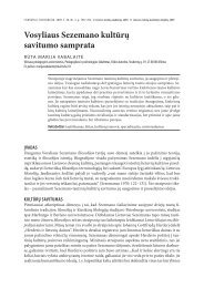
A general view of copper plate surface deterioration after<br />
30 days of exposure to a nutrient medium without contamination<br />
with fungi can be observed in Fig. 8 K 2 . On the surface<br />
147<br />
Fig. 6. FTIR spectra of Cu and Zn<br />
specimens exposed to Cladosporium<br />
herbarum fungi for 30 days<br />
Fig. 7. FTIR spectra of Cu and Zn<br />
specimens exposed to Aspergillus<br />
niger fungi for 30 days<br />
of a Cu plate, there are fine dotted corrosion nidi 0.8–1 µm<br />
in size, localized in places of structural parts. On the surface<br />
of Cu plates, after 30 days of exposure to a nutrient medium<br />
contaminated with Chrysosporium merdarium fungi (Ch 1 ), a<br />
deterioration of the surface oxide layer, film tears and in places<br />
threads of fungal mycelium 2–3 µm wide were spread over<br />
the altered oxidized surface (Fig. 8, Cu 1 ). On the surface of Cu<br />
plates contamined with Penicillium cyclopium fungi (P 2 ), formations<br />
reminiscent of foam, 15–20 µm in size, among them<br />
solitary elements reminiscent of fungal conidia 2–3 µm in<br />
size, were observed. The surface of a Cu plate contaminated<br />
with Arthrinium phaeospermum fungi (Arth 3 ) was covered<br />
with oxide scales 15–20 µm in size, and on them there were<br />
spreading threads of fungal mycelium up to 3 µm in thickness;<br />
there were also plenty of spherical conidia about 5 µm in<br />
size. On the surface of Cu plates contaminated with Cladosporium<br />
herbarum fungi (Cl 4 ), there were plenty of spherical
148<br />
Albinas Lugauskas, Igoris Prosyčevas, Rimantas Ramanauskas, Asta Grigucevičienė, Aušra Selskienė, Vidas Pakštas<br />
formations 2–3 µm in size, reminiscent of fungal conidia. The<br />
surface of a Cu plate contaminated with Aspergillus niger fungi<br />
(A. n. 5 ) was covered with a rather compact oxide film with<br />
the remains of Cu compounds 15–20 µm in size (Fig. 8, Cu 5 ).<br />
A general view of zinc plate surface deterioration after<br />
30 days of exposure to a nutrient medium not contaminated<br />
with fungi is shown in Fig. 9 (K 2 ). Rather large (5 to 30 µm<br />
K 2 – without fungi Cu 1 – Chrysosporium merdarium<br />
Cu 2 – Penicillium cyclopium Cu 3 – Arthrinium phaeospermum<br />
Cu 4 – Cladosporium herbarum Cu 5 – Aspergillus niger<br />
in diameter) surface corrosion nidi, most probably formed<br />
by zinc oxide and zinc hydroxide, are observed. A general<br />
view of surface deterioration of a Zn plate contaminated with<br />
Chrysosporium merdarium fungi (Ch 1 ) is shown in Fig. 9<br />
(Zn 1 ). The zinc surface in a tacyric (clayey) soil form is covered<br />
with scales 10–20 µm in diameter with cracks 1–2 µm<br />
wide among them. On the surface of a zinc plate contami-<br />
Fig. 8. A general view of copper plate surfaces after 30 days of exposure to a nutrient medium: K 2 – without fungi; Cu 1 – contaminated with Chrysosporium<br />
merdarium; Cu 2 – with Penicillium cyclopium; Cu 3 – with Arthrinium phaeospermum; Cu 4 – with Cladosporium herbarum; Cu 5 – with Aspergillus niger
The influence of micromycetes on the corrosion behaviour of metals (Cu, Zn) in environments polluted with organic substances<br />
nated with Penicillium cyclopium fungi (P 2 ), formations in a<br />
lump form 15–20 µm in size are seen. In the bottom part of<br />
the Fig. 9, an accumulation of remains of biogenic origin in<br />
the form of a thread can be seen (Fig. 9, Zn 2 ). The whole surface<br />
of a Zn plate contaminated with Arthrinium phaeospermum<br />
fungi (Arth 3 ) is covered with oblong crystallites 15 µm<br />
in length and up to 1 µm in width. On the surface of a Zn plate<br />
K 2 – without fungi<br />
Zn 2 – Penicillium cyclopium<br />
Zn 4 – Cladosporium herbarum<br />
149<br />
contamindated with Cladosporium herbarum fungi (Cl 4 ), formations<br />
of corrosion products up to 15 µm in size, which are<br />
surrounded by fungal conidia, most often oval up to 2 µm in<br />
size, are observed (Fig. 9, Zn 4 ). The whole surface of a Zn plate<br />
contaminated with Aspergillus niger fungi (A. n. 5 ) is covered<br />
with lumps 20–25 µm in size and separate fungal conidia up<br />
to 30 µm (Fig. 9, Zn 5 ).<br />
Zn 1 – Chrysosporium merdarium<br />
Zn 3 – Arthrinium phaeospermum<br />
Zn 5 – Aspergillus niger<br />
Fig. 9. A general view of zinc plate surfaces after 30 days of exposure to a nutrient medium: K 2 – without fungi; Zn 1 – contaminated with Chrysosporium<br />
merdarium; Zn 2 – with Penicillium cyclopium; Zn 3 – with Arthrinium phaeospermum; Zn 4 – with Cladosporium herbarum; Zn 5 – with Aspergillus niger
150<br />
Albinas Lugauskas, Igoris Prosyčevas, Rimantas Ramanauskas, Asta Grigucevičienė, Aušra Selskienė, Vidas Pakštas<br />
Fig. 10. A topographic view of copper plates after 30 days of exposure to developing fungi of different species: K 2 – plates<br />
without fungi, exposed to the same conditions; Ch 1 – contaminated with Chrysosporium merdarium; P 2 – with Penicillium<br />
cyclopium; Arth 3 – with Arthrinium phaeospermum; Cl 4 – with Cladosporium herbarum; A. n. 5 – with Aspergillus niger<br />
Nanometric evaluation of changes occurring in metal<br />
surfaces. One of the most promising techniques for the characterization<br />
of microbial adhesion and metal surface interaction<br />
is AFM. Topographic surface investigations after 30 days<br />
of exposure to five different fungi were performed with an<br />
Explorer scanning probe microscope (VECO Topometrix,<br />
USA).<br />
The nanomorphological view of copper and zinc specimens<br />
exposed to fungi is shown in Figs. 10 and 11. There<br />
are no significant changes in Cu and Zn surfaces after a<br />
long-term contact with fungi. In the reference K 2 , where<br />
Cu and Zn specimens had been exposed to a nutrient medium<br />
without fungi, both surfaces are less rough. The highest<br />
increase in surface roughness is observed after a long-
The influence of micromycetes on the corrosion behaviour of metals (Cu, Zn) in environments polluted with organic substances<br />
Fig. 11. A topographic view of zinc plates after 30 days of exposure to developing fungi of different species: K 2 – plates<br />
without fungi, exposed to the same conditions; Ch 1 – contaminated with Chrysosporium merdarium; P 2 – with Penicillium<br />
cyclopium; Arth 3 – with Arthrinium phaeospermum; Cl 4 – with Cladosporium herbarum; A. n. 5 – with Aspergillus niger<br />
term contact of copper with Aspergillus niger (A. n. 5 ) and<br />
in the case of zinc with Arthrinium phaeospermum (Arth 3 )<br />
fungi.<br />
The root-mean square roughness (R q , values nm) of the<br />
metals were determined. This statistic parameter characterizes<br />
metal surface roughness. The data obtained are shown<br />
in Fig. 12.<br />
151<br />
The roughness of copper specimens after contact with<br />
a nutrient medium free from fungi increased from 17 to<br />
27.8 nm compared with the initial values of this parameter<br />
(K 1 ), whereas changes of the Cu surface under the influence<br />
of fungi were rather different.<br />
The roughness of plates exposed for 30 days to Aspergillus<br />
niger (A.n. 5 ) and Cladosporium herbarum (Cl 4 ) was higher as
152<br />
Albinas Lugauskas, Igoris Prosyčevas, Rimantas Ramanauskas, Asta Grigucevičienė, Aušra Selskienė, Vidas Pakštas<br />
Fig. 12. Changes in surface roughness (rms, R q , nm) after 30 days of exposure of Cu and Zn to different micromycetes at 26 ± 2 °C<br />
compared to that of reference plates (K 1 , K 2 ) free of fungi. The<br />
nanomorphology of Zn plates exposed to some of the studied<br />
fungi (Arthrinium phaeospermum (Arth 3 ) and Aspergillus niger<br />
(A. n 5 )) was far rougher (R q increased up to 54 and 73, respectively),<br />
and accumulation of large crystals was observed<br />
on the surface. The impact of other fungi was weaker.<br />
Changes in the surface of zinc specimens exposed to the<br />
mentioned fungi differed even more. Under the influence<br />
of Chrysosporium merdarium (Ch 1 ), the surface roughness<br />
markedly decreased from 35.41 (K 1 ) and 38.08 (K 2 ) to 3.99<br />
(Ch 1 ). Thus, according to the data obtained, the surface became<br />
protected from disturbance, while under the influence<br />
of Arth 3 ir A. n. 5 the value of this parameter increased up to<br />
250 and 119, respectively. However, taking into account that<br />
the studies were of a dotted character, they cannot serve as a<br />
guide for considering the obtained data as systematic when<br />
evaluating changes in the Zn and Cu surfaces in all cases of<br />
corrosion; they can only describe the tendencies. Further investigations<br />
are needed.<br />
Table 2. Changes in polarization resistance of Cu and Zn plates after<br />
30 days of exposure to growing fungi of different species at 26 ± 2 °С<br />
Variant<br />
Polarisation resistance<br />
(Rp / Ωcm2 )<br />
Cu Zn<br />
Control (K1) 9.76 0.59<br />
Uncontaminated (K 2) 4.64 9.6<br />
Chrysosporium merdarium (Ch 1) 5.6 3.2<br />
Penicillium cyclopium (P 2) 5.48 11.0<br />
Arthrinium phaeospermum (Arth 3) 6.28 22.8<br />
Cladosporium herbarum (Cl 4) 11.6 11.2<br />
Aspergillus niger (A. n. 5) 34.2 14.4<br />
Corrosion studies. The polarization resistance changes of<br />
Cu and Zn were determined from EIS data and are presented<br />
in Table 2.<br />
The most pronounced changes in polarization resistance<br />
on the surface of Cu were recorded for plates affected by the<br />
A. n. 5 fungus: from 9.76 (K 1 ) up to 34.2 Ωcm 2 . The polarization<br />
resistance under the influence of the Cl 4 fungi changed<br />
less – up to 11.6 Ωcm 2 . Under the influence of some fungi,<br />
a decrease in the polarization resistance of Cu plate surface<br />
was observed, and it decreased to 5.48 under the influence<br />
of P 2 , to 5.6 when affected by Ch 1 and to 6.28 Ωcm 2 when<br />
Cu plates were exposed to Arth 3 . The polarization resistance<br />
of Zn plate surface most often increased under the influence<br />
of the Arth 3 (22.8 Ωcm 2 ) and A. n. 5 (14.4 Ωcm 2 ) fungi. At the<br />
same time, Chrysosporium merdarium (Ch 1 ) least changed<br />
the polarization resistance of the Zn plate, which increased<br />
from 0.59 (K 1 ) to 3.2 Ωcm 2 and markedly decreased (from<br />
9.6 to 3.2 Ωcm 2 ) as compared to that of Zn plates exposed for<br />
30 days to the same conditions without fungi. Such changes<br />
in Cu and Zn polarization resistance values can be associated<br />
with the metabolism pecularities of the fungus and the active<br />
substances released during this process.<br />
A comparison of the R p values of Cu and Zn specimens<br />
suggests that Cu surface corrodes when exposed to a medium<br />
free from fungi. R p decreased from 9.8 (K 1 ) to 4.6 (K 2 ),<br />
whereas the Zn surface became more passive, R p increased<br />
from 0.6 to 9.6 Ωcm 2 . The effect of a medium contaminated<br />
with Aspergillus niger (A. n. 5 ) on copper markedly differed.<br />
Here, R p of Cu surface increased up to 34.2 Ωcm 2 , whereas<br />
the polarization resistance of Zn specimens increased most<br />
notably under the influence of Arthrinium phaeospermum<br />
(Arth 3 ) fungi (R p 22.8).
The influence of micromycetes on the corrosion behaviour of metals (Cu, Zn) in environments polluted with organic substances<br />
CONCLUSIONS<br />
1. Copper and zinc differently respond to the effect of different<br />
micromycetes, depending on the fungal ability to develop<br />
on a metal surface in extreme conditions and produce metabolites<br />
stimulating changes in the metal polarization resistance,<br />
as well as to destroy the surface.<br />
2. Aspergillus niger (A. n. 5 ) and Arthrinium phaeospermum<br />
(Arth 3 ) fungi were most active metal surface disturbers<br />
for copper and zinc, respectively. It is noteworthy that Chrysosporium<br />
merdarium (Ch 1 ) fungi possess the ability to diminish<br />
the surface roughness, which is especially important<br />
in the first stages of metal and metal coating corrosion.<br />
3. The action of different strains of the same fungal species<br />
on metals can differ significantly. Thus, in order to assess<br />
the aggressiveness of individual fungi or their ability to produce<br />
metabolites capable of suppressing metal corrosion, it is<br />
essential to denote specific strains or mycoassociations and<br />
to describe their physiological abilities.<br />
References<br />
Received 9 april 2009<br />
accepted 5 may 2009<br />
1. F. m. Bento, c. Gaylande, Annu. Rev. Microbiol., 27, 192<br />
(1996).<br />
2. G. e. englert, i. l. müller, Proceedings of “Biodegradation<br />
and Biodeterioration in Latin America (LABS 3)”, The British<br />
Phycological Society, UK (1998).<br />
3. i. B. Beech, ch. c. Gaylarde, Rev. Microbiol., 30, 177<br />
(1999).<br />
4. B. little, R. Ray, K. hart, P. Wagner, Mater. Perform., 34, 55<br />
(1995).<br />
5. B. little, R. Staehle, Electrochem. Soc. Interface, 10(4), 44<br />
(2000).<br />
6. B. j. Webster, D. B. Wells, P. j. Bremer, Proceedings of<br />
“NACE Corrosion’96”, houston, USa (1996).<br />
7. Ph. j. Bremer, G. Geesey, Appl. Environ. Microbiol., 57(7),<br />
196 (1991).<br />
8. h-c. Flemming, Proceedings of “Microbially influenced<br />
corrosion of industrial materials”, mülheim an der Ruhr,<br />
Germany (1999).<br />
9. G. Schaule, T. Grieble, h-c. Flemming, Proceedings of<br />
“Microbially influenced corrosion of industrial materials”,<br />
mülheim an der Ruhr, Germany (1999).<br />
10. S. chongdar, G. Gunasekaran, P. Kumar, Electrochim. Acta,<br />
50, 4655 (2005).<br />
11. G. m. Gadd, Adv. Microb. Physiol., 41, 47 (1999).<br />
12. G. m. Gadd, Mycol. Res., 111, 3 (2007).<br />
13. R. S. Dubey, S. N. Upadhyay, Bull. Electrochem., 15(7–8),<br />
266 (1999).<br />
14. Y. Kikuchi, K. R. Srcekumari, Journal of the Iron and Steel<br />
Institute of Japan, 88, 620 (2002).<br />
15. B. muller, W. Burgstaller, h. Strasser, a. Zanella, F. Schinner,<br />
J. Industr. Microbiol., 14, 208 (1995).<br />
153<br />
16. m. v. Dutton, c. S. evans, Can. J. Microbiol., 42, 881<br />
(1996).<br />
17. P. Baldrian, Enzyme Microb. Technol., 32, 78 (2003).<br />
18. m. a. de la Torre, G. Gomez-alarcon, Microb. Ecol., 27,<br />
177 (1994).<br />
19. m. Khan, j. Scullion, Apll. Soil. Ecol., 20, 145 (2000).<br />
20. e. P. Burford, m. Kierans, G. m. Gadd, Mycologist, 17, 98<br />
(2003).<br />
21. a. a. Garbushina, K. Whitenead, T. Dornieden, a. Niesse,<br />
a. Schulte, j. i. hedges, Can. J. Bot., 81, 131 (2003).<br />
22. c. m. muligan, R. Galvez-coultier, Environ. Monit. Assess.,<br />
84, 45 (2003).<br />
23. e. juzeliūnas, R. Ramanauskas, a. lugauskas, m. Samulevičienė,<br />
K. leinartas, Electrochem. Commun., 7, 305<br />
(2005).<br />
24. e. juzeliūnas, R. Ramanauskas, a. lugauskas, K. leinartas,<br />
m. Samulevičienė, a, Sudavičius, R. juškėnas, Corros. Sci.,<br />
49, 4098 (2007).<br />
25. a. lugauskas, D. Bridžiūvienė, a. Narkevičius, e. ivaškevič,<br />
Mycol. Phytopathol., 38(5), 54 (2004).<br />
26. a. lugauskas, D. Pečiulytė, R. Ramanauskas, D. Bučinskienė,<br />
a. Narkevičius, v. Ulevičius, Ekologija, 1, 11 (2005).<br />
27. a. lugauskas, K. leinartas, a. Grigucevičienė, a. Selskienė,<br />
e. Binkauskienė, Ekologija, 3, 149 (2008).<br />
28. v. mohaček-Grošev, R. Božac, G. j. Puppels, Spectrohim.<br />
Acta A, 57, 2815 (2001).<br />
Albinas Lugauskas, Igoris Prosyčevas, Rimantas Ramanauskas,<br />
Asta Grigucevičienė, Aušra Selskienė, Vidas Pakštas<br />
MITOSPORINIŲ GRYBŲ POVEIKIS METALŲ (Cu, Zn)<br />
KOROZIJOS EIGAI ORGANINėMIS MEDžIAGOMIS<br />
UžTERŠTOS APLINKOS SĄLYGOMIS<br />
Santrauka<br />
Tirti 5 rūšių mikromicetai, išskirti nuo metalinių plokštelių, eksponuotų<br />
natūraliomis gamtinėmis sąlygomis: Chrysosporium<br />
merdarium, Penicillium cyclopium, Arthrinium phaeospermum,<br />
Cladosporium herbarum, Aspergillus niger. Nustatyti jiems būdingi<br />
ekologiniai ir biologiniai savitumai. Atrinktais grybais buvo<br />
užkrėstos Cu (>99,5 %) ir Zn (98,5 %) 100 × 150 × 3 mm plokštelės,<br />
patalpintos į Petri lėkšteles ant mitybinės terpės, imituojančios<br />
organinį užterštumą. Nustatyta, kad Cu ir Zn nevienodai reaguoja<br />
į mikromicetų poveikį. Tai priklauso nuo grybų gebos ekstremaliomis<br />
sąlygomis vystytis ant metalų paviršiaus ir gaminti metabolitus,<br />
skatinančius poliarizacinės varžos pakitimus. Tyrimo sąlygomis<br />
aktyviausi vario paviršiaus pažeidėjai buvo Aspergillus niger, cinko<br />
– Arthrinium phaeospermum. Užfiksuota Chrysosporium merdarium<br />
geba mažinti Zn paviršiaus nelygumus. Šis faktas ypač svarbus<br />
metalų bei dangų pirminiais korozijos etapais. Pastebėta, kad tos<br />
pačios rūšies grybų atskirų padermių poveikis metalams ženkliai<br />
skiriasi. Todėl įvertinant grybų agresyvumą ar galimybes gaminti<br />
metabolitus, galinčius slopinti metalų korozijos procesus, būtina<br />
nurodyti konkrečias padermes arba mikobendrijas, apibūdinti jų<br />
fiziologines galias.