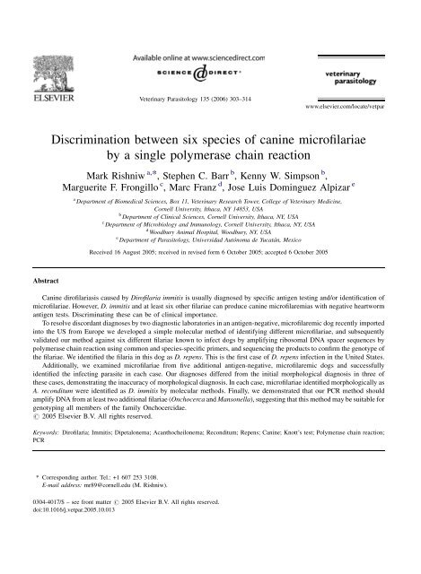
Discrimination between six species of canine microfilariae by a ...
Discrimination between six species of canine microfilariae by a ...
Discrimination between six species of canine microfilariae by a ...

You also want an ePaper? Increase the reach of your titles
YUMPU automatically turns print PDFs into web optimized ePapers that Google loves.
Abstract<br />
Veterinary Parasitology 135 (2006) 303–314<br />
<strong>Discrimination</strong> <strong>between</strong> <strong>six</strong> <strong>species</strong> <strong>of</strong> <strong>canine</strong> micr<strong>of</strong>ilariae<br />
<strong>by</strong> a single polymerase chain reaction<br />
Mark Rishniw a, *, Stephen C. Barr b , Kenny W. Simpson b ,<br />
Marguerite F. Frongillo c , Marc Franz d , Jose Luis Dominguez Alpizar e<br />
a Department <strong>of</strong> Biomedical Sciences, Box 11, Veterinary Research Tower, College <strong>of</strong> Veterinary Medicine,<br />
Cornell University, Ithaca, NY 14853, USA<br />
b Department <strong>of</strong> Clinical Sciences, Cornell University, Ithaca, NY, USA<br />
c Department <strong>of</strong> Microbiology and Immunology, Cornell University, Ithaca, NY, USA<br />
d Woodbury Animal Hospital, Woodbury, NY, USA<br />
e Department <strong>of</strong> Parasitology, Universidad Autónoma de Yucatán, Mexico<br />
Received 16 August 2005; received in revised form 6 October 2005; accepted 6 October 2005<br />
www.elsevier.com/locate/vetpar<br />
Canine dir<strong>of</strong>ilariasis caused <strong>by</strong> Dir<strong>of</strong>ilaria immitis is usually diagnosed <strong>by</strong> specific antigen testing and/or identification <strong>of</strong><br />
micr<strong>of</strong>ilariae. However, D. immitis and at least <strong>six</strong> other filariae can produce <strong>canine</strong> micr<strong>of</strong>ilaremias with negative heartworm<br />
antigen tests. Discriminating these can be <strong>of</strong> clinical importance.<br />
To resolve discordant diagnoses <strong>by</strong> two diagnostic laboratories in an antigen-negative, micr<strong>of</strong>ilaremic dog recently imported<br />
into the US from Europe we developed a simple molecular method <strong>of</strong> identifying different micr<strong>of</strong>ilariae, and subsequently<br />
validated our method against <strong>six</strong> different filariae known to infect dogs <strong>by</strong> amplifying ribosomal DNA spacer sequences <strong>by</strong><br />
polymerase chain reaction using common and <strong>species</strong>-specific primers, and sequencing the products to confirm the genotype <strong>of</strong><br />
the filariae. We identified the filaria in this dog as D. repens. This is the first case <strong>of</strong> D. repens infection in the United States.<br />
Additionally, we examined micr<strong>of</strong>ilariae from five additional antigen-negative, micr<strong>of</strong>ilaremic dogs and successfully<br />
identified the infecting parasite in each case. Our diagnoses differed from the initial morphological diagnosis in three <strong>of</strong><br />
these cases, demonstrating the inaccuracy <strong>of</strong> morphological diagnosis. In each case, micr<strong>of</strong>ilariae identified morphologically as<br />
A. reconditum were identified as D. immitis <strong>by</strong> molecular methods. Finally, we demonstrated that our PCR method should<br />
amplify DNA from at least two additional filariae (Onchocerca and Mansonella), suggesting that this method may be suitable for<br />
genotyping all members <strong>of</strong> the family Onchocercidae.<br />
# 2005 Elsevier B.V. All rights reserved.<br />
Keywords: Dir<strong>of</strong>ilaria; Immitis; Dipetalonema; Acanthocheilonema; Reconditum; Repens; Canine; Knott’s test; Polymerase chain reaction;<br />
PCR<br />
* Corresponding author. Tel.: +1 607 253 3108.<br />
E-mail address: mr89@cornell.edu (M. Rishniw).<br />
0304-4017/$ – see front matter # 2005 Elsevier B.V. All rights reserved.<br />
doi:10.1016/j.vetpar.2005.10.013
304<br />
1. Introduction<br />
Canine heartworm disease is generally diagnosed<br />
<strong>by</strong> antigen testing for Dir<strong>of</strong>ilaria immitis, and/or<br />
identification <strong>of</strong> micr<strong>of</strong>ilariae in the blood <strong>of</strong> infected<br />
dogs. However, other filariae, including Acanthocheilonema<br />
(Dipetalonema) reconditum, Dir<strong>of</strong>ilaria<br />
(Nochtiella) repens, Acanthocheilonema (Dipetalonema)<br />
dracunculoides, Cercopithifilaria grassi, Brugia<br />
malayi, Brugia pahangi, Brugia ceylonensis and<br />
approximately 1% <strong>of</strong> Dir<strong>of</strong>ilaria immitis infestations,<br />
can produce persistent micr<strong>of</strong>ilaremias with negative<br />
heartworm antigen tests (Courtney et al., 1993;<br />
Fischer et al., 2002; Genchi, 2003). In such cases,<br />
diagnosis relies on examination <strong>of</strong> the micr<strong>of</strong>ilariae,<br />
generally using a modified Knott’s test, or alkaline<br />
phosphatase staining (Chalifoux and Hunt, 1971;<br />
Knott, 1935). These methods are imperfect, and rely<br />
on specialist training to accurately differentiate the<br />
filariae. However, in regions where one <strong>of</strong> these<br />
filariae is not enzootic, parasitologists may exclude<br />
that filaria from consideration, or fail to recognize it,<br />
especially if a history <strong>of</strong> potential exposure is not<br />
provided (Cordonnier et al., 2002).<br />
Accurate identification <strong>of</strong> filarial <strong>species</strong> in dogs<br />
can be clinically important, because <strong>of</strong> the zoonotic<br />
concerns and therapeutic implications. While A.<br />
reconditum and A. dracunculoides have few (if any)<br />
clinical consequences, D. repens infestations have<br />
been associated with subcutaneous granulomas and<br />
pruritus in dogs (Baneth et al., 2002; Bredal et al.,<br />
1998; Manuali et al., 2005; Tarello, 2003), and<br />
subcutaneous, conjunctival and pulmonary nodules,<br />
<strong>of</strong>ten confused with neoplastic tumors, in humans<br />
(Cordonnier et al., 2002; Pampiglione et al., 1996,<br />
2001; Romano et al., 1976; Vakalis and Himonas,<br />
1997). To reduce the risk <strong>of</strong> zoonotic transmission,<br />
treatment <strong>of</strong> D. repens infestations with filaricidal<br />
agents, or at least micr<strong>of</strong>ilaricidal agents, is recommended<br />
(adulticidal therapy can be postponed until<br />
clinical signs occur if risk <strong>of</strong> treatment is deemed to<br />
outweigh risk <strong>of</strong> disease). Adult D. repens have been<br />
successfully treated with melarsomine hydrochloride<br />
(Immiticide, Merial; two injections <strong>of</strong> 2.5 mg/kg IM<br />
24 h apart) (Baneth et al., 2002). Micr<strong>of</strong>ilariae are<br />
susceptible to micr<strong>of</strong>ilaricidal doses <strong>of</strong> avermectins<br />
(ivermectin 50 mg/kg PO or SQ; doramectin 400 mg/<br />
kg SQ; effects <strong>of</strong> milbemycin have not been<br />
M. Rishniw et al. / Veterinary Parasitology 135 (2006) 303–314<br />
investigated) (Baneth et al., 2002; Tarello, 2003).<br />
On the other hand, treatment <strong>of</strong> A. dracunculoides or<br />
A. reconditum infestations is not usually required, so<br />
they should be positively identified to avoid inappropriate<br />
adulticide therapy.<br />
Over the last decade, several investigators have<br />
examined the molecular differentiation <strong>of</strong> filariae,<br />
using Southern blotting to identify DNA repeats from<br />
D. repens (Chandrasekharan et al., 1994) or polymerase<br />
chain reaction (PCR) to differentiate either D.<br />
repens or A. reconditum from D. immitis (Bredal et al.,<br />
1998; Favia et al., 1996, 1997; Mar et al., 2002;<br />
Vakalis et al., 1999). However, no existing studies<br />
examined the ability <strong>of</strong> PCR to differentiate <strong>between</strong><br />
more than two filariae. While it would be possible to<br />
run multiple PCR reactions using published primer<br />
sequences to identify a specific filaria, a single<br />
differentiating reaction would provide an expeditious<br />
means <strong>of</strong> obtaining a diagnosis. Additionally,<br />
sequences are not available for all filariae <strong>of</strong> interest<br />
at this time, so <strong>species</strong>-specific reactions may fail in<br />
these cases.<br />
This study was prompted <strong>by</strong> several clinical cases<br />
requiring accurate identification <strong>of</strong> micr<strong>of</strong>ilariae in<br />
dogs. These dogs had negative heartworm antigen<br />
tests and, in several instances, discrepant morphological<br />
identifications <strong>of</strong> the micr<strong>of</strong>ilariae. Using<br />
molecular techniques, we sought to accurately identify<br />
the filarial <strong>species</strong> in each case, and subsequently<br />
developed a PCR-based method that distinguishes<br />
<strong>between</strong> the <strong>six</strong> <strong>species</strong> tested to date (D. immitis, A.<br />
reconditum, D. repens, A. dracunculoides, B. pahangi,<br />
B. malayi) with a single primer pair. Our technique<br />
simplifies filarial identification, compared with previous<br />
studies.<br />
2. Materials and methods<br />
2.1. Case reports<br />
2.1.1. Case 1<br />
A 2-year-old intact male Labrador Retriever, born<br />
in the Czech Republic, but exported as a puppy to The<br />
Netherlands, was presented to the referring veterinarian<br />
(MF) for routine evaluation after importation from<br />
The Netherlands in January 2004 to Canada and<br />
subsequently New York State (United States) in April
2004. The dog was clinically normal and had never<br />
been tested for heartworm or administered heartworm<br />
preventative. The veterinarian identified a micr<strong>of</strong>ilaremia<br />
on routine heartworm screening, and performed<br />
a heartworm antigen test using a Dirocheck ELISA<br />
(Synbiotics Corporation, San Diego, CA), which was<br />
negative. Repeated antigen testing at several diagnostic<br />
laboratories failed to diagnose D. immitis<br />
infestation. Consequently, the veterinarian submitted<br />
blood for morphological classification <strong>of</strong> micr<strong>of</strong>ilariae<br />
to two diagnostic laboratories. Examination <strong>of</strong> the<br />
micr<strong>of</strong>ilariae using modified Knott’s tests resulted in<br />
discrepant diagnoses: one laboratory identified the<br />
micr<strong>of</strong>ilariae as A. reconditum, while the other<br />
identified them as D. repens.<br />
2.1.2. Case 2<br />
An 11-year-old male neutered mixed breed dog<br />
was presented to a referring veterinarian in San Diego,<br />
California, for routine evaluation. As with the first<br />
case, a micr<strong>of</strong>ilaremia was detected, but antigen<br />
testing for D. immitis was negative on multiple<br />
occasions. The micr<strong>of</strong>ilariae were identified as A.<br />
reconditum <strong>by</strong> a commercial diagnostic laboratory.<br />
2.1.3. Case 3<br />
An 8-year-old dog (signalment unknown) was<br />
presented to the referring veterinarian in Utica, New<br />
York, for evaluation <strong>of</strong> hematuria secondary to<br />
prostatitis. Micr<strong>of</strong>ilariae were detected on urinalysis,<br />
and subsequently on examination <strong>of</strong> blood. Antigen<br />
tests for D. immitis were negative. The micr<strong>of</strong>ilariae<br />
were morphologically identified as D. immitis with a<br />
Knott’s test.<br />
2.1.4. Case 4<br />
An 8-year-old female speyed miniature schnauzer<br />
was presented to the referring veterinarian after<br />
adoption from an animal shelter in Clemmons, North<br />
Carolina. Again, a micr<strong>of</strong>ilaremia was detected and<br />
identified morphologically as A. reconditum, but<br />
antigen testing for D. immitis was negative on<br />
multiple occasions.<br />
2.1.5. Case 5<br />
A 1-year-old male intact Pug was presented for<br />
heartworm testing in Modesto, California. Micr<strong>of</strong>ilaremia<br />
was detected and identified morphologically as<br />
M. Rishniw et al. / Veterinary Parasitology 135 (2006) 303–314 305<br />
A. reconditum, but antigen testing for D. immitis was<br />
negative.<br />
2.1.6. Case 6<br />
An 8-year-old male neutered Malamute was<br />
presented for heartworm testing in Vallejo, California.<br />
Micr<strong>of</strong>ilaremia was detected, and identified morphologically<br />
as A. reconditum but antigen testing for D.<br />
immitis was negative.<br />
2.2. Sample preparation<br />
In all <strong>six</strong> clinical cases, 1 ml <strong>of</strong> blood (anticoagulated<br />
with heparin or EDTA) was submitted.<br />
We used blood from a micr<strong>of</strong>ilaremic dog with D.<br />
immitis infestation and from a dog with A. reconditum<br />
infestation as positive controls. In both controls<br />
antigen testing and Knott’s testing provided the<br />
filarial diagnosis. The sample <strong>of</strong> A. recondituminfested<br />
blood was obtained <strong>by</strong> one <strong>of</strong> the authors and<br />
morphologically identified as A. reconditum. Fresh<br />
adult B. malayi and B. pahangi worms were<br />
generously provided <strong>by</strong> T. Klei <strong>of</strong> Louisiana State<br />
University. Blood from a dog infested with A.<br />
dracunculoides was generously provided <strong>by</strong> Francisco<br />
Alonso de Vega <strong>of</strong> Murcia University in Spain after<br />
morphologic diagnosis with alkaline phosphatase<br />
staining.<br />
Finally, we used DNA from a non-infected dog as a<br />
negative control in all PCR reactions.<br />
2.3. DNA extraction<br />
In all clinical cases, the D. immitis positive control<br />
specimen and the Brugia specimens, 500 ml anticoagulated<br />
blood (or five adult Brugia filariae) were<br />
suspended in 500 ml lysis buffer (50 mM Tris, pH 8.0;<br />
100 mM EDTA, 100 mM NaCl, 1% SDS) and<br />
digested with 10 ml proteinase K (10 mg/ml) for<br />
16 h at 55 8C, followed <strong>by</strong> a standard phenol/chlor<strong>of</strong>orm<br />
extraction method, as described previously<br />
(Sambrook and Russell, 2001).<br />
The A. reconditum and A. dracunculoides positive<br />
control specimens were submitted on filter paper as<br />
dried blood spots. DNAwas extracted using a QIAmp<br />
DNA Blood Mini Kit DNA extraction kit (Qiagen<br />
Inc., Valencia, CA) according to manufacturer’s<br />
instructions.
306<br />
2.4. Polymerase chain reaction filarial genotyping<br />
We designed primer pairs that spanned the internal<br />
transcribed spacer region 2 (ITS2) <strong>of</strong> the ribosomal<br />
DNA <strong>of</strong> either D. immitis, orA. reconditum, and a<br />
primer pair spanning the 5S ribosomal intergenic region<br />
<strong>of</strong> D. repens based on previously published sequences<br />
(Table 1) (Favia et al., 2000; Mar et al., 2002). These<br />
primers were used to specifically genotype each filarial<br />
<strong>species</strong>. Additionally, we designed a primer pair that<br />
would amplify fragments <strong>of</strong> different fragment length<br />
from both D. immitis and A. reconditum (DIDR-F1,<br />
DIDR-R1) (referred to as the pan-filarial primers<br />
throughout the remainder <strong>of</strong> the manuscript).<br />
For additional independent genotyping validation,<br />
we designed primers from published sequences <strong>of</strong> the<br />
cytochrome oxidase subunit 1 (COI) gene for each <strong>of</strong><br />
these <strong>species</strong> (Table 1).<br />
Each PCR reaction consisted <strong>of</strong> 1.5 mM MgCl2,<br />
250 mM dNTPs, 20 mM Tris–HCl (pH 8.4), 50 mM<br />
M. Rishniw et al. / Veterinary Parasitology 135 (2006) 303–314<br />
Table 1<br />
Primer sequences used to amplify PCR products from filarial and <strong>canine</strong> DNA<br />
Primer pair Primer sequence Gene target Primer size<br />
(base-pairs)<br />
KCl, 2.5 U <strong>of</strong> Taq polymerase and 1 ml <strong>of</strong> DNA<br />
solution in a total volume <strong>of</strong> 20 ml. The PCR<br />
procedure consisted <strong>of</strong> a denaturing step at 94 8C<br />
for 2 min and 32 cycles <strong>of</strong> denaturing (30 s at 94 8C),<br />
annealing (30 s at 60 8C for ITS2-based primers and<br />
30 s at 58 8C for COI-based primers) and extension<br />
(30 s at 72 8C); a final extension (7 min at 72 8C) and a<br />
soak at 4 8C in an Eppendorf Mastercycler thermal<br />
cycler (Brinkmann Instruments Inc., Westbury, NY).<br />
The PCR product (10 ml) was examined on a 1.5%<br />
agarose gel, and fragments <strong>of</strong> predicted size were<br />
extracted from both reactions and purified using a<br />
QiaQuick PCR purification kit (Qiagen Inc., Valencia,<br />
CA). The PCR products were ligated into pGEM-T<br />
Easy Vector System 1 TA cloning vectors (Promega<br />
US, Madison, WI) and cloned (Sambrook and Russell,<br />
2001). Candidate clones were screened <strong>by</strong> restriction<br />
digests, and clones with expected digestion fragments<br />
were sequenced at the institutional sequencing facility<br />
on an Applied Biosystems Automated 3730 DNA<br />
Product<br />
origin<br />
Product size<br />
(base-pairs)<br />
Genbank<br />
accession #<br />
D.imm-F1 a CAT CAG GTG ATG ATG TGA TGA T ITS2 22 D. immitis 302 AF217800<br />
D.imm-R1 TTG ATT GGA TTT TAA CGT ATC ATT T 25<br />
A.rec-F1 a<br />
CAG GTG ATG GTT TGA TGT GC ITS2 20 A. reconditum 348 AF217801<br />
A.rec-R1 CAC TCG CAC TGC TTC ACT TC 20<br />
D.rep-F1 TGT TTC GGC CTA GTG TTT CGA CCA 5SrRNA 24 D. repens 247 and 153 AJ242966<br />
D.rep-R1 ACG AGA TGT CGT GCT TTC AAC GTG 24 AJ242967<br />
DIDR-F1 AGT GCG AAT TGC AGA CGC ATT GAG 5.8S-ITS2-28S 24 D. immitis 542 AF217800<br />
DIDR-R1 AGC GGG TAA TCA CGA CTG AGT TGA 24 A. reconditum 578 AF217801<br />
D. repens 484 AY693808<br />
A. dracunculoides 584 DQ018785<br />
B. pahangi 664 AY988600<br />
B. malayi 615 AY988599<br />
B. timori 625 b<br />
AF499130<br />
M. ozzardi 430 b<br />
AF228554<br />
O. volvulus 470 b<br />
AF228575<br />
DI COI -F1 AGT GTA GAG GGT CAG CCT GAG TTA COI 24 D. immitis 203 AB192887<br />
DI COI-R1 ACA GGC ACT GAC AAT ACC AAT 21<br />
AR COI-F1 AGT GTT GAG GGA CAG CCA GAA TTG COI 24 A. reconditum 208 AJ544876<br />
AR COI-R1 CCA AAA CTG GAA CAG ACA AAA CAA GC 26<br />
DR COI-F1 AGT GTT GAT GGT CAA CCT GAA TTA COI 24 D. repens 209 AJ271614<br />
DR COI-R1 GCC AAA ACA GGA ACA GAT AAA ACT 24<br />
Actin F2 ACC ACT GGT ATT GTC ATG GAC TCT G Canine b-actin 25 Canis familiaris 276 AF021873<br />
Actin R2 GCT CTT CTC CAG GGA GGA CGA 21<br />
a D.imm-F1, D.imm-R1, A.rec-F1 and A.rec-R1 are identical to ITS2F-Di, ITS2R-Di, ITS2F-Dr and ITSR-Dr primers published <strong>by</strong> Mar et al.<br />
(2002).<br />
b Predicted product size based on Genbank sequence.
Analyzer (Applied Biosystems, Foster City, CA) using<br />
universal M13 forward and reverse primers. Sequence<br />
results were compared with those <strong>of</strong> other <strong>species</strong><br />
within the Genbank data bank at the National Center<br />
for Biomedical Information.<br />
Additionally, we examined DNA quality <strong>by</strong><br />
performing PCR for <strong>canine</strong> actin on each sample,<br />
to ensure sufficient template for the micr<strong>of</strong>ilarial<br />
PCR.<br />
2.5. Micr<strong>of</strong>ilarial phenotyping<br />
In addition to the PCR genotyping, we performed a<br />
modified Knott’s test and wet smear on the blood<br />
submitted from Cases 1–3 and the positive control<br />
samples. The morphological diagnosis for the <strong>six</strong><br />
clinical cases is the preliminary morphological<br />
diagnosis provided with initial sample submission<br />
and not the diagnosis obtained <strong>by</strong> one <strong>of</strong> the authors<br />
(MFF).<br />
3. Results<br />
All blood-derived DNA samples were positive for<br />
<strong>canine</strong> actin (not shown), indicating adequate DNA<br />
extraction.<br />
Primers specific for D. immitis (D.imm-F1 and<br />
D.imm-R1), A. reconditum (A.rec-F1 and A.rec-R1)<br />
and D. repens (D.rep-F1 and D.rep-R1) amplified the<br />
expected products only from D. immitis (302 bp), A.<br />
reconditum (348 bp) and D. repens (247 bp, 153 bp)<br />
DNA, respectively (Fig. 1). The doublet amplicon<br />
obtained with primers specific for D. repens has been<br />
previously reported (Favia et al., 2000). There was<br />
marginal amplification <strong>of</strong> a product from A. reconditum<br />
DNA (247 bp) with D. repens-specific primers.<br />
No filariae-specific primer pairs amplified any<br />
products from DNA <strong>of</strong> the normal dog (negative<br />
control), or from DNA extracted from B. pahangi,<br />
B. malayi or A. dracunculoides (data not shown).<br />
These results confirmed the genotyping <strong>of</strong> D. immitis,<br />
A. reconditum and D. repens for subsequent analysis<br />
and the specificity <strong>of</strong> the primer sets.<br />
The pan-filarial primer pair (DIDR-F1 and DIDR-<br />
R1) amplified the expected product from D. immitis<br />
DNA (542 bp), and A. reconditum DNA (578 bp)<br />
(Fig. 2). Unexpectedly, this primer pair also amplified<br />
M. Rishniw et al. / Veterinary Parasitology 135 (2006) 303–314 307<br />
Fig. 1. Gel electrophoresis <strong>of</strong> filarial PCR products in a 1.5%<br />
agarose gel using filarial-specific ITS2-region primers. Lanes 1,<br />
6, 11, 16: MW marker (KB Plus); lanes 2, 7, 12: D. immitis DNA;<br />
lanes 3, 8, 13: A. reconditum DNA; lanes 4, 9, 14: D. repens (Case 1)<br />
DNA; lanes 5, 10, 15: C. familiaris DNA (negative control). Lanes<br />
2–5: D.immF1/D.immR1 primer pair; lanes 7–10: A.recF1/A.recR1<br />
primer pair; lanes 12–15: D.repF1/D.repR1 primer pair.<br />
a 484 base-pair product from the DNA extracted from<br />
Case 1 (D. repens-infested dog). The different sizes <strong>of</strong><br />
the amplicons for all three filarial <strong>species</strong> allowed us to<br />
use this pan-filarial primer set to genotype the filarial<br />
<strong>species</strong> in Cases 2–6.<br />
Filaria-specific primers for COI demonstrated<br />
specificity for each <strong>of</strong> the filarial <strong>species</strong> (Fig. 3),<br />
further confirming the genotyping <strong>of</strong> micr<strong>of</strong>ilariae<br />
from Case 1 as D. repens and validating the panfilarial<br />
primer set results.<br />
Results <strong>of</strong> PCR with pan-filarial primers from<br />
Cases 2–4 and 6 identified the micr<strong>of</strong>ilariae as D.<br />
immitis (Fig. 4) (confirmed with <strong>species</strong>-specific COI<br />
PCR); PCR from Case 5 identified the micr<strong>of</strong>ilariae as<br />
A. reconditum (also confirmed with <strong>species</strong>-specific<br />
COI PCR).<br />
Fig. 2. Gel electrophoresis <strong>of</strong> filarial PCR products in a 1.5%<br />
agarose gel using pan-filarial ITS2-region primers. Lanes 1, 4:<br />
MW marker (KB Plus); lane 2: A. reconditum DNA; lane 3: D.<br />
immitis DNA; lane 3: D. repens (Case 1) DNA.
308<br />
Fig. 3. Gel electrophoresis <strong>of</strong> filarial PCR products in a 1.5%<br />
agarose gel using filarial-specific COI primers. Lanes 1, 5, 9, 13:<br />
MW marker (KB Plus); lanes 2, 6, 10: D. immitis DNA; lanes 3, 7,<br />
11: A. reconditum DNA; lanes 4, 8, 12: D. repens (Case 1) DNA.<br />
Sequencing <strong>of</strong> the two amplicons obtained with the<br />
D. repens-specific ITS2-region primers (D.rep-F1,<br />
D.rep-R1) unequivocally confirmed the presumptive<br />
diagnosis <strong>of</strong> D. repens micr<strong>of</strong>ilaremia in Case 1.<br />
Sequencing <strong>of</strong> the 484 bp amplicon obtained with the<br />
pan-filarial primers identified a novel sequence that<br />
Fig. 4. Gel electrophoresis <strong>of</strong> filarial PCR products in a 1.5%<br />
agarose gel using pan-filarial ITS2-region primers. Lanes 1, 6:<br />
MW marker (KB Plus); lane 2: Case 4 DNA; lane 3: D. immitis<br />
DNA; lane 4: A. reconditum DNA; lane 5: D. repens (Case 1) DNA.<br />
M. Rishniw et al. / Veterinary Parasitology 135 (2006) 303–314<br />
had high homology with the 5.8S and 28S ribosomal<br />
RNA sequences from D. immitis (present at the 5 0 and<br />
3 0 termini <strong>of</strong> the amplicon), but only moderate<br />
homology with the central D. immitis ITS2 region<br />
(Fig. 5), and no appreciable homology with A.<br />
reconditum ITS2 region. This sequence was consequently<br />
labeled as the ITS2 region <strong>of</strong> D. repens,<br />
submitted to the Genbank data bank at the National<br />
Center for Biomedical Information and assigned the<br />
accession number AY693808.<br />
Morphologic examination <strong>of</strong> the micr<strong>of</strong>ilariae from<br />
Case 1 using a modified Knott’s technique, showed a<br />
lack <strong>of</strong> a cephalic hook, a slightly tapering anterior<br />
end, and no consistent tail bend (Fig. 6). The<br />
concentration <strong>of</strong> micr<strong>of</strong>ilariae (abundant) and the<br />
motility <strong>of</strong> live micr<strong>of</strong>ilariae (not propulsive) on a wet<br />
smear, were most consistent with a diagnosis <strong>of</strong> D.<br />
repens. Examination <strong>of</strong> the micr<strong>of</strong>ilariae from Cases 2<br />
and 3 <strong>by</strong> one <strong>of</strong> the authors (MFF) supported our<br />
molecular diagnosis <strong>of</strong> D. immitis.<br />
Results <strong>of</strong> PCR from B. malayi, B. pahangi and A.<br />
dracunculoides DNA, using the pan-filarial primers,<br />
produced bands at 615 bp (B. malayi), 664 bp (B.<br />
pahangi) and 584 bp (A. dracunculoides) (data not<br />
shown). Sequencing <strong>of</strong> these amplicons revealed<br />
unique sequences, which were subsequently submitted<br />
to Genbank and assigned accession numbers<br />
AY988599 (B. malayi), AY988600 (B. pahangi) and<br />
DQ018785 (A. dracunculoides).<br />
While PCR with pan-filarial primers failed to<br />
discriminate <strong>between</strong> A. reconditum and A. dracunculoides,<br />
restriction fragment length polymorphism<br />
PCR (RFLP-PCR) using MseII resulted in two<br />
fragments (251 bp and 333 bp) only in A. dracunculoides<br />
(data not shown) allowing discrimination<br />
<strong>between</strong> these two Acanthocheilonema <strong>species</strong>.<br />
Sequence comparison <strong>of</strong> the ITS2 region amplified<br />
<strong>by</strong> our pan-filarial primers (DIDR-F1 and DIDR-R1)<br />
and previously published filarial sequences <strong>of</strong> this<br />
region demonstrated 77–86% similarity <strong>between</strong><br />
<strong>species</strong> <strong>of</strong> the same genus (e.g. D. immitis and D.<br />
repens showed 77% similarity, all three Brugia <strong>species</strong><br />
showed 86% similarity), but only 30–60% similarity<br />
<strong>between</strong> <strong>species</strong> <strong>of</strong> different genera (e.g. B. pahangi<br />
and A. reconditum showed only 30% similarity).<br />
Phylogenetic tree analysis also grouped sequences<br />
most closely <strong>by</strong> genus, i.e., <strong>species</strong> <strong>of</strong> the same genus<br />
grouped together.
Fig. 5. Nucleotide sequence alignment <strong>of</strong> 5.8S-ITS2-28S ribosomal DNA region <strong>of</strong> seven filariae. The 5.8S and 28S ribosomal DNA are identified above the 5 0 and 3 0 ends <strong>of</strong> the<br />
sequences; shaded sequences within the majority sequence are primer binding sequences for the pan-filarial primers.<br />
M. Rishniw et al. / Veterinary Parasitology 135 (2006) 303–314 309
310<br />
M. Rishniw et al. / Veterinary Parasitology 135 (2006) 303–314<br />
Fig. 5. (Continued).
4. Discussion<br />
We initiated this study to resolve a discordant<br />
diagnosis <strong>of</strong> filariasis in a dog, and successfully<br />
identified a D. repens infestation in a dog in the United<br />
States <strong>by</strong> PCR genotyping <strong>of</strong> micr<strong>of</strong>ilarial DNA<br />
extracted from the patient’s blood. We also resolved<br />
discordant diagnoses provided <strong>by</strong> standard morphological<br />
examination <strong>of</strong> the micr<strong>of</strong>ilariae (Knott’s test).<br />
Unexpectedly, the pan-filarial primers we designed<br />
(DIDR-F1 and DIDR-R1), based on sequence homology<br />
<strong>of</strong> D. immitis and A. reconditum, amplified not only<br />
the expected 542 and 578 bp products from these<br />
filariae, but also a 484 bp product from D. repens, that<br />
was present in the Case 1 <strong>of</strong> this study, a 584 bp product<br />
from A. dracunculoides, a 615 bp product from B.<br />
malayi, and a 664 bp product from B. pahangi. This<br />
permitted a single PCR reaction to differentiate<br />
<strong>between</strong> D. immitis and five other filariae found in<br />
dogs, removing the need to perform multiple <strong>species</strong>specific<br />
genotyping reactions. We subsequently identified<br />
micr<strong>of</strong>ilariae from an additional four dogs as D.<br />
immitis and as A. reconditum in one dog with this panfilarial<br />
primer set. Our molecular identification was<br />
concordant with independent morphological phenotyping<br />
<strong>of</strong> the micr<strong>of</strong>ilariae <strong>by</strong> an experienced parasitologist<br />
(MFF), but discordant with the initial morphological<br />
diagnosis in 3/5 cases, illustrating the inaccuracy <strong>of</strong><br />
morphological diagnosis <strong>by</strong> minimally trained technicians.<br />
Finally, we were able to provide additional<br />
genotyping information for several filariae that usually<br />
require specialized diagnostic interpretation.<br />
M. Rishniw et al. / Veterinary Parasitology 135 (2006) 303–314 311<br />
Fig. 6. Modified Knott’s stain <strong>of</strong> micr<strong>of</strong>ilaria from Case 1 (D. repens). The head is slightly tapered, there is no cephalic hook, and the tail is<br />
straight, consistent with Dir<strong>of</strong>ilaria repens. Magnification 100 .<br />
Previous investigators have utilized PCR technology<br />
to diagnose filarial infestations in dogs and<br />
humans (Baneth et al., 2002; Favia et al., 1996, 1997;<br />
Mar et al., 2002); however, in each <strong>of</strong> these studies,<br />
only two filarial <strong>species</strong> were compared. We designed<br />
our pan-filarial primers from previously submitted<br />
sequences, and optimized them for efficient amplification.<br />
The need to correctly identify the filarial <strong>species</strong> is<br />
becoming relevant throughout the world, because <strong>of</strong><br />
the increased frequency <strong>of</strong> transportation <strong>of</strong> dogs<br />
<strong>between</strong> continents and countries (Genchi, 2003;<br />
Zahler et al., 1997). While D. repens has been<br />
considered an exotic parasite in some parts <strong>of</strong> the<br />
world, such as the United States and Australia, and<br />
consequently not considered in the differential<br />
diagnosis <strong>of</strong> <strong>canine</strong> filariasis in these continents, this<br />
case illustrates the potential diagnostic complications<br />
that can arise, and provides a simple molecular<br />
strategy for correctly identifying the filaroid. The<br />
potential mosquito vectors <strong>of</strong> D. repens—Aedes<br />
aegypti (Cancrini et al., 2003), Ae. albopictus (Anyanwu<br />
et al., 2000) and Ae. vexans—are enzootic in the<br />
United States and Ae. vexans is enzootic in the region<br />
<strong>of</strong> New York State where this dog currently lives<br />
(Nassau County Department <strong>of</strong> Health, 2003).<br />
Similarly, Ae. aegypti is enzootic in Australia; Ae.<br />
albopictus is not found in Australia. Thus, the<br />
potential exists for spread <strong>of</strong> D. repens in these<br />
regions. Whether the strains in the United States or<br />
Australia can incubate larvae and transmit the<br />
infestations is unknown. Interestingly, Ae. aegypti is
312<br />
relatively resistant to infestation with D. immitis, and<br />
is considered an inefficient intermediate host for<br />
transmission <strong>of</strong> heartworm disease (Apperson et al.,<br />
1989).<br />
The geographic region in which Case 1 acquired<br />
the infection is unknown. Dir<strong>of</strong>ilaria repens has not<br />
been reported in The Netherlands, but has been<br />
identified in the Czech Republic (where this dog was<br />
born), Hungary, Italy, Greece and France (Cordonnier<br />
et al., 2002; Elek et al., 2000; Pampiglione et al., 2001;<br />
Vakalis and Himonas, 1997; Vasilkova et al., 1992).<br />
The micr<strong>of</strong>ilariae from Case 2 were initially<br />
incorrectly identified as A. reconditum, partly based<br />
on a negative antigen test, while the micr<strong>of</strong>ilariae in<br />
Case 3 had been identified correctly as D. immitis. In<br />
Cases 2 and 3, examination <strong>by</strong> an experienced<br />
parasitologist (MFF) correctly identified the micr<strong>of</strong>ilariae<br />
as D. immitis. Causes for antigen-negative<br />
D. immitis micr<strong>of</strong>ilaremias include prior adulticide<br />
therapy without subsequent micr<strong>of</strong>ilaricidal therapy or<br />
natural death <strong>of</strong> adult heartworm with persistence <strong>of</strong><br />
micr<strong>of</strong>ilariae (which have a lifespan <strong>of</strong> approximately<br />
2 years) (Otto, 1977). Additionally, at least one<br />
product insert (Dirocheck Canine Heartworm Antigen<br />
Test Kit Product Insert, Synbiotics, San Diego, CA)<br />
also lists subthreshold antigenemia, immune clearance<br />
<strong>of</strong> antigen–antibody complexes, false positive micr<strong>of</strong>ilarial<br />
results (due to contamination <strong>of</strong> reagents used<br />
for sample preparation), transfusions from micr<strong>of</strong>ilaremic<br />
dogs, or destruction <strong>of</strong> antigen due to<br />
improper storage as potential causes <strong>of</strong> discordant<br />
results. While a Dir<strong>of</strong>ilaria micr<strong>of</strong>ilaremia does not<br />
necessarily endanger the health <strong>of</strong> the infested patient,<br />
the patient can act as a reservoir for further infestation<br />
<strong>of</strong> other dogs if the intermediate mosquito host is<br />
present. Micr<strong>of</strong>ilaricidal therapy can resolve this<br />
situation.<br />
The micr<strong>of</strong>ilaremia in Case 3 was initially<br />
identified during a routine urinalysis for hematuria<br />
(micr<strong>of</strong>ilaruria). We believe this is the first reported<br />
case <strong>of</strong> micr<strong>of</strong>ilaruria in a dog. A prior report exists <strong>of</strong><br />
micr<strong>of</strong>ilaruria in a cat (Beaufils et al., 1991).<br />
We did not test the discriminatory ability <strong>of</strong> our<br />
pan-filarial primer set against all filariae that are<br />
known to infest dogs or cats (Genchi, 2003). At least<br />
one additional ‘‘Dipetalonema-like’’ filaria exists in<br />
Europe and Africa—Cercopithifilaria (Acanthocheilonema)<br />
grassi—but has not been reported in the<br />
M. Rishniw et al. / Veterinary Parasitology 135 (2006) 303–314<br />
United States. It is possible that our pan-filarial primer<br />
set will fail to discriminate this filaria from D. immitis,<br />
however, D. immitis-specific primer sets should<br />
correctly genotype the filarial <strong>species</strong> in question if<br />
this becomes necessary. Further studies are required to<br />
validate the pan-filarial primers against C. grassi.<br />
Additionally, reports exist <strong>of</strong> novel filariae in dogs in<br />
Ireland and the United States (Jackson, 1975; Pacheco<br />
and Tulloch, 1970). Techniques such as those<br />
described in this report might be useful in genotyping<br />
such filariae.<br />
The B. malayi sequence we obtained from adult<br />
filariae showed only 86% similarity with a published<br />
B. malayi sequence (Genbank # AF499130), comparable<br />
to the degree <strong>of</strong> similarity <strong>between</strong> our B. pahangi<br />
sequence and the published B. malayi sequence<br />
(AF499130), or <strong>between</strong> our B pahangi and B. malayi<br />
sequences. This differed substantially from the<br />
similarity <strong>between</strong> the published sequences for B.<br />
malayi (AF499130) and B. timori (AF499132), which<br />
the authors claimed were 99.5% similar, with only 4<br />
base-pair substitutions along the 600 base-pair region<br />
(Fischer et al., 2002). Several reasons exist for these<br />
discrepant results. Fischer et al claimed that this<br />
degree <strong>of</strong> similarity was unique to these two <strong>species</strong>,<br />
however, they did not examine DNA from adult<br />
Brugia <strong>species</strong>, but only from micr<strong>of</strong>ilaria, and <strong>by</strong><br />
supposition based on epidemiological data. Thus, their<br />
initial phenotypic classification, which they used as a<br />
gold standard for that study, could be erroneous. We<br />
could find no study <strong>by</strong> these authors that confirmed<br />
their results using adult B. malayi and B. timori. Given<br />
the much lower degree <strong>of</strong> similarity <strong>between</strong> <strong>species</strong><br />
<strong>of</strong> a single genus detailed in this report (e.g. D. immitis<br />
and D. repens, B. malayi and B. pahangi, or A.<br />
reconditum and A. dracunculoides all showed intragenus,<br />
inter<strong>species</strong> variation <strong>of</strong> approximately 20%,<br />
similar to the difference we observed <strong>between</strong> B.<br />
malayi and the previously published B. timori), we<br />
suspect that the sequence for B. malayi (AF499130)<br />
published <strong>by</strong> Fischer et al. (2002) is incorrect and is in<br />
fact the sequence for B timori. Examination <strong>of</strong> the<br />
data presented <strong>by</strong> these authors indicates a broad<br />
genetic variation <strong>of</strong> the Hha1 tandem repeat in these<br />
<strong>species</strong>, which also suggests a misclassification <strong>of</strong><br />
micr<strong>of</strong>ilariae.<br />
A less plausible alternative is that inter-<strong>species</strong><br />
variation <strong>of</strong> the ITS2 region <strong>between</strong> B. malayi and B.
timori (20%). Several<br />
studies <strong>of</strong> ITS2 regions in various helminths suggest<br />
that while intra-<strong>species</strong> variation can occur, it is less<br />
than inter-<strong>species</strong> variation, and that in most cases<br />
intra-<strong>species</strong> variation is limited to single nucleotide<br />
polymorphisms (Conole et al., 1999; Gasser et al.,<br />
1999a,b; Hu<strong>by</strong>-Chilton et al., 2001; Newton et al.,<br />
1998; Woods et al., 2000) and previous studies <strong>of</strong> other<br />
filariae show similar results (Gasser et al., 1996).<br />
These findings agree with our observations <strong>of</strong> ITS2<br />
variability in filariae and support our genotypic<br />
identification <strong>of</strong> B. malayi. Finally, it is possible that<br />
the sample <strong>of</strong> adult B. malayi provided for our study<br />
was incorrectly identified; however, phenotypic<br />
classification <strong>of</strong> filariae is generally more robust<br />
using adult filariae than micr<strong>of</strong>ilariae. The reason for<br />
the discrepancy <strong>between</strong> our data and that <strong>of</strong> Fischer<br />
et al. (2002) remains to be elucidated.<br />
Finally, we examined the ability <strong>of</strong> our pan-filarial<br />
primer sequences to amplify the 5.8S-ITS2-28S<br />
region from two additional filariae in silico. Analysis<br />
<strong>of</strong> published sequences for Onchocerca volvulus<br />
(Genbank # AF228575), and Mansonella ozzardi<br />
(Genbank # AF228554) demonstrated that our panfilarial<br />
primers should amplify 470 and 430 bp<br />
amplicons, respectively. This suggests that our primer<br />
set might be uniquely capable <strong>of</strong> amplifying DNA that<br />
can be used to genotype all filariae <strong>of</strong> the family<br />
Onchocercidae.<br />
We recommend using the pan-filarial ITS2-region<br />
DIDR-F1 and DIDR-R1 primers with DNA from<br />
D. immitis (as a positive control and amplicon size<br />
standard) against which to compare amplicons from<br />
clinical cases <strong>of</strong> micr<strong>of</strong>ilaremia that test negative for<br />
D. immitis antigen. Amplicons approximately 40 basepairs<br />
larger than the D. immitis amplicon (542 basepairs)<br />
are diagnosed as A. reconditum (578 base-pairs)<br />
or A. dracunculoides (584 base-pairs), while those<br />
approximately 60 base-pairs smaller than D. immitis<br />
are diagnosed as D. repens (484 base-pairs). <strong>Discrimination</strong><br />
<strong>of</strong> Brugia <strong>species</strong> <strong>by</strong> comparing amplicons<br />
is limited because B. malayi (615 base-pairs) is<br />
similar to B. timori (625 base-pairs), but these <strong>species</strong><br />
should be easily differentiated from B. pahangi (664<br />
base-pairs). However, PCR-RFLP will discriminate B.<br />
malayi and B. timori. If further confirmation is required,<br />
either <strong>species</strong>-specific PCR can be performed,<br />
M. Rishniw et al. / Veterinary Parasitology 135 (2006) 303–314 313<br />
using the published primer pairs from this and other<br />
studies, or the amplicons can be sequenced, however,<br />
we feel that simple gel electrophoresis should be<br />
sufficient for diagnosis in most cases. Additionally, we<br />
feel that clinicians and parasitologists should be<br />
alerted to the possibility that D. repens infestation<br />
might be an emerging condition with zoonotic<br />
potential in the United States.<br />
Acknowledgements<br />
The authors would like to thank Dwight Bowman,<br />
Susan Wade and Stephanie Schaaf for their technical<br />
insights and providing D. immitis positive control<br />
samples; Andrew Fei and Fabiano Montiani Ferreira<br />
for assistance in obtaining A. reconditum samples;<br />
Thom Klei for generously providing Brugia filaria;<br />
Francisco Alonso de Vega for generously providing<br />
A. dracunculoides samples and Veterinary Information<br />
Network for providing access to the case that<br />
initiated this study.<br />
References<br />
Anyanwu, I.N., Agbede, R.I., Ajanusi, O.J., Umoh, J.U., Ibrahim,<br />
N.D., 2000. The incrimination <strong>of</strong> Aedes (stegomyia) aegypti as<br />
the vector <strong>of</strong> Dir<strong>of</strong>ilaria repens in Nigeria. Vet. Parasitol. 92,<br />
319–327.<br />
Apperson, C.S., Engber, B., Levine, J.F., 1989. Relative suitability<br />
<strong>of</strong> Aedes albopictus and Aedes aegypti in North Carolina to<br />
support development <strong>of</strong> Dir<strong>of</strong>ilaria immitis. J. Am. Mosq.<br />
Control Assoc. 5, 377–382.<br />
Baneth, G., Volansky, Z., Anug, Y., Favia, G., Bain, O., Goldstein,<br />
R.E., Harrus, S., 2002. Dir<strong>of</strong>ilaria repens infection in a dog:<br />
diagnosis and treatment with melarsomine and doramectin. Vet.<br />
Parasitol. 105, 173–178.<br />
Beaufils, J.P., Martin-Granel, J., Bertrand, F., 1991. Présance de<br />
micr<strong>of</strong>ilaires de Dir<strong>of</strong>ilaria immitis dans les urines d’un chat<br />
occlus. Prat. Med. Chir. An. Com. 26, 467–472 in French.<br />
Bredal, W.P., Gjerde, B., Eberhard, M.L., Aleksandersen, M.,<br />
Wilhelmsen, D.K., Mansfield, L.S., 1998. Adult Dir<strong>of</strong>ilaria<br />
repens in a subcutaneous granuloma on the chest <strong>of</strong> a dog. J.<br />
Small Anim. Pract. 39, 595–597.<br />
Cancrini, G., Romi, R., Gabrielli, S., Toma, L., Di Paolo, M.,<br />
Scaramozzino, P., 2003. First finding <strong>of</strong> Dir<strong>of</strong>ilaria repens in<br />
a natural population <strong>of</strong> Aedes albopictus. Med. Vet. Entomol. 17,<br />
448–451.<br />
Chalifoux, L., Hunt, R.D., 1971. Histochemical differentiation <strong>of</strong><br />
Dir<strong>of</strong>ilaria immitis and Dipetalonema reconditum. J. Am. Vet.<br />
Med. Assoc. 158, 601–605.
314<br />
Chandrasekharan, N.V., Karunanayake, E.H., Franzen, L., Abeyewickreme,<br />
W., Pettersson, U., 1994. Dir<strong>of</strong>ilaria repens: cloning<br />
and characterization <strong>of</strong> a repeated DNA sequence for the diagnosis<br />
<strong>of</strong> dir<strong>of</strong>ilariasis in dogs, Canis familiaris. Exp. Parasitol.<br />
78, 279–286.<br />
Conole, J.C., Chilton, N.B., Jarvis, T., Gasser, R.B., 1999. Intraspecific<br />
and interspecific variation in the second internal transcribed<br />
spacer (ITS-2) sequence for Metastrongylus (Nematoda:<br />
Metastrongyloidea) detected <strong>by</strong> high resolution PCR-RFLP. Int.<br />
J. Parasitol. 29, 1935–1940.<br />
Cordonnier, C., Chatelain, D., Nevez, G., Sevestre, H., Gontier,<br />
M.F., Raccurt, C.P., 2002. Difficulties in diagnosis <strong>of</strong> human<br />
dir<strong>of</strong>ilariasis outside known enzootic areas. Rev. Med. Interne<br />
23, 71–76.<br />
Courtney, C.H., Zeng, Q.Y., MacKinnon, B.R., 1993. Comparison <strong>of</strong><br />
two antigen tests and the modified Knott’s test for detection <strong>of</strong><br />
<strong>canine</strong> heartworm at different worm burdens. Canine Pract. 18,<br />
5–7.<br />
Elek, G., Minik, K., Pajor, L., Parlagi, G., Varga, I., Vetesi, F.,<br />
Zombori, J., 2000. New human Dir<strong>of</strong>ilarioses in Hungary.<br />
Pathol. Oncol. Res. 6, 141–145.<br />
Favia, G., Lanfrancotti, A., Della Torre, A., Cancrini, G., Coluzzi,<br />
M., 1996. Polymerase chain reaction-identification <strong>of</strong> Dir<strong>of</strong>ilaria<br />
repens and Dir<strong>of</strong>ilaria immitis. Parasitology 113 (Pt. 6),<br />
567–571.<br />
Favia, G., Lanfrancotti, A., della Torre, A., Cancrini, G., Coluzzi,<br />
M., 1997. Advances in the identification <strong>of</strong> Dir<strong>of</strong>ilaria repens<br />
and Dir<strong>of</strong>ilaria immitis <strong>by</strong> a PCR-based approach. Parassitologia<br />
39, 401–402.<br />
Favia, G., Bazzocchi, C., Cancrini, G., Genchi, C., Bandi, C., 2000.<br />
Unusual organization <strong>of</strong> the 5S ribosomal spacer in Dir<strong>of</strong>ilaria<br />
repens: absence <strong>of</strong> a canonical spliced leader 1 sequence.<br />
Parasitol. Res. 86, 497–499.<br />
Fischer, P., Wibowo, H., Pischke, S., Ruckert, P., Liebau, E., Ismid,<br />
I.S., Supali, T., 2002. PCR-based detection and identification <strong>of</strong><br />
the filarial parasite Brugia timori from Alor Island, Indonesia.<br />
Ann. Trop. Med. Parasitol. 96, 809–821.<br />
Gasser, R.B., LeG<strong>of</strong>f, L., Petit, G., Bain, O., 1996. Rapid delineation<br />
<strong>of</strong> closely-related filarial parasites using genetic markers in<br />
spacer rDNA. Acta Trop. 62, 143–150.<br />
Gasser, R.B., Rossi, L., Zhu, X., 1999a. Identification <strong>of</strong> Nematodirus<br />
<strong>species</strong> (Nematoda: Molineidae) from wild ruminants in Italy<br />
using ribosomal DNA markers. Int. J. Parasitol. 29, 1809–1817.<br />
Gasser, R.B., Woods, W.G., Huffman, M.A., Blotkamp, J., Polderman,<br />
A.M., 1999b. Molecular separation <strong>of</strong> Oesophagostomum<br />
stephanostomum and Oesophagostomum bifurcum (Nematoda:<br />
Strongyloidea) from non-human primates. Int. J. Parasitol. 29,<br />
1087–1091.<br />
Genchi, C., 2003. Epidemiology and distribution <strong>of</strong> Dir<strong>of</strong>ilaria<br />
and dir<strong>of</strong>ilariosis in Europe: state <strong>of</strong> the art. In: Proceedings<br />
<strong>of</strong> the 2003 Helminthological Colloquium, Vienna, Austria, pp.<br />
6–11., http://www.vu-wien.ac.at/i116/oegtp/downloads_oegtp/<br />
Abstractband.pdf.<br />
Hu<strong>by</strong>-Chilton, F., Beveridge, I., Gasser, R.B., Chilton, N.B., 2001.<br />
Single-strand conformation polymorphism analysis <strong>of</strong> genetic<br />
variation in Labiostrongylus longispicularis from kangaroos.<br />
Electrophoresis 22, 1925–1929.<br />
M. Rishniw et al. / Veterinary Parasitology 135 (2006) 303–314<br />
Jackson, R.F., 1975. A filarial parasite in greyhounds in Southern<br />
Ireland. Vet. Rec. 97, 476–477.<br />
Knott, J., 1935. The periodicity <strong>of</strong> the mf <strong>of</strong> Wuchereia bancr<strong>of</strong>ti,<br />
preliminary report <strong>of</strong> some injection experiments. Trans. Roy.<br />
Soc. Trop. Med. Hyg. 29, 59–64.<br />
Manuali, E., Eleni, C., Giovannini, P., Costarelli, S., Ciorba, A.,<br />
2005. Unusual finding in a nipple discharge <strong>of</strong> a female dog:<br />
dir<strong>of</strong>ilariasis <strong>of</strong> the breast. Diagn. Cytopathol. 32, 108–<br />
109.<br />
Mar, P.H., Yang, I.C., Chang, G.N., Fei, A.C., 2002. Specific<br />
polymerase chain reaction for differential diagnosis <strong>of</strong> Dir<strong>of</strong>ilaria<br />
immitis and Dipetalonema reconditum using primers<br />
derived from internal transcribed spacer region 2 (ITS2). Vet.<br />
Parasitol. 106, 243–252.<br />
Nassau County Department <strong>of</strong> Health, 2003. Nassau County Mosquito<br />
Surveillance and Control Report.<br />
Newton, L.A., Chilton, N.B., Beveridge, I., Gasser, R.B., 1998.<br />
Systematic relationships <strong>of</strong> some members <strong>of</strong> the genera Oesophagostomum<br />
and Chabertia (Nematoda: Chabertiidae) based<br />
on ribosomal DNA sequence data. Int. J. Parasitol. 28, 1781–<br />
1789.<br />
Otto, G.F., 1977. The significance <strong>of</strong> micr<strong>of</strong>ilaremia in the diagnosis<br />
<strong>of</strong> heartworm infection. In: Proceedings <strong>of</strong> the American<br />
Heartworm Society Heartworm Symposium. pp. 22–30.<br />
Pacheco, G., Tulloch, G.S., 1970. Micr<strong>of</strong>ilariae <strong>of</strong> Dir<strong>of</strong>ilaria striata<br />
in a dog. J. Parasitol. 56, 248.<br />
Pampiglione, S., Rivasi, F., Angeli, G., Boldorini, R., Incensati,<br />
R.M., Pastormerlo, M., Pavesi, M., Ramponi, A., 2001. Dir<strong>of</strong>ilariasis<br />
due to Dir<strong>of</strong>ilaria repens in Italy, an emergent zoonosis:<br />
report <strong>of</strong> 60 new cases. Histopathology 38, 344–354.<br />
Pampiglione, S., Rivasi, F., Paolino, S., 1996. Human pulmonary<br />
dir<strong>of</strong>ilariasis. Histopathology 29, 69–72.<br />
Romano, A., Sachs, R., Lengy, J., 1976. Human ocular dir<strong>of</strong>ilariasis<br />
in Israel. Isr. J. Med. Sci. 12, 208–214.<br />
Sambrook, J., Russell, D.W. (Eds.), 2001. Molecular Cloning: A<br />
Laboratory Manual, 3rd ed., vol. 1. Cold Spring Harbor Laboratory<br />
Press, Cold Spring Harbor.<br />
Tarello, W., 2003. Dermatitis associated with Dir<strong>of</strong>ilaria repens<br />
micr<strong>of</strong>ilariae in three dogs in Saudi Arabia. J. Small Anim. Pract.<br />
44, 132–134.<br />
Vakalis, N., Spanakos, G., Patsoula, E., Vamvakopoulos, N.C., 1999.<br />
Improved detection <strong>of</strong> Dir<strong>of</strong>ilaria repens DNA <strong>by</strong> direct polymerase<br />
chain reaction. Parasitol. Int. 48, 145–150.<br />
Vakalis, N.C., Himonas, C.A., 1997. Human and <strong>canine</strong> dir<strong>of</strong>ilariasis<br />
in Greece. Parassitologia 39, 389–391.<br />
Vasilkova, D., Klisenbauer, D., Juhas, T., Moravec, F., Uhlikova, M.,<br />
Hubner, J., Konakova, G., 1992. Isolation <strong>of</strong> Dir<strong>of</strong>ilaria repens<br />
in vitreoretinal findings. Cesk. Oftalmol. 48, 274–277.<br />
Woods, W.G., Whithear, K.G., Richards, D.G., Anderson, G.R.,<br />
Jorgensen, W.K., Gasser, R.B., 2000. Single-strand restriction<br />
fragment length polymorphism analysis <strong>of</strong> the second internal<br />
transcribed spacer (ribosomal DNA) for <strong>six</strong> <strong>species</strong> <strong>of</strong> Eimeria<br />
from chickens in Australia. Int. J. Parasitol. 30, 1019–<br />
1023.<br />
Zahler, M., Glaser, B., Gothe, R., 1997. Imported parasites in dogs:<br />
Dir<strong>of</strong>ilaria repens and Dipetalonema reconditum. Tierarztl.<br />
Prax. 25, 388–392.