Hand atlas of human anatomy - EducationNest
Hand atlas of human anatomy - EducationNest
Hand atlas of human anatomy - EducationNest

Create successful ePaper yourself
Turn your PDF publications into a flip-book with our unique Google optimized e-Paper software.
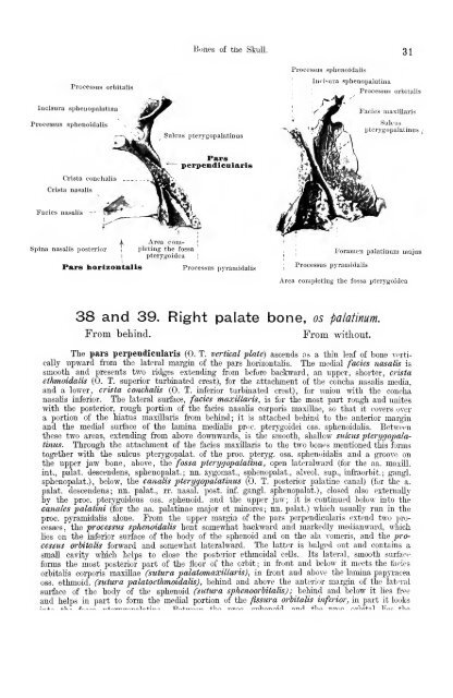
Processus orbitalis<br />
luciaura spheuopalatina<br />
Processus sphenoidalis<br />
Crista conchalis<br />
Crista nasalis<br />
Fades nasalis "<br />
Spina nasalis posterior<br />
Bones <strong>of</strong> the Skull. 31<br />
Sulcus pterygopalatinus<br />
Area completing<br />
the fossa<br />
pterygoidea<br />
Pars<br />
perpendicalaris<br />
I<br />
Pars horizontalis Processus pyramidalis<br />
' Processus<br />
Processus sphenoidalis<br />
j<br />
;<br />
Incisina spheuopalatina<br />
pyramidalis<br />
Processus orbitalis<br />
l-'acies maxlllaris<br />
Sulcus<br />
pterygopalatinus ;<br />
Foramen palatinum majus<br />
Area completing the fossa pterygoidea<br />
38 and 39. Right palate bone, os palatinum.<br />
From behind. From M'ithout.<br />
The pars perpendicnlarig (0. T. vertical plate) ascends as a thin leaf <strong>of</strong> bone verti-<br />
cally upward fi-om the lateral margin <strong>of</strong> the pars horizontalis. The medial fades nasalis is<br />
smooth and presents two ridges extending from before backward, an upper, shorter, crista<br />
ethmoidalis (0. T. superior turbinated crest), for the attachment <strong>of</strong> the concha nasalis media,<br />
and a lower, crista conchalis (0. T. inferior turbinated crest), for union with the concha<br />
nasalis inferior. The lateral surface, fades maxillaris, is for the most part rough and unites<br />
with the posterior, rough portion <strong>of</strong> the fades nasalis corporis maxiUae, so that it covers over<br />
a portion <strong>of</strong> the hiatus maxillaris from behind; it is attached behind to the anterior margin<br />
and the medial surface <strong>of</strong> the lamina medialis pr(jc. pterygoidei oss. sphenoidalis. Between<br />
these two areas, extending from above downwards, is the smooth, shallow sulcus pterygopalatinus.<br />
Through the attachment <strong>of</strong> the fades maxillaris to the two bones mentioned this forms<br />
together with the sulcus pterygopalat. <strong>of</strong> the proc. pteryg. oss. sphenoidalis and a groove on<br />
the upper jaw bone, above, the fossa pterygopalatina , open lateralward (for the aa. maxill.<br />
int., palat. descendens, sphenopalat. ; nn. zygomat., sphenopalat., alveol. sup., infraorbit. ; gangl.<br />
sphenopalat.) , below, the canalis pterygopalatinus (0. T. posterior palatine canal) (for the a.<br />
palat. descendens; nn. palat., rr. nasal, post. inf. gangl. sphenopalat.), closed also externally<br />
by the proc. pterygoideus oss. sphenoid, and the upper jaw; it is continued below into the<br />
canales palatini (for the aa. palatinae major et minores ; nn. palat.) which usually run in the<br />
proc. pyramidahs alone. Prom the upper margin <strong>of</strong> the pars perpendicularis extend two processes,<br />
the processus sphenoidalis bent somewhat backward and markedly medianward, which<br />
lies on the inferior surface <strong>of</strong> the body <strong>of</strong> the sphenoid and on the ala vomeris, and the processus<br />
orbitalis forward and somewhat lateralward. The latter is bulged out and contains a<br />
small cavity whicli helps to close the posterior ethmoidal cells. Its lateral, smooth surface<br />
forms the most posterior part <strong>of</strong> the floor <strong>of</strong> the orbit ; in front and below it meets the fades<br />
orbitalis corporis maxillae (sutura palatomaxillaris), in front and above the lamina papyracea<br />
oss. ethmoid, (sutura palatoeihmoidalis) , behind and above the anterior margin <strong>of</strong> the latn-al<br />
surface <strong>of</strong> the body <strong>of</strong> the sphenoid (sutura sphenoorhitalis) ; behind and below it lies free<br />
and helps in part to form the medial portion <strong>of</strong> the fissura orbitalis inferior, in part it looks