Attachment 2
Attachment 2
Attachment 2

Create successful ePaper yourself
Turn your PDF publications into a flip-book with our unique Google optimized e-Paper software.
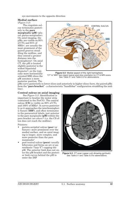
eye movements to the opposite direction<br />
Medial surface<br />
(Figure 5-2)<br />
The cingulate sulcus<br />
terminates posteriorly<br />
in the pars<br />
marginalis (pM) (plural:<br />
partes marginales).<br />
On axial imaging, the<br />
pMs: are visible on 95%<br />
of CTs and 91% of<br />
MRIs 4 , are usually the<br />
most prominent of the<br />
paired grooves straddling<br />
the midline, and<br />
they extend a greater<br />
distance into the<br />
hemispheres 4 . On axial<br />
CT, the pM is located<br />
slightly posterior to the<br />
widest biparietal<br />
diameter 4 ; on the typically<br />
more horizontally<br />
oriented MRI slices the<br />
pM assumes a more<br />
posterior position. The<br />
pMs curve posteriorly in lower slices and anteriorly in higher slices (here, the paired pMs<br />
form the “pars bracket” - a characteristic “handlebar” configuration straddling the midline).<br />
Central sulcus on axial imaging<br />
See Figure 5-3. Identification is<br />
important to localize the motor strip<br />
(contained in the PreCG). The central<br />
sulcus (CS) is visible on 93% of CTs<br />
and 100% of MRIs4 . It curves posteriorly<br />
as it approaches the interhemispheric<br />
fissure (IHF), and often terminates<br />
in the paracentral lobule, just anterior<br />
to the pars marginalis (pM) within the<br />
pars bracket (see above) 4 (i.e. the CS often<br />
does not reach the midline).<br />
Pointers:<br />
• parieto-occipital sulcus (pos) (or<br />
fissure): more prominent over the<br />
medial surface, and on axial imaging<br />
is longer, more complex, and<br />
more posterior than the pars<br />
marginalis5 • post-central sulcus (pocs): usually<br />
bifurcates and forms an arc or parenthesis<br />
(“lazy-Y”) cupping the<br />
pM. The anterior limb does not enter<br />
the pM-bracket and the posterior<br />
limb curves behind the pM to<br />
enter the IHF<br />
SFG<br />
Figure 5-2 Medial aspect of the right hemisphere<br />
“CT” & “MRI” bars depict typical axial slice orientation for CT & MRI scans.<br />
See Table 5-1 and Table 5-2 for abbreviations<br />
NEUROSURGERY 5.1. Surface anatomy 85<br />
CT<br />
CinG<br />
corpus callosum<br />
cs<br />
pocs<br />
cins<br />
pons<br />
cins<br />
prcs CENTRAL SULCUS<br />
PL<br />
SFG<br />
PL<br />
pM<br />
sps<br />
PCu<br />
PreCG<br />
LG<br />
PostCG<br />
pM<br />
Cu<br />
MRI<br />
pos<br />
Figure 5-3 CT scan (upper cut) showing gyri/sulci.<br />
See Table 5-1 and Table 5-2 for abbreviations<br />
cs
Table 5-1 Cerebral sulci<br />
(abbreviations)<br />
cins cingulate sulcus<br />
cs central sulcus<br />
ips-ios intraparietal-intraoccipital<br />
sulcus<br />
los lateral occipital sulcus<br />
pM pars marginalis<br />
pocn pre-occipital notch<br />
pocs post-central sulcus<br />
pof parieto-occipital fissure<br />
pos parieto-occipital sulcus<br />
prcs pre-central sulcus<br />
sfs, ifs superior, inferior frontal<br />
sulcus<br />
sps superior parietal sulcus<br />
sts, its superior, inferior temporal<br />
sulcus<br />
tos trans occipital sulcus<br />
5.1.2. Surface anatomy of the cranium<br />
CRANIOMETRIC POINTS<br />
See Figure 5-4.<br />
Pterion: region<br />
where the following<br />
bones are approximated:<br />
frontal, parietal,<br />
temporal and sphenoid<br />
(greater wing). Estimated<br />
as 2 fingerbreadths<br />
above the zygomatic<br />
arch, and a<br />
thumb’s breadth behind<br />
the frontal process<br />
of the zygomatic<br />
bone (blue circle in Figure<br />
5-4).<br />
Asterion: junction<br />
of lambdoid, occipitomastoid<br />
and<br />
parietomastoid sutures.<br />
Usually lies<br />
within a few millimeters<br />
of the posterior-inferior<br />
edge of the<br />
junction of the transverse<br />
and sigmoid sinuses<br />
(not always<br />
reliable 6 - may overlie<br />
either sinus).<br />
Vertex: the topmost<br />
point of the skull.<br />
Lambda: junction<br />
of the lambdoid<br />
and sagittal sutures.<br />
ophyron<br />
glabella<br />
nasion<br />
rhinion<br />
prosthion<br />
inferior<br />
alveolar point<br />
AG<br />
Table 5-2 Cerebral gyri and lobules<br />
(abbreviations)<br />
angular gyrus<br />
CinG cingulate gyrus<br />
Cu cuneus<br />
LG lingual gyrus<br />
MFG, SFG middle & superior frontal gyrus<br />
OG orbital gyrus<br />
PCu precuneous<br />
PreCG, PostCG pre- and post-central gyrus<br />
PL paracentral lobule (upper SFG and PreCG<br />
and PostCG)<br />
Stephanion: junction of coronal suture and superior temporal line.<br />
IFG<br />
gnathion<br />
or menton<br />
86 5. Neuroanatomy and physiology NEUROSURGERY<br />
POp<br />
PT<br />
POr<br />
inferior frontal gyrus<br />
pars opercularis<br />
pars triangularis<br />
pars orbitalis<br />
STG, MTG, ITG superior, middle & inferior temporal gyrus<br />
SPL, IPL superior & inferior parietal lobule<br />
SMG supramarginal gyrus<br />
stephanion<br />
pterion<br />
NASAL<br />
vertex<br />
bregma<br />
MAXILLA<br />
cs<br />
FRONTAL<br />
GWS<br />
ZYG<br />
MANDIBLE<br />
stl<br />
sqs<br />
TEMPORAL<br />
PARIETAL<br />
sms<br />
asterion<br />
opisthion<br />
gonion<br />
lambda<br />
pms<br />
oms<br />
MASTOID<br />
inion<br />
Figure 5-4 Craniometric points & cranial sutures.<br />
Named bones appear in all upper case letters.<br />
Abbreviations: GWS = greater wing of sphenoid bone, NAS = nasal bone, stl =<br />
superior temporal line, ZYG = zygomatic.<br />
Sutures: cs = coronal, ls = lambdoid, oms = occipitomastoid, pms = parietomastoid,<br />
sms = squamomastoid, sqs = squamosal<br />
ls<br />
OCCIPITAL
Glabella: the most forward projecting point of the forehead at the level of the supraorbital<br />
ridge in the midline.<br />
Opisthion: the posterior margin of the foramen magnum in the midline.<br />
Bregma: the junction of the coronal and sagittal sutures.<br />
Sagittal suture: midline suture from coronal suture to lambdoid suture. Although<br />
often assumed to overlie the superior sagittal sinus (SSS), the SSS lies to the right of the<br />
sagittal suture in the majority of specimens 7 (but never by > 11 mm).<br />
The most anterior mastoid point lies just in front of the sigmoid sinus 8 .<br />
RELATION OF SKULL MARKINGS TO CEREBRAL ANATOMY<br />
Taylor-Haughton lines<br />
Taylor-Haughton (T-H)<br />
lines can be constructed on an<br />
angiogram, CT scout film, or<br />
skull x-ray, and can then be reconstructed<br />
on the patient in the<br />
O.R. based on visible external<br />
landmarks9 . T-H lines are<br />
shown as dashed lines in Figure<br />
5-5.<br />
1. Frankfurt plane, AKA<br />
baseline: line from inferior<br />
margin of orbit<br />
through the upper margin<br />
of the external auditory<br />
meatus (EAM) (as<br />
distinguished from Reid’s<br />
base line: from inferior<br />
orbital margin<br />
through the center of the<br />
EAM)<br />
10 (p 313)<br />
2. the distance from the nasion<br />
to the inion is measured<br />
across the top of<br />
the calvaria and is divided<br />
into quarters (can be<br />
done simply with a piece<br />
of tape which is then<br />
folded in half twice)<br />
©2001 Mark S Greenberg, M.D.<br />
All rights reserved.<br />
Unauthorized use is prohibited.<br />
Figure 5-5 Taylor-Haughton lines<br />
and other localizing methods<br />
3. posterior ear line: perpendicular to the baseline through the mastoid process<br />
4. condylar line: perpendicular to the baseline through the mandibular condyle<br />
5. T-H lines can then be used to approximate the sylvian fissure (see below) and the<br />
motor cortex (also see below)<br />
Sylvian fissure AKA lateral fissure<br />
Approximated by a line connecting the lateral canthus to the point 3/4 of the way<br />
posterior along the arc running over convexity from nasion to inion (T-H lines).<br />
Angular gyrus<br />
Located just above the pinna, important on the dominant hemisphere as part of<br />
Wernicke’s area. Note: there is significant individual variability in the location2 .<br />
Angular artery<br />
Located 6 cm above the EAM.<br />
Motor cortex<br />
Numerous methods utilize external landmarks to locate the motor strip (pre-central<br />
gyrus) or the central sulcus (Rolandic fissure) which separates motor strip anteriorly<br />
from primary sensory cortex posteriorly. These are just approximations since individual<br />
variability causes the motor strip to lie anywhere from 4 to 5.4 cm behind the coronal<br />
suture11 . The central sulcus cannot even be reliably identified visually at surgery12 .<br />
• method 1: the superior aspect of the motor cortex is almost straight up from the<br />
EAM near the midline<br />
• method 213 : the central sulcus is approximated by connecting:<br />
NEUROSURGERY 5.1. Surface anatomy 87<br />
1/2<br />
sylvian fissure<br />
central sulcus<br />
EAM<br />
2 cm<br />
Frankfurt<br />
plane<br />
3/4<br />
posterior ear line<br />
condylar line
A. the point 2 cm posterior to the midposition of the arc extending from nasion<br />
to inion (illustrated in Figure 5-5), to<br />
B. the point 5 cm straight up from the EAM<br />
• method 3: using T-H lines, the central sulcus is approximated by connecting:<br />
A. the point where the “posterior ear line” intersects the circumference of the<br />
skull (see Figure 5-5) (usually about 1 cm behind the vertex, and 3-4 cm behind<br />
the coronal suture), to<br />
B. the point where the<br />
“condylar line” intersects<br />
the line<br />
representing the<br />
sylvian fissure<br />
• method 4: a line drawn<br />
45° to Reid’s base line<br />
starting at the pterion<br />
points in the direction of<br />
the motor strip14 (p 584-5)<br />
RELATIONSHIP OF VENTRICLES<br />
TO SKULL<br />
Figure 5-6 shows the relationship<br />
of non-hydrocephalic<br />
ventricles to the skull in the lateral<br />
view. Some dimensions of interest<br />
are shown in Table 5-315 .<br />
In the non-hydrocephalic<br />
adult, the lateral ventricles lie 4-<br />
5 cm below the outer skull surface.<br />
The center of the body of the<br />
lateral ventricle sits in the midpupillary<br />
line, and the frontal<br />
horn is intersected by a line passing<br />
perpendicular to the calvaria<br />
along this line16 D2<br />
V4<br />
D3<br />
D4<br />
opisthion<br />
baseline<br />
sigmoid sinus<br />
sella turcica<br />
Figure 5-6 Relationship of ventricles to skull landmarks*<br />
* Abbreviations: (F = frontal horn, B = body, A = atrium, O = occipital<br />
horn, T = temporal horn) of lateral ventricle. FM = fora-<br />
. The anterior<br />
horns extend 1-2 cm anterior to<br />
the coronal suture.<br />
men of Monro. Aq = sylvian aqueduct. V3 = third ventricle. V4<br />
= fourth ventricle. cs = coronal suture. Dimensions D1-4 →<br />
see Table 5-3<br />
Average length of third ventricle ≈ 2.8 cm.<br />
Dimension<br />
(see Figure 5-6)<br />
Table 5-3 Dimensions from Figure 5-6<br />
Description Lower limit<br />
(mm)<br />
Average<br />
(mm)<br />
Upper limit<br />
(mm)<br />
D1 length of frontal horn anterior to FM 25<br />
D2 distance from clivus to floor of 4th ventricle at<br />
level of fastigium*<br />
33.3 36.1 40.0<br />
D3 length of 4th ventricle at level of fastigium* 10.0 14.6 19.0<br />
D4 distance from fastigium* to opisthion 30.0 32.6 40.0<br />
* the fastigium is the apex of the 4th ventricle within the cerebellum<br />
B<br />
FM<br />
V3<br />
T<br />
88 5. Neuroanatomy and physiology NEUROSURGERY<br />
cs<br />
D1<br />
F<br />
A<br />
Aq<br />
O<br />
Twining
5.1.3. Surface landmarks of spine levels<br />
Estimates of cervical levels for anterior cervical<br />
spine surgery may be made using the landmarks shown<br />
in Table 5-4. Intra-operative C-spine x-rays are essential<br />
to verify these estimates.<br />
The scapular spine is located at about T2-3.<br />
The inferior scapular pole is ≈ T6 posteriorly.<br />
Intercristal line: a line drawn between the highest<br />
point of the iliac crests across the back will cross the<br />
midline either at the interspace between the L4 and L5<br />
spinous processes, or at the L4 spinous process itself.<br />
Table 5-4 Cervical levels 17<br />
Level Landmark<br />
C1-2 angle of mandible<br />
C3-4 1 cm above thyroid cartilage<br />
(≈ hyoid bone)<br />
C4-5 level of thyroid cartilage<br />
C5-6 crico-thyroid membrane<br />
C6 carotid tubercle<br />
C6-7 cricoid cartilage<br />
5.2. Cranial foramina & their contents<br />
Foramen<br />
Table 5-5 Cranial foramina and their contents*<br />
Contents<br />
nasal slits anterior ethmoidal nn., a. & v<br />
superior orbital fissure Cr. Nn. III, IV, VI, all 3 branches of V1 (ophthalmic division divides into nasociliary, frontal,<br />
and lacrimal nerves); superior ophthalmic vv.; recurrent meningeal br. from lacrimal<br />
a.; orbital branch of middle meningeal a.; sympathetic filaments from ICA plexus<br />
inferior orbital fissure Cr. N. V-2 (maxillary div.), zygomatic n.; filaments from pterygopalatine branch of maxillary<br />
n.; infraorbital a. & v.; v. between inferior ophthalmic v. & pterygoid venous plexus<br />
foramen lacerum usually nothing (ICA traverses the upper portion but doesn’t enter, 30% have vidian a.)<br />
carotid canal internal carotid a., ascending sympathetic nerves<br />
incisive foramen descending septal a.; nasopalatine nn.<br />
greater palatine foramen greater palatine n., a., & v.<br />
lesser palatine foramen lesser palatine nn.<br />
internal acoustic meatus Cr. N. VII (facial); Cr. N. VIII (stato-acoustic) - (see text & Figure 5-7 below)<br />
hypoglossal canal Cr. N. XII (hypoglossal); a meningeal branch of the ascending pharyngeal a.<br />
foramen magnum spinal cord (medulla oblongata); Cr. N. XI (spinal accessory nn.) entering the skull; vertebral<br />
aa.; anterior & posterior spinal arteries<br />
foramen cecum occasional small vein<br />
cribriform plate olfactory nn.<br />
optic canal Cr. N. II (optic); ophthalmic a.<br />
foramen rotundum Cr. N. V2 (maxillary div.), a. of foramen rotundum<br />
foramen ovale Cr. N. V3 (mandibular div.) + portio minor (motor for CrN V)<br />
foramen spinosum middle meningeal a. & v.<br />
jugular foramen internal jugular v. (beginning); Cr. Nn. IX, X, XI<br />
stylomastoid foramen Cr. N. VII (facial); stylomastoid a.<br />
condyloid foramen v. from transverse sinus<br />
mastoid foramen v. to mastoid sinus; branch of occipital a. to dura mater<br />
* Abbreviations: a. = artery, aa. = arteries, v. = vein, vv. = veins, n. = nerve, nn. = nerves, br. = branch, Cr.<br />
N. = cranial nerve, fmn. = foramen, div. = division<br />
Porus acusticus<br />
AKA internal auditory canal (see Figure 5-7)<br />
The filaments of the acoustic portion of VIII penetrate tiny openings of the lamina<br />
cribrosa of the cochlear area 18 .<br />
Transverse crest: separates superior vestibular area and facial canal (above) from<br />
the inferior vestibular area and cochlear area (below) 18 .<br />
Vertical crest (AKA Bill’s bar): separates the meatus to facial canal anteriorly (con-<br />
NEUROSURGERY 5.2. Cranial foramina & their contents 89
taining VII and nervus intermedius) from the vestibular area posteriorly (containing the<br />
superior division of vestibular nerve).<br />
The “5 nerves” of the IAC:<br />
1. facial nerve (VII)<br />
(mnemonic: “7-up”<br />
as VII is in superior<br />
portion)<br />
2. nervus intermedius:<br />
the somatic<br />
sensory branch of<br />
the facial nerve<br />
primarily innervatingmechanoreceptors<br />
of the hair<br />
follicles on the inner<br />
surface of the<br />
pinna and deep<br />
mechanoreceptors<br />
of nasal and buccal<br />
cavities and<br />
chemoreceptors in<br />
the taste buds on<br />
the anterior 2/3 of<br />
the tongue<br />
facial canal (Cr. N. VII with NI*)<br />
vertical crest (”Bill’s bar”)<br />
superior vestibular area (superior<br />
vestibular nerve) (to utricle &<br />
superior & lateral semicircular canals)<br />
transverse crest (crista falciformis)<br />
inferior vestibular area<br />
(to saccule)<br />
foramen singulare (to<br />
posterior semicircular canal)<br />
tractus spiralis foraminosus (cochlear<br />
area) (acoustic portion of Cr. N. VIII)<br />
Figure 5-7 Right internal auditory canal (porus acusticus) & nerves<br />
* NI = nervus intermedius<br />
3. acoustic portion of the VIII nerve (mnemonic: “Coke down” for cochlear portion)<br />
4. superior branch of vestibular nerve: passes through the superior vestibular area<br />
to terminate in the utricle and in the ampullæ of the superior and lateral semicircular<br />
canals<br />
5. inferior branch of vestibular nerve: passes through inferior vestibular area to terminate<br />
in the saccule<br />
5.3. Cerebellopontine angle anatomy<br />
retractor<br />
on cerebellar<br />
hemisphere<br />
foramen of<br />
Luschka<br />
foramen of<br />
Magendie<br />
cerebellar<br />
tonsil<br />
PICA<br />
V<br />
Meckel's<br />
cave<br />
pons<br />
flocculus<br />
choroid<br />
plexus<br />
VII<br />
IAC<br />
VIII<br />
IX<br />
jugular<br />
foramen<br />
X<br />
XI<br />
XII<br />
olive<br />
medulla<br />
Figure 5-8 Normal anatomy of right cerebellopontine angle viewed<br />
from behind (as in a suboccipital approach) 18<br />
(inferior<br />
vestibular<br />
nerve)<br />
90 5. Neuroanatomy and physiology NEUROSURGERY
5.4. Occiptoatlantoaxial-complex anatomy<br />
≈ 50% of head rotation occurs at the C1-2 (atlantoaxial) joint.<br />
Ligaments of the occipito-atlanto-axial complex<br />
apical<br />
odontoid<br />
ligament<br />
cruciate ligament,<br />
ascending band<br />
anterior<br />
atlantooccipital<br />
membrane<br />
anterior<br />
longitudinal<br />
ligament<br />
cruciate ligament,<br />
descending band<br />
tectorial<br />
membrane<br />
transverse<br />
ligament<br />
posterior<br />
C3<br />
longitudinal<br />
ligament<br />
Figure 5-9 Sagittal view of the ligaments of the craniovertebral junction<br />
Modified with permission from “In Vitro Cervical Spine Biomechanical Testing” BNI Quarterly,<br />
Vol.9, No. 4, 1993<br />
Stability of this joint complex is primarily due to ligaments, with little contribution<br />
from bony articulations and joint capsules (see Figure 5-9 through Figure 5-11):<br />
1. ligaments that connect the atlas to the occiput:<br />
A. anterior at-<br />
lanto-occipitalmembrane:cephalad<br />
extension<br />
of the anteriorlongitudinal<br />
ligament.<br />
Extends from<br />
anterior margin<br />
of foramen<br />
magnum<br />
(FM) to anterior<br />
arch of C1<br />
B. posterior atlanto-occipitalmembrane:connects<br />
the posterior<br />
margin<br />
of the FM to<br />
posterior arch<br />
of C1<br />
C1<br />
CRUCIATE<br />
LIGAMENT<br />
C2<br />
spinal<br />
cord<br />
posterior<br />
atlantooccipital<br />
membrane<br />
ligamentum<br />
flavum<br />
C. the ascending band of the cruciate ligament<br />
2. ligaments that connect the axis (viz. the odontoid) to the occiput:<br />
A. tectorial membrane: some authors distinguish 2 components<br />
1. superficial component: cephalad continuation of the posterior longitu-<br />
NEUROSURGERY 5.4. Occiptoatlantoaxial-complex anatomy 91<br />
C1<br />
clivus<br />
Figure 5-10 Dorsal view of the cruciate and alar ligaments<br />
Viewed with tectorial membrane removed.<br />
Modified with permission from “In Vitro Cervical Spine Biomechanical<br />
Testing” BNI Quarterly, Vol.9, No. 4, 1993<br />
C2<br />
ascending<br />
band<br />
right alar<br />
ligament<br />
accessory<br />
(deep) portion<br />
of tectorial<br />
membrane<br />
transverse<br />
band<br />
descending<br />
band
dinal ligament. A strong band connecting the dorsal surface of the<br />
dens to the ventral surface of the FM above, and dorsal surface of C2<br />
& C3 bodies below<br />
2. accessory (deep) portion: located laterally, connects C2 to occipital<br />
condyles<br />
B. alar (“check”) ligaments 19<br />
1. occipito-alar portion: connects side of the dens to occipital condyle<br />
2. atlanto-alar portion: connects side of the dens to the lateral mass of C1<br />
C. apical odontoid liga-<br />
ment: connects tip of<br />
dens to the FM. Little<br />
mechanical strength<br />
3. ligaments that connect the<br />
axis to the atlas:<br />
A. transverse (atlantoaxial)<br />
ligament: the<br />
horizontal component<br />
of the cruciate ligament.<br />
Traps the dens<br />
against the anterior atlas<br />
via a strap-like<br />
mechanism (see Figure<br />
5-11). Provides the majority<br />
of the strength<br />
(“the strongest ligament<br />
of the spine” 20 )<br />
B. atlanto-alar portion of<br />
the alar ligaments (see<br />
above)<br />
C. descending band of the cruciate ligament<br />
The most important structures in maintaining atlanto-occipital stability are the tectorial<br />
membrane and the alar ligaments. Without these, the remaining cruciate ligament<br />
and apical dentate ligament are insufficient.<br />
5.5. Spinal cord anatomy<br />
5.5.1. Spinal cord tracts<br />
odontoid<br />
process<br />
tubercle<br />
right alar<br />
ligament<br />
transverse<br />
ligament<br />
tectorial<br />
membrane<br />
posterior arch C1<br />
Figure 5-11 C1 viewed from above, showing the transverse<br />
and alar ligaments<br />
Modified with permission from “In Vitro Cervical Spine Biomechanical<br />
Testing” BNI Quarterly, Vol.9, No. 4, 1993<br />
Figure 5-12 depicts a cross-section of a typical spinal cord segment, combining some<br />
elements from different levels (e.g. the intermediolateral grey nucleus is only present<br />
from T1 to ≈ L1 or L2 where there are sympathetic (thoracolumbar outflow) nuclei). It is<br />
schematically divided into ascending and descending halves, however, in actuality, ascending<br />
and descending paths coexist on both sides.<br />
Table 5-6 Descending (motor) tracts (↓) in Figure 5-12<br />
Number (see<br />
Figure 5-12)<br />
Path Function Side of body<br />
1 anterior corticospinal tract skilled movement opposite<br />
2 medial longitudinal fasciculus ? same<br />
3 vestibulospinal tract facilitates extensor muscle tone same<br />
4 medullary (ventrolateral) reticulospinal tract automatic respirations? same<br />
5 rubrospinal tract flexor muscle tone same<br />
6 lateral corticospinal (pyramidal) tract skilled movement same<br />
92 5. Neuroanatomy and physiology NEUROSURGERY
Figure 5-12 also depicts some of the laminae according to the scheme of Rexed. Lamina<br />
II is equivalent to the substantia gelatinosa. Laminae III and IV are the nucleus proprius.<br />
Lamina VI is located in the base of the posterior horn.<br />
SENSATION<br />
Table 5-7 Bi-directional tracts in Figure 5-12<br />
Number (see<br />
Figure 5-12)<br />
Path Function<br />
7 dorsolateral fasciculus (of Lissauer)<br />
8 fasciculus proprius short spinospinal connections<br />
Number (see<br />
Figure 5-12)<br />
6<br />
5<br />
Table 5-8 Ascending (sensory) tracts (↑) in Figure 5-12<br />
Path Function Side of<br />
body<br />
same<br />
9 fasciculus gracilis joint position, fine touch,<br />
10 fasciculus cuneatus<br />
vibration<br />
11 posterior spinocerebellar tract stretch receptors same<br />
12 lateral spinothalamic tract pain & temperature opposite<br />
13 anterior spinocerebellar tract whole limb position opposite<br />
14 spinotectal tract unknown, ? nociceptive opposite<br />
15 anterior spinothalamic tract light touch opposite<br />
MOTOR<br />
(descending<br />
paths)<br />
{ 1<br />
4<br />
3<br />
2<br />
STC<br />
bi-directional<br />
paths<br />
{<br />
S = sacral<br />
T = thoracic<br />
C = cervical<br />
2.5-4 cm<br />
anterior spinal<br />
artery<br />
CTS<br />
dentate<br />
ligament<br />
anterior motor<br />
nerve root<br />
Figure 5-12 Schematic cross-section of cervical spinal cord<br />
SENSORY<br />
(ascending<br />
paths)<br />
7 8 9<br />
10<br />
S TC<br />
TC I<br />
II<br />
intermediolateral<br />
grey nucleus<br />
(sympathetic)<br />
{<br />
PAIN & TEMPERATURE: BODY<br />
Receptors: free nerve endings (probable).<br />
1st order neuron: small, finely myelinated afferents; soma in dorsal root ganglion<br />
(no synapse). Enter cord at dorsolateral tract (zone of Lissauer). Synapse: substantia ge-<br />
NEUROSURGERY 5.5. Spinal cord anatomy 93<br />
III<br />
IV<br />
V<br />
VI<br />
VII<br />
X<br />
VIII<br />
IX<br />
IX<br />
11<br />
12<br />
13<br />
14<br />
15
latinosa (Rexed II).<br />
2nd order neuron axon cross obliquely in the anterior white commissure ascending<br />
≈ 1-3 segments while crossing to enter the lateral spinothalamic tract.<br />
Synapse: VPL thalamus. 3rd order neurons pass through IC to postcentral gyrus<br />
(Brodmann’s areas 3, 1, 2).<br />
FINE TOUCH, DEEP PRESSURE & PROPRIOCEPTION: BODY<br />
Fine touch AKA discriminative touch. Receptors: Meissner’s & pacinian corpuscles,<br />
Merkel’s disks, free nerve endings.<br />
1st order neuron: heavily myelinated afferents; soma in dorsal root ganglion (no<br />
synapse). Short branches synapse in nucleus proprius (Rexed III & IV) of posterior gray;<br />
long fibers enter the ipsilateral posterior columns without synapsing (below T6: fasciculus<br />
gracilis; above T6: fasciculus cuneatus).<br />
Synapse: nucleus gracilis/cuneatus (respectively), just above pyramidal decussation.<br />
2nd order neuron axons form internal arcuate fibers, decussate in lower medulla as<br />
medial lemniscus.<br />
Synapse: VPL thalamus. 3rd order neurons pass through IC primarily to postcentral<br />
gyrus.<br />
ANT E R IOR<br />
P OST E R IOR<br />
trigeminal<br />
nerve {<br />
©2001 Mark S Greenberg, M.D.<br />
All rights reserved.<br />
Unauthorized use is prohibited.<br />
V1<br />
V2<br />
V3<br />
superior clavicular occipitals<br />
C2<br />
C2<br />
C3<br />
C5<br />
T2<br />
C3<br />
C4<br />
T3<br />
T4<br />
T6<br />
INTERCOSTALS<br />
posterior<br />
lateral<br />
medial<br />
axillary<br />
RADIAL<br />
post. cutaneous<br />
dorsal cutan.<br />
C4<br />
T2<br />
T4<br />
T6<br />
T8<br />
T10<br />
C5<br />
T2<br />
T12 T1<br />
musculocutan.<br />
C6<br />
L1<br />
medial cutan.<br />
radial<br />
S5<br />
clunials<br />
S3<br />
C8<br />
C7<br />
L2<br />
L3<br />
L4<br />
ilioinguinal<br />
lateral cutan.<br />
nerve of thigh<br />
median<br />
ulnar<br />
FEMORAL<br />
anterior<br />
cutaneous<br />
saphenous<br />
posterior<br />
cutaneous<br />
L3<br />
S1<br />
L4<br />
C8<br />
C7<br />
L5<br />
SCIATIC<br />
COMMON PERONEAL<br />
lat. cutan.<br />
sup. peroneal<br />
deep peroneal<br />
TIBIAL<br />
L4<br />
L5<br />
sural<br />
med.<br />
plantars<br />
{lat.<br />
S1<br />
C6<br />
T1<br />
T8<br />
T10<br />
T12<br />
S1<br />
S4<br />
DERMATO MES<br />
CUT ANEOUS<br />
DERMATO M ES<br />
(anterior) NER VES<br />
(posterior)<br />
Figure 5-13 Dermatomal and sensory nerve distribution<br />
(Redrawn from “Introduction to Basic Neurology”, by Harry D. Patton, John W. Sundsten, Wayne E. Crill and<br />
Phillip D. Swanson, © 1976, pp 173, W. B. Saunders Co., Philadelphia, PA, with permission)<br />
LIGHT (CRUDE) TOUCH: BODY<br />
Receptors: as fine touch (see above), also peritrichial arborizations.<br />
94 5. Neuroanatomy and physiology NEUROSURGERY
1st order neuron: large, heavily myelinated afferents (Type II); soma in dorsal root<br />
ganglion (no synapse). Some ascend uncrossed in post. columns (with fine touch); most<br />
synapse in Rexed VI & VII.<br />
2nd order neuron axons cross in anterior white commissure (a few don’t cross); enter<br />
anterior spinothalamic tract.<br />
Synapse: VPL thalamus. 3rd order neurons pass through IC primarily to postcentral<br />
gyrus.<br />
5.5.2. Dermatomes and sensory nerves<br />
Figure 5-13 shows anterior and posterior view, each schematically separated into<br />
sensory dermatomes (segmental) and peripheral sensory nerve distribution.<br />
5.5.3. Spinal cord vasculature<br />
right vertebral<br />
artery<br />
right common<br />
carotid<br />
right<br />
subclavian<br />
brachiocephalic trunk<br />
aorta<br />
left common<br />
carotid<br />
posterior intercostal<br />
artery<br />
(dorsal branch)<br />
posterior<br />
spinal arteries<br />
radicular<br />
artery<br />
posterior<br />
intercostal<br />
artery<br />
anterior<br />
spinal<br />
aorta<br />
artery<br />
Axial view<br />
left vertebral<br />
artery<br />
basilar artery<br />
spinal cord<br />
radicular artery at C3<br />
anterior spinal artery<br />
radicular artery at C6<br />
deep cervical artery<br />
costocervical trunk<br />
radicular artery at C8<br />
left subclavian<br />
left posterior spinal artery<br />
radicular artery at T5<br />
} intercostal arteries<br />
artery of<br />
Adamkiewicz<br />
(arteria radicularis<br />
anterior magna)<br />
arteria radicularis<br />
magna<br />
(posterior branch)<br />
Figure 5-14 Schematic diagram of spinal cord arterial supply<br />
Modified from Diagnostic Neuroradiology, 2nd ed., Volume II, pp. 1181, Taveras J M, Woods EH, editors, ©<br />
1976, the Williams and Wilkins Co., Baltimore, with permission)<br />
Although a radicular artery from the aorta accompanies the nerve root at many levels,<br />
most of these contribute little flow to the spinal cord itself. The anterior spinal artery<br />
is formed from the junction of two branches, each from one of the vertebral arteries. Major<br />
contributors of blood supply to the anterior spinal cord is from 6-8 radicular arteries<br />
NEUROSURGERY 5.5. Spinal cord anatomy 95
at the following levels (“radiculomedullary arteries”, the levels listed are fairly consistent,<br />
but the side varies 21 (p 1180-1) ):<br />
• C3 - arises from vertebral artery<br />
• C6 - usually arises from deep cervical artery<br />
• C8 - usually from costocervical trunk }<br />
• T4 or T5<br />
• artery of Adamkiewicz AKA arteria radicularis anterior magna<br />
A. the main arterial supply for the spinal cord from ≈ T8 to the conus<br />
B. located on the left in 80% 23<br />
C. situated between T9 & L2 in 85% (between T9 & T12 in 75%); in remaining<br />
15% between T5 & T8 (in these latter cases, there may be a supplemental<br />
radicular artery further down)<br />
D. usually fairly large, gives off cephalic and caudal branch (latter is usually<br />
larger) giving a characteristic hair-pin appearance on angiography<br />
The paired posterior spinal arteries are less well defined than the anterior spinal<br />
artery, and are fed by 10-23 radicular branches.<br />
The midthoracic region has a tenuous vascular supply (“watershed zone”), possessing<br />
only the above noted artery at T4 or T5. It is thus more susceptible to vascular insults.<br />
ANATOMIC VARIANTS<br />
Arcade of Lazorthes: normal variant where the anterior spinal artery joins with<br />
the paired posterior spinal arteries at the conus medullaris.<br />
5.6. Cerebrovascular anatomy<br />
5.6.1. Cerebral vascular territories<br />
MCA<br />
CORONAL VIEW<br />
RAH<br />
AChA<br />
internal carotid<br />
PCommA<br />
basilar artery<br />
anterior cerebral<br />
artery<br />
middle cerebral<br />
artery<br />
anterior choroidal<br />
artery<br />
≈ 10% of population lack an anterior<br />
radicular artery in lower<br />
cervical spine 22<br />
AXIAL VIEW<br />
posterior cerebral<br />
artery<br />
RAH = recurrent artery of Heubner<br />
Figure 5-15 Vascular territories of the cerebral hemispheres<br />
Figure 5-15 depicts approximate vascular distributions of the major cerebral arteries.<br />
There is considerable variability of the major arteries 24 as well as the central distri-<br />
96 5. Neuroanatomy and physiology NEUROSURGERY