A-770041, a Novel and Selective Small-Molecule Inhibitor of Lck ...
A-770041, a Novel and Selective Small-Molecule Inhibitor of Lck ...
A-770041, a Novel and Selective Small-Molecule Inhibitor of Lck ...
You also want an ePaper? Increase the reach of your titles
YUMPU automatically turns print PDFs into web optimized ePapers that Google loves.
0022-3565/05/3151-36–41$20.00<br />
THE JOURNAL OF PHARMACOLOGY AND EXPERIMENTAL THERAPEUTICS Vol. 315, No. 1<br />
Copyright © 2005 by The American Society for Pharmacology <strong>and</strong> Experimental Therapeutics 89169/3052987<br />
JPET 315:36–41, 2005 Printed in U.S.A.<br />
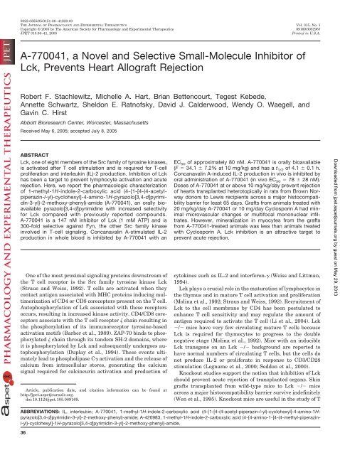
A-<strong>770041</strong>, a <strong>Novel</strong> <strong>and</strong> <strong>Selective</strong> <strong>Small</strong>-<strong>Molecule</strong> <strong>Inhibitor</strong> <strong>of</strong><br />
<strong>Lck</strong>, Prevents Heart Allograft Rejection<br />
Robert F. Stachlewitz, Michelle A. Hart, Brian Bettencourt, Tegest Kebede,<br />
Annette Schwartz, Sheldon E. Ratn<strong>of</strong>sky, David J. Calderwood, Wendy O. Waegell, <strong>and</strong><br />
Gavin C. Hirst<br />
Abbott Bioresearch Center, Worcester, Massachusetts<br />
Received May 6, 2005; accepted July 8, 2005<br />
ABSTRACT<br />
<strong>Lck</strong>, one <strong>of</strong> eight members <strong>of</strong> the Src family <strong>of</strong> tyrosine kinases,<br />
is activated after T cell stimulation <strong>and</strong> is required for T-cell<br />
proliferation <strong>and</strong> interleukin (IL)-2 production. Inhibition <strong>of</strong> <strong>Lck</strong><br />
has been a target to prevent lymphocyte activation <strong>and</strong> acute<br />
rejection. Here, we report the pharmacologic characterization<br />
<strong>of</strong> 1-methyl-1H-indole-2-carboxylic acid (4-{1-[4-(4-acetylpiperazin-l-yl)-cyclohexyl]-4-amino-1H-pyrazolo[3,4-d]pyrimidin-3-yl}-2-methoxy-phenyl)-amide<br />
(A-<strong>770041</strong>), an orally bioavailable<br />
pyrazolo[3,4-d]pyrimidine with increased selectivity<br />
for <strong>Lck</strong> compared with previously reported compounds.<br />
A-<strong>770041</strong> is a 147 nM inhibitor <strong>of</strong> <strong>Lck</strong> (1 mM ATP) <strong>and</strong> is<br />
300-fold selective against Fyn, the other Src family kinase<br />
involved in T-cell signaling. Concanavalin A-stimulated IL-2<br />
production in whole blood is inhibited by A-<strong>770041</strong> with an<br />
One <strong>of</strong> the most proximal signaling proteins downstream <strong>of</strong><br />
the T cell receptor is the Src family tyrosine kinase <strong>Lck</strong><br />
(Straus <strong>and</strong> Weiss, 1992). T cells are activated when they<br />
contact antigen associated with MHC proteins inducing multimerization<br />
<strong>of</strong> CD4 or CD8 coreceptors present on the T cell.<br />
Autophosphorylation <strong>of</strong> <strong>Lck</strong> associated with these receptors<br />
occurs, resulting in increased kinase activity. CD4/CD8 coreceptors<br />
associate with the T cell receptor chain resulting in<br />
the phosphorylation <strong>of</strong> its immunoreceptor tyrosine-based<br />
activation motifs (Barber et al., 1989). ZAP-70 binds to phosphorylated<br />
chain through its t<strong>and</strong>em SH-2 domains, where<br />
it is phosphorylated by <strong>Lck</strong> <strong>and</strong> subsequently undergoes autophosphorylation<br />
(Duplay et al., 1994). These events ultimately<br />
lead to phospholipase C activation <strong>and</strong> the release <strong>of</strong><br />
calcium from intracellular stores, generating the calcium<br />
signal required for calcineurin activation <strong>and</strong> production <strong>of</strong><br />
Article, publication date, <strong>and</strong> citation information can be found at<br />
http://jpet.aspetjournals.org.<br />
doi:10.1124/jpet.105.089169.<br />
EC 50 <strong>of</strong> approximately 80 nM. A-<strong>770041</strong> is orally bioavailable<br />
(F 34.1 7.2% at 10 mg/kg) <strong>and</strong> has a t 1/2 <strong>of</strong> 4.1 0.1 h.<br />
Concanavalin A-induced IL-2 production in vivo is inhibited by<br />
oral administration <strong>of</strong> A-<strong>770041</strong> (in vivo EC 50 78 28 nM).<br />
Doses <strong>of</strong> A-<strong>770041</strong> at or above 10 mg/kg/day prevent rejection<br />
<strong>of</strong> hearts transplanted heterotopically in rats from Brown Norway<br />
donors to Lewis recipients across a major histocompatibility<br />
barrier for least 65 days. Grafts from animals treated with<br />
20 mg/kg/day A-<strong>770041</strong> or 10 mg/day Cyclosporin A had minimal<br />
microvascular changes or multifocal mononuclear infiltrates.<br />
However, mineralization in myocytes from the grafts<br />
from A-<strong>770041</strong>-treated animals was less than animals treated<br />
with Cyclosporin A. <strong>Lck</strong> inhibition is an attractive target to<br />
prevent acute rejection.<br />
cytokines such as IL-2 <strong>and</strong> interferon- (Weiss <strong>and</strong> Littman,<br />
1994).<br />
<strong>Lck</strong> plays a crucial role in the maturation <strong>of</strong> lymphocytes in<br />
the thymus <strong>and</strong> in mature T cell activation <strong>and</strong> proliferation<br />
(Molina et al., 1992; Straus <strong>and</strong> Weiss, 1992). Recruitment <strong>of</strong><br />
<strong>Lck</strong> to the cell membrane by CD4 has been postulated to<br />
enhance T cell sensitivity <strong>and</strong> may regulate the amount <strong>of</strong><br />
antigen required to activate the T cell (Li et al., 2004). <strong>Lck</strong><br />
/ mice have very few circulating mature T cells because<br />
<strong>Lck</strong> is required for thymocytes to progress to the double<br />
negative stage (Molina et al., 1992). Mice with an inducible<br />
<strong>Lck</strong> transgene on an <strong>Lck</strong> / background are reported to<br />
have normal numbers <strong>of</strong> circulating T cells, but the cells do<br />
not produce IL-2 or proliferate in response to CD3/CD28<br />
stimulation (Legname et al., 2000; Seddon et al., 2000).<br />
Knockout studies support the notion that inhibition <strong>of</strong> <strong>Lck</strong><br />
should prevent acute rejection <strong>of</strong> transplanted organs. Skin<br />
grafts transplanted from wild-type mice to <strong>Lck</strong> / mice<br />
across a major histocompatibility barrier survive indefinitely<br />
(Wen et al., 1995). Knockout mice are useful in the study <strong>of</strong> T<br />
ABBREVIATIONS: IL, interleukin; A-<strong>770041</strong>, 1-methyl-1H-indole-2-carboxylic acid (4-{1-[4-(4-acetyl-piperazin-l-yl)-cyclohexyl]-4-amino-1Hpyrazolo[3,4-d]pyrimidin-3-yl}-2-methoxy-phenyl)-amide;<br />
A-420983, 1-methyl-1H-indole-2-carboxylic acid (4-{4-amino-1-[4-(4-methyl-piperazinl-yl)-cyclohexyl]-1H-pyrazolo[3,4-d]pyrimidin-3-yl}-2-methoxy-phenyl)-amide.<br />
36<br />
Downloaded from<br />
jpet.aspetjournals.org by guest on May 29, 2013
cell development but are less valuable in underst<strong>and</strong>ing the<br />
kinase inhibitory effects <strong>and</strong> function in mature T cells;<br />
hence, inhibitors <strong>of</strong> <strong>Lck</strong> are being investigated to prevent<br />
rejection <strong>of</strong> transplanted solid organs. A nonselective <strong>Lck</strong><br />
inhibitor discovered by Abbott Laboratories, A-420983, has<br />
been shown to limit rejection in a nonvascularized neonatal<br />
heart transplantation model <strong>and</strong> prevent acute rejection <strong>of</strong><br />
pancreatic cells transplanted across major histocompatibility<br />
barriers (Waegell et al., 2002; Borhani et al., 2004).<br />
Although A-420983 is a 37 nM <strong>Lck</strong> inhibitor, it lacks selectivity<br />
within the Src family. In particular, it exhibits only<br />
8-fold selectivity against Fyn, a kinase that also plays a role<br />
in T cell signaling (Borhani et al., 2004). Besides the role <strong>of</strong><br />
Fyn in T cell maturation <strong>and</strong> activation, it plays a role in<br />
myelination <strong>of</strong> neurons (Umemori et al., 1994, 1999), Sertoli<br />
cell maintenance <strong>of</strong> germ line stem cells in the testicle<br />
(Maekawa et al., 2002), <strong>and</strong> degranulation <strong>of</strong> mast cells (Parravicini<br />
et al., 2002). A-420983 is also equipotent against Src<br />
<strong>and</strong> only 10-fold selective against Fgr. Src plays a role in bone<br />
formation <strong>and</strong> osteoclast function (Lowe et al., 1993), <strong>and</strong> Src<br />
/ mice develop osteopetrosis as a result <strong>of</strong> the decreased<br />
resorption <strong>of</strong> bone by osteoclasts (Soriano et al., 1991). Hck/<br />
Fgr double knockout mice show defects in neutrophil adhesion<br />
<strong>and</strong> activation leading to defective respiratory burst <strong>and</strong><br />
degranulation that could have consequences innate immunity<br />
(Lowell et al., 1996; Lowell <strong>and</strong> Berton, 1998). We continued<br />
our chemistry effort to find a molecule with greater<br />
selectivity for <strong>Lck</strong> to minimize the possibility that efficacy<br />
was driven by coinhibition <strong>of</strong> both Fyn <strong>and</strong> <strong>Lck</strong>. We also<br />
wanted to find a molecule with greater selectively against Src<br />
<strong>and</strong> Fgr to avoid the potential detrimental effects that have<br />
been implied from studies from knockout mice for these Src<br />
family members.<br />
Subsequent synthetic chemistry efforts resulted in the discovery<br />
<strong>of</strong> A-<strong>770041</strong> (Fig. 1), a compound with increased selectivity<br />
for <strong>Lck</strong> over other Src family members compared<br />
with A-420983. The purpose <strong>of</strong> these studies was to determine<br />
whether A-<strong>770041</strong>, a more selective <strong>Lck</strong> inhibitor that<br />
does not inhibit Fyn, could prevent T cell activation, IL-2<br />
Fig. 1. Structure, molecular weight, <strong>and</strong> ClogP <strong>of</strong> A-<strong>770041</strong>.<br />
A-<strong>770041</strong> Prevents Acute Rejection 37<br />
production in vivo, <strong>and</strong> rejection <strong>of</strong> vascularized transplanted<br />
heart in a rodent model. In the present studies, it is demonstrated<br />
that A-<strong>770041</strong> abolishes the production <strong>of</strong> IL-2 in<br />
vitro <strong>and</strong> in vivo. Furthermore, A<strong>770041</strong> prevents the rejection<br />
<strong>of</strong> fully mismatched vascularized heart allografts in rats<br />
<strong>and</strong> results in prolonged survival <strong>of</strong> grafts, similar to treatment<br />
with Cyclosporin A.<br />
Materials <strong>and</strong> Methods<br />
Animals. Lewis <strong>and</strong> Brown Norway rats (male, 200–300 g) were<br />
obtained from Charles River Laboratories, Inc. (Wilmington, MA),<br />
maintained on a 12-h light/dark cycle, <strong>and</strong> provided food <strong>and</strong> water<br />
ad libitum. All animal studies were reviewed by the Abbott Bioresearch<br />
Center Institutional Animal Care <strong>and</strong> Use Committee <strong>and</strong><br />
complied with Association for Assessment <strong>and</strong> Accreditation <strong>of</strong> Laboratory<br />
Animal Care guidelines <strong>and</strong> the Guide for the Care <strong>and</strong> Use<br />
<strong>of</strong> Laboratory Animals.<br />
Compounds. A-<strong>770041</strong> was synthesized by Abbott Bioresearch<br />
Center (Worcester, MA). Compound was dissolved in a 5% ethanol in<br />
sterile water for all in vivo studies. Cyclosporin A (Neoral) was<br />
purchased from Novartis (Basel, Switzerl<strong>and</strong>). Cyclosporin A was<br />
diluted in 5% ethanol in sterile water for all in vivo studies.<br />
Homogeneous Time-Resolved Fluorescence-Kinase Assay.<br />
The purified recombinant tyrosine kinase to be tested was mixed<br />
with a biotinylated peptide substrate <strong>and</strong> varying inhibitor concentrations<br />
with 1 mM ATP, 10 mM Mg 2 ,<strong>and</strong>2mMMn 2 . After<br />
incubation for 60 min, a europium cryptate-labeled anti-phosphotyrosine<br />
<strong>and</strong> streptavidin-labeled allophycocyanin were added to the<br />
well. The ratio <strong>of</strong> the signal <strong>of</strong> 620 <strong>and</strong> 665 nm was used to calculate<br />
IC 50.<br />
Anti-CD3-Induced IL-2 in Whole Blood. Heparinized human<br />
whole blood was stimulated with CD3 monoclonal antibody <strong>and</strong><br />
phorbol 12-myristate 13-acetate in the presence <strong>of</strong> A-<strong>770041</strong> (0–30<br />
M). IL-2 release into plasma was determined 2 h after stimulation<br />
by an enzyme-linked immunosorbent assay.<br />
Concanavalin A-Induced Cytokine Production. Male Lewis<br />
rats were dosed with 2.5 mg/kg A-<strong>770041</strong> or vehicle (1 ml/kg). At 2,<br />
6, 10, <strong>and</strong> 22 h after dosing <strong>of</strong> the test compound, rats were administered<br />
5 mg/kg concanavalin A (0.5 ml in saline, no. 2000-06; GE<br />
Healthcare, Little Chalfont, Buckinghamshire, UK) i.v. via the tail<br />
vein. Two hours after concanavalin A administration (i.e., 4, 8, 12,<br />
<strong>and</strong> 24 h after test compound dosing), rats were euthanized with<br />
CO 2, blood was collected by cardiac puncture into heparinized tubes,<br />
<strong>and</strong> plasma was separated. The plasma samples were divided into<br />
two aliquots, one for the assay <strong>of</strong> A-<strong>770041</strong> concentration (200 l)<br />
<strong>and</strong> one for the detection <strong>of</strong> IL-2 by enzyme-linked immunosorbent<br />
assay (100 l).<br />
Heterotopic Heart Transplantation. Hearts were transplanted<br />
between Brown Norway (donor) <strong>and</strong> Lewis (recipient) rats<br />
essentially as described by Korecky <strong>and</strong> Masika (1990) with modifications.<br />
Recipient animals were treated with compounds beginning<br />
on day 1, <strong>and</strong> the day <strong>of</strong> surgery was denoted as day 0. The donor<br />
<strong>and</strong> recipient were anesthetized by inhalation <strong>of</strong> is<strong>of</strong>lurane, <strong>and</strong> the<br />
dorsal fur was shaved. The graft harvest in the donor <strong>and</strong> preparation<br />
<strong>of</strong> the recipient for implantation occurred simultaneously, using<br />
one surgeon for each procedure to minimize ischemic time. In the<br />
donor, the superior <strong>and</strong> inferior vena cava, the pulmonary veins, <strong>and</strong><br />
the vena azygous were ligated with 5-0 silk sutures. The pulmonary<br />
artery <strong>and</strong> aorta were isolated <strong>and</strong> transected just proximal to the<br />
first branch <strong>of</strong> the pulmonary artery. The organ was immediately<br />
flushed with 5 ml <strong>of</strong> ice-cold, sterile, lactated Ringer’s solution <strong>and</strong><br />
submerged in the solution. The aorta <strong>and</strong> pulmonary artery were<br />
dissected, <strong>and</strong> surrounding adipose tissue was removed. The recipient<br />
was prepared by making an incision along the midline. The<br />
abdominal contents were wrapped in gauze soaked with warm (37°C)<br />
Ringer’s solution <strong>and</strong> moved to the left. The inferior vena cava <strong>and</strong><br />
Downloaded from<br />
jpet.aspetjournals.org by guest on May 29, 2013
38 Stachlewitz et al.<br />
abdominal aorta were dissected <strong>and</strong> separated. The heart was implanted<br />
by end-to-side anastomoses <strong>of</strong> the graft aorta to the recipient<br />
abdominal aorta <strong>and</strong> the graft pulmonary artery to the vena cava<br />
with 7-0 synthetic suture. The clamp was released, <strong>and</strong> the anastomoses<br />
were compressed with a collagen pad to stop bleeding. Once<br />
the heart began to beat <strong>and</strong> the bleeding was stopped, the abdomen<br />
was closed, <strong>and</strong> anesthesia was withdrawn. The abdomen was palpated<br />
daily to determine whether the graft was beating. Blood was<br />
collected either via the tail vein (200 l) during survival studies or<br />
from the vena cava upon scheduled sacrifice, allowed to clot, <strong>and</strong><br />
serum was separated <strong>and</strong> kept at 70°C until analysis.<br />
Detection <strong>of</strong> A-<strong>770041</strong>. A-<strong>770041</strong> concentrations in plasma or<br />
serum were analyzed by protein precipitation <strong>and</strong> liquid chromatography-mass<br />
spectrometry/mass spectrometry analysis. The drug precipitation<br />
protocol was performed using robotic sample preparation<br />
on a Tecan Genesis workstation. To 50 l <strong>of</strong> plasma or serum was<br />
added 200 l <strong>of</strong> internal st<strong>and</strong>ard solution (100 nM A-420983 in<br />
100% acetonitrile); the resultant precipitate was filtered using a<br />
96-well format 3M filter plate. The filtrate was diluted 1:5 ml with<br />
35% acetonitrile in water, <strong>and</strong> 10 l was injected onto a 3.5 M<br />
Xterra C18 high-performance liquid chromatography column (Waters,<br />
Milford, MA), eluted at 0.85 ml/min using a gradient mobile<br />
phase consisting <strong>of</strong> acetonitrile <strong>and</strong> 0.2% formic acid. The concentrations<br />
were determined by liquid chromatography-mass spectrometry/mass<br />
spectrometry using an API 4000 instrument (Applied Biosystems,<br />
Foster City, CA) in positive electrospray mode. The<br />
calibration curve ranged from 1 to 3 M.<br />
Histology. On day 65 after transplantation, the animal was euthanized<br />
with CO 2, <strong>and</strong> the graft was removed <strong>and</strong> placed in formalin.<br />
The tissue was embedded, cut, <strong>and</strong> stained with hematoxylin <strong>and</strong><br />
eosin. All sections were assessed by a pathologist blinded to the<br />
treatments.<br />
Statistics. Data presented as the mean S.D. <strong>of</strong> at least n 4<br />
measurements. Data from the survival studies are presented as<br />
percentages with at least n 6 per treatment group.<br />
Results<br />
Structure, Physical <strong>and</strong> Chemical Characteristics,<br />
<strong>and</strong> Selectivity <strong>of</strong> A-<strong>770041</strong>. The structure, molecular<br />
weight, <strong>and</strong> ClogP <strong>of</strong> A-<strong>770041</strong>, an ATP-competitive inhibitor<br />
<strong>of</strong> <strong>Lck</strong>, are shown in Fig. 1. A-<strong>770041</strong> is a pyrazolo[3,4d]pyrimidine<br />
with an IC50 <strong>of</strong> 147 nM against recombinant<br />
human <strong>Lck</strong> (64-509) in the presence <strong>of</strong> 1 mM ATP (Km 12<br />
M). Inhibition <strong>of</strong> the kinase activity <strong>of</strong> other Src family<br />
members by A-<strong>770041</strong> is shown in Table 1. A-<strong>770041</strong> is<br />
60-fold selective against Src, 95-fold selective against Fgr,<br />
<strong>and</strong> 300-fold selective against Fyn. A-<strong>770041</strong> was also<br />
greater than 200-fold selective against a secondary battery <strong>of</strong><br />
approximately 20 serine/threonine <strong>and</strong> tyrosine kinases outside<br />
<strong>of</strong> the Src family <strong>and</strong> IC50 values greater than 10 M in<br />
a CEREP panel <strong>of</strong> approximately 70 molecular targets (data<br />
not shown).<br />
Effect <strong>of</strong> A-<strong>770041</strong> on T Cell Activation In Vitro. The<br />
effect <strong>of</strong> A-<strong>770041</strong> on anti-CD3-stimulated IL-2 production in<br />
TABLE 1<br />
Activity <strong>of</strong> A-<strong>770041</strong> against selected Src family kinases<br />
Activity against Src-family kinases was determined as described under Materials<br />
<strong>and</strong> Methods. The ATP concentration was 1 mM.<br />
IC 50<br />
<strong>Lck</strong><br />
M<br />
0.147<br />
Src 9.1<br />
Fgr 14.1<br />
Fyn 44.1<br />
whole blood was determined (Fig. 2). IL-2 production was<br />
decreased in a dose-dependent manner. The EC 50 for inhibition<br />
<strong>of</strong> anti-CD3 induced IL-2 was 80 nM, <strong>and</strong> IL-2 was<br />
suppressed greater than 90% at all concentrations above<br />
1 M.<br />
Single-Dose Pharmacokinetics <strong>of</strong> A-<strong>770041</strong>. Lewis<br />
rats were dosed intragastrically with A-<strong>770041</strong> at 10 mg/kg<br />
or i.v. at 5 mg/kg. Blood was collected from the tail vein at<br />
several different time points throughout a 36-h period. The<br />
plasma concentration <strong>of</strong> A-<strong>770041</strong> was determined <strong>and</strong> is<br />
shown in Fig. 3. A-<strong>770041</strong> has a t 1/2 <strong>of</strong> approximately 4.2 h<br />
<strong>and</strong> a bioavailability <strong>of</strong> 34% with a C max <strong>of</strong> 2186 410 nM at<br />
5.3 h at a dose <strong>of</strong> 10 mg/kg. Single-dose pharmacokinetic<br />
studies showed that the area under the curve scaled linearly<br />
with dose up to 30 mg/kg when the compound was given<br />
orally (data not shown). Pharmacokinetic modeling <strong>of</strong> the<br />
exposures from single oral dose studies was completed to<br />
determine an acceptable dosing regime <strong>of</strong> A-<strong>770041</strong> that<br />
would be expected to maintain C min blood concentrations<br />
above 1 M with repeated dosing. This concentration is expected<br />
to significantly inhibit <strong>Lck</strong> throughout the dosing<br />
period. It was determined that twice daily oral dosing in rats<br />
is required to maintain C min above 1 M.<br />
Effect <strong>of</strong> A-<strong>770041</strong> on Concanavalin A-Induced IL-2<br />
Release in Vivo. Compounds that inhibit <strong>Lck</strong> in vivo should<br />
prevent the activation <strong>of</strong> T cells by concanavalin A <strong>and</strong> the<br />
subsequent release <strong>of</strong> IL-2 in a dose-dependent manner. Animals<br />
were dosed intragastrically with 2.5 mg/kg A-<strong>770041</strong>.<br />
Concanavalin A was administered i.v. via the tail vein at<br />
various times after drug treatment, <strong>and</strong> blood was collected<br />
2 h after concanavalin A, as described under Materials <strong>and</strong><br />
Methods. The correlation <strong>of</strong> concanavalin A-induced IL-2 levels<br />
to A-<strong>770041</strong> concentrations in the serum is shown in Fig.<br />
4. Inhibition <strong>of</strong> concanavalin A-induced IL-2 was shown to be<br />
dependent upon plasma concentration <strong>of</strong> A-<strong>770041</strong> () with<br />
an in vivo EC 50 <strong>of</strong> 78 28 nM. The inhibition <strong>of</strong> IL-2 in vivo<br />
by A-<strong>770041</strong> fits to a sigmoidal curve (r 2 0.79, Fig. 4, line).<br />
Fig. 2. Effect <strong>of</strong> A-<strong>770041</strong> on CD3-mediated IL-2 production in whole<br />
blood. IL-2 released from human whole blood was determined as described<br />
under Materials <strong>and</strong> Methods. A-<strong>770041</strong> was added (0–30 M) at<br />
time 0. IL-2 was determined at 24 h. Data are mean S.D. (n 4).<br />
Downloaded from<br />
jpet.aspetjournals.org by guest on May 29, 2013
Fig. 3. Single-dose pharmacokinetics <strong>of</strong> A-<strong>770041</strong> in the rat. A-<strong>770041</strong><br />
was dosed i.v. (5 mg/kg) or orally (10 mg/kg), <strong>and</strong> blood samples were<br />
collected from the tail vein as described under Materials <strong>and</strong> Methods.<br />
Data are mean S.D. from three animals for each route <strong>of</strong> administration.<br />
Fig. 4. Inhibition <strong>of</strong> concanavalin A-induced IL-2 release in vivo. Rats were<br />
dosed orally with 2.5 mg/kg A-<strong>770041</strong>, <strong>and</strong> concanavalin A was given i.v. at<br />
2, 6, 10, <strong>and</strong> 22 h after A-<strong>770041</strong>. Two hours after concanavalin A, blood was<br />
collected, <strong>and</strong> IL-2 <strong>and</strong> A-<strong>770041</strong> concentrations were determined in serum.<br />
Data points () are representative <strong>of</strong> one blood sample each.<br />
Effect <strong>of</strong> A-<strong>770041</strong> on Rejection <strong>of</strong> Heterotopically<br />
Transplanted Hearts. Recipient animals were treated with<br />
vehicle control or 2.5 to 20 mg/kg/day <strong>of</strong> A-<strong>770041</strong> in equally<br />
A-<strong>770041</strong> Prevents Acute Rejection 39<br />
divided doses 12 h apart beginning the day before surgery.<br />
Hearts were transplanted heterotopically from a Brown Norway<br />
donor to a Lewis recipient, as described under Materials<br />
<strong>and</strong> Methods. Animals were treated for 14 days, <strong>and</strong> graft<br />
viability was assessed by abdominal palpation each day. As<br />
expected, hearts transplanted into animals receiving the vehicle<br />
control ceased beating between days 6 <strong>and</strong> 7 (Fig. 5).<br />
A-<strong>770041</strong> dosed at 2.5 mg/kg/day did not lengthen graft survival<br />
time. There was a dose-dependent increase in survival<br />
with doses <strong>of</strong> 5 <strong>and</strong> 10 mg/kg/day. At doses <strong>of</strong> 10 <strong>and</strong> 20<br />
mg/kg/day <strong>of</strong> A-<strong>770041</strong>, 100% <strong>of</strong> transplanted grafts were<br />
still beating at 14 days.<br />
Plasma concentrations <strong>of</strong> A-<strong>770041</strong> (Table 2) were determined<br />
12 h after compound administration on days 3, 7, <strong>and</strong><br />
14 (C 12h) just before the administration <strong>of</strong> the next dose or<br />
termination <strong>of</strong> the study. As expected, plasma concentration<br />
<strong>of</strong> A-<strong>770041</strong> increased almost linearly with dose <strong>and</strong> had<br />
reached steady state by day 3.<br />
With the positive result from the 14-day study, a subsequent<br />
experiment was completed to determine the effect <strong>of</strong><br />
A-<strong>770041</strong> on long-term survival <strong>of</strong> heart allografts <strong>and</strong> comparison<br />
with Cyclosporin A. Recipients were treated beginning<br />
the day before heterotopic heart transplantation with 10<br />
or 20 mg/kg/day <strong>of</strong> A-<strong>770041</strong> in equally divided doses 12 h<br />
apart or 10 mg/kg/day <strong>of</strong> Cyclosporin A once per day for 65<br />
days. All grafts (n 6/group) in animals receiving 10 or 20<br />
mg/kg/day A-<strong>770041</strong> or 10 mg/kg/day Cyclosporin A survived<br />
to 65 days after transplantation. C 12h serum concentrations<br />
<strong>of</strong> A-<strong>770041</strong> from both groups on days 7, 34, 60, <strong>and</strong> 65 were<br />
comparable with the concentrations seen in the 14-day study<br />
(data not shown).<br />
Animals were sacrificed at day 65 to harvest the transplanted<br />
graft for histological analysis. Representative photomicrographs<br />
<strong>of</strong> allografts are shown in Fig. 6. Allografts<br />
from rats dosed with 10 mg/kg/day A-<strong>770041</strong> (A <strong>and</strong> B) or 10<br />
Fig. 5. Effect <strong>of</strong> A-<strong>770041</strong> on 14-day survival <strong>of</strong> heterotopically transplanted<br />
heart allografts. Recipient Lewis rats were treated with<br />
A-<strong>770041</strong> (0–20 mg/kg/day) starting the day before surgery <strong>and</strong> for 14<br />
days after transplantation <strong>of</strong> a heart from a Brown Norway donor. The<br />
abdomen was palpated daily to assess graft viability. Data are plotted as<br />
percentage <strong>of</strong> grafts surviving daily (n 6/group).<br />
Downloaded from<br />
jpet.aspetjournals.org by guest on May 29, 2013
40 Stachlewitz et al.<br />
TABLE 2<br />
Serum concentrations <strong>of</strong> A-<strong>770041</strong> in the 14-day transplantation study<br />
Serum concentrations <strong>of</strong> A-<strong>770041</strong> were measured as described under Materials <strong>and</strong><br />
Methods. Data are presented as mean S.D. (n 6).<br />
Study Day<br />
C 12h<br />
2.5 mg/kg/day 5 mg/kg/day 10 mg/kg/day 20 mg/kg/day<br />
nM<br />
Day 0 128 18 217 55 574 114 994 174<br />
Day 3 222 63 581 236 1467 446 3133 1583<br />
Day 7 278 58 526 35 1369 848 3072 1300<br />
Day 14 191 29 343 130 1065 224 2963 1583<br />
Fig. 6. Histology <strong>of</strong> transplanted heart grafts at 65 days after transplantation.<br />
Recipient Lewis rats were treated with A-<strong>770041</strong> (10–20 mg/kg/<br />
day) or Cyclosporin A (10 mg/kg/day) starting the day before surgery <strong>and</strong><br />
for 65 days after transplantation <strong>of</strong> a heart from a Brown Norway donor.<br />
Sections <strong>of</strong> the transplanted graft were collected <strong>and</strong> processed as described<br />
under Materials <strong>and</strong> Methods. A <strong>and</strong> B, A-<strong>770041</strong>, 10 mg/kg/day;<br />
C <strong>and</strong> D, A-<strong>770041</strong>, 20 mg/kg/day; E <strong>and</strong> F, Cyclosporin A, 10/mg/kg/day.<br />
A, C, <strong>and</strong> E, low magnification; B, D, <strong>and</strong> F, high magnification. ,<br />
representative areas <strong>of</strong> inflammation; e, representative areas <strong>of</strong> edema.<br />
mg/kg/day Cyclosporin A (E <strong>and</strong> F) had minimal multifocal<br />
mononuclear infiltrates in allografts (arrows). The allografts<br />
from animals dosed with 10 mg/kg/day A-<strong>770041</strong> had an<br />
increased incidence <strong>of</strong> minimal vascular changes characterized<br />
by tunica media hypertrophy, vacuolated myocytes, <strong>and</strong><br />
reactive endothelial cells (vessel, Fig. 6B) compared with the<br />
Cyclosporin A or 20 mg/kg/day A-<strong>770041</strong> groups. In addition,<br />
one graft from the 10 mg/kg/day A-<strong>770041</strong> group had minimal<br />
vasculitis (data not shown). There was also increased<br />
incidence <strong>of</strong> minimal edema (e) within the 10 mg/kg/day<br />
A-<strong>770041</strong> <strong>and</strong> 10 mg/kg/day Cyclosporin A groups. Mineralization<br />
was present in allografts from the Cyclosporin A<br />
group, with minimal to mild scores in three <strong>of</strong> five allografts<br />
but was not seen in grafts from animals treated with<br />
A-<strong>770041</strong> (data not shown). In addition, allografts from the<br />
Cyclosporin A group had neovascularization extending intramurally<br />
from the pericardium.<br />
Discussion<br />
A-<strong>770041</strong> is a selective inhibitor <strong>of</strong> <strong>Lck</strong> that shows comparable<br />
efficacy with Cyclosporin A in preventing acute transplant<br />
rejection in solid organs. This compound has prototypical<br />
activity <strong>of</strong> an immunosuppressive agent by blocking T<br />
cell activation <strong>and</strong> IL-2 production in vitro <strong>and</strong> in vivo. Pharmacokinetic<br />
data show that A-<strong>770041</strong> is amenable to chronic,<br />
oral, twice daily dosing in rats with compound reaching a<br />
steady-state C 12h in blood by day 3 <strong>and</strong> maintaining it<br />
through day 65. Oral doses <strong>of</strong> A-<strong>770041</strong> at 10 <strong>and</strong> 20 mg/kg/<br />
day prevented acute rejection for 65 days after transplantation,<br />
at which point the experiment was concluded. The 10<br />
mg/kg/day dose limited rejection but did not completely prevent<br />
the infiltration <strong>of</strong> mononuclear cells into the allograft.<br />
These data show that a specific inhibitor <strong>of</strong> <strong>Lck</strong> is efficacious<br />
in preventing acute rejection in a rodent model.<br />
The serum concentrations <strong>of</strong> A-<strong>770041</strong> measured in the<br />
dose-response experiments <strong>of</strong> concanavalin A-mediated IL-2<br />
production in vivo compared with the results <strong>of</strong> 14- <strong>and</strong><br />
65-day transplantation studies provide some interesting predictions<br />
concerning the efficacious plasma concentrations <strong>of</strong><br />
A-<strong>770041</strong> needed to prevent acute rejection. One hundred<br />
percent survival <strong>of</strong> grafts at either 14 or 65 days after transplantation<br />
only occurred when the C min concentrations for<br />
A-<strong>770041</strong> were maintained above the EC 90 for inhibiting IL-2<br />
production in vivo. This point is most clearly illustrated by<br />
the lack <strong>of</strong> efficacy <strong>of</strong> A-<strong>770041</strong> when dosed at 2.5 <strong>and</strong> 5<br />
mg/kg/day. In both <strong>of</strong> these treatment groups, the C min serum<br />
concentrations are well below the EC 90 for inhibition <strong>of</strong><br />
concanavalin-A-induced IL-2. These data suggest, similar to<br />
the data published with Cyclosporin A, that IL-2 production<br />
must be nearly maximally inhibited to prevent acute rejection<br />
<strong>of</strong> transplanted organs.<br />
<strong>Lck</strong> is an attractive target to block T cell activation for a<br />
number <strong>of</strong> reasons (Tsutsui et al., 2003). First, <strong>Lck</strong> is crucial<br />
in the activation <strong>of</strong> T cells causing clonal expansion <strong>and</strong><br />
generation <strong>of</strong> a cytotoxic T cell response (Weiss <strong>and</strong> Littman,<br />
1994). Second, it is expected that inhibitors <strong>of</strong> <strong>Lck</strong> should<br />
prevent rejection <strong>of</strong> transplanted organs as evidenced by<br />
prolonged skin allograft survival in <strong>Lck</strong> / mice (Wen et al.,<br />
1995). The data presented here <strong>and</strong> in a previous publication<br />
with a first generation <strong>Lck</strong> inhibitor (A-420893) show the<br />
potential <strong>of</strong> an inhibitor <strong>of</strong> <strong>Lck</strong> to prevent acute rejection<br />
(Waegell et al., 2002; Borhani et al., 2004).<br />
In addition, the expression <strong>of</strong> <strong>Lck</strong> is thought to be limited<br />
to T cells, natural killer cells, B1 cells, <strong>and</strong> heart, brain, <strong>and</strong><br />
retinal neurons (Omri et al., 1998; Ping et al., 2002). Although<br />
expression <strong>of</strong> <strong>Lck</strong> in the brain <strong>and</strong> heart has been<br />
reported, <strong>Lck</strong> / mice have not been reported to have any<br />
cognitive or cardiovascular deficits. The only reported detrimental<br />
effect <strong>of</strong> knocking out <strong>Lck</strong> in nonimmune tissues is<br />
retinal dysplasia <strong>and</strong> retinal detachment (Omri et al., 1998).<br />
Whether this is a developmental or functional defect from the<br />
lack <strong>of</strong> <strong>Lck</strong> expression will need to be explored with potent,<br />
selective inhibitors <strong>of</strong> <strong>Lck</strong> such as A-<strong>770041</strong>.<br />
Downloaded from<br />
jpet.aspetjournals.org by guest on May 29, 2013
<strong>Lck</strong> / mice show thymic atrophy from a reduction in<br />
double-positive thymocytes <strong>and</strong> pr<strong>of</strong>ound lymphopenia with<br />
a 100-fold reduction in mature CD4 cells <strong>and</strong> a 3-fold reduction<br />
in mature CD8 cells (Molina et al., 1992). Although the<br />
lack <strong>of</strong> <strong>Lck</strong> expression through development <strong>of</strong> the knockout<br />
mouse certainly affects thymopoiesis, lack <strong>of</strong> <strong>Lck</strong> expression<br />
in peripheral mature T cells does not alter survival (Legname<br />
et al., 2000; Seddon et al., 2000). In a doxycycline-inducible<br />
<strong>Lck</strong>-transgenic mouse on an <strong>Lck</strong>/ background, when<br />
doxycycline is fed through development, there is a relatively<br />
normal repertoire <strong>of</strong> mature T cells. Cessation <strong>of</strong> doxycycline<br />
treatment <strong>and</strong> loss <strong>of</strong> <strong>Lck</strong> expression results in decreased<br />
thymopoiesis but no effects on the numbers <strong>of</strong> circulating<br />
mature T cells for several months. Circulating T cells from<br />
the <strong>Lck</strong> / mice have a significantly decreased proliferative<br />
response, calcium mobilization, <strong>and</strong> IL-2 production (Straus<br />
<strong>and</strong> Weiss, 1992; Trobridge <strong>and</strong> Levin, 2001). These data<br />
suggest that the inhibition <strong>of</strong> <strong>Lck</strong> with a small molecule<br />
should have no effect on the number <strong>of</strong> mature circulating T<br />
cells or survival <strong>of</strong> these cells but should be expected to block<br />
activation <strong>and</strong> proliferation.<br />
<strong>Inhibitor</strong>s <strong>of</strong> <strong>Lck</strong> may also be efficacious in other inflammatory<br />
diseases, including rheumatoid arthritis, multiple<br />
sclerosis, inflammatory bowel diseases, type 1 diabetes, systemic<br />
lupus erythematosus, <strong>and</strong> psoriasis (Kamens et al.,<br />
2001). In conclusion, we have discovered A-<strong>770041</strong>, a selective<br />
inhibitor <strong>of</strong> <strong>Lck</strong> that is orally active, inhibits T cell<br />
activation <strong>and</strong> proliferation <strong>and</strong> prevents allograft rejection<br />
in transplanted hearts similar to Cyclosporin A for up to 65<br />
days. These data show that selective inhibitors <strong>of</strong> <strong>Lck</strong> have<br />
the potential to be efficacious in preventing acute rejection.<br />
These inhibitors would be expected to avoid the renal effects<br />
<strong>of</strong> long-term therapy with Cyclosporin A.<br />
Acknowledgments<br />
We thank Elizabeth O’Connor <strong>and</strong> Jamie Erickson for expert<br />
processing <strong>of</strong> the tissue sections from this study <strong>and</strong> ABC Bioresources<br />
for help with dosing <strong>and</strong> pr<strong>of</strong>essional care <strong>of</strong> animals on this<br />
study.<br />
References<br />
Barber EK, Dasgupta JD, Schlossman SF, Trevillyan JM, <strong>and</strong> Rudd CE (1989) The<br />
CD4 <strong>and</strong> CD8 antigens are coupled to a protein-tyrosine kinase (p56lck) that<br />
phosphorylates the CD3 complex. Proc Natl Acad Sci USA 86:3277–3281.<br />
Borhani DW, Calderwood DJ, Friedman MM, Hirst GC, Li B, Leung AK, McRae B,<br />
Ratn<strong>of</strong>sky S, Ritter K, <strong>and</strong> Waegell W (2004) A-420983: a potent, orally active<br />
inhibitor <strong>of</strong> lck with efficacy in a model <strong>of</strong> transplant rejection. Bioorg Med Chem<br />
Lett 14:2613–2616.<br />
A-<strong>770041</strong> Prevents Acute Rejection 41<br />
Duplay P, Thome M, Herve F, <strong>and</strong> Acuto O (1994) p56lck interacts via its src<br />
homology 2 domain with the ZAP-70 kinase. J Exp Med 179:1163–1172.<br />
Kamens JS, Ratn<strong>of</strong>sky SE, <strong>and</strong> Hirst GC (2001) <strong>Lck</strong> inhibitors as a therapeutic<br />
approach to autoimmune disease <strong>and</strong> transplant rejection. Curr Opin Investig<br />
Drugs 2:1213–1219.<br />
Korecky B <strong>and</strong> Masika M (1990) Use <strong>of</strong> a heterotopic cardiac isotransplant for<br />
pharmacological <strong>and</strong> toxicological studies. Toxicol Pathol 18:541–546.<br />
Legname G, Seddon B, Lovatt M, Tomlinson P, Sarner N, Tolaini M, Williams K,<br />
Norton T, Kioussis D, <strong>and</strong> Zamoyska R (2000) Inducible expression <strong>of</strong> a p56<strong>Lck</strong><br />
transgene reveals a central role for <strong>Lck</strong> in the differentiation <strong>of</strong> CD4 SP thymocytes.<br />
Immunity 12:537–546.<br />
Li QJ, Dinner AR, Qi S, Irvine DJ, Huppa JB, Davis MM, <strong>and</strong> Chakraborty AK<br />
(2004) CD4 enhances T cell sensitivity to antigen by coordinating <strong>Lck</strong> accumulation<br />
at the immunological synapse. Nat Immunol 5:791–799.<br />
Lowe C, Yoneda T, Boyce BF, Chen H, Mundy GR, <strong>and</strong> Soriano P (1993) Osteopetrosis<br />
in Src-deficient mice is due to an autonomous defect <strong>of</strong> osteoclasts. Proc Natl<br />
Acad Sci USA 90:4485–4489.<br />
Lowell CA <strong>and</strong> Berton G (1998) Resistance to endotoxic shock <strong>and</strong> reduced neutrophil<br />
migration in mice deficient for the Src-family kinases Hck <strong>and</strong> Fgr. Proc Natl<br />
Acad Sci USA 95:7580–7584.<br />
Lowell CA, Fumagalli L, <strong>and</strong> Berton G (1996) Deficiency <strong>of</strong> Src family kinases<br />
p59/61hck <strong>and</strong> p58c-fgr results in defective adhesion-dependent neutrophil functions.<br />
J Cell Biol 133:895–910.<br />
Maekawa M, Toyama Y, Yasuda M, Yagi T, <strong>and</strong> Yuasa S (2002) Fyn tyrosine kinase<br />
in Sertoli cells is involved in mouse spermatogenesis. Biol Reprod 66:211–221.<br />
Molina TJ, Kishihara K, Siderovski DP, van Ewijk W, Narendran A, Timms E,<br />
Wakeham A, Paige CJ, Hartmann KU, Veillette A, et al. (1992) Pr<strong>of</strong>ound block in<br />
thymocyte development in mice lacking p56lck. Nature (Lond) 357:161–164.<br />
Omri B, Blancher C, Neron B, Marty MC, Rutin J, Molina TJ, Pessac B, <strong>and</strong> Crisanti<br />
P (1998) Retinal dysplasia in mice lacking p56lck. Oncogene 16:2351–2356.<br />
Parravicini V, Gadina M, Kovarova M, Odom S, Gonzalez-Espinosa C, Furumoto Y,<br />
Saitoh S, Samelson LE, O’Shea JJ, <strong>and</strong> Rivera J (2002) Fyn kinase initiates<br />
complementary signals required for IgE-dependent mast cell degranulation. Nat<br />
Immunol 3:741–748.<br />
Ping P, Song C, Zhang J, Guo Y, Cao X, Li RC, Wu W, Vondriska TM, Pass JM, Tang<br />
XL, et al. (2002) Formation <strong>of</strong> protein kinase C(epsilon)-<strong>Lck</strong> signaling modules<br />
confers cardioprotection. J Clin Investig 109:499–507.<br />
Seddon B, Legname G, Tomlinson P, <strong>and</strong> Zamoyska R (2000) Long-term survival but<br />
impaired homeostatic proliferation <strong>of</strong> naive T cells in the absence <strong>of</strong> p56lck.<br />
Science (Wash DC) 290:127–131.<br />
Soriano P, Montgomery C, Geske R, <strong>and</strong> Bradley A (1991) Targeted disruption <strong>of</strong> the<br />
c-src proto-oncogene leads to osteopetrosis in mice. Cell 64:693–702.<br />
Straus DB <strong>and</strong> Weiss A (1992) Genetic evidence for the involvement <strong>of</strong> the lck<br />
tyrosine kinase in signal transduction through the T cell antigen receptor. Cell<br />
70:585–593.<br />
Trobridge PA <strong>and</strong> Levin SD (2001) <strong>Lck</strong> plays a critical role in Ca 2 mobilization <strong>and</strong><br />
CD28 costimulation in mature primary T cells. Eur J Immunol 31:3567–3579.<br />
Tsutsui H, Adachi K, Seki E, <strong>and</strong> Nakanishi K (2003) Cytokine-induced inflammatory<br />
liver injuries. Curr Mol Med 3:545–559.<br />
Umemori H, Kadowaki Y, Hirosawa K, Yoshida Y, Hironaka K, Okano H, <strong>and</strong><br />
Yamamoto T (1999) Stimulation <strong>of</strong> myelin basic protein gene transcription by Fyn<br />
tyrosine kinase for myelination. J Neurosci 19:1393–1397.<br />
Umemori H, Sato S, Yagi T, Aizawa S, <strong>and</strong> Yamamoto T (1994) Initial events <strong>of</strong><br />
myelination involve Fyn tyrosine kinase signalling. Nature (Lond) 367:572–576.<br />
Waegell W, Babineau M, Hart M, Dixon K, McRae B, Wallace C, Leach M, Ratn<strong>of</strong>sky<br />
S, Belanger A, Hirst G, et al. (2002) A420983, a novel, small molecule inhibitor <strong>of</strong><br />
<strong>Lck</strong> prevents allograft rejection. Transplant Proc 34:1411–1417.<br />
Weiss A <strong>and</strong> Littman DR (1994) Signal transduction by lymphocyte antigen receptors.<br />
Cell 76:263–274.<br />
Wen T, Zhang L, Kung SK, Molina TJ, Miller RG, <strong>and</strong> Mak TW (1995) Allo-skin graft<br />
rejection, tumor rejection <strong>and</strong> natural killer activity in mice lacking p56lck. Eur<br />
J Immunol 25:3155–3159.<br />
Address correspondence to: Dr. Robert F. Stachlewitz, Abbott Bioresearch<br />
Center, 100 Research Drive, Worcester, MA 01605. E-mail: robert.stachlewitz@<br />
abbott.com<br />
Downloaded from<br />
jpet.aspetjournals.org by guest on May 29, 2013


