Anatomy and histology of the denture bearing area - Dentistry ...
Anatomy and histology of the denture bearing area - Dentistry ...
Anatomy and histology of the denture bearing area - Dentistry ...

Create successful ePaper yourself
Turn your PDF publications into a flip-book with our unique Google optimized e-Paper software.
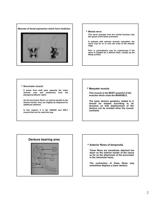
Muscles <strong>of</strong> facial expression which form modiolus<br />
• Buccinator muscle<br />
It arises from both jaws opposite <strong>the</strong> molar<br />
alveolar <strong>area</strong> <strong>and</strong> posteriorly from <strong>the</strong><br />
pterygom<strong>and</strong>ibular raphé.<br />
As <strong>the</strong> buccinator fibres run almost parallel to <strong>the</strong><br />
<strong>denture</strong> border, <strong>the</strong>y can slightly be displaced for<br />
additional retention.<br />
In this respect, it is <strong>the</strong> UNIQUE <strong>and</strong> ONLY<br />
muscle that can be used this way.<br />
Denture <strong>bearing</strong> <strong>area</strong><br />
• Mental nerve<br />
This nerve emerges from <strong>the</strong> mental foramen near<br />
<strong>the</strong> apices <strong>of</strong> <strong>the</strong> lower premolars.<br />
In patients with extreme alveolar resorption, <strong>the</strong><br />
nerve may lie on or near <strong>the</strong> crest <strong>of</strong> <strong>the</strong> alveolar<br />
ridge.<br />
Pain or paraes<strong>the</strong>sia may be experienced if <strong>the</strong><br />
nerve is trapped by a <strong>denture</strong> base, usually by <strong>the</strong><br />
fitting surface.<br />
• Massater muscle<br />
This muscle is <strong>the</strong> MOST powerful <strong>of</strong> <strong>the</strong><br />
muscles which close <strong>the</strong> MANDIBLE.<br />
The lower <strong>denture</strong> periphery related to it<br />
should be shaped according to its<br />
structure so that displacement <strong>of</strong> <strong>the</strong><br />
<strong>denture</strong> can be avoided when <strong>the</strong> muscle<br />
contracts.<br />
• Anterior fibres <strong>of</strong> temporalis<br />
These fibres are sometimes attached low<br />
down on <strong>the</strong> anterior border <strong>of</strong> <strong>the</strong> ramus<br />
as far as <strong>the</strong> attachment <strong>of</strong> <strong>the</strong> buccinator<br />
in <strong>the</strong> retromolar fossa.<br />
The contraction <strong>of</strong> <strong>the</strong>se fibres may<br />
sometimes displace a lower <strong>denture</strong>.<br />
2