Anatomy and histology of the denture bearing area - Dentistry ...
Anatomy and histology of the denture bearing area - Dentistry ...
Anatomy and histology of the denture bearing area - Dentistry ...
Create successful ePaper yourself
Turn your PDF publications into a flip-book with our unique Google optimized e-Paper software.
• Fovea palatinae<br />
These are a pair <strong>of</strong> mucous gl<strong>and</strong> duct orifices<br />
near <strong>the</strong> midline at <strong>the</strong> junction <strong>of</strong> <strong>the</strong> hard <strong>and</strong><br />
s<strong>of</strong>t palate.<br />
These l<strong>and</strong>marks provide a guide to <strong>the</strong> position<br />
<strong>of</strong> <strong>the</strong> posterior palatal border <strong>of</strong> a <strong>denture</strong>.<br />
• Incisive papilla<br />
Incisive papilla is a mass <strong>of</strong> fibrous tissue about 1<br />
cm behind <strong>the</strong> upper incisors.<br />
Its position in <strong>the</strong> edentulous mouth indicates<br />
where <strong>the</strong> incisors <strong>and</strong> canines should be set.<br />
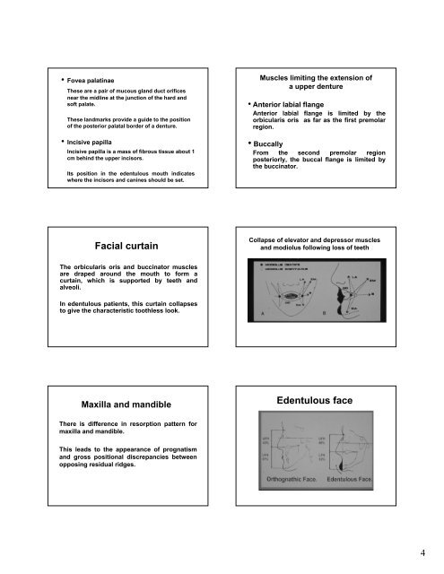
Facial curtain<br />
The orbicularis oris <strong>and</strong> buccinator muscles<br />
are draped around <strong>the</strong> mouth to form a<br />
curtain, which is supported by teeth <strong>and</strong><br />
alveoli.<br />
In edentulous patients, this curtain collapses<br />
to give <strong>the</strong> characteristic toothless look.<br />
Maxilla <strong>and</strong> m<strong>and</strong>ible<br />
There is difference in resorption pattern for<br />
maxilla <strong>and</strong> m<strong>and</strong>ible.<br />
This leads to <strong>the</strong> appearance <strong>of</strong> prognatism<br />
<strong>and</strong> gross positional discrepancies between<br />
opposing residual ridges.<br />
Muscles limiting <strong>the</strong> extension <strong>of</strong><br />
a upper <strong>denture</strong><br />
• Anterior labial flange<br />
Anterior labial flange is limited by <strong>the</strong><br />
orbicularis oris as far as <strong>the</strong> first premolar<br />
region.<br />
• Buccally<br />
From <strong>the</strong> second premolar region<br />
posteriorly, <strong>the</strong> buccal flange is limited by<br />
<strong>the</strong> buccinator.<br />
Collapse <strong>of</strong> elevator <strong>and</strong> depressor muscles<br />
<strong>and</strong> modiolus following loss <strong>of</strong> teeth<br />
Edentulous face<br />
4


