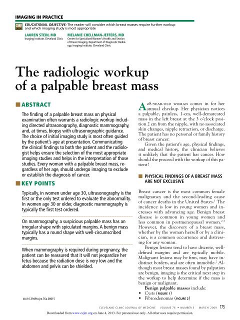
The radiologic workup of a palpable breast mass - Cleveland Clinic ...
The radiologic workup of a palpable breast mass - Cleveland Clinic ...
The radiologic workup of a palpable breast mass - Cleveland Clinic ...

Create successful ePaper yourself
Turn your PDF publications into a flip-book with our unique Google optimized e-Paper software.
imAging in PrActice<br />
CME<br />
CREDIT<br />
EDUCATIONAL OBJECTIVE: <strong>The</strong> reader will consider which <strong>breast</strong> <strong>mass</strong>es require further <strong>workup</strong><br />
and which imaging study is most appropriate<br />
Lauren Stein, MD<br />
Imaging Institute, <strong>Cleveland</strong> <strong>Clinic</strong><br />
<strong>The</strong> <strong>radiologic</strong> <strong>workup</strong><br />
<strong>of</strong> a <strong>palpable</strong> <strong>breast</strong> <strong>mass</strong><br />
■■AbstrAct<br />
<strong>The</strong> finding <strong>of</strong> a <strong>palpable</strong> <strong>breast</strong> <strong>mass</strong> on physical<br />
examination <strong>of</strong>ten warrants a <strong>radiologic</strong> <strong>workup</strong> including<br />
directed ultrasonography, diagnostic mammography,<br />
and, at times, biopsy with ultrasonographic guidance.<br />
<strong>The</strong> choice <strong>of</strong> initial imaging study is most <strong>of</strong>ten guided<br />
by the patient’s age at presentation. Communicating<br />
the clinical findings to both the patient and the radiologist<br />
helps ensure the selection <strong>of</strong> the most appropriate<br />
imaging studies and helps in the interpretation <strong>of</strong> those<br />
studies. Every woman with a <strong>palpable</strong> <strong>breast</strong> <strong>mass</strong>, regardless<br />
<strong>of</strong> her age, should undergo imaging to exclude<br />
or establish the diagnosis <strong>of</strong> cancer.<br />
■■Key<br />
Points<br />
Typically, in women under age 30, ultrasonography is the<br />
first or the only test ordered to evaluate the abnormality.<br />
In women age 30 or older, diagnostic mammography is<br />
typically the first test ordered.<br />
On mammography, a suspicious <strong>palpable</strong> <strong>mass</strong> has an<br />
irregular shape with spiculated margins. A benign <strong>mass</strong><br />
typically has a round shape with well-circumscribed<br />
margins.<br />
When mammography is required during pregnancy, the<br />
patient can be reassured that it will not jeopardize her<br />
fetus because the radiation dose is very low and the<br />
abdomen and pelvis can be shielded.<br />
doi:10.3949/ccjm.76a.08015<br />
MeLanie CheLLMan-JefferS, MD<br />
Center for Specialized Women’s Health and Section<br />
<strong>of</strong> Breast Imaging, Department <strong>of</strong> Diagnostic Radiology,<br />
Imaging Institute, <strong>Cleveland</strong> <strong>Clinic</strong><br />
28-year-old woman comes in for her<br />
A annual checkup. Her physician notices<br />
a <strong>palpable</strong>, painless, 1-cm, well-demarcated<br />
<strong>mass</strong> in the left <strong>breast</strong> at the 3 o’clock position<br />
2 cm from the nipple, with no associated<br />
skin changes, nipple retraction, or discharge.<br />
<strong>The</strong> patient has no personal or family history<br />
<strong>of</strong> <strong>breast</strong> cancer.<br />
Given the patient’s age, physical findings,<br />
and medical history, the clinician believes<br />
it unlikely that the patient has cancer. How<br />
should she proceed with the <strong>workup</strong> <strong>of</strong> this patient?<br />
■ Physical findings <strong>of</strong> a <strong>breast</strong> <strong>mass</strong><br />
are not exclusive<br />
Breast cancer is the most common female<br />
malignancy and the second-leading cause<br />
<strong>of</strong> cancer deaths in the United States. 1 <strong>The</strong><br />
incidence is low in young women and increases<br />
with advancing age. Benign <strong>breast</strong><br />
disease is common in young women and<br />
less common in postmenopausal women. 2,3<br />
However, the discovery <strong>of</strong> a <strong>breast</strong> <strong>mass</strong>,<br />
whether by the woman herself or by a clinician,<br />
is a common occurrence and distressing<br />
for any woman.<br />
Benign lesions tend to have discrete, welldefined<br />
margins and are typically mobile.<br />
Malignant lesions may be firm, may have indistinct<br />
borders, and are <strong>of</strong>ten immobile. 2 Although<br />
most <strong>breast</strong> <strong>mass</strong>es found by palpation<br />
are benign, imaging is the critical next step in<br />
the <strong>workup</strong> to help determine if the <strong>mass</strong> is<br />
benign or malignant.<br />
Benign <strong>palpable</strong> <strong>mass</strong>es include:<br />
• Cysts ( figure 1)<br />
• Fibroadenomas ( figure 2)<br />
CLEVELAND CLINIC JOURNAL OF MEDICINE VOLUME 76 • NUMBER 3 MARCH 2009 175<br />
Downloaded from<br />
www.ccjm.org on June 4, 2013. For personal use only. All other uses require permission.
on examination,<br />
note the clockface<br />
location,<br />
size, texture,<br />
tenderness,<br />
and mobility<br />
<strong>of</strong> the lump<br />
PAlPAble breAst mAss<br />
A<br />
figure 1. A simple cyst in the left <strong>breast</strong>.<br />
All three mammographic views—craniocaudal<br />
(A), mediolateral oblique (B), and<br />
spot-compression (C)—show a round,<br />
well-circumscribed <strong>mass</strong> in the mid-<strong>breast</strong>.<br />
Ultrasonography (D) shows a round, wellcircumscribed<br />
anechoic lesion with a sharply<br />
defined posterior wall and posterior acoustic<br />
enhancement.<br />
• Prominent fat lobules<br />
• Lymph nodes<br />
• Oil cysts<br />
• Lipomas<br />
• Hamartomas ( figure 3)<br />
• Hematomas<br />
• Fat necrosis<br />
• Galactoceles.<br />
Malignant <strong>palpable</strong> <strong>mass</strong>es include:<br />
• Invasive ductal and lobular carcinoma ( fig-<br />
•<br />
■<br />
ure 4)<br />
Ductal carcinoma in situ (which rarely<br />
presents as a <strong>palpable</strong> <strong>mass</strong>.)<br />
history and Physical examination<br />
To ensure that imaging provides the most useful<br />
information about a <strong>palpable</strong> <strong>breast</strong> lump,<br />
176 CLEVELAND CLINIC JOURNAL OF MEDICINE VOLUME 76 • NUMBER 3 MARCH 2009<br />
B<br />
D<br />
C<br />
it is important to first do a careful history and<br />
physical examination. Important aspects <strong>of</strong> the<br />
history include family history, personal history<br />
<strong>of</strong> <strong>breast</strong> cancer, and any previous <strong>breast</strong> biopsies.<br />
<strong>The</strong> onset and duration <strong>of</strong> the <strong>palpable</strong><br />
<strong>mass</strong>, changes in its size, the relationship <strong>of</strong><br />
these changes to the menstrual cycle, and the<br />
presence or lack <strong>of</strong> tenderness are additional<br />
important elements <strong>of</strong> the history.<br />
On examination, it is important to note<br />
the clock-face location, size, texture, tender-<br />
Downloaded from<br />
www.ccjm.org on June 4, 2013. For personal use only. All other uses require permission.
A<br />
ness, and mobility <strong>of</strong> the lump. Accompanying<br />
nipple discharge and skin erythema or retraction<br />
are also important to report. In addition<br />
to conveying the location <strong>of</strong> the <strong>mass</strong> to the<br />
radiologist, it is equally important that the patient<br />
know what features the physician feels.<br />
This way, if the clinical information from the<br />
ordering physician is not available at the time<br />
<strong>of</strong> the <strong>radiologic</strong> evaluation, the patient will<br />
be able to guide the radiologist to the region<br />
<strong>of</strong> concern.<br />
■<br />
imaging techniques<br />
Mammography and ultrasonography are the<br />
primary imaging studies for evaluating <strong>palpable</strong><br />
<strong>breast</strong> <strong>mass</strong>es. Typically, in women under<br />
age 30, ultrasonography is the first or the<br />
only test ordered to evaluate the abnormality. 4<br />
In women age 30 or older, diagnostic mammography<br />
is typically the first test ordered.<br />
If mammography indicates that the <strong>palpable</strong><br />
<strong>mass</strong> is not benign, then ultrasonography is<br />
the next study to be done. 3 Although a powerful<br />
tool, magnetic resonance imaging <strong>of</strong> the<br />
<strong>breast</strong> does not currently have a role in the<br />
<strong>workup</strong> <strong>of</strong> a <strong>palpable</strong> abnormality and should<br />
B<br />
stein AnD cHellmAn-JeFFers<br />
C<br />
figure 2. Fibroadenoma. On mammography,<br />
the craniocaudal (A) and mediolateral<br />
oblique (B) views with a bright metallic<br />
marker (arrows) show a round, well-circumscribed<br />
<strong>mass</strong> in the upper outer quadrant<br />
<strong>of</strong> the left <strong>breast</strong>. Ultrasonography (C)<br />
shows an oval, well-circumscribed, mildly<br />
heterogeneous, hypoechoic <strong>mass</strong> that is<br />
wider than tall, indicating a benign <strong>mass</strong>.<br />
not be used as a decision-delaying tactic or in<br />
place <strong>of</strong> biopsy.<br />
screening or diagnostic mammography?<br />
Mammography is used in both screening and<br />
diagnosis. Screening mammography consists<br />
<strong>of</strong> two standard views <strong>of</strong> each <strong>breast</strong>—craniocaudal<br />
and mediolateral oblique—and is appropriate<br />
for asymptomatic women.<br />
Women age 30 or older who present with a<br />
<strong>palpable</strong> <strong>breast</strong> <strong>mass</strong> require diagnostic mammography,<br />
in which standard mammographic<br />
views are obtained, as well as additional views<br />
(eg, tangential or spot-compression views) to<br />
better define the area <strong>of</strong> clinical concern. In<br />
a tangential view, a metallic skin marker is<br />
placed on the skin overlying the site <strong>of</strong> the<br />
<strong>palpable</strong> abnormality.<br />
On mammography, a suspicious <strong>palpable</strong><br />
<strong>mass</strong> has an irregular shape with spiculated<br />
margins. A benign <strong>mass</strong> typically has a round<br />
shape with well-circumscribed margins. If the<br />
<strong>palpable</strong> abnormality is not mammographically<br />
benign (eg, if it does not look like a lymph<br />
node, lipoma, or degenerating fibroadenoma),<br />
then ultrasonography is performed.<br />
Mammography is less sensitive in younger<br />
Women over<br />
age 30 who<br />
present with a<br />
<strong>palpable</strong> <strong>breast</strong><br />
<strong>mass</strong> require<br />
diagnostic<br />
mammography<br />
CLEVELAND CLINIC JOURNAL OF MEDICINE VOLUME 76 • NUMBER 3 MARCH 2009 177<br />
Downloaded from<br />
www.ccjm.org on June 4, 2013. For personal use only. All other uses require permission.
core-needle<br />
biopsy has<br />
become more<br />
popular than<br />
fine-needle<br />
aspiration,<br />
as it includes<br />
surrounding<br />
tissue<br />
PAlPAble breAst mAss<br />
A<br />
figure 3. Hamartoma. Craniocaudal (A) and medio lateral oblique (B) mammographic<br />
views <strong>of</strong> the left <strong>breast</strong> show an apparently encapsulated, heterogeneous <strong>mass</strong> that contains<br />
fat mixed with fibroglandular tissue.<br />
women (ie, under age 30) because their <strong>breast</strong> tissue<br />
tends to be dense and glandular, whereas the<br />
tissue becomes more “fat-replaced” with age. 3<br />
ultrasonography plays<br />
a complementary role<br />
Ultrasonography complements diagnostic<br />
mammography and can be used as a first imaging<br />
study to evaluate a <strong>palpable</strong> <strong>breast</strong> <strong>mass</strong> in<br />
a young woman (ie, under age 30) with dense<br />
<strong>breast</strong> tissue. Ultrasonography is helpful in<br />
distinguishing cystic lesions from solid <strong>mass</strong>es.<br />
It helps the radiologist delineate the shape,<br />
borders, and acoustic properties <strong>of</strong> the <strong>mass</strong>.<br />
It is also performed when a <strong>palpable</strong> <strong>mass</strong> is<br />
mammographically occult. When a <strong>mass</strong> appears<br />
suspicious on either mammography or<br />
ultrasonography, ultrasonography can be used<br />
to guide biopsy.<br />
A suspicious <strong>mass</strong> on ultrasonography classically<br />
appears “taller than wide” and has posterior<br />
acoustic shadowing. Microlobulations<br />
and a spiculated margin also raise concern for<br />
178 CLEVELAND CLINIC JOURNAL OF MEDICINE VOLUME 76 • NUMBER 3 MARCH 2009<br />
B<br />
malignancy. A benign sonographic appearance<br />
<strong>of</strong> a <strong>palpable</strong> <strong>mass</strong> includes a “wider than<br />
tall” (ellipsoid) shape, with homogeneous<br />
echogenicity, and four or fewer gentle lobulations.<br />
A thin, echogenic capsule also suggests<br />
the <strong>mass</strong> is benign.<br />
core-needle biopsy<br />
with ultrasonographic guidance<br />
Core-needle biopsy is performed with a largediameter<br />
(14-gauge to 18-gauge) needle to<br />
obtain tissue cores for histologic analysis. It<br />
has gained popularity over fine-needle aspiration<br />
because it includes surrounding tissue<br />
architecture, thus providing a more definitive<br />
histologic diagnosis.<br />
Pathologic information obtained from<br />
core-needle biopsy allows the radiologist and<br />
surgeon to counsel the patient and determine<br />
the best surgical management or follow-up imaging<br />
study. If a clinician performs fine-needle<br />
biopsy in the <strong>of</strong>fice, it should be preceded by<br />
an imaging <strong>workup</strong> <strong>of</strong> the <strong>palpable</strong> finding.<br />
Downloaded from<br />
www.ccjm.org on June 4, 2013. For personal use only. All other uses require permission.
A<br />
■ What is aPProPriate<br />
for our 28-year-old Patient?<br />
Because she is under age 30, ultrasonography<br />
is the initial study <strong>of</strong> choice to evaluate the<br />
<strong>mass</strong>. If a simple cyst is detected, she can be<br />
reassured that the lesion is benign, and no<br />
subsequent follow-up is required. If the lesion<br />
is a solid <strong>mass</strong> with benign features, mammography<br />
may be considered, the patient may be<br />
followed with short-interval imaging (every 6<br />
months) depending on patient-specific factors<br />
such as family history, or the <strong>mass</strong> can be biopsied.<br />
If the lesion is a solid <strong>mass</strong> with suspicious<br />
or malignant features, mammography with<br />
spot-compression views should be performed,<br />
and the patient should undergo core-needle<br />
biopsy with ultrasonographic guidance.<br />
In a patient age 30 or older, diagnostic<br />
mammography is the imaging study <strong>of</strong> first<br />
choice. 3 If the <strong>mass</strong> is clearly benign on mammography,<br />
no additional imaging would be<br />
necessary. If mammography fails to image the<br />
<strong>mass</strong> or shows it to have benign features such<br />
as fat, then the patient can undergo ultrasono-<br />
B<br />
stein AnD cHellmAn-JeFFers<br />
figure 4. Infiltrating ductal carcinoma. Craniocaudal (A) and mediolateral oblique (B)<br />
mammographic views <strong>of</strong> the right <strong>breast</strong> show an irregular, mildly spiculated, high-density<br />
lesion in the posterior, medial <strong>breast</strong>. Ultrasonography (C) shows an irregularly shaped<br />
hypoechoic <strong>mass</strong> which is taller than wide (a pr<strong>of</strong>ile tending to indicate malignancy) and<br />
has mild posterior acoustic shadowing.<br />
C<br />
graphy for further evaluation and confirmation<br />
<strong>of</strong> the clinical and mammographic findings. If<br />
the <strong>mass</strong> appears suspicious or malignant on<br />
mammography, ultrasonography is the next<br />
step, as it can help characterize the lesion and<br />
be used to guide core-needle biopsy.<br />
■ if a Pregnant Woman<br />
has a PalPable <strong>breast</strong> <strong>mass</strong><br />
Most publications on <strong>breast</strong> cancer in pregnancy<br />
report a prevalence <strong>of</strong> 3 per 10,000<br />
pregnancies, accounting for 3% <strong>of</strong> all <strong>breast</strong><br />
cancers diagnosed. 5 <strong>The</strong>refore, imaging evaluation<br />
<strong>of</strong> a <strong>palpable</strong> <strong>mass</strong> should not be postponed.<br />
Hormonal changes throughout pregnancy<br />
may increase the nodularity <strong>of</strong> <strong>breast</strong> tissue,<br />
raising the concern <strong>of</strong> <strong>palpable</strong> <strong>mass</strong>es. Additionally,<br />
there is a higher prevalence <strong>of</strong> galactoceles<br />
and lactating adenomas in these patients.<br />
Because contrasting fatty <strong>breast</strong> tissue is<br />
lost during pregnancy and because <strong>of</strong> the need<br />
to minimize radiation exposure, ultrasonography<br />
is <strong>of</strong>ten the imaging test <strong>of</strong> first choice. If<br />
Ultrasound is<br />
<strong>of</strong>ten the study<br />
used first in<br />
pregnancy, but<br />
mammography<br />
can also be used<br />
CLEVELAND CLINIC JOURNAL OF MEDICINE VOLUME 76 • NUMBER 3 MARCH 2009 179<br />
Downloaded from<br />
www.ccjm.org on June 4, 2013. For personal use only. All other uses require permission.
PAlPAble breAst mAss<br />
mammography is required, the radiation dose<br />
is very low and the patient’s abdomen and pelvis<br />
can be shielded. 6 In this situation, the patient<br />
can be reassured that the imaging test is<br />
not jeopardizing her fetus.<br />
■<br />
What WorkuP is required in men?<br />
Breast cancer in men is rare, accounting for<br />
less than 0.5% <strong>of</strong> all cases. 7 Most <strong>of</strong>ten, a <strong>palpable</strong><br />
<strong>breast</strong> <strong>mass</strong> in a man presents as unilateral<br />
gynecomastia. Gynecomastia occurs in a<br />
bimodal age distribution (in the 2nd and 7th<br />
decades) and has a variety <strong>of</strong> hormonal and<br />
drug-related causes. Despite the low prevalence<br />
<strong>of</strong> <strong>breast</strong> cancer in men, the combination<br />
<strong>of</strong> mammography and ultrasonography is<br />
recommended for evaluation at all ages. ■<br />
■<br />
references<br />
1. Klein S. Evaluation <strong>of</strong> <strong>palpable</strong> <strong>breast</strong> <strong>mass</strong>es. Am Fam<br />
Physician 2005; 71:1731–1738.<br />
2. Pruthi S. Detection and evaluation <strong>of</strong> a <strong>palpable</strong> <strong>breast</strong><br />
<strong>mass</strong>. Mayo Clin Proc 2001; 76:641–648.<br />
3. Harvey JA. Sonography <strong>of</strong> <strong>palpable</strong> <strong>breast</strong> <strong>mass</strong>es. Semin<br />
Ultrasound CT MR 2006; 27:284–297.<br />
4. Mehta TS. Current uses <strong>of</strong> ultrasound in the evaluation <strong>of</strong><br />
the <strong>breast</strong>. Radiol Clin North Am 2003; 41:841–856.<br />
5. gallenberg MM, Lopines CL. Breast cancer and pregnancy.<br />
Semin Oncol 1989; 16:369–376.<br />
6. Barnavon Y, Wallack MK. Management <strong>of</strong> the pregnant<br />
patient with carcinoma <strong>of</strong> the <strong>breast</strong>. Surg Gynecol Obstet<br />
1990; 171:347–352.<br />
7. Cardenosa g. <strong>The</strong> Core Curriculum: Breast Imaging. Philadelphia:<br />
Lippincott Williams and Wilkins, 2003;304.<br />
ADDRESS: Melanie Chellman-Jeffers, MD, Imaging Institute,<br />
Section <strong>of</strong> Breast Imaging, A10, <strong>Cleveland</strong> <strong>Clinic</strong>, 9500 Euclid<br />
Avenue, <strong>Cleveland</strong>, OH 44195; e-mail chellmm@ccf.org.<br />
Visit our web site at http://<br />
www.ccjm.org<br />
Contact us by e-mail at<br />
ccjm@ccf.org<br />
180 CLEVELAND CLINIC JOURNAL OF MEDICINE VOLUME 76 • NUMBER 3 MARCH 2009<br />
Downloaded from<br />
www.ccjm.org on June 4, 2013. For personal use only. All other uses require permission.