The Clinical Picture - Cleveland Clinic Journal of Medicine
The Clinical Picture - Cleveland Clinic Journal of Medicine
The Clinical Picture - Cleveland Clinic Journal of Medicine

You also want an ePaper? Increase the reach of your titles
YUMPU automatically turns print PDFs into web optimized ePapers that Google loves.
<strong>The</strong> CliniCal PiCTure<br />
Giulia Rech, MD<br />
Internal <strong>Medicine</strong>, Aging and Nephrologic<br />
Disease Department, Dermatology Division,<br />
Ospedale Sant’Orsola-Malpighi, Università degli<br />
Studi di Bologna, Italy<br />
Bianca MaRia PiRaccini, MD, PhD<br />
Internal <strong>Medicine</strong>, Aging and Nephrologic Disease<br />
Department, Dermatology Division, Ospedale Sant’Orsola-<br />
Malpighi, Università degli Studi di Bologna, Italy<br />
RiccaRDo BalestRi, MD<br />
Internal <strong>Medicine</strong>, Aging and Nephrologic<br />
Disease Department, Dermatology Division,<br />
Ospedale Sant’Orsola-Malpighi, Università degli<br />
Studi di Bologna, Italy<br />
FeDeRico BaRDazzi, MD<br />
Internal <strong>Medicine</strong>, Aging and Nephrologic<br />
Disease Department, Dermatology Division,<br />
Ospedale Sant’Orsola-Malpighi, Università degli<br />
Studi di Bologna, Italy<br />
<strong>The</strong> <strong><strong>Clinic</strong>al</strong> <strong>Picture</strong><br />
Scar reactivation and dry cough<br />
52-year-old woman is referred to our<br />
A dermatology clinic by her primary care<br />
physician for swelling and redness <strong>of</strong> seven old<br />
scars. <strong>The</strong> swelling began 3 months ago.<br />
She is on no regular medications. She has<br />
never smoked. She underwent liposuction 7<br />
years ago and appendectomy at age 15.<br />
Physical examination reveals indurated<br />
and painful brownish-red plaques on the<br />
thighs and lower trunk, extending beyond the<br />
borders <strong>of</strong> six scars from the liposuction surgery<br />
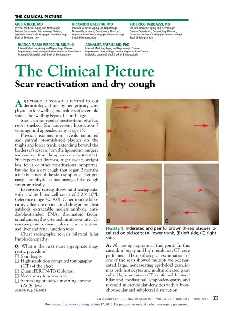
and one scar from the appendectomy (Figure 1).<br />
She reports no dyspnea, night sweats, weight<br />
loss, fever, or other constitutional symptoms,<br />
but she has a dry cough that began 2 months<br />
after the onset <strong>of</strong> the skin symptoms. Her primary<br />
care physician has managed the cough<br />
symptomatically.<br />
Laboratory testing shows mild leukopenia,<br />
with a white blood cell count <strong>of</strong> 3.0 × 10⁹ /L<br />
(reference range 4.2–9.0). Other routine laboratory<br />
values are normal, including antinuclear<br />
antibody, extractable nuclear antibody, antidouble-stranded<br />
DNA, rheumatoid factor,<br />
urinalysis, erythrocyte sedimentation rate, Creactive<br />
protein, serum calcium concentration,<br />
and liver and renal function tests.<br />
Chest radiography reveals bilateral hilar<br />
lymphadenopathy.<br />
Q: What is the next most appropriate diagnostic<br />
procedure?<br />
□ Skin biopsy<br />
□ High-resolution computed tomography<br />
(CT) <strong>of</strong> the chest<br />
□ QuantiFERON-TB Gold test<br />
□ Ventilatory function tests<br />
□ Serum angiotensin-converting enzyme<br />
(ACE) level<br />
doi:10.3949/ccjm.78a.10132<br />
annalisa PatRizi, MD, PhD<br />
Internal <strong>Medicine</strong>, Aging and Nephrologic Disease<br />
Department, Dermatology Division, Ospedale Sant’Orsola-<br />
Malpighi, Università degli Studi di Bologna, Italy<br />
Figure 1. Indurated and painful brownish-red plaques localized<br />
on old scars: (A) lower trunk, (B) left side, (C) right<br />
side.<br />
A: All are appropriate at this point. In this<br />
case, skin biopsy and high-resolution CT were<br />
performed. Histopathologic examination <strong>of</strong><br />
one <strong>of</strong> the scars showed multiple well-demarcated,<br />
large, noncaseating epitheloid granulomas<br />
with histiocytes and multinucleated giant<br />
cells. High-resolution CT confirmed bilateral<br />
hilar and mediastinal lymphadenopathy and<br />
revealed micronodular densities with a bronchovascular<br />
and subpleural distribution.<br />
CLEVELAND CLINIC JOURNAL OF MEDICINE VOLUME 78 • NUMBER 6 JUNE 2011 375<br />
Downloaded from<br />
www.ccjm.org on June 17, 2013. For personal use only. All other uses require permission.
Treatment:<br />
oral prednisone<br />
1 mg/kg/day<br />
for 6 weeks,<br />
then tapered<br />
until skin and<br />
hilar lesions<br />
resolved<br />
SCar SarCoidoSiS<br />
Figure 2. Noncaseating granuloma seen on<br />
skin biopsy study.<br />
An interferon-gamma-release assay for tuberculosis—QuantiFERON-TB<br />
Gold (Cellestis,<br />
Carnegie, Australia)—was negative.<br />
Ventilatory function tests showed a normal<br />
pattern, while the serum ACE level, electrocardiography,<br />
and an eye examination revealed<br />
no pathologic findings.<br />
Q: What is the diagnosis?<br />
□ Keloids<br />
□ Scar sarcoidosis<br />
□ Paraneoplastic sign<br />
□ Dermat<strong>of</strong>ibrosarcoma protuberans<br />
□ Tubercolosis<br />
A: Based on the data outlined above, we<br />
made the diagnosis <strong>of</strong> scar sarcoidosis with<br />
involvement <strong>of</strong> hilar and mediastinal lymph<br />
nodes. <strong>The</strong> patient began systemic treatment<br />
with oral prednisone 1 mg/kg/day for 6 weeks,<br />
which was then gradually withdrawn, until<br />
the skin and hilar lesions resolved completely.<br />
■ scar sarcoidosis<br />
Sarcoidosis is a multisystem disorder <strong>of</strong> unknown<br />
cause characterized by the formation<br />
<strong>of</strong> noncaseating granulomas in the affected<br />
organs. Patients may present with symptoms<br />
related to the specific organ affected, but they<br />
may have no symptoms or only general symptoms<br />
such as fever or general malaise.<br />
<strong>The</strong> skin is involved in 25% <strong>of</strong> cases and<br />
presents so many polymorphous manifestations<br />
that sarcoidosis has become known as<br />
one <strong>of</strong> the “great imitators” in dermatology. 1,2<br />
Although sarcoidosis on liposuction scars has<br />
not been reported previously, the reactivation <strong>of</strong><br />
old scars is well known on sites <strong>of</strong> previous injections,<br />
tattoos, herpes zoster, and burns. 2,3<br />
<strong>The</strong> finding <strong>of</strong> granuloma is not specific for<br />
sarcoidosis (Figure 2). <strong>The</strong> histologic differential<br />
diagnosis <strong>of</strong> sarcoidosis includes tuberculosis,<br />
atypical mycobacteriosis, fungal infection,<br />
reaction to a foreign body, rheumatoid<br />
nodules, leishmaniasis, Crohn disease, and<br />
necrobiosis lipoidica diabeticorum.<br />
<strong>The</strong> diagnosis <strong>of</strong> scar sarcoidosis is confirmed<br />
only by excluding other conditions via<br />
a comprehensive evaluation <strong>of</strong> clinical manifestations,<br />
histology, history, and radiologic<br />
and laboratory findings.<br />
It has been suggested that the most satisfying<br />
therapy for the patient and physician in<br />
sarcoidosis is no treatment at all, 4 and in fact<br />
sarcoidosis <strong>of</strong>ten remits spontaneously. Currently,<br />
the choice <strong>of</strong> treatment depends on<br />
the degree <strong>of</strong> systemic involvement, and the<br />
oral corticosteroid prednisone remains the<br />
first-line treatment. If the condition does not<br />
respond, the use <strong>of</strong> other systemic agents has<br />
been reported, but their effectiveness has not<br />
been evaluated in controlled clinical trials.<br />
Recurrence is common after the suspension<br />
<strong>of</strong> treatment; therefore, treatment may<br />
need to be continued for several years, with<br />
frequent checkups.<br />
Skin lesions are a visible clue to the diagnosis.<br />
Reactivation <strong>of</strong> old scars may be the<br />
single manifestation <strong>of</strong> cutaneous sarcoidosis,<br />
but it may also precede or accompany systemic<br />
involvement, <strong>of</strong>ten representing the main sign<br />
<strong>of</strong> an exacerbation or a relapse <strong>of</strong> systemic sarcoidosis,<br />
as in our patient. 5 ■<br />
■ rEFErENcEs<br />
1. Marchell rM, Judson MA. Chronic cutaneous lesions <strong>of</strong><br />
sarcoidosis. Clin Dermatol 2007; 25:295–302.<br />
2. Tchernev g. Cutaneous sarcoidosis: the "great imitator":<br />
etiopathogenesis, morphology, differential diagnosis,<br />
and clinical management. Am J Clin Dermatol 2006;<br />
7:375–382.<br />
3. Fernandez-Faith e, McDonnell J. Cutaneous sarcoidosis:<br />
differential diagnosis. Clin Dermatol 2007; 25:276–287.<br />
4. Baughman rP, Lower ee, du Bois rM. Sarcoidosis. Lancet<br />
2003; 361:1111–1118.<br />
5. Sorabjee JS, garje r. Reactivation <strong>of</strong> old scars: inevitably<br />
sarcoid. Postgrad Med J 2005; 81:60–61.<br />
ADDRESS: Riccardo Balestri, MD, Via Massarenti 1, <strong>Clinic</strong>a<br />
Dermatologica, 40138 Bologna, Italy; e-mail ilsabo@libero.it.<br />
376 CLEVELAND CLINIC JOURNAL OF MEDICINE VOLUME 78 • NUMBER 6 JUNE 2011<br />
Downloaded from<br />
www.ccjm.org on June 17, 2013. For personal use only. All other uses require permission.