How to Interpret Borderline Hip and Elbow radiographs. - Love My Pet
How to Interpret Borderline Hip and Elbow radiographs. - Love My Pet
How to Interpret Borderline Hip and Elbow radiographs. - Love My Pet

Create successful ePaper yourself
Turn your PDF publications into a flip-book with our unique Google optimized e-Paper software.
<strong>How</strong> <strong>to</strong> <strong>Interpret</strong> <strong>Borderline</strong> <strong>Hip</strong> <strong>and</strong> <strong>Elbow</strong><br />
<strong>radiographs</strong>.<br />
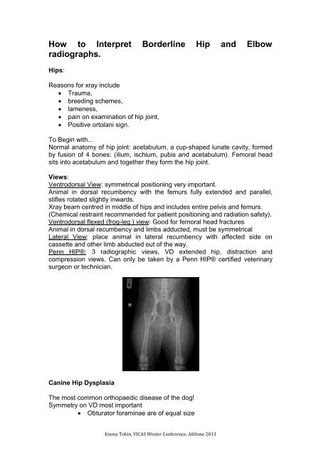
<strong>Hip</strong>s:<br />
Reasons for xray include<br />
Trauma,<br />
breeding schemes,<br />
lameness,<br />
pain on examination of hip joint,<br />
Positive or<strong>to</strong>lani sign.<br />
To Begin with...<br />
Normal ana<strong>to</strong>my of hip joint: acetabulum, a cup-shaped lunate cavity, formed<br />
by fusion of 4 bones: (ilium, ischium, pubis <strong>and</strong> acetabulum). Femoral head<br />
sits in<strong>to</strong> acetabulum <strong>and</strong> <strong>to</strong>gether they form the hip joint.<br />
Views:<br />
Ventrodorsal View: symmetrical positioning very important.<br />
Animal in dorsal recumbency with the femurs fully extended <strong>and</strong> parallel,<br />
stifles rotated slightly inwards.<br />
Xray beam centred in middle of hips <strong>and</strong> includes entire pelvis <strong>and</strong> femurs.<br />
(Chemical restraint recommended for patient positioning <strong>and</strong> radiation safety).<br />
Ventrodorsal flexed (frog-leg ) view: Good for femoral head fractures<br />
Animal in dorsal recumbency <strong>and</strong> limbs adducted, must be symmetrical<br />
Lateral View: place animal in lateral recumbency with affected side on<br />
cassette <strong>and</strong> other limb abducted out of the way.<br />
Penn HIP®: 3 radiographic views, VD extended hip, distraction <strong>and</strong><br />
compression views. Can only be taken by a Penn HIP® certified veterinary<br />
surgeon or technician.<br />
Canine <strong>Hip</strong> Dysplasia<br />
The most common orthopaedic disease of the dog!<br />
Symmetry on VD most important<br />
Obtura<strong>to</strong>r foraminae are of equal size<br />
Emma Tobin, VICAS Winter Conference, Athlone 2013
The ilial wings have same diameter<br />
Line drawn thro lumbar vertebrae continues straight through the<br />
pelvic symphsis<br />
VD radiograph can be insenstive. Increased incidence of detection as dog<br />
gets older!<br />
Radiological Findings<br />
Femoral head subluxation or luxation<br />
>50% or more of femoral head (FH) is not within acetabulum or<br />
Norberg angle abnormal<br />
Osteoarthrosis/itis<br />
Remodelling of FH <strong>and</strong> neck<br />
Remodelling of acetabulum<br />
Periarticular osteophytes<br />
Morgans line<br />
Osteophyte formation on FH parallel <strong>to</strong> physeal scar (FH<br />
rimming)<br />
Subchondral sclerosis of the acetabular margin.<br />
Measurement of the Norberg angle : Draw a line between the centres of the<br />
femoral heads, <strong>and</strong> a line extending cranially from the centre of the femoral<br />
head <strong>to</strong> the craniolateral acetabular margin. The norberg angle is the angle of<br />
intersection between the two. Normal dogs have an angle of 105 degrees or<br />
more (95 in the cat). Values less than this indicate subluxation, the lower the<br />
angle the more severe the subluxation. The Norberg angle does not correlate<br />
as well as the distraction index (DI) with regard <strong>to</strong> the subsequent<br />
develpoment of osteoarthrosis.<br />
<strong>Hip</strong> Control Schemes:<br />
BVA, OFA, PennHIP®<br />
<strong>Elbow</strong> Joints<br />
Humero-radial-ulnar joint.<br />
Mediolateral Extended<br />
Lateral recumbency on affected limb. Upper limb retracted caudally <strong>and</strong> head<br />
<strong>and</strong> neck extended. Angle of 120 degrees between the humerus <strong>and</strong> the<br />
radius <strong>and</strong> ulna. Beam centred on medial epicondyle.<br />
Evaluates incongruity<br />
Osteophytes (O/ps) on cranial aspect of joint<br />
Emma Tobin, VICAS Winter Conference, Athlone 2013
Medial Coronoid Process (MCP) is superimposed on radial<br />
head.<br />
Mediolateral Flexed (maximally)<br />
Lateral recumbency on affected limb. Upper limb retracted caudally <strong>and</strong> head<br />
<strong>and</strong> neck extended. Distal limb pulled <strong>to</strong>wards head <strong>and</strong> neck so that angle<br />
between humerus <strong>and</strong> radius/ulna is
UAP<br />
Osteochondritis Dissecans (OCD) of the medial humeral<br />
condyle<br />
<strong>Elbow</strong> incongruity<br />
Ununited Medial Humeral Epicondyle<br />
Medial Epicondylar Spur<br />
Incomplete Ossification of the Humeral Condyle<br />
Patella Cubiti<br />
Metabolic Disorders<br />
Hypervitaminosis A in cats<br />
Osteoarthrosis<br />
Usually as a result of elbow dysplasia in predisposed breeds<br />
Secondary <strong>to</strong> trauma, do joint tap if suddenly very lame<br />
Infection<br />
Bacterial, viral, pro<strong>to</strong>zoal or fungal<br />
STS, subchondral erosion, later changes of periosteal<br />
proliferation<br />
Neoplasia<br />
Synovial cell carcinoma most common joint neoplasm<br />
Emma Tobin, VICAS Winter Conference, Athlone 2013