TARSOCRURAL LUXATION - Love My Pet
TARSOCRURAL LUXATION - Love My Pet
TARSOCRURAL LUXATION - Love My Pet
- No tags were found...

You also want an ePaper? Increase the reach of your titles
YUMPU automatically turns print PDFs into web optimized ePapers that Google loves.
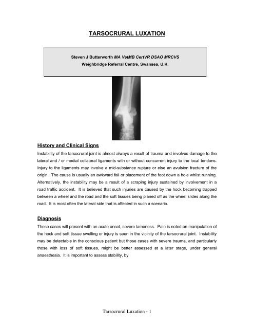
<strong>TARSOCRURAL</strong> <strong>LUXATION</strong>Steven J Butterworth MA VetMB CertVR DSAO MRCVSWeighbridge Referral Centre, Swansea, U.K.History and Clinical SignsInstability of the tarsocrural joint is almost always a result of trauma and involves damage to thelateral and / or medial collateral ligaments with or without concurrent injury to the local tendons.Injury to the ligaments may involve a mid-substance rupture or else an avulsion fracture of theorigin. The cause is usually an awkward fall or placement of the foot down a hole whilst running.Alternatively, the instability may be a result of a scraping injury sustained by involvement in aroad traffic accident. It is believed that such injuries are caused by the hock becoming trappedbetween a wheel and the road and the soft tissues being planed off as the wheel slides along theroad. It is most often the lateral side that is affected in such a scenario.DiagnosisThese cases will present with an acute onset, severe lameness. Pain is noted on manipulation ofthe hock and soft tissue swelling or injury is seen in the vicinity of the tarsocrural joint. Instabilitymay be detectable in the conscious patient but those cases with severe trauma, and particularlythose with loss of soft tissues, might be better assessed at a later stage, under generalanaesthesia. It is important to assess stability, byTarsocrural Luxation - 1
stressing the joint, in both flexion and extension since it is necessary to establish whether one orboth components of each collateral ligament is/are injured.Radiography may be helpful in showing the presence of avulsion fractures and joint spacewidening due to instability, which might require stressed views to become apparent. It may alsoshow the extent of bone loss in "shearing" injuries, although this is usually known after clinicalexamination.TreatmentNon-surgical management is indicated in cases with only partial rupture of a collateral ligamentor an avulsion fracture of a malleolus, which is amenable to closed reduction. The application ofa cast for 6 to 8 weeks may allow satisfactory healing and a return to normal function.Surgical management is indicated in the following situations :-(a) Where avulsion fractures of the medial and/or lateral malleoli are responsible for theinstability and the fragment is considered large enough to accomodate implants. Surgicalmanagement may involve re-attachment of the fragment using a lagged bonescrew or pin and tension band wire.(b) If the injury involves complete rupture of the collateral ligament, and itis treated early, it may be possible to suture the two ends together using anappropriate material such as monofilament nylon or polydioxanone (PDS,Eticon Ltd.). Injuries to the local tendons should also be evaluated andsutured appropriately. Postoperatively, a cast should be applied to the hockfor 6 to 8 weeks.Tarsocrural Luxation - 2
(c) If the instability is marked and/or conservative management has failed then it may not bepossible to reconstruct the ligaments due to thedegree of tearing or fibrosis. In such cases it may benecessary to place prosthetic collateral ligaments ofmonofilament nylon, braided polyester (Ethibond,Ethicon Ltd.) or wire. Many techniques have beendescribed to achieve this (Holt, 1976; Holt, 1977),whereby bone tunnels or bone screws have beenused to anchor the prosthesis in place. The earliermethods described did not really take into accountthe collateral ligaments having two components andthus may not have been able to stabilise the joint inboth flexion and extension. A more recentlydeveloped method (Aron & Purinton, 1985)addresses this problem and is probably the best oneto use, particularly in patients that are large enoughto allow placement of the bone screws. On themedial side, a screw is placed in the medialmalleolus and then two are placed in the talus, one proximally and one distally, thus allowingplacement of a long and a short collateral ligament. The technique is the same on the lateralaspect except that the screws are placed into the lateral malleolus and calcaneus in a similarfashion. Whichever method is used, the hock should be supported with a cast for 4 to 8 weekspostoperatively.Such injuries in cats can prove problematic to treat because placement of implants to supportcollateral prostheses is difficult (due to size) and maintaining external coaptation can also befrustrating. The use of a transarticular pin may be considered whilst soft tissue healing takesplace. Such a pin is placed from the proximal tibia and across the joint with the latter held inreduction and at a functional angle.Tarsocrural Luxation - 3
(d) Open luxations should be considered emergencies. If the joint can be thoroughlydebrided within the "golden period" of 6 to 8 hours (possibly extended to 12 to 18 hours ifantibiotics are given immediately) then it may be possible toprevent the contamination from becoming established asinfection. The presence of the latter in a joint, particularlyone that is as tight-fitting as the tarsocrural joint, is likely toseverely compromise the end result. After this it might bepossible to consider ligament reconstruction or prostheticreplacement. However, it is often the case that much softtissue has been lost and collateral replacement wouldrequire a great deal of foreign material to be left in apotentially infected site. Instead it may be worth consideringthe application of a transarticular external skeletal fixator.This will stabilise the joint whilst soft tissue healing takesplace whilst at the same time allowing access to the woundsfor daily topical treatment. Such a frame is most easilyapplied to the medial aspect which works quite well in mostcases since the soft tissue loss is usually on the lateralaspect. In small patients, placement of the fixation pins intothe metatarsi can be difficult and the use of a Rudy bootmay side-step such a problem. The principle behind such a boot is to incorporate the distal pinsinto a cast applied to the pes rather than drilling them into the metatarsi. The fixator is left inplace for about 8 weeks and then, once soft tissue healing has taken place, surgery to correctany instability (as discussed above) can be reconsidered. It is the experience of many surgeonsthat the soft tissue healing is sufficient to restore stability and that a second operation is oftenunnecessary.(e) In cases where the techniques mentioned above have failed or where there is concurrentarticular damage which would compromise function in the long-term, it may be necessary toconsider tarsocrural arthrodesis as a salvage procedure. Several techniques to achieve this havebeen described (Stoll & others, 1975; Klause & others, 1989; Sumner-Smith & Kuzma, 1989) buttheir differences revolve around the method of stabilisation. In all cases, the articular cartilage isremoved either by making parallel cuts with an oscillating saw across the distal tibia and trochlearof the talus or by burring the cartilage off the subchondral bone whilst maintaining the articularcontours which then provide inherent postoperative stability. If an oscillating saw is used thenattention has to be paid to the angle of the cuts since this will determine the fixed angulation ofthe joint. The aim is to produce a weight-bearing angle which is considered to be 135-145 o in thedog and 115-125 o in the cat. Stability may then be achieved using a transarticular bone screw orTarsocrural Luxation - 4
pin, placed through the talus and up into the tibial medullary cavity, with or without the addition ofa figure-of-eight wire from the calcaneus to the tibia. Alternatively, several lagged bone screwscan be placed across the joint in varying directions or a plate may be applied to the lateral aspect,after resection of the distal fibula, and contoured around the plantar aspect of the talus which thenpermits a bone screw to be placed through the plateand across the joint. All these methods are directedat arthrodesis of the tarsocrural joint alone andeach has its advantages and disadvantages.However, there has been some concern over theincidence of lameness associated with osteoarthritisof the more distal joints of the hock after successfultarsocrural arthrodesis (Doverspike & Vasseur,1991; Gorse & others, 1991). It has, therefore,been suggested that the optimum method ofstabilising a tarsocrural arthrodesis would be toapply a plate extending from the distal tibia rightdown to the proximal metatarsi (Klause & others,1989). This stabilises all the joints of the hock andshould promote ankylosis of the distal joints as wellas arthrodesis of the tarsocrural joint. In the pastsuch a plate has always been applied to the dorsalaspect of the joint and the main long-term complication of this technique is that of implantloosening (DeCamp & others, 1993). More recently, it has been suggested that a bespoke platecan be applied to the medial aspect of the joint. The latter may have certain mechanicaladvantages but there is the additional involvement of producing a customized implant.Any such arthrodesis requires external support (cast or splinting) postoperatively untilradiographic fusion is evident. This may take 6 to 12 weeks depending on the patient's age.PrognosisLuxation of this joint carries the worst prognosis of all the hock luxations. There is little in the wayof natural laxity in the joint but a good range of movement is essential for painfree use of thehock. Unfortunately, periarticular fibrosis and post-traumatic osteoarthritis may cause apersistent lameness even when stability has been restored. In dealing with such cases it must beremembered that they carry a guarded prognosis. However, the outlook is related, to someextent, to the degree of injury and it is often possible to return a dog to reasonable function, evenif this requires arthrodesis.Tarsocrural Luxation - 5
In the cat it has been suggested that tarsocrural luxation has a poor long-term outlook (Schmökel& others, 1994) and unless stable reconstruction can be achieved, to allow early return tofunction, then it may be better to consider arthrodesis from the outset.REFERENCES / FURTHER READINGARON, D.A. & PURINTON, P.T. (1985a) Collateral ligaments of the tarsocrural joint - Ananatomic and functional study. Veterinary Surgery 14, 173-177ARON, D.A. & PURINTON, P.T. (1985b) Replacement of the Collateral Ligaments of the CanineTarsocrural Joint - A Proposed Technique. Veterinary Surgery 14, 178-184DeCAMP, C.E., MARTINEZ, S.A. & JOHNSTON, S.A. (1993) Pantarsal arthrodesis in dogs anda cat: 11 cases (1983-1991). Journal of the American Veterinary Medical Association 203, 1705-1707DOVERSPIKE, M. & VASSEUR, P.B. (1991) Clinical Findings and Complications AfterTalocrural Arthrodesis in Dogs: Experience with Six Cases. Journal of the American AnimalHospital Association 26, 403-410GORSE, M.J., EARLEY, T.D. & ARON, D.N. (1991) Tarsocrural Arthrodesis: Long-termFunctional Results. Journal of the American Animal Hospital Association 27, 231-235HOLT, P.E. (1976) Fracture-dislocation of the tibio-tarsal joint in a dog. TheVeterinary Record99, 335-336HOLT, P.E. (1977) Treatment of tibio-tarsal instability in small animals. Journal of Small AnimalPractice 18, 415-422HOLT, P.E. (1979) Collateral Ligament Prostheses in the Canine Tarsus. Canine Practice 6,53-60HURTER, K., SCHAWALDER, P. AND SCHMOKEL, H.G. (2003) Talocalcaneocentral luxationcombined with lateral instability of the talocrural joint in a dog and a cat. . Veterinary andComparative Orthopaedics and Traumatology 17, 53-56KLAUSE, S.E., PIERMATTEI, D.L. & SCHWARZ, P.D. (1989) Tarsocrural Arthrodesis:Complications and Recommendations. Veterinary and Comparative Orthopaedics andTraumatology 3, 119-124SCHMOKEL, H.G. AND EHRISMANN, G. (2001) The surgical treatment of talocrural luxation innine cats. . Veterinary and Comparative Orthopaedics and Traumatology 14, 46-50SJOSTROM, L. & HAKANSON, N. (1994) Traumatic injuries associated with the short lateralcollateral ligaments of the talocrural joint of the dog. Journal of Small Animal Practice 35, 163-168Tarsocrural Luxation - 6
STOLL, S.G., SINIBALDI, K.R., DeANGELIS, M.P. & ROSEN, H. (1975) A Technique forTibiotarsal Arthrodesis Utilizing Cancellous Bone Screws in Small Animals. Journal of theAmerican Animal Hospital Association 11, 185-191SUMNER-SMITH, G. & KUZMA, A. (1989) A technique for arthrodesis of the canine tarsocruraljoint. Journal of Small Animal Practice 30, 65-67Tarsocrural Luxation - 7