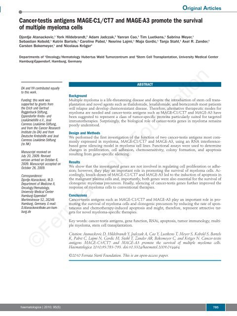
Cancer-testis antigens MAGE-C1/CT7 and MAGE-A3 promote the ...
Cancer-testis antigens MAGE-C1/CT7 and MAGE-A3 promote the ...
Cancer-testis antigens MAGE-C1/CT7 and MAGE-A3 promote the ...

You also want an ePaper? Increase the reach of your titles
YUMPU automatically turns print PDFs into web optimized ePapers that Google loves.
<strong>Cancer</strong>-<strong>testis</strong> <strong>antigens</strong> <strong>MAGE</strong>-<strong>C1</strong>/<strong>CT7</strong> <strong>and</strong> <strong>MAGE</strong>-<strong>A3</strong> <strong>promote</strong> <strong>the</strong> survival<br />
of multiple myeloma cells<br />
Djordje Atanackovic, 1 York Hildebr<strong>and</strong>t, 2 Adam Jadczak, 2 Yanran Cao, 1 Tim Luetkens, 1 Sabrina Meyer, 1<br />
Sebastian Kobold, 1 Katrin Bartels, 1 Caroline Pabst, 1 Nesrine Lajmi, 1 Maja Gordic, 1 Tanja Stahl, 1 Axel R. Z<strong>and</strong>er, 2<br />
Carsten Bokemeyer, 1 <strong>and</strong> Nicolaus Kröger 2<br />
Departments of 1 Oncology/Hematology Hubertus Wald Tumorzentrum <strong>and</strong> 2 Stem Cell Transplantation, University Medical Center<br />
Hamburg-Eppendorf, Hamburg, Germany<br />
DA <strong>and</strong> YH contributed equally<br />
to this work.<br />
Funding: this work was<br />
supported by grants from<br />
<strong>the</strong> Erich und Gertrud<br />
Roggenbuck-Stiftung,<br />
Eppendorfer Krebs- und<br />
Leukämiehilfe e.V., José<br />
Carreras Leukämie-Stiftung,<br />
<strong>and</strong> from <strong>the</strong> <strong>Cancer</strong> Research<br />
Institute (to DA) <strong>and</strong> from<br />
Deutsche Krebshilfe <strong>and</strong> José<br />
Carreras Leukämie-Stiftung<br />
(to NK)<br />
Manuscript received on<br />
July 20, 2009. Revised<br />
version arrived on October 6,<br />
2009. Manuscript accepted on<br />
October 26, 2009.<br />
Correspondence:<br />
Djordje Atanackovic, M.D.<br />
Department of Medicine II,<br />
Oncology/Hematology,<br />
University Medical Center<br />
Hamburg-Eppendorf<br />
Martinistrasse 52, 20246<br />
Hamburg, Germany. E-mail:<br />
D.Atanackovic@uke.uni-hamburg.de<br />
ABSTRACT<br />
Background<br />
Multiple myeloma is a life-threatening disease <strong>and</strong> despite <strong>the</strong> introduction of stem cell transplantation<br />
<strong>and</strong> novel agents such as thalidomide, lenalidomide, <strong>and</strong> bortezomib most patients<br />
will relapse <strong>and</strong> develop chemoresistant disease. Therefore, alternative <strong>the</strong>rapeutic modes for<br />
myeloma are needed <strong>and</strong> cancer-<strong>testis</strong> <strong>antigens</strong> such as <strong>MAGE</strong>-<strong>C1</strong>/<strong>CT7</strong> <strong>and</strong> <strong>MAGE</strong>-<strong>A3</strong> have<br />
been suggested to represent a class of tumor-specific proteins particularly suited for targeted<br />
immuno<strong>the</strong>rapies. Surprisingly, <strong>the</strong> biological role of cancer-<strong>testis</strong> genes in myeloma remains<br />
poorly understood.<br />
Design <strong>and</strong> Methods<br />
We performed <strong>the</strong> first investigation of <strong>the</strong> function of two cancer-<strong>testis</strong> <strong>antigens</strong> most commonly<br />
expressed in myeloma, <strong>MAGE</strong>-<strong>C1</strong>/<strong>CT7</strong> <strong>and</strong> <strong>MAGE</strong>-<strong>A3</strong>, using an RNA interferencebased<br />
gene silencing model in myeloma cell lines. Functional assays were used to determine<br />
changes in proliferation, cell adhesion, chemosensitivity, colony formation, <strong>and</strong> apoptosis<br />
resulting from gene-specific silencing.<br />
Results<br />
We show that <strong>the</strong> investigated genes are not involved in regulating cell proliferation or adhesion;<br />
however, <strong>the</strong>y play an important role in promoting <strong>the</strong> survival of myeloma cells. Accordingly,<br />
knock-down of <strong>MAGE</strong>-<strong>C1</strong>/<strong>CT7</strong> <strong>and</strong> <strong>MAGE</strong>-<strong>A3</strong> led to <strong>the</strong> induction of apoptosis in<br />
<strong>the</strong> malignant plasma cells <strong>and</strong>, importantly, both genes were also essential for <strong>the</strong> survival of<br />
clonogenic myeloma precursors. Finally, silencing of cancer-<strong>testis</strong> genes fur<strong>the</strong>r improved <strong>the</strong><br />
response of myeloma cells to conventional <strong>the</strong>rapies.<br />
Conclusions<br />
<strong>Cancer</strong>-<strong>testis</strong> <strong>antigens</strong> such as <strong>MAGE</strong>-<strong>C1</strong>/<strong>CT7</strong> <strong>and</strong> <strong>MAGE</strong>-<strong>A3</strong> play an important role in promoting<br />
<strong>the</strong> survival of myeloma cells <strong>and</strong> clonogenic precursors by reducing <strong>the</strong> rate of spontaneous<br />
<strong>and</strong> chemo<strong>the</strong>rapy-induced apoptosis <strong>and</strong> might, <strong>the</strong>refore, represent attractive targets<br />
for novel myeloma-specific <strong>the</strong>rapies.<br />
Key words: cancer-<strong>testis</strong> <strong>antigens</strong>, gene function, RNAi, apoptosis, tumor immunology, multiple<br />
myeloma, stem cell transplantation.<br />
Citation: Atanackovic D, Hildebr<strong>and</strong>t Y, Jadczak A, Cao Y, Luetkens T, Meyer S, Kobold S, Bartels<br />
K, Pabst C, Lajmi N, Gordic M, Stahl T, Z<strong>and</strong>er AR, Bokemeyer C, <strong>and</strong> Kröger N. <strong>Cancer</strong>-<strong>testis</strong><br />
<strong>antigens</strong> <strong>MAGE</strong>-<strong>C1</strong>/<strong>CT7</strong> <strong>and</strong> <strong>MAGE</strong>-<strong>A3</strong> <strong>promote</strong> <strong>the</strong> survival of multiple myeloma cells.<br />
Haematologica 2010;95:785-793. doi:10.3324/haematol.2009.014464<br />
©2010 Ferrata Storti Foundation. This is an open-access paper.<br />
Original Articles<br />
©Ferrata Storti Foundation<br />
haematologica | 2010; 95(5) 785
D. Atanackovic et al.<br />
Introduction<br />
Multiple myeloma (MM), <strong>the</strong> second most common<br />
hematologic malignancy, is a B-cell neoplasia characterized<br />
by <strong>the</strong> clonal proliferation of malignant plasma cells<br />
in <strong>the</strong> bone marrow, with clinical consequences that<br />
include anemia, lytic bone lesions, renal dysfunction,<br />
hypercalcemia, hypogammaglobulinemia, <strong>and</strong> increased<br />
risk of infection. 1 Clinical remissions of MM are commonly<br />
achieved by high-dose <strong>the</strong>rapy followed by autologous<br />
stem cell transplantation as well as by <strong>the</strong> administration<br />
of recently introduced drugs such as bortezomib,<br />
thalidomide, <strong>and</strong> lenalidomide. However, <strong>the</strong><br />
median survival has still not improved beyond 4-5 years, 2<br />
mainly because of <strong>the</strong> persistence of <strong>the</strong>rapy-resistant<br />
minimal residual disease eventually leading to relapse<br />
<strong>and</strong> death in <strong>the</strong> vast majority of MM patients.<br />
Accordingly, alternative <strong>the</strong>rapeutic modes, which more<br />
specifically <strong>and</strong> effectively target remaining myeloma<br />
cells, are needed.<br />
The development of <strong>the</strong>rapies specifically aiming at<br />
MM requires <strong>the</strong> identification of appropriate myelomaspecific<br />
structures <strong>and</strong> cancer-<strong>testis</strong> (CT) <strong>antigens</strong> have<br />
been suggested to represent a class of tumor proteins particularly<br />
suited for targeted <strong>the</strong>rapies such as T-cell-mediated<br />
immuno<strong>the</strong>rapy. 3 CT <strong>antigens</strong> are typically<br />
expressed in a wide range of tumor types but not in any<br />
healthy tissues o<strong>the</strong>r than <strong>testis</strong> <strong>and</strong> placenta; <strong>the</strong> expression<br />
of <strong>the</strong>se <strong>antigens</strong> is regulated by epigenetic mechanisms<br />
such as <strong>promote</strong>r methylation <strong>and</strong> histone acetylation.<br />
4 Among CT <strong>antigens</strong>, <strong>the</strong> melanoma antigen genes<br />
(<strong>MAGE</strong>) represent <strong>the</strong> largest gene family. The original<br />
<strong>MAGE</strong> genes included <strong>MAGE</strong>-A, -B, <strong>and</strong> –C, <strong>the</strong> so-called<br />
CT-X-<strong>MAGE</strong> proteins, which are localized in clusters on<br />
<strong>the</strong> X chromosome <strong>and</strong> encode proteins with 50 to 99%<br />
sequence identity to each o<strong>the</strong>r. 5 In addition, a number of<br />
autosomal gene families named <strong>MAGE</strong>-D through<br />
<strong>MAGE</strong>-L appear to be more widely expressed in normal<br />
tissues. 3 Generally, <strong>MAGE</strong> genes are defined by a shared<br />
<strong>MAGE</strong> homology domain, a well-conserved domain of<br />
about 200 amino acids. 6 The <strong>MAGE</strong> homology domain<br />
does not contain any regions with significant homology<br />
with o<strong>the</strong>r known proteins, but it has been indicated that<br />
this domain represents an important site for protein-protein<br />
interactions. 7<br />
CT antigen expression is a rare event in most hematologic<br />
malignancies including leukemias <strong>and</strong> non-<br />
Hodgkin’s B-cell lymphomas. 8,9 However, MM probably<br />
represents <strong>the</strong> malignancy with <strong>the</strong> richest CT antigen<br />
expression of all human cancers <strong>and</strong> <strong>the</strong> two CT <strong>antigens</strong><br />
most frequently expressed in MM are <strong>MAGE</strong>-<strong>A3</strong> <strong>and</strong><br />
<strong>MAGE</strong>-<strong>C1</strong>/<strong>CT7</strong>. 10-15 <strong>MAGE</strong>-<strong>A3</strong> is also expressed in a wide<br />
variety of solid tumors. It is a cytoplasmic protein with a<br />
molecular weight of 35 kDa 16 which was discovered ana-<br />
lyzing CD8 + T-cell reactivity of a cancer patient against an<br />
autologous melanoma cell line. 17 <strong>MAGE</strong>-<strong>C1</strong>/<strong>CT7</strong> was<br />
identified simultaneously by representational difference<br />
analysis <strong>and</strong> serological analysis of recombinant cDNA<br />
expres-sion libraries (SEREX). 18,19 <strong>MAGE</strong>-<strong>C1</strong>/<strong>CT7</strong> is about<br />
800 amino acids longer than o<strong>the</strong>r <strong>MAGE</strong> proteins <strong>and</strong><br />
contains a large number of unique short repetitive<br />
sequences in front of <strong>the</strong> <strong>MAGE</strong> homology domain. 19 The<br />
<strong>MAGE</strong>-<strong>C1</strong>/<strong>CT7</strong> gene is located on b<strong>and</strong> Xq26 whereas<br />
<strong>the</strong> <strong>MAGE</strong>-A genes are located on Xq28. 18-20 Like o<strong>the</strong>r<br />
<strong>MAGE</strong> genes, <strong>MAGE</strong>-<strong>C1</strong>/<strong>CT7</strong> is specifically expressed in<br />
a variety of solid tumors, although less frequently than<br />
<strong>MAGE</strong>-<strong>A3</strong>. 19<br />
As in <strong>the</strong> case of most solid tumors, expression of CT<br />
<strong>antigens</strong> is increased in a coordinate manner in advanced<br />
stages of myeloma 10,11,21,22 <strong>and</strong> we have recently shown<br />
that once CT antigen expression occurs in MM, it persists<br />
throughout <strong>the</strong> whole course of <strong>the</strong> disease. 22 These<br />
observations point to a possible role of CT <strong>antigens</strong> in<br />
promoting <strong>the</strong> progression of MM, an idea which is fur<strong>the</strong>r<br />
supported by recent microarray analyses showing an<br />
association between CT antigen expression, including <strong>the</strong><br />
expression of <strong>MAGE</strong> genes, <strong>and</strong> a more aggressive course<br />
of <strong>the</strong> disease as well as reduced survival. 14,21,23 Comparable<br />
results were described in a study focusing on <strong>the</strong><br />
expression of CT <strong>antigens</strong> belonging to <strong>the</strong> SSX gene<br />
family in which <strong>the</strong> expression of <strong>the</strong>se genes was related<br />
to reduced overall survival of MM patients. 24 Notably,<br />
our findings <strong>and</strong> <strong>the</strong> observations of o<strong>the</strong>rs indicate that,<br />
of all CT <strong>antigens</strong>, <strong>the</strong> expression of <strong>MAGE</strong>-<strong>A3</strong> <strong>and</strong>, particularly,<br />
of <strong>MAGE</strong>-<strong>C1</strong>/<strong>CT7</strong>, has <strong>the</strong> strongest impact on<br />
<strong>the</strong> prognosis of myeloma. 22,25<br />
Although <strong>the</strong>se collected data suggest that <strong>the</strong> expression<br />
of CT <strong>antigens</strong> might contribute to tumor progression,<br />
particularly in MM, <strong>the</strong>ir biological role in germ line<br />
cells <strong>and</strong> malignancies remains poorly understood. In this<br />
study, we performed <strong>the</strong> first investigation of <strong>the</strong> function<br />
of <strong>the</strong> two CT <strong>antigens</strong> most commonly expressed in<br />
myeloma, <strong>MAGE</strong>-<strong>C1</strong>/<strong>CT7</strong> <strong>and</strong> <strong>MAGE</strong>-<strong>A3</strong>, describing an<br />
important role of <strong>the</strong>se proteins in promoting <strong>the</strong> survival<br />
of myeloma cells. We believe that targeting such proteins<br />
might hit an Achilles’ heel of <strong>the</strong> malignancy leading to<br />
an improved outcome or even cures following st<strong>and</strong>ard<br />
<strong>the</strong>rapy.<br />
Design <strong>and</strong> Methods<br />
Cell lines<br />
Myeloma cell lines MOLP-8, RPMI-8226, KMS-12-BM, EJM;<br />
IM-9, NCI-H929, OPM-2, <strong>and</strong> LP-1 were obtained from <strong>the</strong><br />
German Collection of Microorganisms <strong>and</strong> Cell Cultures<br />
(DSMZ, Braunschweig, Germany). Cell lines Brown <strong>and</strong> SK-007<br />
were provided by <strong>the</strong> New York branch of <strong>the</strong> Ludwig Institute<br />
for <strong>Cancer</strong> Research. Lines were maintained in RPMI 1640 with<br />
10% fetal calf serum (FCS) <strong>and</strong> penicillin/streptomycin.<br />
Western blot<br />
For some experiments myeloma cell lines were sorted based on<br />
<strong>the</strong>ir expression of CD138 using a FACSAria cell-sorting system<br />
(BD Biosciences, Heidelberg, Germany). Whole cell protein<br />
extracts were prepared from cell lines <strong>and</strong> western blotting was<br />
performed as previously described. 26 For all target proteins analyzed,<br />
appropriate blocking studies were performed using recombinant<br />
proteins in order to confirm specificity of <strong>the</strong> staining.<br />
©Ferrata Storti Foundation<br />
Gene silencing using transfection with stealth<br />
RNA interference<br />
BLOCK-iT fluorescein-labeled double-str<strong>and</strong>ed DNA, scrambled<br />
control short interfering RNA (siRNA), <strong>and</strong> stealth siRNA<br />
targeting <strong>MAGE</strong>-<strong>C1</strong>/<strong>CT7</strong> or <strong>MAGE</strong>-<strong>A3</strong> were purchased from<br />
Invi-trogen (Karlsruhe, Germany). Myeloma cell lines were<br />
transfected using Lipofectamine 2000 (Invitrogen) according to<br />
<strong>the</strong> manufacturer’s recommendations <strong>and</strong> transfection efficiency<br />
was determined by fluorescence microscopy using fluoresceinlabeled<br />
double-str<strong>and</strong>ed DNA.<br />
786 haematologica | 2010; 95(5)
3-(4,5-dimethylthiaxol-2-yl)-2,5-diphenyltetrazolium<br />
bromide viability assay<br />
Relative numbers of viable cells were assessed by a 3-(4,5dimethylthiaxol-2-yl)-2,5-diphenyltetrazolium<br />
bromide (MTT)<br />
assay 27 72 h after transfection with CT antigen-specific siRNA or<br />
with scrambled control siRNA. Myeloma cells were cultured<br />
overnight <strong>and</strong> subsequently treated with bortezomib or melphalan<br />
12 h before analyses. MTT was added to each well <strong>and</strong> cells<br />
were incubated for 4 h. Absorbance was read at 570 nm using a<br />
spectrophotometer.<br />
Measurement of apoptosis<br />
The terminal deoxynucleotidyl transferase dUTP nick end labeling<br />
(TUNEL) assay was done according to <strong>the</strong> manufacturer’s recommendations<br />
(Millipore, Billerica, MA, USA). Briefly, cells were<br />
fixed in 1% paraformaldehyde, <strong>and</strong> frozen until staining at -20°C<br />
using 70% ethanol. For staining, cells were incubated sequentially<br />
in staining solution for 1 h, <strong>and</strong> propidium iodide/RNAse A solution<br />
for 30 min. For annexin V staining, cells were incubated with<br />
annexin V-fluorescein isothiocyanate in annexin binding buffer.<br />
Analysis by flow cytometry was performed within <strong>the</strong> next 3 h.<br />
Colony formation assay<br />
Myeloma cell lines Molp-8 <strong>and</strong> RPMI-8226 were plated at 1000<br />
cells/mL of methylcellulose medium (StemCell Technologies,<br />
Cologne, Germany) in a 6-well culture dish. Plates were incubated<br />
at 37°C <strong>and</strong> colonies consisting of more than 40 cells were<br />
counted 7 to 10 days after <strong>the</strong> culture had been started.<br />
Analysis of cell proliferation<br />
Cell proliferation was measured by staining with carboxyfluorescein<br />
diacetate succinimidyl ester (CFSE) or using a plate-based<br />
system. Myeloma cells were pulsed with bromodeoxyuridine<br />
(BrdU) <strong>and</strong> peroxidase-labeled anti-BrdU was added to <strong>the</strong> cells.<br />
Resulting immune complexes were detected by a substrate reaction,<br />
<strong>and</strong> absorbance was read at 450 nm. For FACS analysis,<br />
myeloma cells were stained intracellularly with CFSE <strong>and</strong> CFSErelated<br />
green fluorescence intensity was measured by flow cytometry.<br />
Cell adhesion assay<br />
The myeloma cells were resuspended 72 h post-transfection in<br />
RPMI-1640 + 0.2% bovine serum albumin (adhesion medium)<br />
<strong>and</strong> were incubated for 30 min at 37°C. Cells were <strong>the</strong>n resuspended<br />
in adhesion medium <strong>and</strong> were incubated for 4 h in 96-well<br />
plates coated with fibronectin. Unbound cells were removed by<br />
four washes <strong>and</strong> absorbance with Chromogen Substrate Solution<br />
(Alpco, Salem, NH, USA) was read at 450/620nm.<br />
Statistical analysis<br />
Statistical analyses were performed using SPSS software. The<br />
Mann-Whitney U test was used to calculate differences between<br />
different experimental conditions. Differences were regarded significant<br />
if <strong>the</strong> P value was less than 0.05.<br />
The Design <strong>and</strong> Methods of <strong>the</strong> study are described in more<br />
detail in <strong>the</strong> Online Supplementary Appendix.<br />
Results<br />
<strong>Cancer</strong>-<strong>testis</strong> <strong>antigens</strong> <strong>MAGE</strong>-<strong>A3</strong>/A6 <strong>and</strong> <strong>MAGE</strong>-<strong>C1</strong>/<strong>CT7</strong><br />
are constitutively expressed in multiple myeloma cell<br />
lines <strong>and</strong> expression is specifically suppressed by<br />
transfection with interfering RNA<br />
We have previously described very frequent expression<br />
Biological role of <strong>MAGE</strong>-<strong>C1</strong>/<strong>CT7</strong> <strong>and</strong> <strong>MAGE</strong>-<strong>A3</strong> in myeloma<br />
of CT <strong>antigens</strong> in myeloma cell lines as well as in patients’<br />
samples at <strong>the</strong> RNA level. 10 As a prerequisite for <strong>the</strong> functional<br />
analysis of <strong>MAGE</strong>-<strong>A3</strong> <strong>and</strong> <strong>MAGE</strong>-<strong>C1</strong>/<strong>CT7</strong> expression<br />
in MM, in this study we investigated <strong>the</strong> protein<br />
expression of both genes in ten different MM cell lines.<br />
Considering high sequence homology of <strong>MAGE</strong>-A proteins,<br />
we first confirmed, in a western blot assay, specific<br />
binding of antibody M3H67 to recombinant full-length<br />
<strong>MAGE</strong>-<strong>A3</strong> protein, but not to <strong>MAGE</strong>-A2, <strong>MAGE</strong>-A4, or<br />
<strong>MAGE</strong>-A12 recombinant proteins (data not shown).<br />
Recombinant <strong>MAGE</strong>-A6 protein was not available to us;<br />
however, as <strong>MAGE</strong>-<strong>A3</strong> <strong>and</strong> <strong>MAGE</strong>-A6 show 95% protein<br />
sequence homology, binding of M3H67 to <strong>MAGE</strong>-A6<br />
seems very likely <strong>and</strong>, <strong>the</strong>refore, <strong>the</strong> protein recognized in<br />
<strong>the</strong> respective immunoassays was designated <strong>MAGE</strong>-<br />
<strong>A3</strong>/A6. Western blot analyses revealed that <strong>MAGE</strong>-<strong>A3</strong>/A6<br />
<strong>and</strong> <strong>MAGE</strong>-<strong>C1</strong>/<strong>CT7</strong> were constitutively expressed in<br />
every single cell line (Figure 1A) fur<strong>the</strong>r supporting a functional<br />
role of <strong>the</strong>se genes in myeloma. A comparably high<br />
transfection efficiency (70-80%) was generally achieved<br />
using <strong>the</strong> <strong>MAGE</strong>-<strong>A3</strong>/A6- <strong>and</strong> <strong>MAGE</strong>-<strong>C1</strong>/<strong>CT7</strong>-expressing<br />
cell lines Molp-8 <strong>and</strong> RPMI-8226; <strong>the</strong>se two cell lines were,<br />
<strong>the</strong>refore, preferably used for subsequent experiments.<br />
Since <strong>MAGE</strong>-<strong>A3</strong>/A6 <strong>and</strong> <strong>MAGE</strong>-<strong>C1</strong>/<strong>CT7</strong> were present<br />
in all MM cell lines available to us, we could not investigate<br />
<strong>the</strong> function of <strong>the</strong>se genes transfecting <strong>the</strong>m into <strong>MAGE</strong>negative<br />
MM lines. We, <strong>the</strong>refore, decided to perform<br />
knockdown experiments using interfering RNA <strong>and</strong><br />
observed that two of <strong>the</strong> three siRNA constructs targeting<br />
different sequences of <strong>the</strong> respective gene’s mRNA<br />
showed a strong inhibitory effect leading to down-regulation<br />
of <strong>MAGE</strong>-<strong>A3</strong>/A6 <strong>and</strong> <strong>MAGE</strong>-<strong>C1</strong>/<strong>CT7</strong> protein expression<br />
(Figure 1B). The two respective siRNA constructs<br />
showing an inhibitory effect were used in all subsequent<br />
experiments <strong>and</strong> <strong>the</strong> effects of transfection of myeloma cell<br />
lines Molp-8 <strong>and</strong> RPMI-8226 with one interfering RNA<br />
(RNAi) <strong>and</strong> scrambled control RNAi, respectively, are<br />
shown as examples in each figure. Knock-down efficiency<br />
for both genes reached its maximum at 72 h after transfection<br />
<strong>and</strong> lasted for at least 7 days. Importantly, analyzing<br />
whe<strong>the</strong>r silencing of <strong>MAGE</strong>-<strong>A3</strong> or <strong>MAGE</strong>-<strong>C1</strong>/<strong>CT7</strong> would<br />
have an effect on o<strong>the</strong>r <strong>MAGE</strong> family members or non-<br />
<strong>MAGE</strong> CT <strong>antigens</strong>, we found that RNAi transfection<br />
specifically affected <strong>the</strong> expression of <strong>the</strong> target genes <strong>and</strong><br />
not <strong>the</strong> presence of o<strong>the</strong>r CT <strong>antigens</strong> such as <strong>MAGE</strong>-A4,<br />
ropporin-1, or NY-ESO-1 (Figure 1C). However, knockdown<br />
of <strong>MAGE</strong>-<strong>C1</strong>/<strong>CT7</strong> seemed to have a slight downregulating<br />
effect on <strong>the</strong> expression of <strong>MAGE</strong>-C2/CT10, a<br />
protein that shows a significant degree of homology with<br />
<strong>MAGE</strong>-<strong>C1</strong>/<strong>CT7</strong>. 28<br />
<strong>Cancer</strong>-<strong>testis</strong> <strong>antigens</strong> <strong>MAGE</strong>-<strong>A3</strong> <strong>and</strong> <strong>MAGE</strong>-<strong>C1</strong>/<strong>CT7</strong><br />
protect myeloma cells from undergoing spontaneous<br />
apoptosis<br />
Performing knockdown experiments we noticed a significant<br />
growth delay in cultures of both myeloma cell<br />
lines after transfection with both <strong>MAGE</strong>-specific RNAi<br />
constructs. Performing a classical MTT assay we confirmed<br />
that <strong>the</strong> numbers of viable cells were significantly<br />
lower in <strong>the</strong> cultures containing MM cells transfected with<br />
<strong>MAGE</strong>-<strong>A3</strong>- <strong>and</strong> <strong>MAGE</strong>-<strong>C1</strong>/<strong>CT7</strong>-specific RNAi but not<br />
with scrambled control RNAi (Figure 2A). In order to<br />
determine whe<strong>the</strong>r this was due to an anti-proliferative<br />
effect of gene knockdown or based on a pro-apoptotic<br />
effect of <strong>MAGE</strong> silencing, we next performed a TUNEL<br />
©Ferrata Storti Foundation<br />
haematologica | 2010; 95(5) 787
D. Atanackovic et al.<br />
assay to demonstrate <strong>the</strong> proportion of apoptotic cells in<br />
cultures 72 h post-transfection. This assay revealed that,<br />
compared to untreated cells <strong>and</strong> myeloma cells transfected<br />
with control RNAi, silencing of <strong>MAGE</strong>-<strong>A3</strong> <strong>and</strong> <strong>MAGE</strong>-<br />
<strong>C1</strong>/<strong>CT7</strong> did indeed lead to <strong>the</strong> induction of spontaneous<br />
apoptosis in <strong>the</strong> malignant cells (Figure 2B). Remarkably,<br />
loss of <strong>the</strong> anti-apoptotic effect of <strong>MAGE</strong>-<strong>A3</strong> <strong>and</strong> <strong>MAGE</strong>-<br />
<strong>C1</strong>/<strong>CT7</strong> resulted in rates of apoptosis as high as 80%<br />
(Figure 2B). This finding was confirmed by microscopic<br />
A<br />
<strong>MAGE</strong>-<strong>A3</strong>/A6<br />
B<br />
<strong>MAGE</strong>-<strong>C1</strong><br />
ACTB<br />
<strong>MAGE</strong>-<strong>A3</strong><br />
RNAi<br />
Control<br />
RNAi<br />
<strong>MAGE</strong>-<strong>C1</strong><br />
RNAi<br />
Control<br />
RNAi<br />
C<br />
<strong>MAGE</strong>-<strong>C1</strong><br />
<strong>MAGE</strong>-C2<br />
<strong>MAGE</strong>-<strong>A3</strong>/A6<br />
<strong>MAGE</strong>-A4<br />
ROPN1<br />
NY-ESO-1<br />
ACTB<br />
Myeloma cell lines<br />
KMS-12-BM<br />
IM-9<br />
RPMI-8226<br />
OPM-2<br />
LP-1<br />
EJM<br />
NCI-H929<br />
Molp-8<br />
SK-007<br />
Brown<br />
Medium<br />
Testis<br />
Molp-8<br />
35 kDa<br />
124 kDa<br />
42 kDa<br />
RPMI-8266<br />
24h 48h 72h 7d 24h 48h 72h 7d<br />
Control<br />
<strong>MAGE</strong>-<strong>A3</strong><br />
RNAi<br />
Control<br />
<strong>MAGE</strong>-<strong>C1</strong><br />
124 kDa<br />
41 kDa<br />
35 kDa<br />
35 kDa<br />
24 kDa<br />
23 kDa<br />
42 kDa<br />
<strong>MAGE</strong>-<strong>A3</strong>/A6 (35 kDa)<br />
ACTB (42 kDa)<br />
<strong>MAGE</strong>-<strong>A3</strong>/A6 (35 kDa)<br />
ACTB (42 kDa)<br />
<strong>MAGE</strong>-<strong>C1</strong> (124 kDa)<br />
ACTB (42 kDa)<br />
<strong>MAGE</strong>-<strong>C1</strong> (124 kDa)<br />
ACTB (42 kDa)<br />
Figure 1. <strong>Cancer</strong>-<strong>testis</strong> genes <strong>MAGE</strong>-<strong>A3</strong> <strong>and</strong> <strong>MAGE</strong>-<strong>C1</strong>/<strong>CT7</strong> are constitutively<br />
expressed in myeloma cells <strong>and</strong> transfection with inhibitory<br />
RNAi results in gene-specific down-regulation of both proteins. (A)<br />
ten myeloma cell lines were investigated for <strong>the</strong> expression of<br />
<strong>MAGE</strong>-<strong>A3</strong> <strong>and</strong> <strong>MAGE</strong>-<strong>C1</strong>/<strong>CT7</strong> protein by western blot. β-actin (ACTB)<br />
served as an internal control for protein quality <strong>and</strong> protein lysate<br />
of normal human <strong>testis</strong> was used as a positive control. Notably, all<br />
ten myeloma cell lines showed constitutive expression of both CT<br />
<strong>antigens</strong>. (B) Myeloma cell lines Molp-8 (left column) <strong>and</strong> RPMI-<br />
8226 (right column) were transfected using two RNAi constructs<br />
specific for CT genes <strong>MAGE</strong>-<strong>A3</strong> <strong>and</strong> <strong>MAGE</strong>-<strong>C1</strong>/<strong>CT7</strong>, respectively, or<br />
with scrambled control RNAi. Treatment resulted in knockdown of<br />
<strong>the</strong> expression of <strong>MAGE</strong>-<strong>A3</strong> <strong>and</strong> <strong>MAGE</strong>-<strong>C1</strong>/<strong>CT7</strong> protein in both cell<br />
lines starting 48-72 h after transfection as indicated by<br />
immunoblotting. Gene-silencing lasted at least until day 7 after<br />
transfection. (C) Importantly, treatment with gene-specific RNAi only<br />
affected <strong>the</strong> expression of <strong>the</strong> target genes <strong>and</strong> not <strong>the</strong> expression<br />
of o<strong>the</strong>r CT <strong>antigens</strong> tested (<strong>MAGE</strong>-A4, Ropporin-1, NY-ESO-1). The<br />
only exception was CT gene <strong>MAGE</strong>-C2/CT10, <strong>the</strong> expression of which<br />
was suppressed following transfection with RNAi targeting <strong>MAGE</strong>-<br />
<strong>C1</strong>/<strong>CT7</strong>. Comparable findings were observed following gene-silencing<br />
in cell line RPMI-8226 <strong>and</strong> using a second gene-specific RNAi<br />
construct (data not shown).<br />
analysis of MM cells 72 h post-transfection (Figure 2C). In<br />
order to determine whe<strong>the</strong>r <strong>MAGE</strong> genes had been efficiently<br />
downregulated in viable cells we transfected cell<br />
lines Molp-8 <strong>and</strong> RPMI-8226 with siRNA constructs specific<br />
for ei<strong>the</strong>r <strong>MAGE</strong> gene. After 72 h we sorted viable<br />
<strong>and</strong> apoptotic cells according to annexin V-staining by<br />
FACS. Analyzing both populations by immunoblot we<br />
detected high levels of both <strong>MAGE</strong> genes in viable cells<br />
but no expression in apoptotic cells (Figure 2B). This suggests<br />
that <strong>the</strong> survival of a minority of myeloma cells is<br />
most likely based on <strong>the</strong> fact that not all cells could efficiently<br />
be transfected with <strong>the</strong> <strong>MAGE</strong>-specific siRNA.<br />
This idea is also supported by <strong>the</strong> average efficiency of<br />
siRNA transfection, which we had determined earlier.<br />
When we analyzed which factors might be involved in<br />
mediating spontaneous apoptosis following knockdown<br />
of <strong>MAGE</strong>-<strong>A3</strong> <strong>and</strong> <strong>MAGE</strong>-<strong>C1</strong>/<strong>CT7</strong>, we observed specific<br />
up-regulation of caspase-9 <strong>and</strong> caspase-12 expression<br />
starting 48 h after transfection with gene-specific RNAi<br />
but not with control RNAi (Figure 2D). Interestingly, we<br />
did not detect upregulated expression of caspases 3 or 8,<br />
pointing to a dominant role of <strong>the</strong> former factors in mediating<br />
apoptosis in myeloma cells following silencing of<br />
<strong>MAGE</strong>-<strong>A3</strong> <strong>and</strong> <strong>MAGE</strong>-<strong>C1</strong>/<strong>CT7</strong>.<br />
<strong>Cancer</strong>-<strong>testis</strong> <strong>antigens</strong> <strong>MAGE</strong>-<strong>A3</strong>/A6 <strong>and</strong> <strong>MAGE</strong>-<strong>C1</strong>/<strong>CT7</strong><br />
are expressed in clonogenic myeloma precursors <strong>and</strong><br />
protect <strong>the</strong>m from apoptosis<br />
It has long been suggested that in <strong>the</strong> case of MM, a<br />
small number of cycling precursors generates <strong>and</strong> replenishes<br />
a large number of non-dividing myeloma cells.<br />
Dividing precursor cells are thought to be resistant to<br />
chemo<strong>the</strong>rapy <strong>and</strong> to be responsible for relapses commonly<br />
seen in this malignancy. Clonogenic precursors are<br />
present in <strong>the</strong> patients’ bone marrow <strong>and</strong> also among<br />
myeloma cell lines <strong>and</strong> can be sorted based on <strong>the</strong> fact that<br />
<strong>the</strong>y, in contrast to <strong>the</strong> bulk of malignant cells, do not<br />
express surface molecule CD138. 29 We analyzed <strong>the</strong><br />
expression of CT <strong>antigens</strong> in CD138-positive MM cells<br />
<strong>and</strong> CD138-negative myeloma precursors sorted from<br />
bulk cultures of cell lines Molp-8 <strong>and</strong> KMS-12-BM <strong>and</strong> we<br />
found that both fractions strongly expressed <strong>MAGE</strong>-<br />
<strong>A3</strong>/A6 <strong>and</strong> <strong>MAGE</strong>-<strong>C1</strong>/<strong>CT7</strong> (Figure 3A). Importantly, following<br />
silencing of <strong>MAGE</strong>-<strong>A3</strong>/A6 <strong>and</strong> <strong>MAGE</strong>-<strong>C1</strong>/<strong>CT7</strong> in<br />
two different myeloma cell lines using two inhibitory<br />
RNAi constructs, we observed a significantly reduced<br />
number of cell colonies in a st<strong>and</strong>ard assay measuring<br />
clonogenic growth of myeloma precursors (Figure 3B).<br />
These results demonstrate that both <strong>MAGE</strong> genes not<br />
only have a promoting effect on <strong>the</strong> survival of common<br />
myeloma cells but also support anti-apoptotic effects in<br />
myeloma precursor cells replenishing <strong>the</strong> bulk of myeloma<br />
cells.<br />
©Ferrata Storti Foundation<br />
<strong>MAGE</strong>-<strong>A3</strong> <strong>and</strong> <strong>MAGE</strong>-<strong>C1</strong>/<strong>CT7</strong> <strong>promote</strong> multiple myeloma<br />
growth mainly by preventing apoptosis <strong>and</strong> not by<br />
promoting proliferation or cell adhesion<br />
Next, we investigated whe<strong>the</strong>r effects on <strong>the</strong> proliferative<br />
function of myeloma cells or on <strong>the</strong>ir adhesion capacity<br />
might contribute to <strong>the</strong> growth-promoting effect of<br />
<strong>MAGE</strong>-<strong>A3</strong> <strong>and</strong> <strong>MAGE</strong>-<strong>C1</strong>/<strong>CT7</strong>. CT <strong>antigens</strong>, such as<br />
CAGE, <strong>promote</strong> <strong>the</strong> motility of tumor cells 30 <strong>and</strong> it has<br />
been shown that knockdown of CT <strong>antigens</strong> belonging to<br />
<strong>the</strong> SSX family of genes in a melanoma cell line results in<br />
reduced migratory capacity of <strong>the</strong> tumor cells. 31 However,<br />
788 haematologica | 2010; 95(5)
performing an adhesion assay following silencing of<br />
<strong>MAGE</strong>-<strong>A3</strong> or <strong>MAGE</strong>-.<strong>C1</strong>/<strong>CT7</strong> in <strong>the</strong> myeloma cell line<br />
Molp-8 we did not observe any difference in adhesion<br />
capacity between cells transfected with gene-specific<br />
RNAi <strong>and</strong> cells transfected with control RNAi (data not<br />
shown).<br />
Analyzing <strong>the</strong> proliferative activity of myeloma cells<br />
following knockdown of <strong>MAGE</strong>-<strong>A3</strong> or <strong>MAGE</strong>-<strong>C1</strong>/<strong>CT7</strong><br />
(Figure 4) we observed only a slight suppressive effect of<br />
gene-specific RNAi transfection on cellular growth.<br />
However, we cannot completely rule out <strong>the</strong> possibility<br />
that increased rates of apoptosis during <strong>the</strong> culture period<br />
might have contributed to this effect on cell proliferation.<br />
In agreement with this idea, we found that gating on nonapoptotic<br />
cells in flow cytometry-based assay, myeloma<br />
cells transfected with <strong>MAGE</strong>-specific RNAi or control<br />
RNAi had both undergone multiple cell divisions at 72 h<br />
after transfection (data not shown).<br />
A<br />
Untreated<br />
Control RNAi<br />
<strong>MAGE</strong>-<strong>A3</strong> RNAi<br />
B<br />
C<br />
Untreated<br />
Control RNAi<br />
<strong>MAGE</strong>-<strong>C1</strong> RNAi<br />
D<br />
Untreated<br />
Control RNAi<br />
<strong>MAGE</strong>-<strong>A3</strong> RNAi<br />
Untreated<br />
Control RNAi<br />
<strong>MAGE</strong>-<strong>C1</strong> RNAi<br />
Specific<br />
RNAi<br />
Control<br />
RNAi<br />
Specific RNAi<br />
Control RNAi<br />
Specific RNAi<br />
Molp-8<br />
0 20 40 60 80 100<br />
Apoptotic cells (%)<br />
<strong>MAGE</strong>-<strong>A3</strong><br />
<strong>MAGE</strong>-<strong>A3</strong><br />
<strong>MAGE</strong>-<strong>C1</strong><br />
<strong>MAGE</strong>-<strong>C1</strong><br />
24h 48h 72h 7d 24h 48h 72h 7d<br />
RPMI-8226<br />
0 20 40 60 80 100 0 20 40 60 80 100<br />
Relative viability (%)<br />
Relative viability (%)<br />
TUNEL Annexin V<br />
Caspase-12 (48 kDa)<br />
Caspase-9 (64 kDa)<br />
Caspase-8 (55 kDa)<br />
Caspase-3 (32 kDa)<br />
Caspase-9<br />
ACTB (42 kDa)<br />
Biological role of <strong>MAGE</strong>-<strong>C1</strong>/<strong>CT7</strong> <strong>and</strong> <strong>MAGE</strong>-<strong>A3</strong> in myeloma<br />
Targeting of <strong>MAGE</strong>-<strong>A3</strong> <strong>and</strong> <strong>MAGE</strong>-<strong>C1</strong>/<strong>CT7</strong> fur<strong>the</strong>r<br />
increases <strong>the</strong> <strong>the</strong>rapeutic efficacy of conventional<br />
modes of <strong>the</strong>rapy for multiple myeloma<br />
Resistance to conventional myeloma-specific <strong>the</strong>rapies<br />
represents a severe problem in this hematologic malignancy.<br />
In order to explore whe<strong>the</strong>r <strong>MAGE</strong>-specific interventions<br />
could complement established modes of <strong>the</strong>rapy for<br />
MM, we combined silencing of <strong>MAGE</strong>-<strong>A3</strong> <strong>and</strong> <strong>MAGE</strong>-<br />
<strong>C1</strong>/<strong>CT7</strong> with chemo<strong>the</strong>rapeutic treatment or application<br />
of a ‘novel’ drug, namely <strong>the</strong> proteasome inhibitor bortezomib.<br />
A slight decrease of <strong>the</strong> relative number of viable<br />
cells following transfection with scrambled control RNAi<br />
was likely caused by an additional cytotoxic effect of <strong>the</strong><br />
lipofection reagent used in this assay. Never<strong>the</strong>less, we<br />
observed that specific targeting of <strong>MAGE</strong>-<strong>A3</strong> or <strong>MAGE</strong>-<br />
<strong>C1</strong>/<strong>CT7</strong> more than doubled <strong>the</strong> <strong>the</strong>rapeutic efficacy exerted<br />
by <strong>the</strong> cytotoxic agent melphalan (Figure 5A) or bortezomib<br />
(Figure 5B) alone in comparison to control RNAi.<br />
+<br />
<strong>MAGE</strong>-<strong>A3</strong>/A6<br />
Figure 2. Silencing of CT genes <strong>MAGE</strong>-<strong>A3</strong> or<br />
<strong>MAGE</strong>-<strong>C1</strong>/<strong>CT7</strong> exerts a strong pro-apoptotic<br />
effect on myeloma cells. (A) Myeloma cell<br />
lines Molp-8 (left column) <strong>and</strong> RPMI-8226<br />
(right column) were transfected with two RNAi<br />
constructs specific for CT genes <strong>MAGE</strong>-<strong>A3</strong> or<br />
<strong>MAGE</strong>-<strong>C1</strong>/<strong>CT7</strong>. Numbers of viable myeloma<br />
cells transfected with RNAi specific for <strong>the</strong> target<br />
genes or of cells transfected with scrambled<br />
control RNAi were normalized to <strong>the</strong><br />
number of viable untreated cells. Silencing of<br />
<strong>MAGE</strong>-<strong>A3</strong> or <strong>MAGE</strong>-<strong>C1</strong>/<strong>CT7</strong> resulted in significantly<br />
decreased cell viability at 72 h after<br />
transfection as indicated by an MTT assay.<br />
Results show mean values [+ st<strong>and</strong>ard error<br />
of means (SEM)] of three separate experiments<br />
<strong>and</strong> asterisks indicate statistically significant<br />
differences between untreated cells<br />
<strong>and</strong> cells transfected with scrambled control<br />
or one gene-specific RNAi (*P
D. Atanackovic et al.<br />
These findings indicate that a <strong>MAGE</strong>-specific <strong>the</strong>rapy<br />
could represent an ideal adjunct to conventional modes of<br />
<strong>the</strong>rapy for MM, preventing recurrences <strong>and</strong> leading to<br />
persistent responses in higher proportions of patients with<br />
this disease.<br />
Discussion<br />
In this study we performed <strong>the</strong> first analysis of <strong>the</strong> function<br />
of CT <strong>antigens</strong> in MM. Observing that knockdown of<br />
<strong>MAGE</strong>-<strong>A3</strong>/A6 <strong>and</strong> <strong>MAGE</strong>-<strong>C1</strong>/<strong>CT7</strong> led to a reduced num-<br />
A<br />
B<br />
Cell clusters/culture<br />
Cell clusters/culture<br />
70<br />
60<br />
50<br />
40<br />
30<br />
20<br />
10<br />
0<br />
70<br />
60<br />
50<br />
40<br />
30<br />
20<br />
10<br />
0<br />
CD 138+<br />
CD 138-<br />
CD 138+<br />
CD 138-<br />
Testis<br />
Molp-8 KMS-12-BM<br />
<strong>MAGE</strong>-<strong>A3</strong> (35 kDa)<br />
<strong>MAGE</strong>-<strong>C1</strong> (124 kDa)<br />
ACTB<br />
Untreated Control <strong>MAGE</strong>-<strong>A3</strong><br />
RNAi RNAi<br />
Untreated Control <strong>MAGE</strong>-<strong>C1</strong><br />
RNAi RNAi<br />
Untreated Control <strong>MAGE</strong>-<strong>C1</strong><br />
RNAi RNAi<br />
Molp-8 RPMI-8226<br />
Figure 3. Silencing of CT genes <strong>MAGE</strong>-<strong>A3</strong> or <strong>MAGE</strong>-<strong>C1</strong>/<strong>CT7</strong> results<br />
in a decreased outgrowth of myeloma precursors in a colony formation<br />
assay. Previous studies have shown that myeloma precursors<br />
are also present in <strong>the</strong> bulk population of myeloma cell lines <strong>and</strong><br />
that <strong>the</strong>se cells can be identified by <strong>the</strong>ir lack of CD138 expression.<br />
29 FACS-sorting CD138 + <strong>and</strong> CD138- subpopulations we show<br />
here by western blot analysis that myeloma precursors derived from<br />
cell lines Molp-8 <strong>and</strong> KMS-12-BM as well as conventional myeloma<br />
plasma cells express <strong>MAGE</strong>-<strong>A3</strong> <strong>and</strong> <strong>MAGE</strong>-<strong>C1</strong>/<strong>CT7</strong> (A). In a clono-<br />
genic growth assay, colonies of myeloma precursors were counted<br />
7-10 days after culture initiation using myeloma cell lines Molp-8<br />
(left column) <strong>and</strong> RPMI-8226 (right column) separately transfected<br />
with two RNAi constructs specific for CT genes <strong>MAGE</strong>-<strong>A3</strong> or <strong>MAGE</strong>-<br />
<strong>C1</strong>/<strong>CT7</strong>, respectively (B). Bars indicate mean values (+SEM) of three<br />
separate experiments. Numbers of colonies produced by myeloma<br />
cells transfected with RNAi specific for <strong>the</strong> target genes or of cells<br />
transfected with scrambled control RNAi were compared with<br />
colony numbers in cultures with untreated cells <strong>and</strong> asterisks indicate<br />
statistically significant differences (*P
might contribute to <strong>the</strong>rapy-resistance <strong>and</strong> persistence of<br />
residual disease in human cancers. Accordingly, studies<br />
have shown that expression of <strong>MAGE</strong> <strong>and</strong> GAGE genes in<br />
cancer cell lines induces a chemo<strong>the</strong>rapy-resistant phenotype<br />
in vitro 26 <strong>and</strong> tumor expression of <strong>MAGE</strong>-A1 seems to<br />
correlate with clinical responses to chemo<strong>the</strong>rapy in gastric<br />
cancer. 38 Our finding of an additive effect of <strong>MAGE</strong><br />
knockdown on <strong>the</strong> cytotoxicity exerted by drugs used for<br />
myeloma <strong>the</strong>rapy, melphalan <strong>and</strong> bortezomib, indicates<br />
that targeting CT genes might open a new <strong>the</strong>rapeutic<br />
route for patients with disease resistant to conventional<br />
<strong>the</strong>rapies.<br />
Most malignancies probably arise from a rare population<br />
of “cancer stem cells” having <strong>the</strong> ability to self-renew<br />
<strong>and</strong> maintain <strong>the</strong> tumor. In <strong>the</strong> case of MM <strong>the</strong> malignant<br />
plasma cells seem to arise from less differentiated cells<br />
that resemble post-germinal center, CD138-negative B<br />
cells. 29 Novel anti-myeloma agents bortezomib <strong>and</strong><br />
lenalidomide inhibit malignant plasma cells but, at least in<br />
vitro, have little activity against <strong>the</strong>se myeloma precursors.<br />
27 This differential activity may explain why <strong>the</strong>se<br />
compounds yield significant clinical responses but not<br />
cures. 39 Interestingly, it has been hypo<strong>the</strong>sized that CT<br />
genes, which are often heterogeneously expressed by only<br />
few cells within <strong>the</strong> tumor mass, might represent a hallmark<br />
of cancer stem cells 40 <strong>and</strong> we demonstrate here for<br />
<strong>the</strong> first time expression of CT genes <strong>MAGE</strong>-<strong>A3</strong> <strong>and</strong><br />
<strong>MAGE</strong>-<strong>C1</strong>/<strong>CT7</strong> in CD138 + myeloma cells <strong>and</strong> <strong>the</strong>ir 138negative<br />
precursors.<br />
Colony growth assays have previously been used to<br />
assess <strong>the</strong> number <strong>and</strong> function of myeloma precursors<br />
present in <strong>the</strong> tumor bulk since <strong>the</strong>se precursors represent<br />
an independent predictive factor for poor prognosis in<br />
MM. 41 We found that knockdown of CT genes <strong>MAGE</strong>-<strong>A3</strong><br />
or <strong>MAGE</strong>-<strong>C1</strong>/<strong>CT7</strong> led to a significantly delayed outgrowth<br />
of clonogenic myeloma precursors. These findings<br />
Untreated<br />
Specific RNAi<br />
Melphalan<br />
Melphalan<br />
+ control RNAi<br />
Melphalan<br />
+specific RNai<br />
0 20 40 60 80 100<br />
Untreated<br />
Specific RNAi<br />
Bortezomib<br />
Bortezomib<br />
+ control RNAi<br />
Bortezomib<br />
+specific RNai<br />
<strong>MAGE</strong>-<strong>A3</strong> <strong>MAGE</strong>-<strong>C1</strong><br />
0 20 40 60 80 100<br />
Relative viability (%)<br />
Biological role of <strong>MAGE</strong>-<strong>C1</strong>/<strong>CT7</strong> <strong>and</strong> <strong>MAGE</strong>-<strong>A3</strong> in myeloma<br />
suggest that targeting CT <strong>antigens</strong> might not only have<br />
<strong>the</strong> potential to affect fully differentiated myeloma cells<br />
but might also contribute to eradicating <strong>the</strong>ir clonogenic<br />
precursors in MM, a malignancy which is still incurable<br />
due to inevitable relapse following st<strong>and</strong>ard <strong>the</strong>rapy.<br />
How could CT <strong>antigens</strong> be targeted in MM? Myeloma<br />
is a malignancy which is thought to be controlled, at least<br />
Untreated<br />
Control RNAi<br />
<strong>MAGE</strong>-<strong>A3</strong> RNAi<br />
Untreated<br />
Control RNAi<br />
<strong>MAGE</strong>-<strong>C1</strong> RNAi<br />
0 0.5 1.0 1.5 2.0 2.5<br />
Proliferation (OD)<br />
Figure 4 Silencing of CT genes <strong>MAGE</strong>-<strong>A3</strong> or <strong>MAGE</strong>-<strong>C1</strong>/<strong>CT7</strong> only<br />
exerts a minor influence on <strong>the</strong> proliferation of myeloma cells.<br />
Proliferation of myeloma cell line Molp-8 following silencing of<br />
<strong>MAGE</strong>-<strong>A3</strong>- or <strong>MAGE</strong>-<strong>C1</strong>/<strong>CT7</strong> was assessed by a BrdU incorporation<br />
assay. Proliferation was measured 72 h after transfection with RNAi<br />
specific for <strong>the</strong> target gene or with scrambled control RNAi <strong>and</strong><br />
results were compared to untreated cells. Bars show mean values<br />
(+SEM) of three separate experiments <strong>and</strong> asterisks indicate statistically<br />
significant differences between untreated cells <strong>and</strong> scrambled<br />
control or gene specific RNAi (*P
D. Atanackovic et al.<br />
to a certain extent, by <strong>the</strong> adaptive immune system, a<br />
view which is supported by <strong>the</strong> fact that <strong>the</strong> <strong>the</strong>rapeutic<br />
effect of allogeneic stem cell transplantation is partly<br />
mediated by donor-derived T cells <strong>and</strong> that infusions of<br />
donor T cells are capable of inducing remission in relapsed<br />
MM patients. 42,43 We <strong>and</strong> o<strong>the</strong>rs have recently shown that<br />
spontaneous antibody <strong>and</strong> T-cell immunity against<br />
<strong>MAGE</strong>-<strong>A3</strong> <strong>and</strong> <strong>MAGE</strong>-<strong>C1</strong>/<strong>CT7</strong> is present in patients with<br />
MM 10,44-46 <strong>and</strong> that <strong>the</strong> presence of such immunity might<br />
be associated with an improved prognosis. 44 Our finding<br />
that immune responses against CT <strong>antigens</strong> are induced<br />
by allogeneic stem cell transplantation 10 suggests that<br />
<strong>the</strong>se tumor <strong>antigens</strong> might indeed represent natural targets<br />
for donor-derived allo-immune or even spontaneous<br />
anti-myeloma immune responses. Collectively, <strong>the</strong>se findings<br />
indicate that T-cell-based immuno<strong>the</strong>rapies might<br />
represent one <strong>the</strong>rapeutic route for effective targeting of<br />
CT <strong>antigens</strong>. Importantly, immunization with <strong>MAGE</strong>-<strong>A3</strong><br />
protein has previously been performed in melanoma <strong>and</strong><br />
lung cancer patients <strong>and</strong> has proven to be safe, to be capable<br />
of evoking specific humoral <strong>and</strong> T-cell responses, 47,48<br />
<strong>and</strong> to have some clinical activity in patients in <strong>the</strong> adjuvant<br />
setting. 49 However, in addition to applying<br />
immuno<strong>the</strong>rapy directed against <strong>MAGE</strong>-<strong>C1</strong>/<strong>CT7</strong> or<br />
<strong>MAGE</strong>-<strong>A3</strong> <strong>the</strong>se genes might also be targeted by more<br />
“immediate” <strong>the</strong>rapeutic modes such as small molecules,<br />
antisense oligonucleotides, or classical gene <strong>the</strong>rapy.<br />
While <strong>the</strong> exact molecular mechanisms mediating<br />
effects of CT genes on myeloma cells still need to be elu-<br />
References<br />
1. Kyle RA, Rajkumar SV. Multiple myeloma.<br />
N Engl J Med. 2004;351(18):1860-73.<br />
2. Kumar SK, Rajkumar SV, Dispenzieri A,<br />
Lacy MQ, Hayman SR, Buadi FK, et al.<br />
Improved survival in multiple myeloma<br />
<strong>and</strong> <strong>the</strong> impact of novel <strong>the</strong>rapies. Blood.<br />
2008;111(5):2516-20.<br />
3. Simpson AJ, Caballero OL, Jungbluth A,<br />
Chen YT, Old LJ. <strong>Cancer</strong>/<strong>testis</strong> <strong>antigens</strong>,<br />
gametogenesis <strong>and</strong> cancer. Nat Rev <strong>Cancer</strong>.<br />
2005;5(8):615-25.<br />
4. Wischnewski F, Pantel K, Schwarzenbach<br />
H. Promoter demethylation <strong>and</strong> histone<br />
acetylation mediate gene expression of<br />
<strong>MAGE</strong>-A1, -A2, -<strong>A3</strong>, <strong>and</strong> -A12 in human<br />
cancer cells. Mol <strong>Cancer</strong> Res. 2006;4(5):<br />
339-49.<br />
5. Chomez P, De Backer O, Bertr<strong>and</strong> M, De<br />
Plaen E, Boon T, Lucas S. An overview of<br />
<strong>the</strong> <strong>MAGE</strong> gene family with <strong>the</strong> identification<br />
of all human members of <strong>the</strong> family.<br />
<strong>Cancer</strong> Res. 2001;61(14):5544-51.<br />
6. Barker PA, Salehi A. The <strong>MAGE</strong> proteins:<br />
emerging roles in cell cycle progression,<br />
apoptosis, <strong>and</strong> neurogenetic disease. J<br />
Neurosci Res. 2002;67(6):705-12.<br />
7. Taniura H, Kobayashi M, Yoshikawa K.<br />
Functional domains of necdin for proteinprotein<br />
interaction, nuclear matrix targeting,<br />
<strong>and</strong> cell growth suppression. J Cell<br />
Biochem. 2005;94(4):804-15.<br />
8. Chambost H, van Baren N, Brasseur F,<br />
Olive D. <strong>MAGE</strong>-A genes are not expressed<br />
in human leukemias. Leukemia. 2001;<br />
15(11):1769-71.<br />
9. Lim SH, Austin S, Owen-Jones E, Robinson<br />
L. Expression of testicular genes in haema-<br />
cidated, our findings strongly suggest that <strong>MAGE</strong>-<br />
<strong>C1</strong>/<strong>CT7</strong> <strong>and</strong>/or <strong>MAGE</strong>-<strong>A3</strong> are important for <strong>the</strong> survival<br />
<strong>and</strong> progression of myeloma cells <strong>and</strong> maybe also of<br />
o<strong>the</strong>r malignancies. Toge<strong>the</strong>r with our previous observation<br />
that myeloma represents <strong>the</strong> malignancy with <strong>the</strong><br />
richest expression of CT <strong>antigens</strong>, <strong>the</strong> persistence <strong>and</strong><br />
prognostic relevance of CT gene expression in MM, <strong>and</strong><br />
<strong>the</strong> ability of CT <strong>antigens</strong> to induce spontaneous <strong>and</strong><br />
<strong>the</strong>rapy-induced immune responses, our current study<br />
provides a strong rationale for employing <strong>MAGE</strong>-<br />
<strong>C1</strong>/<strong>CT7</strong> <strong>and</strong> <strong>MAGE</strong>-<strong>A3</strong> as <strong>the</strong>rapeutic targets in MM.<br />
We believe that such a strategy might prove particularly<br />
fruitful when CT antigen-specific approaches are used in<br />
a setting of minimal residual disease following conventional<br />
myeloma <strong>the</strong>rapy.<br />
Authorship <strong>and</strong> Disclosures<br />
tological malignancies. Br J <strong>Cancer</strong>. 1999;<br />
81(7):1162-4.<br />
10. Atanackovic D, Arfsten J, Cao Y, Gnjatic S,<br />
Schnieders F, Bartels K, et al. <strong>Cancer</strong>-<strong>testis</strong><br />
<strong>antigens</strong> are commonly expressed in multiple<br />
myeloma <strong>and</strong> induce systemic immunity<br />
following allogeneic stem cell transplantation.<br />
Blood. 2007;109(3):1103-12.<br />
11. Jungbluth AA, Ely S, DiLiberto M,<br />
Niesvizky R, Williamson B, Frosina D, et al.<br />
The cancer-<strong>testis</strong> <strong>antigens</strong> <strong>CT7</strong> (<strong>MAGE</strong>-<strong>C1</strong>)<br />
<strong>and</strong> <strong>MAGE</strong>-<strong>A3</strong>/6 are commonly expressed<br />
in multiple myeloma <strong>and</strong> correlate with<br />
plasma-cell proliferation. Blood. 2005;<br />
106(1):167-74.<br />
12. Pellat-Deceunynck C, Mellerin MP,<br />
Labarriere N, Jego G, Moreau-Aubry A,<br />
Harousseau JL, et al. The cancer germ-line<br />
genes <strong>MAGE</strong>-1, <strong>MAGE</strong>-3 <strong>and</strong> PRAME are<br />
commonly expressed by human myeloma<br />
cells. Eur J Immunol. 2000;30(3):803-9.<br />
13. De Vos J, Thykjaer T, Tarte K, Ensslen M,<br />
Raynaud P, Requir<strong>and</strong> G, et al. Comparison<br />
of gene expression profiling between<br />
malignant <strong>and</strong> normal plasma cells with<br />
oligonucleotide arrays. Oncogene. 2002;<br />
21(44):6848-57.<br />
14. Condomines M, Hose D, Raynaud P,<br />
Hundemer M, De Vos J, Baudard M, et al.<br />
<strong>Cancer</strong>/<strong>testis</strong> genes in multiple myeloma:<br />
expression patterns <strong>and</strong> prognosis value<br />
determined by microarray analysis. J<br />
Immunol. 2007;178(5):3307-15.<br />
15. Tinguely M, Jenni B, Knights A, Lopes B,<br />
Korol D, Rousson V, et al. <strong>MAGE</strong>-<strong>C1</strong>/CT-7<br />
expression in plasma cell myeloma: subcellular<br />
localization impacts on clinical outcome.<br />
<strong>Cancer</strong> Sci. 2008;99(4):720-5.<br />
16. Gaugler B, Van den Eynde B, van der<br />
Bruggen P, Romero P, Gaforio JJ, De Plaen E,<br />
DA designed research, analyzed data, <strong>and</strong> wrote <strong>the</strong><br />
paper; YH contributed vital new analytical tools, performed<br />
research, analyzed data, <strong>and</strong> wrote <strong>the</strong> paper; AJ<br />
performed research; YC performed research; TL designed<br />
research <strong>and</strong> analyzed data; SM performed research; SK<br />
analyzed data; KB performed research; CP analyzed data;<br />
NL performed research; MJ performed research; TS performed<br />
research; ARZ analyzed data; CB analyzed data;.<br />
NK designed research <strong>and</strong> analyzed data.<br />
The authors declare no potential conflicts of interest.<br />
et al. Human gene <strong>MAGE</strong>-3 codes for an<br />
antigen recognized on a melanoma by<br />
autologous cytolytic T lymphocytes. J Exp<br />
Med. 1994;179(3):921-30.<br />
17. Kocher T, Schultz-Thater E, Gudat F,<br />
Schaefer C, Casorati G, Juretic A, et al.<br />
Identification <strong>and</strong> intracellular location of<br />
<strong>MAGE</strong>-3 gene product. <strong>Cancer</strong> Res. 1995;<br />
55(11):2236-9.<br />
18. Chen YT, Gure AO, Tsang S, Stockert E,<br />
Jager E, Knuth A, et al. Identification of<br />
multiple cancer/<strong>testis</strong> <strong>antigens</strong> by allogeneic<br />
antibody screening of a melanoma cell<br />
line library. Proc Natl Acad Sci USA.<br />
1998;95(12):6919-23.<br />
19. Lucas S, De Smet C, Arden KC, Viars CS,<br />
Le<strong>the</strong> B, Lurquin C, et al. Identification of a<br />
new <strong>MAGE</strong> gene with tumor-specific<br />
expression by representational difference<br />
analysis. <strong>Cancer</strong> Res. 1998;58(4):743-52.<br />
20. Kruger S, Ola V, Feller AC, Fischer D,<br />
Friedrich M. Expression of cancer-<strong>testis</strong><br />
antigen <strong>CT7</strong> (<strong>MAGE</strong>-<strong>C1</strong>) in breast cancer:<br />
an immunohistochemical study with<br />
emphasis on prognostic utility. Pathol<br />
Oncol Res. 2007;13(2):91-6.<br />
21. Chng WJ, Kumar S, Vanwier S, Ahmann G,<br />
Price-Troska T, Henderson K, et al.<br />
Molecular dissection of hyperdiploid multiple<br />
myeloma by gene expression profiling.<br />
<strong>Cancer</strong> Res. 2007;67(7):2982-9.<br />
22. Atanackovic D, Luetkens T, Hildebr<strong>and</strong>t Y,<br />
Arfsten J, Bartels K, Horn C, et al.<br />
Longitudinal analysis <strong>and</strong> prognostic effect<br />
of cancer-<strong>testis</strong> antigen expression in multiple<br />
myeloma. Clin <strong>Cancer</strong> Res. 2009;15(4):<br />
1343-52.<br />
23. Mattioli M, Agnelli L, Fabris S, Baldini L,<br />
Morabito F, Bicciato S, et al. Gene expression<br />
profiling of plasma cell dyscrasias<br />
©Ferrata Storti Foundation<br />
792 haematologica | 2010; 95(5)
eveals molecular patterns associated with<br />
distinct IGH translocations in multiple<br />
myeloma. Oncogene. 2005;24(15):2461-73.<br />
24. Taylor BJ, Reiman T, Pittman JA, Keats JJ,<br />
de Bruijn DR, Mant MJ, et al. SSX cancer<br />
<strong>testis</strong> <strong>antigens</strong> are expressed in most multiple<br />
myeloma patients: co-expression of<br />
SSX1, 2, 4, <strong>and</strong> 5 correlates with adverse<br />
prognosis <strong>and</strong> high frequencies of SSX-positive<br />
PCs. J Immuno<strong>the</strong>r. 2005;28(6):564-75.<br />
25. Andrade VC, Vettore AL, Felix RS, Almeida<br />
MS, Carvalho F, Oliveira JS, et al.<br />
Prognostic impact of cancer/<strong>testis</strong> antigen<br />
expression in advanced stage multiple<br />
myeloma patients. <strong>Cancer</strong> Immun.<br />
2008;8:2.<br />
26. Duan Z, Duan Y, Lamendola DE, Yusuf RZ,<br />
Naeem R, Penson RT, et al. Overexpression<br />
of <strong>MAGE</strong>/GAGE genes in paclitaxel/doxorubicin-resistant<br />
human cancer cell lines.<br />
Clin <strong>Cancer</strong> Res. 2003;9(7):2778-85.<br />
27. Matsui W, Wang Q, Barber JP, Brennan S,<br />
Smith BD, Borrello I, et al. Clonogenic multiple<br />
myeloma progenitors, stem cell properties,<br />
<strong>and</strong> drug resistance. <strong>Cancer</strong> Res.<br />
2008;68(1):190-7.<br />
28. Gure AO, Stockert E, Arden KC, Boyer AD,<br />
Viars CS, Scanlan MJ, et al. CT10: a new<br />
cancer-<strong>testis</strong> (CT) antigen homologous to<br />
<strong>CT7</strong> <strong>and</strong> <strong>the</strong> <strong>MAGE</strong> family, identified by<br />
representational-difference analysis. Int J<br />
<strong>Cancer</strong>. 2000;85(5):726-32.<br />
29. Matsui W, Huff CA, Wang Q, Malehorn<br />
MT, Barber J, Tanhehco Y, et al.<br />
Characterization of clonogenic multiple<br />
myeloma cells. Blood. 2004;103(6):2332-6.<br />
30. Cilensek ZM, Yehiely F, Kular RK, Deiss LP.<br />
A member of <strong>the</strong> GAGE family of tumor<br />
<strong>antigens</strong> is an anti-apoptotic gene that confers<br />
resistance to Fas/CD95/APO-1, interferon-gamma,<br />
taxol <strong>and</strong> gamma-irradiation.<br />
<strong>Cancer</strong> Biol Ther. 2002;1(4):380-7.<br />
31. Cronwright G, Le Blanc K, Go<strong>the</strong>rstrom C,<br />
Darcy P, Ehnman M, Brodin B.<br />
<strong>Cancer</strong>/<strong>testis</strong> antigen expression in human<br />
mesenchymal stem cells: down-regulation<br />
of SSX impairs cell migration <strong>and</strong> matrix<br />
metalloproteinase 2 expression. <strong>Cancer</strong><br />
Res. 2005;65(6):2207-15.<br />
Biological role of <strong>MAGE</strong>-<strong>C1</strong>/<strong>CT7</strong> <strong>and</strong> <strong>MAGE</strong>-<strong>A3</strong> in myeloma<br />
32. Cho B, Lim Y, Lee DY, Park SY, Lee H, Kim<br />
WH, et al. Identification <strong>and</strong> characterization<br />
of a novel cancer/<strong>testis</strong> antigen gene<br />
CAGE. Biochem Biophys Res Commun.<br />
2002;292(3):715-26.<br />
33. Shim E, Shim H, Bae J, Lee H, Jeoung D.<br />
CAGE displays oncogenic potential <strong>and</strong><br />
induces cytolytic T lymphocyte activity.<br />
Biotechnol Lett. 2006;28(7):515-22.<br />
34. Yang B, O'Herrin S, Wu J, Reagan-Shaw S,<br />
Ma Y, Nihal M, et al. Select cancer testes<br />
<strong>antigens</strong> of <strong>the</strong> <strong>MAGE</strong>-A, -B, <strong>and</strong> -C families<br />
are expressed in mast cell lines <strong>and</strong> <strong>promote</strong><br />
cell viability in vitro <strong>and</strong> in vivo. J<br />
Invest Dermatol. 2007;127(2):267-75.<br />
35. Yang B, O’Herrin SM, Wu J, Reagan-Shaw<br />
S, Ma Y, Bhat KM, et al. <strong>MAGE</strong>-A, mMageb,<br />
<strong>and</strong> <strong>MAGE</strong>-C proteins form complexes<br />
with KAP1 <strong>and</strong> suppress p53-dependent<br />
apoptosis in <strong>MAGE</strong>-positive cell lines.<br />
<strong>Cancer</strong> Res. 2007;67(20):9954-62.<br />
36. Morishima N, Nakanishi K, Takenouchi H,<br />
Shibata T, Yasuhiko Y. An endoplasmic<br />
reticulum stress-specific caspase cascade in<br />
apoptosis. Cytochrome c-independent activation<br />
of caspase-9 by caspase-12. J Biol<br />
Chem. 2002;277(37):34287-94.<br />
37. Cho HJ, Caballero OL, Gnjatic S, Andrade<br />
VC, Colleoni GW, Vettore AL, et al.<br />
Physical interaction of two cancer-<strong>testis</strong><br />
<strong>antigens</strong>, <strong>MAGE</strong>-<strong>C1</strong> (<strong>CT7</strong>) <strong>and</strong> NY-ESO-1<br />
(CT6). <strong>Cancer</strong> Immun. 2006;6:12.<br />
38. Suzuki T, Yoshida K, Wada Y, Hamai Y,<br />
Sentani K, Oue N, et al. Melanoma-associated<br />
antigen-A1 expression predicts resistance<br />
to docetaxel <strong>and</strong> paclitaxel in<br />
advanced <strong>and</strong> recurrent gastric cancer.<br />
Oncol Rep. 2007;18(2):329-36.<br />
39. Huff CA, Matsui W, Smith BD, Jones RJ.<br />
The paradox of response <strong>and</strong> survival in<br />
cancer <strong>the</strong>rapeutics. Blood. 2006;107(2):<br />
431-4.<br />
40. Costa FF, Le Blanc K, Brodin B. Concise<br />
review: cancer/<strong>testis</strong> <strong>antigens</strong>, stem cells,<br />
<strong>and</strong> cancer. Stem Cells. 2007;25(3):707-11.<br />
41. Takahashi T, Lim B, Jamal N, Tritchler D,<br />
Lockwood G, McKinney S, et al. Colony<br />
growth <strong>and</strong> self renewal of plasma cell precursors<br />
in multiple myeloma. J Clin Oncol.<br />
1985;3(12):1613-23.<br />
42. Tricot G, Vesole DH, Jagannath S, Hilton J,<br />
Munshi N, Barlogie B. Graft-versus-myeloma<br />
effect: proof of principle. Blood.<br />
1996;87(3):1196-8.<br />
43. Orsini E, Bellucci R, Alyea EP, Schlossman<br />
R, Canning C, McLaughlin S, et al.<br />
Expansion of tumor-specific CD8+ T cell<br />
clones in patients with relapsed myeloma<br />
after donor lymphocyte infusion. <strong>Cancer</strong><br />
Res. 2003;63(10):2561-8.<br />
44. Goodyear OC, Pratt G, McLarnon A, Cook<br />
M, Piper K, Moss P. Differential pattern of<br />
CD4+ <strong>and</strong> CD8+ T-cell immunity to<br />
<strong>MAGE</strong>-A1/A2/<strong>A3</strong> in patients with monoclonal<br />
gammopathy of undetermined significance<br />
(MGUS) <strong>and</strong> multiple myeloma.<br />
Blood. 2008;112(8):3362-72.<br />
45. Goodyear O, Piper K, Khan N, Starczynski<br />
J, Mahendra P, Pratt G, et al. CD8+ T cells<br />
specific for cancer-germline gene <strong>antigens</strong><br />
are found in many patients with multiple<br />
myeloma <strong>and</strong> <strong>the</strong>ir frequency correlates<br />
with disease burden. Blood. 2005;106(13):<br />
4217-24.<br />
46. Curioni-Fontecedro A, Knights AJ,<br />
Tinguely M, Nuber N, Schneider C,<br />
Thomson CW, et al. <strong>MAGE</strong>-<strong>C1</strong>/<strong>CT7</strong> is <strong>the</strong><br />
dominant cancer-<strong>testis</strong> antigen targeted by<br />
humoral immune responses in patients<br />
with multiple myeloma. Leukemia.<br />
2008;22(8):1646-8.<br />
47. Atanackovic D, Altorki NK, Stockert E,<br />
Williamson B, Jungbluth AA, Ritter E, et al.<br />
Vaccine-induced CD4+ T cell responses to<br />
<strong>MAGE</strong>-3 protein in lung cancer patients. J<br />
Immunol. 2004;172(5):3289-96.<br />
48. Atanackovic D, Altorki NK, Cao Y, Ritter E,<br />
Ferrara CA, Ritter G, et al. Booster vaccination<br />
of cancer patients with <strong>MAGE</strong>-<strong>A3</strong> protein<br />
reveals long-term immunological<br />
memory or tolerance depending on priming.<br />
Proc Natl Acad Sci USA. 2008;105(5):<br />
1650-5.<br />
49. Brichard VG, Lejeune D. GSK’s <strong>antigens</strong>pecific<br />
cancer immuno<strong>the</strong>rapy programme:<br />
pilot results leading to phase III<br />
clinical development. Vaccine. 2007;25<br />
(Suppl 2):B61-71.<br />
©Ferrata Storti Foundation<br />
haematologica | 2010; 95(5) 793