Dramatic clinical efficacy of cladribine in Rosai-Dorfman disease ...
Dramatic clinical efficacy of cladribine in Rosai-Dorfman disease ...
Dramatic clinical efficacy of cladribine in Rosai-Dorfman disease ...

Create successful ePaper yourself
Turn your PDF publications into a flip-book with our unique Google optimized e-Paper software.
A. Aouba et al.<br />
<strong>Dramatic</strong> <strong>cl<strong>in</strong>ical</strong> <strong>efficacy</strong> <strong>of</strong> <strong>cladrib<strong>in</strong>e</strong> <strong>in</strong><br />
<strong>Rosai</strong>-<strong>Dorfman</strong> <strong>disease</strong> and evolution <strong>of</strong> the<br />
cytok<strong>in</strong>e pr<strong>of</strong>ile: towards a new therapeutic<br />
approach<br />
<strong>Rosai</strong>-<strong>Dorfman</strong> <strong>disease</strong> (RDD) is a rare disorder,<br />
<strong>of</strong>ten benign but with possible life-threaten<strong>in</strong>g<br />
prognosis. In most cases, specific treatment is not<br />
necessary; when required, the management <strong>of</strong><br />
RDD is not codified to date, and various<br />
chemotherapies have been shown to be <strong>in</strong>effective.<br />
Here, we report a patient with RDD who presented<br />
a dramatic and susta<strong>in</strong>ed response with <strong>cladrib<strong>in</strong>e</strong>.<br />
Analysis <strong>of</strong> the cytok<strong>in</strong>e pr<strong>of</strong>ile evolution<br />
shows a clear correlation between serum levels <strong>of</strong><br />
TNF-alpha and IL-6 and <strong>disease</strong> activity. Our f<strong>in</strong>d<strong>in</strong>gs<br />
show the promis<strong>in</strong>g <strong>efficacy</strong> <strong>of</strong> <strong>cladrib<strong>in</strong>e</strong> and<br />
suggest that therapies target<strong>in</strong>g specifically<br />
cytok<strong>in</strong>es might be useful <strong>in</strong> some cases <strong>of</strong> active<br />
RDD.<br />
Haematologica 2006; 91:(11)e144-e145<br />
<strong>Rosai</strong>-<strong>Dorfman</strong> <strong>disease</strong> (RDD) or s<strong>in</strong>us histiocytosis with<br />
massive lymphadenopathy (SHML) is a rare disorder <strong>of</strong><br />
unknown etiology characterized by a nonmalignant proliferation<br />
<strong>of</strong> dist<strong>in</strong>ctive histiocytic/phagocytic cells with<strong>in</strong> lymph<br />
node s<strong>in</strong>uses and lymphatics <strong>in</strong> extranodal sites. 1 RDD<br />
shows variable evolution, most <strong>of</strong>ten as prolonged <strong>cl<strong>in</strong>ical</strong><br />
course with exacerbation and spontaneous remission phases.<br />
1,2 Despite its usual benign prognosis with<br />
relaps<strong>in</strong>g/remitt<strong>in</strong>g but f<strong>in</strong>ally self-limit<strong>in</strong>g course, RDD<br />
may be complicated by organ compression and/or extranodal<br />
<strong>in</strong>volvement responsible for a wide spectrum <strong>of</strong> <strong>cl<strong>in</strong>ical</strong><br />
manifestations; it also may be life-threatened because <strong>of</strong> secondary<br />
organs <strong>in</strong>volvement responsible for dysfunction and<br />
pr<strong>of</strong>ound impairment <strong>of</strong> general health status. 1,3 Because<br />
prospective therapeutic studies are lack<strong>in</strong>g, management <strong>of</strong><br />
RDD is not codified yet. Various chemotherapies, <strong>in</strong>terferon<br />
and radiotherapy are usually <strong>in</strong>effective to improve general<br />
status and reduce tumor mass. When required, surgical<br />
debulk<strong>in</strong>g might be useful but debilitat<strong>in</strong>g, and should be<br />
proposed as the last option. 2 Here we report a patient with<br />
RDD who presented a dramatic and susta<strong>in</strong>ed response<br />
after treatment with <strong>cladrib<strong>in</strong>e</strong>, and discuss new therapeutic<br />
approaches regard<strong>in</strong>g cytok<strong>in</strong>e pr<strong>of</strong>ile evolution.<br />
Study design<br />
Case report<br />
A 45-year-old woman was admitted for massive bilateral<br />
cervical lymph nodes (LN). LN biopsy <strong>in</strong> March 2002<br />
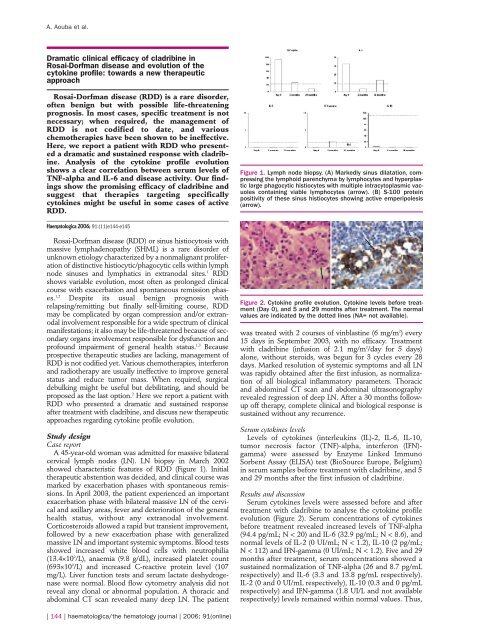
showed characteristic features <strong>of</strong> RDD (Figure 1). Initial<br />
therapeutic abstention was decided, and <strong>cl<strong>in</strong>ical</strong> course was<br />
marked by exacerbation phases with spontaneous remissions.<br />
In April 2003, the patient experienced an important<br />
exacerbation phase with bilateral massive LN <strong>of</strong> the cervical<br />
and axillary areas, fever and deterioration <strong>of</strong> the general<br />
health status, without any extranodal <strong>in</strong>volvement.<br />
Corticosteroids allowed a rapid but transient improvement,<br />
followed by a new exacerbation phase with generalized<br />
massive LN and important systemic symptoms. Blood tests<br />
showed <strong>in</strong>creased white blood cells with neutrophilia<br />
(13.4×10 9 /L), anaemia (9.8 g/dL), <strong>in</strong>creased platelet count<br />
(693×10 9 /L) and <strong>in</strong>creased C-reactive prote<strong>in</strong> level (107<br />
mg/L). Liver function tests and serum lactate deshydrogenase<br />
were normal. Blood flow cytometry analysis did not<br />
reveal any clonal or abnormal population. A thoracic and<br />
abdom<strong>in</strong>al CT scan revealed many deep LN. The patient<br />
| 144 | haematologica/the hematology journal | 2006; 91(onl<strong>in</strong>e)<br />
Figure 1. Lymph node biopsy. (A) Markedly s<strong>in</strong>us dilatation, compress<strong>in</strong>g<br />
the lymphoid parenchyma by lymphocytes and hyperplastic<br />
large phagocytic histiocytes with multiple <strong>in</strong>tracytoplasmic vacuoles<br />
conta<strong>in</strong><strong>in</strong>g viable lymphocytes (arrow). (B) S-100 prote<strong>in</strong><br />
positivity <strong>of</strong> these s<strong>in</strong>us histiocytes show<strong>in</strong>g active emperipolesis<br />
(arrow).<br />
Figure 2. Cytok<strong>in</strong>e pr<strong>of</strong>ile evolution. Cytok<strong>in</strong>e levels before treatment<br />
(Day 0), and 5 and 29 months after treatment. The normal<br />
values are <strong>in</strong>dicated by the dotted l<strong>in</strong>es (NA= not available).<br />
was treated with 2 courses <strong>of</strong> v<strong>in</strong>blast<strong>in</strong>e (6 mg/m 2 ) every<br />
15 days <strong>in</strong> September 2003, with no <strong>efficacy</strong>. Treatment<br />
with <strong>cladrib<strong>in</strong>e</strong> (<strong>in</strong>fusion <strong>of</strong> 2.1 mg/m 2 /day for 5 days)<br />
alone, without steroids, was begun for 3 cycles every 28<br />
days. Marked resolution <strong>of</strong> systemic symptoms and all LN<br />
was rapidly obta<strong>in</strong>ed after the first <strong>in</strong>fusion, as normalization<br />
<strong>of</strong> all biological <strong>in</strong>flammatory parameters. Thoracic<br />
and abdom<strong>in</strong>al CT scan and abdom<strong>in</strong>al ultrasonography<br />
revealed regression <strong>of</strong> deep LN. After a 30 months followup<br />
<strong>of</strong>f therapy, complete <strong>cl<strong>in</strong>ical</strong> and biological response is<br />
susta<strong>in</strong>ed without any recurrence.<br />
Serum cytok<strong>in</strong>es levels<br />
Levels <strong>of</strong> cytok<strong>in</strong>es (<strong>in</strong>terleuk<strong>in</strong>s (IL)-2, IL-6, IL-10,<br />
tumor necrosis factor (TNF)-alpha, <strong>in</strong>terferon (IFN)gamma)<br />
were assessed by Enzyme L<strong>in</strong>ked Immuno<br />
Sorbent Assay (ELISA) test (BioSource Europe, Belgium)<br />
<strong>in</strong> serum samples before treatment with <strong>cladrib<strong>in</strong>e</strong>, and 5<br />
and 29 months after the first <strong>in</strong>fusion <strong>of</strong> <strong>cladrib<strong>in</strong>e</strong>.<br />
Results and discussion<br />
Serum cytok<strong>in</strong>es levels were assessed before and after<br />
treatment with <strong>cladrib<strong>in</strong>e</strong> to analyse the cytok<strong>in</strong>e pr<strong>of</strong>ile<br />
evolution (Figure 2). Serum concentrations <strong>of</strong> cytok<strong>in</strong>es<br />
before treatment revealed <strong>in</strong>creased levels <strong>of</strong> TNF-alpha<br />
(94.4 pg/mL; N < 20) and IL-6 (32.9 pg/mL; N < 8.6), and<br />
normal levels <strong>of</strong> IL-2 (0 UI/mL; N < 1.2), IL-10 (2 pg/mL;<br />
N < 112) and IFN-gamma (0 UI/mL; N < 1.2). Five and 29<br />
months after treatment, serum concentrations showed a<br />
susta<strong>in</strong>ed normalization <strong>of</strong> TNF-alpha (26 and 8.7 pg/mL<br />
respectively) and IL-6 (3.3 and 13.8 pg/mL respectively).<br />
IL-2 (0 and 0 UI/mL respectively), IL-10 (0.3 and 0 pg/mL<br />
respectively) and IFN-gamma (1.8 UI/L and not available<br />
respectively) levels rema<strong>in</strong>ed with<strong>in</strong> normal values. Thus,
analysis <strong>of</strong> the cytok<strong>in</strong>e pr<strong>of</strong>ile evolution clearly shows a<br />
positive correlation between serum cytok<strong>in</strong>es levels <strong>of</strong><br />
TNF-alpha and IL-6 and <strong>disease</strong> activity. The pathophysiology<br />
<strong>of</strong> RDD is poorly understood. However, a<br />
cytok<strong>in</strong>es and/or chemok<strong>in</strong>es-mediated migration <strong>of</strong><br />
monocytes might be <strong>in</strong>volved <strong>in</strong> histiocytes accumulation<br />
and activation. This functional activation may be triggered<br />
by different stimuli, as suggested by the coexistence <strong>of</strong><br />
RDD and hematological malignancies, autoimmune <strong>disease</strong>s<br />
or post-<strong>in</strong>fectious conditions. 1,4-6 Although both<br />
Langerhans cells histiocytosis (LCH) and RDD appear as<br />
<strong>disease</strong>s related to the monocytes/ macrophagic system <strong>of</strong><br />
differenciation, which are associated with <strong>in</strong>creased<br />
cytok<strong>in</strong>es production, their pr<strong>of</strong>ile <strong>of</strong> cytok<strong>in</strong>e expression<br />
is different. Histiocytes <strong>of</strong> RDD strongly expressed both<br />
transcripts <strong>of</strong> TNF-alpha and IL-1beta and also moderate<br />
IL-6 specific signals, while <strong>in</strong> LCH, transcript expression is<br />
variable for TNF-alpha, weak and occasional for IL-1beta<br />
and negative for IL-6. 7 Reactive <strong>in</strong>flammatory cells accompany<strong>in</strong>g<br />
the pathologic cells also expressed <strong>in</strong> variable<br />
amounts both <strong>of</strong> these cytok<strong>in</strong>es. 7 Complex <strong>in</strong>teractions<br />
between RDD histiocytes and these reactive cells, notably<br />
polyclonal plasma cells, probably occur such as autocr<strong>in</strong>e<br />
and/or paracr<strong>in</strong>e loop mechanisms with enhanced<br />
cytok<strong>in</strong>es stimulation and activation. 6,7 This hypothesis is<br />
supported by the <strong>efficacy</strong> <strong>of</strong> rituximab reported <strong>in</strong> one<br />
case. 8 We thus formulate the hypothesis that RDD could<br />
be a good candidate for therapies target<strong>in</strong>g TNF-alpha, IL-<br />
6 and IL-1 cytok<strong>in</strong>es, which are probably directly responsible<br />
for its systemic symptoms. The orig<strong>in</strong> <strong>of</strong> histiocytes<br />
<strong>of</strong> RDD and LCH from a common bone marrow precursor<br />
related to the monocyte/macrophage l<strong>in</strong>eage and the<br />
promis<strong>in</strong>g results <strong>of</strong> <strong>cladrib<strong>in</strong>e</strong> for recurrent and/or high<br />
risk LCH 9-11 provide a strong rationale to use this drug<br />
<strong>in</strong> RDD. Cladrib<strong>in</strong>e is a pur<strong>in</strong>e analog with an important<br />
toxicity for both mature non-divid<strong>in</strong>g lymphocytes and<br />
monocytes. 12 Cladrib<strong>in</strong>e decreases viability and impairs<br />
functions <strong>of</strong> monocytes <strong>in</strong>clud<strong>in</strong>g <strong>in</strong>hibition <strong>of</strong> IL-6 production,<br />
that probably play a central role on RDD activity.<br />
13 Cladrib<strong>in</strong>e was previously reported <strong>in</strong> RDD <strong>in</strong> 4<br />
patients, and showed a very good and susta<strong>in</strong>ed response<br />
<strong>in</strong> 2 patients, as observed <strong>in</strong> our patient, a moderate and<br />
transient response <strong>in</strong> one case and an absence <strong>of</strong> <strong>efficacy</strong><br />
<strong>in</strong> an another case. 14,15 Analysis <strong>of</strong> the cytok<strong>in</strong>e pr<strong>of</strong>ile evolution<br />
may suggest that other immunomodulators and/or<br />
biotherapies target<strong>in</strong>g powerfully and more specifically<br />
TNF-alpha and/or IL-6 such as thalidomide 16, lenalidomide,<br />
anti-TNF-alpha blockers or anti-IL-6 antibody could<br />
be promis<strong>in</strong>g therapies <strong>in</strong> this <strong>disease</strong>, at least <strong>in</strong> symptoms<br />
relieve, and should be evaluated <strong>in</strong> prospective studies.<br />
However, these cytok<strong>in</strong>es may only represent markers<br />
<strong>of</strong> <strong>disease</strong> activity and help <strong>in</strong> monitor<strong>in</strong>g <strong>efficacy</strong> <strong>of</strong> therapy.<br />
All these treatments, however, should be used with<br />
caution because they may <strong>in</strong>duce serious adverse events<br />
<strong>in</strong>clud<strong>in</strong>g myelosuppression, <strong>in</strong>fections, autoimmune <strong>disease</strong>s<br />
and lymphoproliferative disorders, which may be<br />
also observed spontaneously <strong>in</strong> the course <strong>of</strong> RDD.<br />
Therefore, s<strong>in</strong>ce RDD had most <strong>of</strong>ten a benign prognosis,<br />
the use <strong>of</strong> these therapies must be limited to recurrent,<br />
refractory and/or life-threaten<strong>in</strong>g <strong>disease</strong>. In conclusion,<br />
our study suggests that pro-<strong>in</strong>flammatory cytok<strong>in</strong>es such<br />
as IL-6 and TNF-alpha may play a role <strong>in</strong> the pathophysiology<br />
<strong>of</strong> RDD and may be used as biomarkers <strong>of</strong> RDD<br />
activity. Furthermore, beside cytotoxic drugs <strong>of</strong> the monocytic<br />
l<strong>in</strong>eage, biotherapies or drugs specifically target<strong>in</strong>g<br />
these cytok<strong>in</strong>es <strong>in</strong> recurrent, relaps<strong>in</strong>g and/or life-threaten<strong>in</strong>g<br />
<strong>disease</strong> might be used. Further prospective studies<br />
are warranted to confirm our f<strong>in</strong>d<strong>in</strong>gs and to assess the<br />
safety and effectiveness <strong>of</strong> these treatments <strong>in</strong> a larger<br />
group <strong>of</strong> patients with RDD.<br />
Achille Aouba1,2 * Benjam<strong>in</strong> Terrier, 1 * Viorel Vasiliu, 3 Sophie Candon, 4 Nicole<br />
Brousse, 3 Bruno Varet, 1 Olivier Herm<strong>in</strong>e1,5 1 3 Department <strong>of</strong> Adult Hematology, Department <strong>of</strong> Pathology, 4epartment<br />
<strong>of</strong> Immunology, 5CNRS UMR 8147, Hôpital Necker Enfants-Malades,<br />
Paris V University, Assistance Publique-Hôpitaux de Paris (AP-HP), 49-<br />
161 rue de Sèvres, 75743 Paris Cedex 15, France.<br />
2Department <strong>of</strong> Internal Medic<strong>in</strong>e, Hôpital Coch<strong>in</strong>, AP-HP, 27 rue du<br />
faubourg Sa<strong>in</strong>t-Jacques, 75014 Paris, France.<br />
* AA and BT contributed equally to this work. A.A., B.T. and O.H. wrote<br />
the paper, B.V. participated to the follow-up <strong>of</strong> the patient, V.V. and N.B.<br />
provided the figures, and S.C. performed the cytok<strong>in</strong>es dosage.<br />
Keywords: <strong>Rosai</strong>-<strong>Dorfman</strong> <strong>disease</strong>, s<strong>in</strong>us histiocytosis with massive lymphadenopathy,<br />
<strong>cladrib<strong>in</strong>e</strong>, TNF-alpha, <strong>in</strong>terleuk<strong>in</strong>e-6<br />
References<br />
haematologica onl<strong>in</strong>e 2006<br />
1. McCla<strong>in</strong> KL, Natkunam Y, Swerdlow SH. Atypical cellular disorders.<br />
Hematology (Am Soc Hematol Educ Program).<br />
2004:283-296.<br />
2. Pulsoni A, Anghel G, Falcucci P, Matera R, Pescarmona E,<br />
Ribersani M, Villiva N, Mandelli F. Treatment <strong>of</strong> s<strong>in</strong>us histiocytosis<br />
with massive lymphadenopathy (<strong>Rosai</strong>-<strong>Dorfman</strong> <strong>disease</strong>):<br />
report <strong>of</strong> a case and literature review. Am J Hematol.<br />
2002;69:67-71.<br />
3. Foucar E, <strong>Rosai</strong> J, <strong>Dorfman</strong> R. S<strong>in</strong>us histiocytosis with massive<br />
lymphadenopathy (<strong>Rosai</strong>-<strong>Dorfman</strong> <strong>disease</strong>): review <strong>of</strong> the<br />
entity. Sem<strong>in</strong> Diagn Pathol. 1990;7:19-73.<br />
4. <strong>Rosai</strong> J, <strong>Dorfman</strong> RF. S<strong>in</strong>us histiocytosis with massive lymphadenopathy.<br />
A newly recognized benign cl<strong>in</strong>icopathological<br />
entity. Arch Pathol. 1969;87:63-70.<br />
5. Lev<strong>in</strong>e PH, Jahan N, Murari P, Manak M, Jaffe ES. Detection <strong>of</strong><br />
human herpesvirus 6 <strong>in</strong> tissues <strong>in</strong>volved by s<strong>in</strong>us histiocytosis<br />
with massive lymphadenopathy (<strong>Rosai</strong>-<strong>Dorfman</strong> <strong>disease</strong>). J<br />
Infect Dis. 1992;166:291-295.<br />
6. Maia DM, <strong>Dorfman</strong> RF. Focal changes <strong>of</strong> s<strong>in</strong>us histiocytosis<br />
with massive lymphadenopathy (<strong>Rosai</strong>-<strong>Dorfman</strong> <strong>disease</strong>)<br />
associated with nodular lymphocyte predom<strong>in</strong>ant Hodgk<strong>in</strong>'s<br />
<strong>disease</strong>. Hum Pathol. 1995;26:1378-1382.<br />
7. Foss HD, Herbst H, Araujo I, Hummel M, Berg E, Schmitt-<br />
Graff A, Ste<strong>in</strong> H. Monok<strong>in</strong>e expression <strong>in</strong> Langerhans' cell histiocytosis<br />
and s<strong>in</strong>us histiocytosis with massive lymphadenopathy<br />
(<strong>Rosai</strong>-<strong>Dorfman</strong> <strong>disease</strong>). J Pathol.<br />
1996;179:60-65.<br />
8. Petschner F, Walker UA, Schmitt-Graff A, Uhl M, Peter HH.<br />
["Catastrophic systemic lupus erythematosus" with <strong>Rosai</strong>-<br />
<strong>Dorfman</strong> s<strong>in</strong>us histiocytosis. Successful treatment with anti-<br />
CD20/rutuximab]. Dtsch Med Wochenschr. 2001;126:998-1001.<br />
9. Rodriguez-Gal<strong>in</strong>do C, Kelly P, Jeng M, Presbury GG, Rieman<br />
M, Wang W. Treatment <strong>of</strong> children with Langerhans cell histiocytosis<br />
with 2-chlorodeoxyadenos<strong>in</strong>e. Am J Hematol.<br />
2002;69:179-184.<br />
10. Weitzman S, Wayne AS, Arceci R, Lipton JM, Whitlock JA.<br />
Nucleoside analogues <strong>in</strong> the therapy <strong>of</strong> Langerhans cell histiocytosis:<br />
a survey <strong>of</strong> members <strong>of</strong> the histiocyte society and<br />
review <strong>of</strong> the literature. Med Pediatr Oncol. 1999;33:476-481.<br />
11. Watts J, Files B. Langerhans cell histiocytosis: central nervous<br />
system <strong>in</strong>volvement treated successfully with 2chlorodeoxyadenos<strong>in</strong>e.<br />
Pediatr Hematol Oncol. 2001;18:199-<br />
204.<br />
12. Guchelaar HJ, Richel DJ, Schaafsma MR. Cl<strong>in</strong>ical and toxicological<br />
aspects <strong>of</strong> the ant<strong>in</strong>eoplastic drug <strong>cladrib<strong>in</strong>e</strong>: a review.<br />
Ann Hematol. 1994;69:223-230.<br />
13. Carrera CJ, Terai C, Lotz M, Curd JG, Piro LD, Beutler E,<br />
Carson DA. Potent toxicity <strong>of</strong> 2-chlorodeoxyadenos<strong>in</strong>e<br />
toward human monocytes <strong>in</strong> vitro and <strong>in</strong> vivo. A novel<br />
approach to immunosuppressive therapy. J Cl<strong>in</strong> Invest.<br />
1990;86:1480-1488.<br />
14. Rodriguez-Gal<strong>in</strong>do C, Helton KJ, Sanchez ND, Rieman M,<br />
Jeng M, Wang W. Extranodal <strong>Rosai</strong>-<strong>Dorfman</strong> <strong>disease</strong> <strong>in</strong> children.<br />
J Pediatr Hematol Oncol. 2004;26:19-24.<br />
15. Tasso M, Esquembre C, Blanco E, Moscardo C, Niveiro M,<br />
Paya A. S<strong>in</strong>us histiocytosis with massive lymphadenopathy<br />
(<strong>Rosai</strong>-<strong>Dorfman</strong> <strong>disease</strong>) treated with 2-chlorodeoxyadenos<strong>in</strong>e.<br />
Pediatr Blood Cancer. 2005.<br />
16. Tjiu JW, Hsiao CH, Tsai TF. Cutaneous <strong>Rosai</strong>-<strong>Dorfman</strong> <strong>disease</strong>:<br />
remission with thalidomide treatment. Br J Dermatol.<br />
2003;148:1060-1061.<br />
haematologica/the hematology journal | 2006; 91(onl<strong>in</strong>e) | 145 |