PROGRESS IN PROTOZOOLOGY
PROGRESS IN PROTOZOOLOGY
PROGRESS IN PROTOZOOLOGY

Create successful ePaper yourself
Turn your PDF publications into a flip-book with our unique Google optimized e-Paper software.
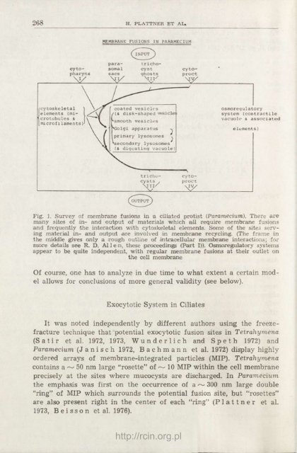
268 H. PLATTNER ET AL»<br />
cy topharynx<br />
V/<br />
cytoskeletal<br />
elements (microtubules<br />
&<br />
microfi laments)<br />
MEMBRANE FUSIONS <strong>IN</strong> PARAMECIUM<br />
parasomal<br />
sacs<br />
trichocyst<br />
ghosts<br />
Mil/<br />
coated vesicles<br />
{(&. disk-shaped vesicles<br />
smooth vesicles<br />
*Golgi apparatus ^<br />
primary lysosomes ^<br />
secondary lysosomes<br />
(& digesting vacuole)<br />
^OUTPUT<br />
trichocys<br />
Ls ,<br />
' a 11/<br />
cy toproct<br />
—\<br />
cy toproct<br />
My<br />
osmoregulatory<br />
system (contractile<br />
vacuolo & associated<br />
elements)<br />
Fig. 1. Survey of membrane fusions in a ciliated protist (Paramecium). There are<br />
many sites of in- and output of materials which all require membrane fusions<br />
and frequently the interaction with cytoskeletal elements. Some of the sites serving<br />
material in- and output are involved in membrane recycling. (The frame in<br />
the middle gives only a rough outline of intracellular membrane interactions; for<br />
moore details see R. D. Al 1 e n, these proceedings (Part I)). Osmoregulatory systems<br />
appear to be quite independent, with regular membrane fusions at their outlet on<br />
the cell membrane<br />
Of course, one has to analyze in due time to what extent a certain model<br />
allows for conclusions of more general validity (see below).<br />
Exocytotic System in Ciliates<br />
It was noted independently by different authors using the freezefracture<br />
technique that potential exocytotic fusion sites in Tetrahymena<br />
(Satir et al. 1972, 1973, Wunderlich and Speth 1972) and<br />
Paramecium (J a n i s c h 1972, B a c h m a n n et al. 1972) display highly<br />
ordered arrays of membrane-integrated particles (MIP). Tetrahymena<br />
contains a ~ 50 nm large "rosette" of ~ 10 MIP within the cell membrane<br />
precisely at the sites where mucocysts are discharged. In Paramecium<br />
the emphasis was first on the occurrence of a~ 300 nm large double<br />
"ring" of MIP which surrounds the potential fusion site, but "rosettes"<br />
are also present right in the center of each "ring" (P 1 a 11 n e r et al.<br />
1973, B ei sso n et al. 1976).<br />
http://rcin.org.pl