Download (2189Kb) - NERC Open Research Archive - Natural ...
Download (2189Kb) - NERC Open Research Archive - Natural ...
Download (2189Kb) - NERC Open Research Archive - Natural ...

Create successful ePaper yourself
Turn your PDF publications into a flip-book with our unique Google optimized e-Paper software.
A<br />
- -<br />
-. S.<br />
S 110<br />
a ll II r I<br />
II<br />
I. to 0<br />
ae -
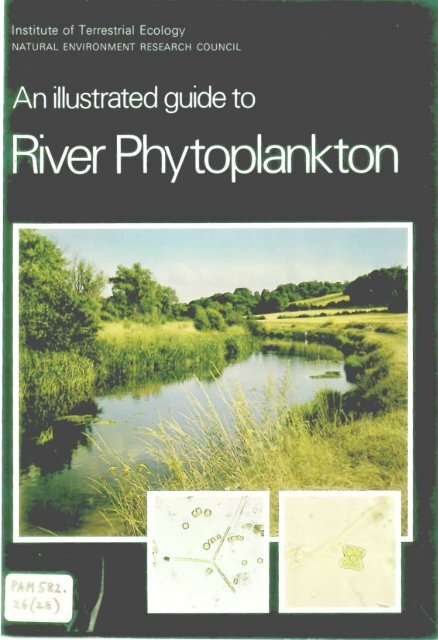
Institute of Terrestrial Ecology<br />
NATURAL ENVIRONMENT RESEARCH COUNCIL<br />
An illustrated guide to<br />
River Phytoplankton<br />
Hilary Belcher Et Erica Swale<br />
Culture Centre of Algae and Protozoa<br />
Cambridge<br />
London<br />
Her Majesty's Stationery Office<br />
INSTITUTE OF TEFP,ESTRIAL ECOLOGY<br />
U3RARY SEVIl:CE<br />
ED INDJRGH- LABORATOFJES<br />
BUSH ESTATE, PENICU1K<br />
MIDLOTHIAN EH26 OQB
© Crown copyright 1979<br />
First published 1979<br />
Institute of Terrestrial Ecology<br />
Culture Centre of Algae and Protoz a<br />
36 Storey's Way<br />
Cambridge<br />
CB3 0 DT<br />
0223 (Cambridge) 61378<br />
The cover shows the River Mole N ar Leatherh ad<br />
(Photograph by Kenneth Scowen)<br />
ISBN 0 11 886602 8<br />
%i::::::;r1TUTE OF<br />
T ESTRIAL<br />
ECO LOGY<br />
Li Di:RA RY<br />
13 JUL1981<br />
7.41-1<br />
1,14s (“2..z6<br />
Inset are Diatoma elongatum (left), Tetraddron minimum (right).<br />
(Photographs by the authors)<br />
The Institute of Terrestrial Ecology (ITE) was established in 1973, from<br />
the former Nature Conservancy's research stations and staff, joined<br />
later by the Institute of Tree Biology and the Culture Centre of Algae and<br />
Protozoa. ITE contributes to and draws upon the collective knowledge<br />
of the fourteen sister institutes which make up the National<br />
Environment <strong>Research</strong> Council, spanning all the environmental<br />
sciences.<br />
The Institute studies the factors determining the structure, composition<br />
and processes of land and freshwater systems, and of individual plant<br />
and animal species. It is developing a sounder scientific basis for<br />
predicting and modelling environmental trends arising from natural or<br />
man-made change. The results of this research are available to those<br />
responsible for the protection, management and wise use of our<br />
natural resources.<br />
Nearly half of ITE's work is research commissioned by customers, such<br />
as the Nature Conservancy Council who require information for wildlife<br />
conservation, the Forestry Commission and the Department of the<br />
Environment. The remainder is fundamental research supported by<br />
<strong>NERC</strong>.<br />
ITE's expertise is widely used by international organisations in overseas<br />
projects and programmes of research..
Foreword<br />
The purpose of this guide is to enable a non-specialist to<br />
identify most of the algae occurring in the plankton of rivers.<br />
In addition it will be found helpful in identifying algae in<br />
canals, broads and lowland pools, the plankton of which<br />
contains many of the same organisms.<br />
We hope to produce a future guide including the commoner<br />
benthic diatoms of rivers. A few of these, present frequently<br />
in the plankton during periods of high rate of flow, have<br />
been included in the present publication.<br />
Many readers, in search of a name, will want to look at the<br />
pictures first, hence these are placed before the key and<br />
descriptions. Numbers following the names opposite the<br />
plates refer to page numbers of the descriptions.<br />
3
Contents<br />
Introduction<br />
Methods<br />
Plates<br />
Key to genera<br />
Descriptions<br />
Glossary<br />
References<br />
Index<br />
4<br />
Page<br />
5<br />
8<br />
12<br />
32<br />
37<br />
58<br />
60<br />
64
Introduction<br />
During the spring and summer, rivers which flow through<br />
lowland areas generally contain microscopic algae in<br />
suspension. In these regions, the addition of non-toxic<br />
substances from agricultural land and of sewage effluent<br />
from towns leads to increased algal growth, and this often<br />
causes the water to be noticeably brown or green. In several<br />
papers dealing with the ecology of small algae in British<br />
rivers, R. W. Butcher concluded that those found in suspension<br />
were there only temporarily, and they consisted almost<br />
entirely of individuals either accidentally removed from their<br />
usual habitat on the bottom or washed out of lakes or<br />
backwaters*. He was convinced that to develop a 'true'<br />
plankton, a river needed to be far larger than any in Great<br />
Britain, for example, the bigger European rivers such as the<br />
Rhine or the Volga. It is unfortunate that his views were<br />
influential in forming the opinion, widely held among<br />
ecologists, that there is no appreciable phytoplankton in<br />
British rivers.<br />
The fact that relatively small riverst can and do contain<br />
substantial reproducing populations of suspended algae is of<br />
importance both ecologically and from the practical aspect of<br />
human use. For the purpose of this guide we include under<br />
the word 'plankton' all those algae which can reasonably be<br />
expected to be found fairly commonly in suspension in .<br />
rivers. In addition to those which reproduce extensively<br />
while in the stream itself, namely, the 'true' plankton, there<br />
are others, easily recognized by the experienced observer,<br />
which are normally bottom-living or attached cells swept<br />
into this situation by chance. These algae will probably sink<br />
fairly rapidly out of suspension, but while suspended it seems<br />
reasonable to consider them part of the plankton, and here<br />
they will contribute to its whole chlorophyll content and<br />
animal nutrient potential.<br />
A knowledge of the plankton algae in rivers is becoming of<br />
increasing practical importance in view of the need to develop<br />
*See references on page 60<br />
frhe terms 'small' and 'large' are open to considerable difference of<br />
interpretation.<br />
5
and maintain adequate supplies of water of suitable quality<br />
for domestic and industrial use. Any treatment plant designed<br />
for raw river water must be able to deal with dense growths<br />
of phytoplankton. According to their size and shape,different<br />
algae have different capacities for filter-clogging, and heavy<br />
populations can be extremely troublesome. The various water<br />
transfer schemes now being carried out are likely to affect<br />
algal populations in the rivers concerned. A relatively troublefree<br />
river might produce unexpectedly large populations of<br />
phytoplankton if suitable inocula were introduced with<br />
'foreign' water. Problems resulting from enrichment by<br />
nitrates and phosphates derived from agricultural run-off<br />
and sewage have become commonplace. To tackle these<br />
problems it is helpful to be able to recognize the dominant<br />
forms in populations.<br />
Plankton algae are part of the food chain or web involving<br />
the higher animals. Relatively little appears to be known in<br />
detail of the complex relationships existing, but, for the<br />
smaller invertebrates, algae are a major source of food, and<br />
such animals as Daphnia and a variety of rotifers are frequently<br />
seen with their guts stuffed with green cells. The<br />
young stages of some fish (e.g. perch) feed largely on diatoms<br />
and the enormous populations of freshwater mussels living<br />
in muddy river beds (as in the Thames at Reading) must<br />
take in large numbers of algae during the course of their<br />
filter feeding.<br />
COMPOSITION OF THE PHYTOPLANKTON<br />
The algae to be found in suspension in rivers normally<br />
reach significant numbers only during the spring and summer.<br />
In spring diatoms are predominant, the characteristic plankton<br />
species belonging mainly to the centric sub-group, when<br />
they often occur in concentrations of five million cells per<br />
litre, or more. Centric diatoms are cylindrical or discoid in<br />
shape, and typical members of this group in rivers belong to<br />
the genera Cyclotella and Stephanodiscus. The valves of<br />
these genera bear a fringe of extremely fine, long spines<br />
which can beseen with phase contrast illumination. The<br />
spines are shed when the material is 'cleaned' for detailed<br />
examination, but their presence probably enhances the high<br />
filter clogging capacity of the centric diatoms.<br />
6
Pennate diatoms (the other major sub-group) seem to be<br />
much less well adapted for a planktonic existence, although a<br />
few species, especially of the genera Nitzschia and Synedra,<br />
do form appreciable populations while in suspension and<br />
are widely distributed in rivers. Members of this group are<br />
characteristically elongated and bilaterally symmetrical, and<br />
in general they are less troublesome as regards filter clogging<br />
than the centrics whose cells can build up into dense masses.<br />
Apart from diatoms, other algae belonging to many taxonomic<br />
groups can also be abundant, with a preponderance<br />
of green ones in summer. These include motile forms like<br />
Chlamydomonas and non-motile examples such as Scenedesmus.<br />
A peculiarity of the river flora is that a usually infrequent<br />
species may suddenly have a burst of growth and become<br />
temporarily dominant.<br />
FACTORS CONTROLLING THE GROWTH OF PHYTOPLANKTON<br />
The development of a phytoplankton community in a river<br />
depends directly upon the physical factors of flow and<br />
turbidity, and when either or both of these is too great, no<br />
appreciable populations can be formed. Day length and<br />
temperature, particularly the former, seem also to be important,<br />
and the highest numbers of algae occur during prolonged<br />
periods of bright dry weather, when the rate of flow and<br />
silt are also at a minimum.<br />
In most lowland rivers nitrates and phosphates, derived from<br />
agriculture and from sewage, are present in abundance for<br />
algal growth. Deficiency of silica, however, may lead to the<br />
end of vigorous populations of diatoms in spring, which are<br />
then often succeeded by a mixed plankton, mainly of green<br />
algae, throughout the summer.<br />
NOTE ON EUTROPHY AND EUTROPHICATION<br />
In discussions of aquatic biology the words 'eutrophic' and<br />
'eutrophication' frequently occur, and something should be<br />
said as to their meaning. 'Eutrophy' and 'eutrophic' have<br />
been in use as medical terms since the mid-18th century to<br />
mean good (i.e. healthy) nutrition, and the promoting of good<br />
nutrition, respectively (Shorter Oxford English Dictionary,<br />
1965). In a limnological sense 'eutrophic' was first used by<br />
7
E. Naumann in 1919, when he contrasted this condition<br />
(i.e. of being rich in plant nutrients) with 'oligotrophic'<br />
(poor in nutrients). Naumann later defined the process of<br />
'eutrophication' as an increase in nutrients, especially nitrogen<br />
and phosphorus. In recent years more or less any form of<br />
enrichment, either natural or as a result of human activity,<br />
has been described as eutrophication. This can be somewhat<br />
confusing as the meaning may be extended to overlap the<br />
non-toxic, or rather, positively beneficial-to-organisms,<br />
forms of pollution. A different but related criterion of eutrophication<br />
(appearing in the more specialized literature) takes<br />
into account the effect of nutrient enrichment upon water<br />
chemistry, bodies of water (usually lakes) being categorized<br />
as eutrophic in which regular depletion of oxygen takes place<br />
in summer.<br />
'Eutrophic' can thus generally be taken to mean simply<br />
'nutrient enriched' and 'eutrophication' as 'nutrient enrichment'.<br />
There seems to be no generally accepted borderline between<br />
'eutrophication' and 'pollution'.<br />
METHODS<br />
The following suggestions are kept as uncomplicated as<br />
possible; more sophisticated techniques are to be found in<br />
the references.<br />
Collection: i The simplest method is to use a jug and line.<br />
It is best to collect well away from the river bank, as conditions<br />
at the edge are likely to be different from those in the main<br />
stream. The jug can either be thrown from the bank or from a<br />
bridge or jetty. Polythene jugs, unless weighted, are too<br />
light, and the most satisfactory kinds are the old fashioned<br />
enamelled metal ones. With practice, it is possible to fill a<br />
jug with surface water only, and thus have roughly comparable<br />
samples from different rivers. 2 A piece of hosepipe<br />
may be used to provide a mixed water sample down to a<br />
given depth. A line is tied round one end of the pipe and<br />
this end is pushed down vertically into the water, while<br />
holding the free end of the line. At its full depth, the upper<br />
end of the pipe is closed with the hand and the lower end is<br />
pulled up. The tube then contains a column of water of its<br />
own length and diameter. A disadvantage of this method is<br />
that the operator needs to be at water level, though it is<br />
8
possible, but difficult, to manipulate both ends of the pipe at<br />
a distance. A hosepipe, moreover, is only practicable where<br />
the current is weak.<br />
Examination: Living material is to be preferred, but as some<br />
algae die fairly rapidly, especially in hot weather, it is as well<br />
to fix part of the sample immediately on collection. As a<br />
general preservative, Lugol's iodine is recommended. This is a<br />
saturated aqueous solution of potassium iodide, to which is<br />
added crystalline iodine to saturate it again. From this stock<br />
solution, only a few drops are needed, enough to colour the<br />
water sample a pale brown. After a few weeks or months of<br />
storage the samples may become decolourized, when more<br />
iodine should be added.<br />
The presence of mucilage surrounding individual cells or<br />
colonies is of taxonomic significance. It may be difficult to<br />
see, but shows up clearly on adding a little Indian ink to a<br />
liquid mount, when the particles of pigment do not penetrate<br />
the mucilage which appears as a bright halo.<br />
As collected, a drop of water on a microscope slide may<br />
contain few organisms, and some form of concentration<br />
is indicated. The best and quickest method is to centrifuge,<br />
using 10 ml tapering tubes. A method for constructing an<br />
efficient miniature centrifuge is given by Belcher (1978).<br />
If no centrifuge is available, the sample can be passed<br />
through a fine filter paper, washing down the sides of the<br />
funnel and thus concentrating a good proportion of the<br />
plankton into a volume of about 0.5 ml. A small drop of the<br />
concentrate is put on to a microscope slide and left to settle<br />
out for a minute or so before carefully laying on the coverslip.<br />
This prevents loss of cells to the sides and it is also helpful<br />
to examine the uncovered drop with a low power objective.<br />
This gives an immediate impression of the overall density<br />
and composition of the sample, and it draws attention<br />
especially to the larger forms which are often less conspicuous<br />
when dispersed by the pressure of the coverslip. When<br />
mounted, examination should not be too prolonged because<br />
of drying out and because some delicate flagellates die<br />
rapidly on a slide. For keeping temporary mounts for several<br />
hours, or even until the next day, an effective method is to<br />
seal the edges of the coverslip with a petroleum jelly such as<br />
PR-A*<br />
9
'Vaseline'. This can be heated in a small flat tin lid with a<br />
handle like a miniature frying pan, and applied molten with a<br />
paint brush.<br />
Fixed samples may also be centrifuged, but as iodineimpregnated<br />
cells are heavy, they are easily concentrated by<br />
sedimentation (settling). The sample in its original container<br />
may simply be allowed to stand for 24 hours and the water<br />
siphoned off to leave a sediment for examination. For<br />
quantitative work, known volumes are sedimented (100 ml<br />
tubes are convenient) and a factor calculated. The least<br />
disturbance to the sediment takes place if the lower end of<br />
the glass or polythene siphon tube is bent upwards.<br />
If algal cells have been too darkly pigmented by the iodine,<br />
they can be decolourized by a trace of sodium thiosulphate<br />
solution (or photographic fixer). In seasons of vigorous<br />
growth, there may be too many cells in the original sample<br />
to be able to count readily in the residue from 100 ml. In this<br />
case, a series of sub-dilutions is set up and a suitable<br />
concentration chosen to count.<br />
Counting: There are two main methods. A haemacytometer<br />
of the Fuchs-Rosenthal type is suitable for the smaller more<br />
compact cells, and is especially useful for the centric diatoms.<br />
To count the larger colonial or filamentous forms an inverted<br />
microscope is invaluable. Here small sedimentation tubes are<br />
placed on the stage and the counts made directly through<br />
their glass bottoms. A counting chamber of the Lund type<br />
(Lund 1959, 1962) enables all the algae in a small volume of<br />
water to be counted. This is more accurate but considerably<br />
more time-consuming than the other methods.<br />
In quantitative work, the problem arises of how many cells to<br />
count. For statistical respectability, about 100 cells of all<br />
sorts are considered reasonable. With exceptionally 'thin'<br />
samples one has sometimes to make do with fewer, but the<br />
smaller the number of cells counted the less comparable are<br />
different samples. The converse does not apply. Increased<br />
'accuracy' flattens off rapidly the greater the number counted<br />
above 100, and the extra effort is seldom worth the time<br />
expended.<br />
A word on the use of haemacytometers for counting algal<br />
cells may be appropriate. The centre of the slide bears an<br />
10
engraved rectangular area of given size, subdivided by<br />
intersecting lines. When a drop of liquid is mounted, the<br />
special coverslip is supported at a known height above the<br />
central area by the slightly raised 'shoulders' of the slide.<br />
A simple calculation then gives the volume of liquid contained<br />
over the grid and a conversion factor can be worked out to<br />
find the concentration of cells per ml of the original unconcentrated<br />
water.<br />
Boney (1975) should be referred to for a helpful section on<br />
preservation, sedimentation and counting methods.<br />
Recording: A preliminary check-list of British freshwater<br />
algae has been produced (Whitton, Holmes Et Sinclair,<br />
1978), available from the Department of the Environment<br />
Water Data Unit, Reading Bridge House, Reading. 1000<br />
species are listed and the authors explain in the introduction<br />
that 'Each species .... has a unique 6-digit code for the<br />
presentation of records in a standard manner and for use in<br />
computing'. Where more than one name is in use for an alga the<br />
one chosen in this guide agrees with that in the check-list.<br />
11
UniceHular green flageHates<br />
1 Chlamydomonas monadina (C. cingulata) (p. 37) ; 18-35 pm<br />
long.<br />
2 Chlamydomonas pertusa (p. 37) ; up to 25 pm long.<br />
3 Chlamydomonas isogama (p. 37) ; up to 12 pm long.<br />
4 Haematococcus droebakensis (p.38) ; up to 70 pm long.<br />
5 Haematococcus pluvialis (p. 38) ; up to 65 pm long, a, b<br />
show individual variation in shape.<br />
6 Mesostigma viride (p. 38) ; up to 18 pm long, a anterior<br />
view; b side view.<br />
7 Lobomonas ampla (p. 38) ; up to 40 pm long.<br />
8 Phacotus lenticularis (p. 38) ; up to 20 pm in diameter, a, b<br />
side views at right angles to each other.<br />
9 Pteromonas angulosa (p. 38) ; up to 20 pm long, a, b side<br />
views at right angles to each other.<br />
10 Platymonas cordiformis (Tetraselmis cordiformis) (p. 38) ;<br />
up to 25 pm long, a side view; b diagram of anterior view.<br />
11 Nephroselmis angulata (Heteromastix angulata); (p. 39)<br />
up to 15 pm long, a side view; b, c cells of two different<br />
widths (anterior view) ; d side view at right angles to a.<br />
12
C13<br />
CY)<br />
4442id0<br />
; .;,11r77.7::4!<br />
-A<br />
C.)<br />
1:3
Unicellular green flagellates (cont.)<br />
and Colonial green flagellates<br />
1 2 Pandorina morum (p. 39) ; colonies up to 50 pm in diameter.<br />
1 3 Gonium pectorale (p. 39) colonies up to 100 pm across.<br />
14 Volvox aureus (p.39) ; colonies up to 500 pm in diameter,<br />
a colony with daughter colonies; b part of surface of colony<br />
to show cells joined by protoplasmic connexions.<br />
15 Volvox globator (p.39) ; colonies up to 600 pm across,<br />
details of cells to showdifferences from those of V. aureus.<br />
16 Eudorina elegans (p.39) ; colonies up to 200 pm in diameter.<br />
17 Chlorogonium tetragamum (p. 37) ; up to 33 pm long,<br />
single pyrenoid.<br />
18 Chlorogonium elongatum (p.37) ; up to 45 pm long, two<br />
pyrenoids.<br />
19 Spermatozopsis exsultans (p.39) ; up to 12 pm long, a, b two<br />
individuals differing slightly in shape.<br />
14
Colonial green flagellates (cont.)<br />
20 Gonium sociale (p.39) ; colonies up to 50 pm across,<br />
lateral view showing remains of mother cell wall attached<br />
below, a character of the var. sacculum (Basichlamys<br />
sacculifera): b anterior view.<br />
21 Pascherina tetras (p.39) ; colonies up to 25 pm long.<br />
Green non-motile unicells<br />
22 Ankistrodesmus angustus (A, contortus; A. falcatus var.<br />
spirilliformis; Monoraphidium contortum) (p. 40) ; three cells,<br />
length up to 35 pm.<br />
23 Ankistrodesmus subcapitatus (p. 41) ; cells up to 18 pm<br />
long (up to 9 pm across 'ring'), group of five cells, one<br />
showing cross walls prior to division.<br />
24 Ankistrodesmus gracifis (Selenastrum gracile) (p. 40) ; up to<br />
40 pm long.<br />
25 Tetra&dron incus (p.46); up to 30 pm across,a older and b<br />
younger cells to show differences in shape.<br />
26 Tetra6dron caudatum (p.46) ; up to 25 pm across,a older<br />
and b younger cells showing differences in shape.<br />
27 Tetra6dron minimum (p.46) ; up to 20 pm across.<br />
28 Unicellular stages of a species of Scenedesmus,resembling<br />
Chodatella subsalsa (p.42); up to 10 pm long.<br />
29 Goniochloris mutica (N.B. Xanthophyceae) (p. 47) ; up to<br />
10 pm across.<br />
30 Trachychloron circulare (Xanthophyceae) (p.47) ; up to<br />
10 pm in diameter,a face view; b edge view.<br />
31 Lagerheimia wratislaviensis (p.41) ; up to 14 pm long<br />
(excluding spines).<br />
32 Lagerheimia genevensis (p.41) ; up to 10 pm long (excluding<br />
spines).<br />
33 Chodatella quadriseta (p.42) ; up to 15 pm long, a, b two<br />
cells showing variation in shape. (N.B. see comments on<br />
unicellular Scenedesmus stages, p. 42).<br />
34 Chodatella citriformis (p.41) ; up to 25 pm long (excluding<br />
spines).<br />
35 Chodatella ciliata (p.41) ; up to 25 pm long (excluding<br />
spines), a mother cell with young cells which have not yet<br />
grown their spines ; b vegetative cell.<br />
16
PR-A"<br />
17
Unicellular (mostly) non-motile<br />
green unicells (cont.)<br />
36 Didymogenes palatina (p.42) ; up to 8 pm long, a, b two<br />
pairs of cells,<br />
37 Scenedesmus grahneisii (Didymocystis inconspicua (p.45);<br />
up to 8 pm long.<br />
38 Polyedriopsis spinulosa (p.44) ; up to 20 pm across<br />
(excluding spines).<br />
39 Chlorella vulgaris (p.41) ; 2-10 pm, a young cells; b four<br />
daughter cells within mother cell wall.<br />
40 Siderocelis ornata (p.45); up to 15 pm long, a young cell;<br />
younger and older cells.<br />
41 Oocystis pseudocoronata (p.44); up to 15 pm long, a<br />
daughter cells inside mother cell wall; b young cell which<br />
could belong to a number of unornamented species.<br />
42 Stichococcus bacillaris (p.45); up to 4 pm wide, a single<br />
cell; b dividing cell—the daughter cells often remained joined<br />
for'some time to form short filaments.<br />
43 Siderocystopsis fusca (p.45) ; up to 20 pm long, a, b<br />
b newly released daughter cells lying within remains of<br />
mother cell wall.<br />
44 Golenkiniopsis parvula (p.43) ; about 6 pm in diameter.<br />
45 Golenkinia radiata (p.43); up to 18 pm in diameter.<br />
18
45<br />
43a<br />
19
Elongated non-motile green unicells<br />
46 Ankistrodesmus acicula ris (Monoraphidium aciculare) (p.40);<br />
up to 200 pm long, a dividing cell; kbundle of young cells;<br />
c vegetative cell.<br />
47 Koliella longiseta (Raphidonema longiseta) (p.43); up to<br />
150 pm long, a, b vegetative cells; c cells about to divide;<br />
d young cell before elongation.<br />
48 Ankistrodesmus fusiformis (p.40) ; up to 100 pm long,<br />
a group of cells in characieristic arrangement; b single cell<br />
enlarged.<br />
49 Ankistrodesmus falcatus (p.40) ; up to 80 pm long, a single<br />
cell; b, c two-celled and four-celled bundles.<br />
50 Elakatothrix gelatinosa (p.43); up to 30 pm long, usually<br />
two cells in mucilage envelope.<br />
51 Gloeotila pelagica (p.43); filament about 2 pm wide.<br />
52 Ankyra judayi (p.41) ; up to 100 pm long.<br />
53 Sdenedesmus acuminatus (p.44) ; unicellular stage, up to<br />
30 pm long.
(mostly) Non-motile green colonies<br />
54 Closterium moniliferum (p.46) ; up to 400 pm long.<br />
55 Cosmarium botrytis (p.47) ; up to 90 pm long.<br />
56 Coelastrum microporum (p.42); cells up to 10 pm, colonies<br />
up to 60 pm across.<br />
57 Botryococcus braunii (p.41) ; cells up to 10 pm long,<br />
colonies variable in shape and size, up to 1 mm across.<br />
Colour green to reddish brown.<br />
58 Scenedesmus acuminatus (p.44) ; cells up to 30 pm long,<br />
a eight-celled colony; b four-celled colony. Shape of cells<br />
variable.<br />
59 Scenedesmus protruberans (p.45) ; cells up to 30 pm long.<br />
60 Scenedesmus obliquus (S. acutus) (p.44); cells up to<br />
20 pm long.<br />
61 Scenedesmus quadricauda (p.45) ; cells up to 25 pm long,<br />
a 'typical' four-celled colony; b, c "Chodatella' stages.<br />
62 Actinastrum hantzschii (p.40) ; cells up to 25 pm long.<br />
63 Tetrastrum staurogeniaeforme (p.46) ; cells up to about 6 pm.<br />
22
'-<br />
v;,.<br />
0<br />
4t<br />
.<br />
,<br />
'<br />
;<br />
LO<br />
II<br />
,
24<br />
Non-motile green colonies<br />
64 Dictyosphaerium pulchellum (p.42) ; cells up to 10 pm<br />
across.<br />
65 Micractinium pusillum (p.43) ; cells up to 10 pm across.<br />
66 Crucigenia tetrapedia (p. 42); each cell of four-celled'<br />
packet up to 8 pm across.<br />
67 Crucigenia fenestrata (p.42) ; individual cells up to 8 pm<br />
across, a older colony dividing ; b young colony.<br />
68 Crucigenia apiculata (p.42) ; cells up to 8 pm long.<br />
69 Crucigenia rectangularis (p.42) ; cells up to 8 pm long.<br />
70 Pediastrum boryanum (p.44); colonies up to 200 pm in<br />
diameter.<br />
71 Pediastrum duplex (p.44) ; colonies up to 200 pm in<br />
diameter.<br />
72 Pediastrurn tetras (p.44) ; colonies up to 30 pm across,<br />
a eight-celled colony; b four-celled colony.
PR-A***<br />
25
Diatoms<br />
73 Stephanodiscus astraea (p.48) ; valves up to 70 pm in<br />
diameter.<br />
74 Stephanodiscus invisitatus (p.48) ; valves up to 20 pm in<br />
diameter, a cleaned valve; b, c living cells.<br />
75 Stephanodiscus tenuis (p.48) valves up to 30 pm in<br />
diameter.<br />
76 Cyclotella kuetzingiana (p.49); valves up to 45 pm in<br />
diameter.<br />
77 Cyclotella comta (p.49) ; valves up to 30 pm in diameter.<br />
78 Cyclotella meneghiniana (p.49); valves up to 30 pm in<br />
diameter.<br />
79 Cyclotella pseudostelligera (p.49) valves up to 10 pm in<br />
diameter.<br />
80 Thalassiosira fluviatilis (p.49) ; valves up to 25 pm in<br />
diameter.<br />
81 Melosira varians (p.50); cells up to 30 pm wide. Part of<br />
living filament.<br />
82 Melosira granulata (p.50) ; cells 5-20 pm wide. Part of<br />
living filament.<br />
83 Navicula viridula (p. 51); up to 80 pm long, a living cell ;<br />
b cleaned valve.<br />
84 Nitzschia acicularis (p.51); up to 150 pm long. Living cell.<br />
85 Synedra acus (p.51) ; up to 300 pm long. Living cell.<br />
86 Navicula cryptocephala (p.51) ; valves up to 40 pm long.<br />
87 Navicula gracilis (p.51); up to 60 pm long.<br />
88 Diatoma vulgare (p.50); 10-60 pm long, a living cell,<br />
girdle view ; b, c, d valve views to show differences in shape<br />
and size.<br />
89 Nitzschia palea (p.52) ; up to 65 pm long, a cleaned valve;<br />
b living cell.<br />
26
o.)<br />
03<br />
j/f111111110111-111\\WTIOMMTIM71111<br />
1111111111111"1/1;7 Cj°<br />
cl
Diatoms (cont.); Chrysophyceae<br />
90 Nitzchia sigmoidea (p.52); up to 500 pm long. Living cell.<br />
91 Asterionella formosa (p.50) ; individual cells of colony up to<br />
130 pm long. Living colony.<br />
92 Diatoma elongatum (p.50); individual cells up to 120 pm<br />
long. Living 3-celled colony.<br />
93 Dinobryon sertularia (p.52) ; 'flasks' 30-100 pm long.<br />
Living colony.<br />
94 Mallomonas spp. (p. 52); cells 5-50 pm long, a cell of<br />
species with scales but no spines; b cell with both scales<br />
and spines.<br />
95 Synura petersenii (p.53); colonies up to 500 pm in diameter,<br />
a whole colony; b individual cell, 20-40 pm long.<br />
96 Ochromonas tuberculatus (p.53) ; up to 20 pm long.<br />
28<br />
-
Miscellaneous<br />
97 Cryptomonas sp. (p.53) ; in the genus cells range from<br />
10-80 pm in length.<br />
98 Chroornonas sp. (p.54) ; small cells not more than about<br />
10 pm long.<br />
99 Rhodomonailacustris var. nannoplanktica (R. minuta v.<br />
nannoplanktica) (p.54) ; up to 10 pm long.<br />
100 Trachelomonas volvocinopsis (p.55) ; up to 25 pm in diameter,<br />
a living cell in optical section, b diagram of exterior of 'pot'<br />
(lorica).<br />
101 Euglena viridis (p.55) ; up to 75 pm long.<br />
102 Peridinium sp. (p.54) ; large species are up to 70 pm long.<br />
103 Phacus pleuronectes (p.55) ; up to 80 pm long.<br />
104 Microcystis aeruginosa (p.55) ; cells 3-9 pm across,<br />
colonies variable in shape and up to macroscopic size.<br />
105 Aphanizomenon flos-aquae (p.56) ; a bundle of filaments,<br />
each up to 6 pm wide, bundles may reach macroscopic size;<br />
b cells of filaments showing spore (upper), end of filament<br />
(middle) and heterocyst (lower).<br />
106 Oscillatoria limnetica (p.56) ; filaments about 1.5 pm wide.<br />
107 Oscillatoria agardhii (p.56) ; filaments approx. 7 pm wide.<br />
108 Oscillatoria limosa (p.56) ; filaments about 15 pm wide.<br />
109 Merismopedia glauca (p.56) ; each cell of 'packet' about<br />
4 pm across.<br />
30
97<br />
109<br />
102<br />
108<br />
107<br />
fre .105a<br />
77.71"17<br />
10 5 b<br />
106<br />
101<br />
31<br />
ta:t"<br />
4g.
Key to genera<br />
CELLS SWIM (WITH FLAGELLA)<br />
Unicellular<br />
Colonial<br />
CELLS DO NOT SWIM (NO FLAGELLA, BUT<br />
Unicellular<br />
Green elongated<br />
Green 'chunky'<br />
Yellow or brown<br />
Colonial<br />
Green .<br />
Brown or other colour<br />
Filamentous .<br />
32<br />
—<br />
Section A : single motile cells<br />
Cell with wide envelope enclosing a<br />
smaller protoplast<br />
Envelope + thick and strong, pale<br />
yellow to brown or black<br />
Envelope spherical or flask-shaped,<br />
smooth or ornamented, one<br />
flagellum<br />
Envelope lenticular, patterned, two<br />
flagella .<br />
Envelope thin and delicate, two flagella<br />
Outline lobed .<br />
Outline smoothly oval, not winged .<br />
With longitudinal wings, side view<br />
flattened .<br />
MAY CREEP)<br />
Trachelomonas<br />
Phacotus<br />
Lobomonas<br />
Haematococcus<br />
Pteromonas<br />
Section<br />
Cell without wide envelope, wall fits closely<br />
Chloroplast brownish or yellowish<br />
Cells drop- or heart-shaped . Cryptomonas<br />
Cells lemon- or egg-shaped, sometimes<br />
with long 'tail', often having<br />
hairy appearance due to covering of<br />
scales or spines . . Mallomonas<br />
single Synura cells<br />
A
Cells armour-plated, helmet-like,<br />
often "large" .<br />
Cells irregularly rounded, fragile,<br />
chloroplast often pale .<br />
Chloroplast bright blue, drop-shaped .<br />
Chloroplast green<br />
Cells spherical to elongated, not<br />
flattened<br />
Cells spherical or oval, two flagella<br />
Cells broadly spindle-shaped, can<br />
change shape, one flagellum<br />
Cells narrowly spindle-shaped,<br />
rigid, two flagella .<br />
Cells banana-shaped, two or four<br />
flagella, swim in spirals<br />
Cells flattened<br />
Cells oval, four flagella from<br />
anterior depression .<br />
Cells bean-shaped, two flagella,<br />
swims sideways .<br />
Cells shaped like rubber dinghy,<br />
two flagella arise from middle .<br />
Purple cells, sausage-, cigar- or<br />
screw-shaped (not illustrated) .<br />
Section B : motile colonies<br />
Peridiniurn<br />
Ochrornonas<br />
Chroomonas<br />
Chlamydomonas<br />
Euglena<br />
Chlorogoniurn<br />
Spermatozopsis<br />
Platymonas<br />
Nephroselmis<br />
Mesostigma<br />
Purple sulphur<br />
bacteria<br />
Cells brownish or yellowish<br />
Cells densely packed into spherical or<br />
somewhat elongated colonies . . Synura<br />
Cells in flask-like envelopes, colony<br />
branching . Dinobryon<br />
Cells green<br />
Colony flat, four or sixteen cells . Goniurn<br />
Colony ± spherical<br />
Four cells in a small bunch . . Pascherina<br />
Sixteen, thirty-two or sixty-four cells<br />
Cells closely packed so that centre<br />
of colony + solid . . Pandorina<br />
Cells loosely packed round outside<br />
of hollow sphere . . Eudorina<br />
Hundreds of cells forming hollow<br />
sphere . Volvox<br />
33
Section C : non-motile elongated<br />
green unicells<br />
Cells straight<br />
Cells single or in pairs,<br />
mucilaginous coating .<br />
with wide<br />
Cells curved<br />
Cells very large, with several pyrenoids<br />
Cells smaller, slender, single or in<br />
bunches, usually no pyrenoids .<br />
34<br />
Cells without wide mucilage coat<br />
Spindle-shaped with processes at<br />
one end like feet or handlebars<br />
Needle-shaped, extremely long anCI<br />
thin<br />
More than six spines<br />
Spines in tufts at angles of irregular<br />
cell<br />
Spines at each end of ellipsoidal cell<br />
Spines delicate, over whole surface<br />
of ellipsoidal cell<br />
Spines scattered over surface of<br />
spherical cell .<br />
Without spines<br />
Cells flattened<br />
Cells of two identical halves, with<br />
'waist', often large<br />
Cells triangular .<br />
Cells four orfive-angled<br />
Cells round, discoid .<br />
Elakatothrix<br />
Ankyra<br />
Ankistrodesmus<br />
Raphidonema<br />
Closterium<br />
Section D : non-motile 'chunky'<br />
green unicells<br />
Ankistrodesmus<br />
Selenastrum<br />
With spines<br />
Six spines or less<br />
Spines short, cells four orfive-angled Tetra6dron<br />
Spines long, cells ellipsoidal . Lagerheimia<br />
Chodatella<br />
Scenedesmus<br />
(single cells)<br />
Polyedriopsis<br />
Chodatella<br />
Siderocystopsis<br />
Golenkinia<br />
Golenkiniopsis<br />
Cosmarium<br />
Goniochloris<br />
Tetra6dron<br />
Trachychloron
Cells not flattened<br />
Wall with brown spots or dots . Siderocelis<br />
Siderocystopsis<br />
Oocystis<br />
Wall without brown spots or dots<br />
Cells cylindrical, sometimes loose<br />
filaments of two or three cells . Stichococcus<br />
Cells spherical or ovoid . Chlorella<br />
Cells ellipsoidal, lemon- or barrelshaped<br />
Oocystis<br />
Cells crescent-shaped Ankistrodesmus<br />
Cells short, spindle-shaped Scenedesmus<br />
(single cells)<br />
Section E : non-motile yellow<br />
or brown unicells<br />
Cells cylindrical or disc-shaped, rectangular<br />
in side view . Cyclotella<br />
Stephanodiscus<br />
Thalassiosira<br />
Cells long and narrow<br />
Two chloroplasts, arranged fore and<br />
aft, plant can creep Nitzschia<br />
Several chloroplasts, not arranged as<br />
above, plant cannot creep . Synedra<br />
Cells like flat-bottomed boats, narrowly<br />
rectangular in side view<br />
Two chloroplasts, arranged longitudinally,<br />
side by side . Navicula<br />
Two chloroplasts, arranged fore and aft Nitzschia<br />
A number of chloroplasts, not arranged<br />
as above, wall with heavy transverse<br />
ridges going right across Diatoma<br />
Section F : green non-motile colonies<br />
Flat plate of two, four or eight elongated<br />
parallel cells . . Scenedesmus<br />
Didymogenes<br />
Flat plate of cells in foursquare or radial<br />
pattern Crucigenia<br />
Tetrastrum<br />
Pediastrum<br />
35
Colonies of shapes other than flat<br />
Cells in irregular solid mass .<br />
Cells closely packed into a ball .<br />
Cells more loosely held together<br />
Cells attached distantly by narrow<br />
strands of mucilage, like balloons on<br />
strings .<br />
Cells spherical, attached side by side<br />
in irregular ring or square, or in<br />
smaller groupings, with long thick<br />
spines .<br />
Cells elongated, attached to centre<br />
of colony by one end and radiating<br />
outwards<br />
Filament of cells firmly fastened together<br />
Cells banded with green, about 2 pm<br />
across<br />
Cells yellow-brown, sometimes with<br />
spikes at ends .<br />
Cells blue-green, blackish, purple,<br />
greyish or pale, not green or brown .<br />
36<br />
Botryococcus<br />
Coelastrum<br />
Dictyosphaerium<br />
Micractinium<br />
Actinastrum<br />
Section G : non-motile colonies,<br />
not green<br />
Cells blue-green or blackish green<br />
Colony irregular in shape, often large,<br />
many small spherical cells Microcystis<br />
Colony a small flat plate, with spherical<br />
pale blue-green cells in rows . Merismopedia<br />
Colony of small filaments in tufted<br />
bundles . . Aphanizornenon<br />
Cells or colony brownish<br />
Colony a dense irregular mass of green<br />
cells often masked by brown pigment Botryococcus<br />
Colony of long narrow cells arranged<br />
in stars or zig -zags Asterionella<br />
Diatoma<br />
Section H : planktonic filaments<br />
Short filaments of a few cells loosely<br />
joined together<br />
Cells green, c. 4pm wide .<br />
Cells yellow-brown .<br />
Stichococcus<br />
Stephanodiscus<br />
Cyclotella<br />
Thalassiosira<br />
Gloeotila<br />
Melosira<br />
Oscillatoria<br />
Aphanizornenon
Descriptions of species illustrated<br />
(Numbers in brackets refer to illustrations)<br />
Class Chlorophyceae<br />
These, the green algae, have bright green chloroplasts, but<br />
among them is a great variety of form and organization.<br />
Planktonic genera range from the apparently simple Chlorella<br />
to the elaborate colonies of Volvox. More freshwater algae<br />
belong to the Chlorophyceae than to any other group.<br />
ORDER VOLVOCALES<br />
Flagellated forms, unicellular or colonial. For simplicity we<br />
include here four genera, Platymonas, Nephroselmis, Mesostigma<br />
and Spermatozopsis, which since the advent of<br />
electron microscopy are now generally held to belong to a<br />
separate group of green flagellates, the Prasinophyceae.<br />
Chlamydomonas (1-3) Spherical, ovoid or elongated cells<br />
with a smooth close-fitting wall and two equal flagella.<br />
Chloroplasts and pyrenoids variable. A vast genus, difficult<br />
to identify. The three species illustrated are common in rivers<br />
and have well-marked characteristics.<br />
C. monadina Stein (1) (C. cingulata Pascher) Spherical,<br />
18-35 pm diameter, with a cup-shaped chloroplast and a<br />
pyrenoid which may be a flattened sphere, a doughnut-like<br />
ring, or in two or three separate pieces. This species is often<br />
dominant or semi-dominant in a plankton population'.<br />
C. pertusa Chodat (2) Ovoid cells up to 25 pm long, with<br />
two pyrenoids placed fore and aft, each enclosed in a<br />
stellate chloroplast.<br />
C. isogama Korshikov (3) Small fusiform cells up to 12 pm<br />
long, with a lateral chloroplast and single pyrenoid.<br />
Chlorogonium (17, 18) Green fusiform cells with two equal<br />
flagella. Two species are common in rivers.<br />
C. tetragamum Bohlin (17) Up to 33 pm long, with one<br />
central conspicuous pyrenoid.<br />
C. elongatum Dangeard (18) Up to 45 pm long, with two<br />
pyrenoids, one anterior and one posterior.<br />
Haematococcus (4, 5) The characteristic wide gap between<br />
the outer wall and the central chloroplast is crossed by<br />
readily visible fine strands of protoplasm. Two equal flagella.<br />
37
Two species found in rivers (also, more often, in standing<br />
water).<br />
H. pluvialis Flotow em. Wille (5a, b) Up to 65 pm long and<br />
has several pyrenoids, but these and the green pigment<br />
often masked by an accumulation of red oil. Frequent in<br />
rivers. Also responsible for the red colouration common in<br />
bird baths and small water bodies.<br />
H. droebakensis Wollenweber (4) A striking object, up to<br />
70 pm long, with only two pyrenoids and more conspicuous,<br />
branching protoplasmic strands than in H. pluvialis. Occasional<br />
in British rivers, but in Central Europe has given much<br />
trouble, dense growths causing problems of smell in water<br />
supplies.<br />
Lobomonas (7) Chlamydomonas-type cells, but with a wide<br />
transparent envelope whose surface is thrown into lobes.<br />
L. ampla Pascher (7) Up to 40 pm long, occasional in rivers.<br />
Mesostigma (6) (Prasinophyceae) Small flagellates, flattened<br />
and shaped like a rubber dinghy, the chloroplast forming a<br />
green rim, with a red eye spot in the centre and a pyrenoid<br />
at each end. The two flagella stand up from the centre. The<br />
cells have a characteristic appearance when swimming.<br />
M. viride Lauterborn (6a, b) Up to 18 pm long. This is the<br />
the only British species.<br />
Phacotus (8) Cells like those of Chlamydomonas, but<br />
enclosed in a wall resembling two shallow dishes placed<br />
together. Wall sometimes thin and transparent, but usually<br />
thick and sculptured.<br />
P. lenticularis (Ehrenb.) Stein (8a, b) Up to 20 pm in diameter.<br />
Sometimes frequent.<br />
Platymonas (10) (Prasinophyceae) Flattened green heartshaped<br />
cells with four flagella and a basal pyrenoid.<br />
P. cordiformis (N. Carter) Korshikov (10a, b) Up to 25 pm<br />
long and often found in rivers.<br />
Pteromonas (9) Cells like those of Chlamydomonas but<br />
flattened, with a wing on either side like an elm fruit.<br />
P. angulosa (Carter) Lemmermann (9a, b) Up to 20 pm<br />
long, common.<br />
Nephroselmis (11) (Prasinophyceae) Bean-or kidney-shaped<br />
green flagellates with two flagella. Swim jerkily with one end<br />
of the 'bean' going forwards.<br />
38
N. angulata (Korshikov) Skuja (11a-d) (Heteromastix<br />
angulata Korshikov) Up to 15 pm long, common.<br />
Spermatozopsis (19) (Prasinophyceae) Small flagellates<br />
with two or four flagella, shaped like slender fish and swim<br />
rapidly in spirals.<br />
S. exsultans Korshikov (19) Up to 12 pm in length and<br />
sometimes common. Its swift darting movements make it<br />
noticeable in a mixed sample.<br />
Eudorina (16) Colonial, with thirty-two or sixty-four green<br />
cells widely separated at the surface of a gelatinous sphere.<br />
E. elegans Ehrenberg (16) with cells 5-15 pm long and<br />
colonies up to 200 pm across, is often abundant.<br />
Pandorina (12) Colonial. Differs from Eudorina in that the<br />
cells fit closely together without a large central space.<br />
P. morum (Muller) Bory (12) Cells 8-20 pm long, colonies<br />
up to 50 pm across. Often abundant.<br />
Gonium (13, 20) Colonies form a flat plate composed of 14<br />
or 16 cells. Two species are common.<br />
G. pectorale M011er (13) Sixteen-celled colonies up to 50 pm<br />
across.<br />
G. sociale (Dujardin) Warming (20a, b) four-celled colonies up<br />
to 50 pm across. The colony illustrated in 20b shows the<br />
remains of the mother cell wall below, which is characteristic<br />
of the variety sacculum J. Stein. This is sometimes distinguished<br />
under the name Basichlamys sacculifera (Scherffel)<br />
Skuja.<br />
Pascherina (21) Colonies of four cells like those of Chlamydomonas,<br />
arranged in a tetrad and readily breaking apart<br />
into single cells. One species only.<br />
P. tetras (Korsh.) Silva (21) Colonies up to 25 pm across,<br />
frequent in rivers.<br />
Volvox (14, 15) Macroscopic spherical colonies containing<br />
hundreds of cells. Two species are occasionally found in<br />
British rivers, a third one being much rarer.<br />
V. aureus Ehrenberg (14a, b) has colonies up to 500 pm in<br />
diameter and the individual cells appear round (b).<br />
V. globator Linn. em. Ehrenb. (15) Even larger colonies than<br />
V. aureus, which are superficially like the previous species,<br />
but the cells are stellate.<br />
39
ORDER CHLOROCOCCALES<br />
Green algae in which the vegetative states are non-motile.<br />
Unicellular or colonial but not filamentous.<br />
N.B. For convenience, three members of the Ulotrichales are<br />
included here (Gloeotila, Koliella and Stichococcus).<br />
Actinastrum (61) Stellate colonies of four or eight elongated,<br />
cigar-shaped cells which radiate outwards from a central<br />
point of attachment.<br />
A. hantzschii Lagerheim (61) Cells up to 25 pm long, common<br />
in rivers.<br />
Ankistrodesmus (including Monoraphidium) (22-24, 46, 48,<br />
49) A large genus of elongated cells, straight or curved, which<br />
may live singly or be united loosely into colonies. The following<br />
species are frequently seen.<br />
A.acicularis (A. Braun) Korshikov (46) Cells needle-shaped<br />
(acicular), straight, up to 200pm in length. Differs from<br />
Koliella (Raphidonema) in tapering gradually from near the<br />
centre towards the ends, and in producing several daughter<br />
cells within the mother envelope. Koliella is almost parallelsided<br />
for much of its length and breaks across the middle to<br />
form two daughter cells.<br />
A. fusiformis Corda (48a, b, c) Cells thicker in proportion<br />
and more 'stumpy' than those of A. acicularis, with ends<br />
narrowly tapered but not needle-like. Up to 100 pm long.<br />
Young cells often in crossed arrangement (48a), while older<br />
cells are solitary (48b).<br />
A. falcatus (Corda) Ralfs (49a, b, c) Cells narrowly fusiform,<br />
curved into a smooth wide arc, not spiral, up to 80 pm long.<br />
Sometimes joined in loose bundles as illustrated.<br />
A. gracilis (Reinsch) Korshikov (Selenastrum gracile Reinsch)<br />
(24) Cells narrowly fusiform, up to 40 pm long, sharply<br />
curved into semicircles, often united into loose colonies as<br />
shown.<br />
A. angustus Bernard (A. contortus Thuret falcatus<br />
(Corda) Ralfs var. spirilliformis G. S. West (22a, b,c) Cells<br />
narrowly fusiform, up to 35 pm long with needle points, twisted<br />
into a part of a spiral, giving to the cell in many positions the<br />
characteristic appearance of having one sharp and one blunt<br />
end.<br />
40
A. subcapitatus Korshikov (23) Small blunt-ended sausageshaped<br />
cells up to 18 pm long, curved through half a circle<br />
or more (about 9 pm across), and may be slightly twisted.<br />
Ankyra (52) Fusiform cells extended into a long narrow<br />
point at each end, one tip bearing two small processes like<br />
horns or handle-bars.<br />
A. judayi (Smith) Fott (52) Cells up to 100 pm long ; seen<br />
occasionally in rivers.<br />
Botryococcus (56) Forms irregular lumpy colonies up to 1 mm<br />
across, with.numerous green cells 5-10 pm long embedded<br />
in an oily matrix. This may be colourless, when the colony<br />
looks green, or brown with carotenoids which mask the<br />
green cells so that the colonies appear rust-red. May float on<br />
the surface and form a conspicuous scum or bloom.<br />
B. braunii Kützing (56) occurs occasionally in rivers.<br />
Chlorella (39) Small spherical or ovoid cells with a single<br />
chloroplast .Two or four daughter cells enclosed for a time<br />
within the parent wall.<br />
C. vulgaris Beijerinck (39a, b) Cells 2-10 pm in diameter,<br />
b, four young cells inside mother cell wall. Sometimes<br />
abundant in rivers.<br />
Chodatella and Lagerheimia (28, 31-35) These genera will be<br />
considered together as there is much confusion between<br />
them, some species being assigned to either one or the other<br />
by different authorities. Both have small single cells which<br />
bear regularly arranged spines. The system of Fott (1948) is<br />
followed here, in which the main criterion is the presence in<br />
Lagerheimia of a tubercle or knob at the base of each spine.<br />
But this is not easy to see and it is best to recognise the<br />
species by their distinctive shapes.<br />
Chodatella (Lagerh.) Lemmermann (35a, b) Smoothly<br />
ellipsoidal cells up to 25 pm long with about six long spines<br />
at each end. Frequent.<br />
C. citriformis Snow (34) Resembles C. ciliata in position and<br />
number of spines, but cells lemon-shaped. Occasional.<br />
Lagerheimia wratislaviensis Schräder (31) Ellipsoidal, up to<br />
15 pm long with four strong spines arranged one at either<br />
end and two at the equator to form a cross. Frequent.<br />
L. genevensis Chodat (32) Cells regularly oblong, up to<br />
10 pm long with a spine at each corner, all in same plane.<br />
The spines have fairly visible basal tubercles. Frequent.<br />
41
Chodatella quadriseta Lemmermann (33a, b) Oblong cells<br />
up to 15 pm long, bearing four spines more or less at the<br />
corners, but without basal tubercles. The pairs at each end<br />
are usually not in the same plane, but when they are the cells<br />
are superficially like those of Lagerheimia genevensis but<br />
usually less regular in shape. A further difficulty is that cells of<br />
this kind could be single cell stages of Scenedesmus<br />
quadricauda which occur in cultures of some strains of that<br />
species. Fig. 28a and b illustrates two such Scenedesmus<br />
cells, these resembling Chodatella subsalsa Lemmermann,<br />
(up to 10 pm in length). The above comments may be<br />
unhelpful, but it seems better to point out the problems than<br />
to give over simplified and misleading information.<br />
Coelastrum (56) Regular spherical colonies composed of up<br />
to thirty-two cells, each of which can produce within itself a<br />
daughter colony which is released as a whole.<br />
C. microporum Naegeli (56) with cells up to 30 pm across<br />
is common.<br />
Crucigenia (66-69) After dividing, cells of this genus stay<br />
attached to each other in groups of four, in one plane; often<br />
two generations are grouped together. Frequent.<br />
C. tetrapedia (Kirchner) W. 8- G. S. West (66) Individual<br />
cells up to 8 pm across. The primary groups of cells fit<br />
together closely, with no central space.<br />
C. fenestrata Schmid le (67) Individual cells more rounded<br />
than in C. tetrapedia and the groups of four surround a<br />
central gap.<br />
C. apiculata Schmid le (68) Cells up to 8 pm long in a closely<br />
packed rectangle; characteristic points, like those on orange<br />
pips.<br />
C. rectangularis (Nageli) Gay (69) Smooth ovoid cells up to<br />
8 pm long, lying together in rectangles with what appears<br />
to be minimal attachment.<br />
Dictyosphaerium (64) Colonies of small ovoid cells held<br />
loosely together in a clear mucilage by fine branching<br />
strands which are often barely visible. The latter<br />
are probably formed from the mother cell walls.<br />
D. pulchellum Wood (63) Rounded cells up to 10 pm across.<br />
D. ehrenbergianum Nag., with transversley elongated cells of<br />
the same size, also occurs, but less frequently.<br />
Didymogenes (36) Colonies of small, elongated, round-<br />
42
ended cells lying crosswise and joined by mucilage.<br />
D. palatine Schmid le (36) Two curved sausage-shaped cells<br />
up to 8 pm long.<br />
Elakatothrix (50) Fusiform cells up to about 40 pm in length,<br />
in Pairs, or more, enclosed in a mucilage envelope. They are<br />
sometimes found in rivers but are difficult to identify to<br />
species. The one illustrated is probably E. acuta Pascher.<br />
Gloeotila (51) A green filamentous alga belonging to the<br />
Ulotrichales.<br />
G. pelagica Skuja (51) forms filaments only about 2 pm<br />
wide, but the green chloroplasts distinguish it immediately<br />
from planktonic blue-green algal filaments. Has been seen in<br />
several rivers.<br />
Golenkinia (45) Spherical cells, usually solitary, covered<br />
with numerous long radiating spines. The green chloroplast<br />
contains a cup-shaped pyrenoid.<br />
G. radiate Chodat (45) is up to 20 pm in diameter.<br />
Golenkiniopsis (44) Similar to Golenkinia, but has a spherical<br />
pyrenoid.<br />
G. parvula (Woronich.) Korshikov (44) is found in rivers<br />
and can be distinguished from Golenkinia radiate by its small<br />
size, only about 6 pm in diameter. As the spines are thin and<br />
difficult to see, it may also be confused with Chlorella (39).<br />
Koliella (Raphidonema) (47) Extremely long narrow straight<br />
cells with a superficial resemblance to some species of<br />
Ankistrodesmus. However, they belong to the Ulotrichales<br />
(Ankistrodesmus is placed in the Chlorococcales) and divide<br />
by breaking into halves across the middle and not by the<br />
production of daughter cells within a mother cell wall.<br />
K.Iongiseta (Vischer) Hindåk (Raphidonema longiseta Vischer)<br />
(47 a, b, c, d) Cells up to 150 pm long and 1.5-3.0 pm wide.<br />
a, b vegetative cells ; c dividing cell ; dyoung cell. Common in<br />
rivers.<br />
Micractinium (65) Round spiny cells usually in opencentred<br />
colonies, each cell having 1 or 2 long conspicuous<br />
spines.<br />
M. pusillurn Fresenius (65) with cells up to 10 pm across is<br />
frequent.<br />
Oocystis (41) Ellipsoidal cells without spines, containing<br />
1, 2 or 4 chloroplasts according to the state of division. In<br />
43
L..<br />
some species the wall is ornamented with raised spots. After<br />
division the mother cell wall may enlarge and retain the<br />
daughter cells for some time.<br />
0. pseudocoronata Korshikov (41a) is shown with daughter<br />
cells inside the mother cell wall. Cells normally up to 15 pm<br />
long. (41b) is a young cell which could belong to any one of<br />
a number of unornamented species.<br />
Pediastrum (70, 71, 72) A colonial form in the shape of<br />
flat green plates containing varying numbers of cells.<br />
P. boryanum (Turp.) Menegh. (70) has no gaps between<br />
individual cells, of which there are usually 16 but sometimes<br />
32 or 64. The peripheral cells each have 2 points. Colonies<br />
up to 200 pm in diameter. The commonest species.<br />
P. duplex Meyen (71) is similar but there are distinct gaps<br />
between the cells. Colonies up to 200 pm across. Occasional.<br />
P. tetras (Ehrenb.) Ralfs (72) is in general much smaller, with<br />
only 2, 4 or 8 cells per colony, which are up to 30 pm across<br />
and have a more angular appearance than P. boryanum.<br />
(72a) and (72b), 8-celled and 4-celled colonies respectively.<br />
Polyedriopsis (38) Cells of irregular shape with rounded<br />
angles from which arise tufts of long strong spines.<br />
P. spinulosa Schmid le (38) has polyhedral cells up to 20 pm<br />
across. Occasional in rivers.<br />
Scenedesmus (58-60) In this genus the elongated cells are<br />
joined side by side to make flat rectangular plate-like colonies<br />
of two, four or eight cells, often with spines at the corners<br />
and elsewhere, Some species show brownish markings due<br />
to iron compounds. The usual method of reproduction is by a<br />
daughter colony being formed inside each cell of the old one.<br />
In some species, however, single celled stages may be<br />
produced under certain conditions. These often look like<br />
species of Chodatella and can cause much nomenclatural<br />
confusion.<br />
S. acuminatus (Lagerh.) Chodat (58a, b; 53) Fusiform cells,<br />
the outermost crescentic with long points. Eight-celled<br />
colonies frequent (b). Cells up to 30 pm long. Common in<br />
plankton. Unicellular stages also occur (53).<br />
S. obliquus (Turp.) Kiitzing (60) (S. acutus Meyen) Fusiform<br />
cells without spines, the outer cells of the colony not<br />
crescent-shaped as in S. acuminatus. Cells up to 20 pm long.<br />
Common in plankton.<br />
44
S. quadricauda (Turp.) Bréb. (61a, b, c) In this common and<br />
variable species, or aggregate of species, the cells are<br />
cylindrical and not drawn out at the ends. The colony has a<br />
long spine at each corner, typically as in a, but there may be<br />
extra shorter spines as in b and c. The latter are from a clone<br />
which regularly produced the Chodatella-like stages illustrated<br />
in (28). (33) is of Chodatella-stages from another form of<br />
S. quadricauda. In the wild these stages are impossible to<br />
distinguish from such species as Chodatella quadriseta.<br />
The cells of S. quadricauda are usually up to 25 pm long,<br />
but are occasionally much bigger.<br />
S. protruberans Fritsch (59) Cells fusiform, the outer ones<br />
with ends drawn out into rounded or capitate tips, ending<br />
in long strong spines, often somewhat bent. Cells up to<br />
30 pm long. A frequent, characteristic form.<br />
S grahneisii (Heynig) Fott (37) Belongs to a group of<br />
species with small two-celled colonies. The cells are usually<br />
decorated with brown spots or stripes and are without<br />
spines. Sometimes included in the genus Didymocystis.<br />
Cells up to 8 pm long. Common.<br />
Siderocelis (40) Small spherical or ovoid cells rather like<br />
Chlorella, but the wall is covered with raised brown spots.<br />
S. ornate Fott (40a, b) Cells ovoid, up to 15 pm long but<br />
often much smaller. Evenly dispersed brown spots. Frequent.<br />
Siderocystopsis (43) Ellipsoidal cells like those of Oocystis,<br />
but with numerous long fine spines radiating outwards. Walls<br />
covered with minute brown spots..<br />
S. fusca (Korsh.) Swale (Siderocystis fusca Korsh.) (43a, b)<br />
Cells up to 20 pm long. b shows smooth-walled young cells<br />
within remains of mother cell wall. The spines of this<br />
organism are often hard to see.<br />
Stichococcus (42) Small cylindrical cells with rounded ends,<br />
sometimes forming short chains. Each cell contains a single<br />
chloroplast, except before a cross wall is formed at division.<br />
The genus is placed among the filamentous members of the<br />
Ulotrichales.<br />
S. bacillaris NI6geli (42) Cells up to 4 pm in diameter,<br />
frequent.<br />
Tetra6dron (25-27) Comprises a number of species with<br />
small angular cells, some with a short spine arising from<br />
each angle. (Spines never in bundles).<br />
45
T. minimum (A. Braun) Hansgirg (27) Cells flat and more or<br />
less square, often with rather concave sides like a cushion.<br />
Up to 20 pm across. Common.<br />
T. incus (Teilung) G. M. Smith (25a, b) Four-cornered cells<br />
with the corners produced into relatively narrow points<br />
ending in a spine. Cells often slightly twisted so that the<br />
points are not all in the same plane. Up to 30 pm across.<br />
Young cells, b, are more deeply indented than older ones.<br />
Occasionally found in rivers.<br />
T. caudatum (Corda) Hansgirg (26a, b) Cells with five<br />
spine-tipped angles. As with T. incus the young cells are<br />
more deeply indented, but here one side is always much<br />
deeper than the others. Up to 25 pm across. Occasional.<br />
T. muticum (A. Br.) Hansg. See Goniochloris mutica Fig. 29<br />
p. 47.<br />
Tetrastrum (62) Forms four-celled flat colonies like some<br />
species of Crucigenia (p.42) above, but differs in having<br />
spines (often only small) and in not producing compound<br />
colonies of more than one generation.<br />
T. staurogeniaeforme (Schroeder) Lemm. (62) Each cell has a<br />
rounded external wall bearing a row of spines, the other<br />
sides being faceted due to mutual compression. Cells up to<br />
6 pm across. Occasional.<br />
ORDER DESMIDIALES<br />
The desmids are a sharply defined group of non-flagellate<br />
green algae distinguished by their characteristic shape and in<br />
having peculiar sexual and asexual reproduction. Bipolar<br />
symmetry is almost always shown and the transverse 'equator'<br />
is often marked by a constriction (the 'isthmus'). Although tiny<br />
ones occur, desmids tend to be much bigger than members of<br />
the Chlorococcales.<br />
Closterium (54) The cells vary from crescent-shaped to long<br />
and thin with slightly curved pointed ends. A row of<br />
conspicuous pyrenoids can often be seen up the longitudinal<br />
axis. Of the many species, only a few are common in lowland<br />
districts. Individuals are occasionally found in rivers.<br />
C. moniliferum (Bory) Ehrenberg (53) is perhaps the most<br />
likely in this habitat. Cells crescent-shaped, thicker than<br />
most species, with an axial row of pyrenoids, sometimes<br />
difficult to see. Up to 400 pm long.<br />
46
Cosmarium (55) A large and varied genus found mostly in<br />
soft water districts. Elliptical or angular cells with a strong<br />
constriction across the centre and without spines.<br />
C. botrytis (Bow) Menegh. (54) Elliptical with truncated<br />
ends: pyrenoids may be obscured by dense storage products.<br />
Up to 90 pm in length. Occasional in river plankton.<br />
Class Xanthophyceae<br />
A group containing green or yellowish algae, distinguished<br />
from the Chlorophyceae by the form of their zoospores and<br />
by their pigments. As well as the conspicuous filamentous<br />
genera Tribonema and Vaucheria the class includes many<br />
small unicellular flagellates, some with sculptured walls.<br />
Only two species are frequent in the plankton of rivers.<br />
Goniochloris (29) Small triangular or polyhedral cells, some of<br />
them described originally as species of Tetra &Iron.<br />
G. mutica (A. Braun) Fott (Tetraddron muticum (A. Br.)<br />
Hansgirg (29) Triangular cells up to 10 pm across, the walls<br />
with a delicate honeycomb pattern. Usually two yellowish<br />
green chloroplasts without pyrenoids (the clearest distinction<br />
from Tetraëdron). Frequent.<br />
Trachychloron (30) Discoid cells with honeycomb pattern<br />
on walls like Goniochloris.<br />
T. circulare (Bourrelly Et Georges) Fott (Pseudostaurastrum<br />
circulare Bourrelly Et Georges) (30a, b) Circular cells flattened<br />
like a coin, or slightly bent. two yellowish green chromatophores.<br />
About 10 pm in diameter. Frequent.<br />
Class Bacillariophyceae (Diatoms)<br />
A group of unicellular, colonial or filamentous algae (freshwater<br />
and marine) with brown chloroplasts and glass-like<br />
silica walls. This enclosing frustule has the form of a box,<br />
and is usually sculptured, the pattern being of much importance<br />
taxonomically. The view of the lid or base of the box (which<br />
is in two halves fitting together) is known as the valve view,<br />
and that of the side the girdle view. Though some species<br />
are recognisable at a superficial glance, identification often<br />
depends on the preparation of 'cleaned' material (i.e. freed<br />
from organic matter by heat or oxidising agents), which is<br />
then mounted in a medium of high refractive index.<br />
47
A detailed treatment of cleaning and mounting is beyond the<br />
scope of this guide, but may be found in Lund (1961).<br />
The diatoms fall into two main groups, the centric and<br />
pennate, the former showing radial symmetry like a petri dish,<br />
while the latter are bilaterally symmetrical about a longitudinal<br />
axis. The centric species will be described first.<br />
CENTRIC DIATOMS (CENTRICAE)<br />
The following eight species are difficult if not impossible to<br />
determine unless cleaned. All have the shape of a shallow<br />
tin can, and thus appear rectangular or round in girdle and<br />
valve views respectively. They have two or several brown or<br />
brownish green chloroplasts, often pale, and when multiplying<br />
fast may be joined into short loose filaments. The living cells<br />
of many centric diatoms have a crown of long delicate<br />
spines radiating from the valve edges. These are visible with<br />
the light microscope but are always lost in prepared material.<br />
Stephanodiscus (73-75) In this genus a valve pattern of<br />
rows of puncta (or spots) radiates from the centre to the<br />
edge of the frustule. Towards the periphery the rows become<br />
double or multiple, forming a wedge like a narrow slice of<br />
pie. Identifying species of Stephanodiscus, apart from<br />
S. astraea, is a specialist's job, but two of the smaller species<br />
are given here as examples.<br />
S. astraea (Ehrenb.) Grunow (S. rotula (Kutz.) Hendey) (73)<br />
A relatively large and coarse species, 25-70 pm in diameter,<br />
with numerous chloroplasts, and conspicuous spines around<br />
the margin. The puncta can be seen easily with a 1/6 inch<br />
or 4 mm objective. Common in reservoirs and often found in<br />
rivers.<br />
S. invisitatus Hohn Hellerman (74) This is usually, but<br />
incorrectly, referred to in ecological studies as S. hantzschii<br />
Grun., which also occurs, although less frequently. The<br />
markings are almost invisible with the light microscope, even<br />
with an oil immersion objective, and are generally composed<br />
of double rows of spots forming faint stripes. As shown in<br />
the figure, a single larger spot near the centre may be seen<br />
with phase contrast illumination. Cells up to 15 pm across.<br />
Abundant in rivers and canals.<br />
S. tenuis Hustedt (75) Markings more distinct than in<br />
S. invisitatus and are made up of rows of puncta about<br />
48
three wide at the margin. Up to 30 pm in diameter. Abundant<br />
and in our experience the most frequently occurring species.<br />
Cyclotella (76-79) Small diatoms resemblingStephanodiscus<br />
when alive, but the valve pattern shows distinct outer and<br />
inner zones.<br />
C. meneghiniana Kützing (78) Up to 30 pm in diameter.<br />
This is by far the commonest species of Cyclotella and is<br />
often abundant. It is confused with small species of<br />
Stephanodiscus, from which it cannot be distinguished<br />
when alive. The centre of the valve is plain, except for one or<br />
two small puncta, while around the edge is a broad band of<br />
heavy striae, arranged so that eight or ten occupy 10 pm of<br />
the circumference.<br />
C. kuetzingiana Thwaites (76) Much less common than<br />
C. meneghiniana and has finer and more closely packed<br />
marginal striae. (c. fifteen per 10 pm) Up to 45 pm in diameter<br />
C. comta (Ehrenb.) Kütz. (77) The marginal striae form a<br />
wider border than in the two previous species and the centre<br />
of the valve is heavily punctate. Up to 30 pm across.<br />
Occasional in rivers, more frequent in reservoirs and pools.<br />
C. pseudostelligera Hustedt (79) A small species, 10 pm or<br />
less in diameter. Valve pattern variable. Outside the marginal<br />
zone of striae is a ring of small puncta. A single punctum at<br />
the centre is usually surrounded by an inner ring of small<br />
striae. Common in rivers.<br />
Thalassiosira (80) Species of this genus are more common<br />
in the sea than in freshwater. Valve pattern variable. Cells<br />
often joined by a thread of mucilage to form short loose<br />
chains.<br />
Hustedt (T. weissflogii (Grunow) G. Fryxell& Hasle)<br />
(80) Radiating lines of extremely fine spots give the valves a<br />
characteristic mottled appearance. Three to eight large puncta<br />
lie irregularly in the centre and around the periphery is a ring<br />
of puncta broken by a single short radial slit, like a hole made by<br />
a screwdriver. Up to 25 pm in diameter. Common in rivers.<br />
A rarer species, T.guillardii Hasle, lacks the mottled appearance<br />
and the central puncta, and the slit is shorter.<br />
Melosira (81-82) Cells always in filaments. Individual cells<br />
held together much more firmly than in the loose, temporary<br />
chains occurring during rapid growth in the previous eight<br />
species.<br />
49
M. varians C. A. Agardh (81) Filaments up to 30 pm wide,<br />
the individual cells being generally a little-longer than wide.<br />
In each cell are several lobed brown chloroplasts. With the<br />
light microscope the valves appear completely without<br />
markings, like the bottom of a glass beaker. A common<br />
benthic form, often swept into the plankton.<br />
M. granulate (Ehrenb.) Ra Ifs (82) Cells 5-20 pm wide and<br />
up to 20 pm long. Fig. 82 is part of a filament of narrow<br />
elongated cells, but the species is extremely variable and it<br />
is possible to find both long and narrow and short and broad<br />
celled filaments mixed together in the same sample. However,<br />
the sculpturing of fine puncta like a nutmeg grater (visible<br />
sometimes in living cells) and the terminal spines (one long<br />
and several short) is highly characteristic. Frequent.<br />
PENNATE DIATOMS (PENNATAE)<br />
Species in this group have cells bilaterally symmetrical about<br />
a long axis. They are abundant in the benthos, from which<br />
they are often disturbed and swept up into the plankton. A<br />
few, in particular Nitzschia acicularis, seem to be more or<br />
less 'true' planktonic forms, this species being almost<br />
ubiquitous in the surface water of lowland rivers. Pennate<br />
diatoms having the longitudinal structure known as a raphe<br />
are able to glide over solid surfaces.<br />
Asterionella (91) The long narrow cells are joined by pads of<br />
mucilage at one end to form radiating star-shaped colonies.<br />
A. formosa Hassell (91) Cells up to 130 pm long united<br />
into star-shaped colonies (rarely in zig-zags) of four, eight<br />
or sixteen individuals. The cells are somewhat expanded at<br />
each end, the inner ends being larger. A well-known alga<br />
abundant in lakes and reservoirs and also frequently seen in<br />
the bigger rivers.<br />
Diatoma (88, 92) The species show much variation in shape<br />
and size, but all have characteristic heavy transverse ridges<br />
in valve view.<br />
D. vulgare Bory (88a, b, c, d) Greatly variable in shape and<br />
in size ranges from 10-60 pm long. Common in river plankton<br />
in winter.<br />
D. elongatum Agardh (92) Narrow elongated cells up to<br />
120 pm in length which tend to remain attached to one<br />
another to form either zig-zags or radial colonies which can<br />
50
e confused with Asterione Ila. There are usually not more<br />
than three cells in the radiating colonies, however, and the<br />
inner ends are not enlarged as in Asterionella. In valve view<br />
the strong ribs can be seen, and in girdle view (the position<br />
usually visible in living samples when examined) the ends of<br />
the ribs give a rough, thickened appearance to each margin.<br />
Synedra (85) In this genus the cells are usually long and<br />
narrow.<br />
S. acus Kützing (85) Needle-shaped cells up to 300 pm<br />
long. Frequent in rivers as scattered individuals, but seems<br />
not to divide rapidly in the plankton as does Nitzschia<br />
acicularis. Living cells can be distinguished from the latter<br />
in having several (instead of only two) chloroplasts, and in<br />
not being able to glide over a solid surface.<br />
S. ulna (Nitzsch.) Ehrenb. var. danica (Katz.) Grun. is similar<br />
to the above, and common in lake plankton. It is distinguished<br />
by the spacing of the transverse striae, about ten in 10 pm<br />
rather than twelve to fourteen in S. acus.<br />
Navicula (83, 86, 87) A large genus whose members are<br />
usually oblong or boat-shaped in girdle view. All species can<br />
move and have two chloroplasts, one on each side of the<br />
cell seen in valve view. The three species described here<br />
represent common benthic forms often seen in suspension.<br />
As there are many species of Navicula looking much alike to<br />
the non-specialist, this must be taken as only a rough guide<br />
to nomenclature.<br />
N. viridula Kützing (83) Cells up to 80 pm long, diamondshaped<br />
in valve view, with ends not noticeably pulled out.<br />
N. gracilis (Ehrenb.) Kützing (87) Cells up to 60 pm in<br />
length. Differs from N. viridula in being boat-shaped in valve<br />
view, with the sides curved more gently and regularly.<br />
N. cryptocephala Katzing (86) A smaller species, up to 40 pm<br />
long, with knob-like ends in valve view.<br />
Nitzschia (84, 89, 90) A large genus of elongated diatoms<br />
having transverse fine striae right across the valve and a<br />
characteristic row of coarse spots down one margin. They<br />
move actively on a slide and can be distinguished in life<br />
from Navicula and Synedra by having two chloroplasts<br />
arranged fore and aft instead of side by side in Navicula or<br />
several along the cell as in Synedra.<br />
N. acicularis W. Smith (84) Narrow elongated cells up to<br />
150 pm long, with ends drawn out like glass rods. One of the<br />
51
commonest diatoms in rivers and appears to be 'truly'<br />
planktonic, dividing and building up populations rapidly<br />
while suspended.<br />
N. palea (Utz.) W. Smith (89) One of a large group of<br />
species all looking much alike. In waterworks practice several<br />
of these tend to be 'lumped' as N. palea as it is difficult, even<br />
for a specialist, to sort out the differences. Cells in valve<br />
view have parallel straight sides and tapered ends often<br />
slightly knobbed. 20-65 pm long. Very common.<br />
N. sigmoidea (Ehrenb.) W. Smith (90) An abundant benthic<br />
species occasionally found in the plankton. Immediately<br />
recognizable by its large size, up to 500 pm long, and<br />
sigmoid shape.<br />
Class Chrysophyceae<br />
Mainly motile forms, unicellular or colonial, with yellowbrown<br />
chloroplasts. Although common, they are often hard<br />
to examine as the cells tend to be fragile and die and break<br />
up readily when centrifuged or mounted on a microscope<br />
slide. It is therefore quite possible to underestimate or even<br />
completely overlook members of this group in a sample. The<br />
examples illustrated are relatively robust. For detailed identification<br />
see Huber-Pestalozzi 1941.<br />
Dinobryon (93) Cells with brown chloroplasts and two unequal<br />
flagella, contained within a transparent flask-shaped lorica.<br />
Individuals are joined into apparently branching colonies of<br />
highly distinctive shape. At times some or most of the loricas<br />
contain spherical smooth walled spores instead of vegetative<br />
cells. Found occasionally in rivers and (like Synura, below)<br />
may be responsible for unpleasant taste and odour in drinking<br />
water.<br />
D.sertularia Ehrenb. (93) with individual 'flasks' 30-100 pm<br />
long is the commonest species.<br />
Mallomonas (94a, b) A genus with many species, which are<br />
difficult to identify with the light microscope. The cells are<br />
5-50 pm in length, with one flagellum, yellow-brown<br />
chloroplasts, and are covered all over with scales of silica (a),<br />
or with these plus long bristles (b), which give a roughness<br />
to the cell outline. Using an electron microscope, the<br />
52
structure of the scales provides a means of distinguishing<br />
species.<br />
Synura (95a, b) Swimming colonies, usually spherical but<br />
occasionally elongated, of pear-shaped cells joined together<br />
by their 'tails'. Individual cells rather like those of Mallomonas,<br />
scaly and with brownish chloroplasts, but with two unequal<br />
flagella. Colonies up to 500 pm across, which often break up<br />
into separate cells after collection (and nearly always on<br />
centrifuging). Electron microscopy may be necessary to<br />
identify some of the species, but S. petersenii Korshikov,<br />
figured here, is by far the commonest in rivers.<br />
Synura is a well-known cause of taste and odour in water<br />
supplies.<br />
Ochromonas (96) Motile unicells with one or two chloro-<br />
plasts, and with a long and a short flagellum. Up to 25 pm<br />
long. Many species have been described, but they are often<br />
difficult to identify. Occasional in rivers. Fig. 96 is of 0.<br />
tuberculatus Hibberd, which is fairly representative of the<br />
bigger species.<br />
N OTE<br />
Cells of Chromulina are similar to those of Ochromonasbut<br />
have only one visible flagellum. They too are to be seen<br />
occasionally in rivers. Another similar genus is Chrysococcus,<br />
with small cells up to 10 pm in diameter but enclosed in a<br />
spherical lorica from which a single flagellum protrudes like<br />
a miniature Trachelomonas(Fig. 99b). This is sometimes<br />
common in river plankton.<br />
Class Cryptophyceae<br />
The Cryptophyceae is a small group \Aihose members are<br />
widespread and often abundant. They are generally heart- or<br />
leaf-shaped, swimming with two flagella and mostly brownish<br />
or olive green in colour. There is an opening known as the<br />
'gullet' at the anterior end of the cell near the base of the<br />
flagella (although they do not take in solid food). This is<br />
lined with spots called 'trichocysts'. The genera are easy to<br />
recognize, but the species are usually difficult to determine.<br />
Cryptomonas (97) Cells 10-80 pm long, of a brown or olive<br />
green colour and highly characteristic shape. Mostly two<br />
53
chloroplasts arranged as in the figure. Species of Cryptornonas<br />
are extremely hard to determine, but they are widespread<br />
and common, the bigger ones being conspicuous.<br />
Chroomonas (98) Unmistakable on account of the bright<br />
blue-green colour of the one or two chloroplasts. Species<br />
difficult to identify, usually about 10 pm long. Frequent as<br />
odd individuals in the plankton.<br />
Rhodornonas (99) The common representative of the genus<br />
is R. lacustris Pascher et Ruttner var nannoplanktica (Skuja)<br />
Javornicky (R. minute Skuja var. nannoplanktica Skuja),<br />
which has cells about 10 pm long and is frequent in most<br />
types of water body. The cells are heart-shaped, pale olive<br />
brown in colour, and have a single chloroplast and pyrenoid.<br />
A conspicuous refractive granule in the 'tail' is a useful aid<br />
to recognition.<br />
Class Dinophyceae<br />
The Dinophyceae form a large and complex group, reaching<br />
their greatest diversity in the marine plankton. These<br />
flagellates are often covered with armour-like plates, whose<br />
arrangement is used in classification. Of the two flagella, one<br />
is directed backwards and the other lies in a groove around<br />
the centre of the cell. In general the species are hard to<br />
identify and need specialist keys.<br />
Peridiniurn (102) A large and difficult genus of more or less<br />
spherical or flattened armoured forms without horns, up to<br />
70 pm long and usually brown in colour. Species of Peridiniurn<br />
are the only flagellates of this type common in rivers.<br />
Similarly shaped but unarmoured cells are likely to belong to<br />
the genus Gymnodinium. A colourless species of Gymnodinium<br />
containing an orange oil drop sometimes occurs in noticeable<br />
numbers.<br />
Class Euglenophyceae<br />
Another large and complex group of flagellates which includes<br />
many colourless forms. In most euglenoids only one thick<br />
flagellum is visible. The three members mentioned below all<br />
have bright green chloroplasts, one apparent flagellum and a<br />
conspicuous red eye spot at the front end.<br />
54
Euglena (101) Spindle-shaped green flagellates which swim<br />
vigorously. Examples of the many species are common in<br />
polluted or slightly polluted (or enriched) water.<br />
E. viridis Ehrenberg (101) Up to 70 pm in length, with a<br />
central pyrenoid from which arms of the chloroplast radiate<br />
to most parts of the cell. Reserve products often obscure<br />
details of the cell contents. Found occasionally in rivers.<br />
Phacus (103) Flattened and leaf-like green flagellates, often<br />
twisted and with spiked 'tails'. As with Euglena a large<br />
number of species has been described.<br />
P. pleuronectes (0. F. M.) Dujardin (103) Up to 80 pm long.<br />
This and other species sometimes occur in river plankton.<br />
Trachelomonas (100) Cells similar to those of Euglena, but<br />
enclosed in a 'pot' with a hole through which the flagellum<br />
emerges. The pot or lorica varies in colour from yellow to<br />
brown and from spherical to vase-shape according to the<br />
species, which also differ in the degree of ornamentation of<br />
the wall.<br />
T. volvocinopsis Swirenko (100a, b) Lorica brown, spherical<br />
and smooth, with no collar and up to 25 pm in diameter.<br />
The cell contains several green chloroplasts quite clearly to<br />
be seen through the wall. T. volvocina Ehrenberg is similar<br />
but has only two chloroplasts. Both are sometimes found in<br />
river plankton.<br />
Class Myxophyceae (Cyanophyceae)<br />
These, the blue-green algae, are considered to be more<br />
closely related to bacteria than to the other algal classes.<br />
The nuclear material and photosynthetic apparatus are not<br />
organized in membrane-bounded organelles and there are no<br />
flagellated stages. The gliding movements of the filamentous<br />
forms are caused by the extrusion of mucilage. In colour<br />
they may be bluish-green, grey, brownish, blackish or even<br />
purple, but never bright green.<br />
Microcystis (104) Cells small, rounded and embedded in<br />
mucilage to form irregular floating masses of various shapes.<br />
M. aeruginosa Kützing (104) Cells 3-9 pm across. Colonies<br />
often perforated with holes, variable in size and may be<br />
55
macroscopic. Occurs mainly in eutrophic lakes and sometimes<br />
in the more enriched rivers.<br />
Merismopedia (109) This genus is easily recognized by its<br />
characteristic colonies of small rounded cells which divide in<br />
only two planes to produce flat rectangular rafts. Cells in<br />
multiples of four.<br />
M. glauca (Ehrenb.) Nägeli (109) Rafts of pale bluish green<br />
cells, each about 4 pm across. Found occasionally in rivers.<br />
Aphanizomenon (105) Cells in fine filaments aggregated<br />
together into bundles of irregular size.<br />
A. flos-aquae (L.) Ralfs (105a, b) Individual colonies reach<br />
macroscopic size and consist of filaments up to 6 pm wide<br />
which taper towards the ends. In 104b a spore and heterocyst<br />
(specialized cell) are shown, together with the tip of a<br />
filament. This is one of the species which form dense blooms<br />
in eutrophic pools and lakes, and may be found occasionally<br />
in river plankton.<br />
Oscillatoria (106-108) Unbranched filaments with neither<br />
spores nor heterocysts. A large genus, many of the species<br />
need specialist knowledge for identification. Often difficult<br />
to distingui,sh from the related generaPhormidium and<br />
Lyngbya. The following are representative of those which<br />
may be found in river plankton.<br />
0. limnetica Lemm. (106) Extremely fine pale filaments about<br />
1.5 pm in width, slightly constricted between the cells like a<br />
string of sausages. Occasional.<br />
0. limosa Agardh (108) One of the most characteristic<br />
species, with thick greyish green filaments about 15 pm wide.<br />
The cells are short in proportion to their width, rather like the<br />
segments of an earthworm. Filaments creep rapidly over the<br />
surface of a microscope slide. 0. limosa is found living on the<br />
bottom mud of slow flowing rivers, but pieces of filament<br />
may be swept into suspension.<br />
0. agardhii Gomont (107) One of the many species inter-<br />
mediate in size between0.1imnetica and0.1imosa.<br />
The cells are about 7 pm in width, greyish in colour and<br />
mottled with elongated black markings which are gascontaining<br />
vacuoles. Found as single filaments in the plankton<br />
of lakes and is occasionally carried into rivers.<br />
56
NOTE<br />
Though not part of the plankton, the conspicuous flocculent<br />
masses, usually several inches across, seen floating on the<br />
surface of many lowland rivers in spring and early summer,<br />
are composed largely of matted filaments of Oscillatoria.<br />
These grow as a felt on the bottom mud and during vigorous<br />
photosynthesis bubbles of oxygen are trapped among the<br />
filaments, causing whole patches of algae and mud to break<br />
away and float upwards.<br />
57
Acicular<br />
Axile<br />
Benthic<br />
Capitate<br />
Carotenoid<br />
Centric<br />
Chloroplast<br />
Flagella<br />
Frustule<br />
Fusiform<br />
Girdle-view<br />
Heterocyst<br />
Lorica<br />
Macroscopic<br />
Parietal<br />
Pennate<br />
Plankton<br />
58<br />
Glossary<br />
needle-shaped<br />
in the centre of the cell<br />
bottom-living (as distinct from planktonic)<br />
terminating in a head or knob<br />
orange-coloured fat soluble pigment<br />
applied to a sub-division of the diatoms in<br />
which the cells are usually shaped like short<br />
cylinders<br />
(or chromatophore) the photosynthetic organ<br />
in plant cells, usually green or brown, but also<br />
red or blue-green in algae<br />
(singular flagellum) whip-like structures used<br />
by cells for swimming, hence "flagellates'—<br />
a general term for cells which move by flagella<br />
cell wall of diatom, formed of silica<br />
spindle- or cigar-shaped<br />
applied to view of diatom cell in which the<br />
junction between the two valves can be seen<br />
(as distinct from valve-view)<br />
specialized non-pigmented thick-walled cell<br />
occurring in blue-green algal filaments<br />
pot- or vase-shaped structure enclosing<br />
various algal cells<br />
term loosely applied to anything visible with<br />
the unaided eye<br />
lying against the wall of a cell (as distinct<br />
from axile)<br />
applied to a sub-division of the diatoms in<br />
which the cells are mostly bilaterally symmetrical<br />
and usually elongated<br />
general term for floating organisms which are<br />
either non-motile or unable to move strongly<br />
enough to be independent of water movements
Puncta (singular punctum) spots on diatom frustules<br />
used in identification together with other<br />
markings<br />
Pyrenoid colourless structure associated with the<br />
chloroplast and concerned with the formation<br />
of starch<br />
Raphe the linear structure (an elaborate slit) along<br />
the valve side of some pennate diatoms; only<br />
those with a raphe can glide over surfaces<br />
Sigmoid of an elongated S-shape, like Gyrosigma<br />
Spore general term applied to asexually produced<br />
resting cell<br />
Stellate star-shaped<br />
Striae the striped markings on diatom frustules, used<br />
in identification<br />
Tetrahedral applied to cells arranged in a group of four to<br />
fit into the shape of a regular triangular<br />
pyramid (a tetrahedron)<br />
Valve applied to the wall of one half of the box-like<br />
diatom frustule; "valve-view' is 'top-view" and<br />
contrasts with the lateral "girdle-view'<br />
59
References<br />
1. General<br />
BELCHER, H. (1978) A miniature<br />
battery-operated centrifuge.<br />
Microscopy 33, 278-279.<br />
BELCHER, H. Et SWALE, E. (1976)<br />
A Beginner's Guide to Freshwater<br />
Algae. London, HMSO.<br />
BLUM, J. L. (1956) The ecology of<br />
river algae. Bot. Rev. 22, 291-314<br />
BONEY, A. D. (1975) Phytoplankton.<br />
London, Edward Arnold.<br />
GEORGE, E. A. (1976) A guide to<br />
algal keys (excluding seaweeds).<br />
Br. phycol. J. 11, 49-55.<br />
JANE, F. W. (1942) Methods for the<br />
collection and examination of<br />
fresh-water algae, with special<br />
references to flagellates.J. Quekett<br />
microsc. Club, Ser. 4, 1, 217-229.<br />
LUND, J. W. G. (1951) A sedimentation<br />
technique for counting algae<br />
and other organisms.Hydrobiologia<br />
3, 390-394<br />
LUND, J. W. G. (1959) A simple<br />
counting chamber for nannoplankon.<br />
Limnol. Oceanogr., 4, 57-65<br />
LUND, J. W. G. (1961) The algae of<br />
the Malham Tarn district. Fld.<br />
Stud. 1, 85-119.<br />
LUND, J. W. G. (1962) Concerning<br />
a counting chamber for nannoplankton<br />
described previously.<br />
Limnol. Oceanogr. 7, 261-262.<br />
LUND, J. W. G., KIPLING, C Er LE<br />
CREN, E. D. (1958) The inverted<br />
microscope method of estimating<br />
algal numbers and the statistical<br />
basis of estimations by counting.<br />
Hydrobiologia 11, 143-170.<br />
LUND, J. W. G. Er TALLING, J. F.<br />
(1957) Botanical limnological<br />
methods with special reference to<br />
the algae. Bot. Rev. 23, 489-583.<br />
PRESCOTT, G. W. (1954) How to<br />
know the Freshwater Algae. Iowa,<br />
Brown.<br />
WEBER, C. I. (ed.) (1966) A guide<br />
to the common Diatoms at Water<br />
Pollution Surveillance System<br />
Stations. Cincinnati, U. S. Dept. of<br />
the Interior.<br />
60<br />
WEST, G. S. Er FRITSCH, F. E. (1927)<br />
A treatise on the British Freshwater<br />
Algae. Cambridge, University<br />
Press.<br />
WHITTON, B. A. (ed.) (1975)<br />
River Ecology. Oxford, Blackwell.<br />
WHITTON, B. A., HOLMES, N. T. H.<br />
Et SINCLAIR, C. (1978) A coded<br />
list of 1000 freshwater algae of the<br />
British Isles. Reading, Department<br />
of the Environment Water Data<br />
Unit.<br />
2. Some studies on British rivers<br />
and canals<br />
BOWLES, B QUENNELL, S (1971)<br />
Some quantitative algal studies in<br />
the River Thames.J. Soc. Wat.<br />
Treat. Exam. 20, 35-51.<br />
BUTCHER, R. W. (1924) The plankton<br />
of the River Wharfe. <strong>Natural</strong>ist,<br />
Hull 175-180, 211-214.<br />
BUTCHER, R. W. (1932) Studies in<br />
the ecology of rivers. II. The<br />
microflora of rivers with special<br />
reference to the algae of the river<br />
bed. Ann. Bot., Lond., 46, 813-861.<br />
FRITSCH, F. E. (1902) Algological<br />
notes. 111. Preliminary report on the<br />
phytoplankton of the Thames.Ann.<br />
Bot. 16, 1-9.<br />
FRITSCH, F. E. (1903) Further<br />
observations on the phytoplankton<br />
of the River Thames.Ann. Bot.<br />
17, 631-646.<br />
FRITSCH, F. E. (1905) The plankton<br />
of some English rivers.Ann. Bot.<br />
19, 163-167<br />
HOUGHTON, G. U. (1972) Longterm<br />
increases in planktonic growth<br />
in the Essex Stour. Wat. Treat.<br />
Exam. 21, 299-308.<br />
LACK, T. J. (1971) Quantitative<br />
studies on the phytoplankton of<br />
the Rivers Thames and Kennet at<br />
Reading.Freshwat. Biol. 1, 312-224.<br />
LACK, T. J. Et BERRIE, A. D. (1976)<br />
Phytoplankton production in the<br />
RiversThames and Kennet at Reading
during 1970. In : Light as an<br />
ecological factor II. (G. C. Evans,<br />
R. Bainbridge Et 0. Rackham eds.)<br />
pp. 43-62. Oxford, Blackwell.<br />
(Symp. Br. ecol. Soc. 16, 43-62).<br />
MOORE. J. W. (1976) Seasonal<br />
succession of algae in rivers. I.<br />
Examples from the Avon, a large<br />
slow-flowing river. J. Phycol. 12,<br />
342-349.<br />
MOORE, J. W. (1977) Seasonal<br />
succession of planktonic and<br />
epiphytic algae in a canal in<br />
southern England. Hydrobiologia<br />
53, 213-219.<br />
MOSS, B. (1977) Conservation<br />
problems in the Norfolk Broads<br />
and rivers of East Anglia, England<br />
—phytoplankton, boats and the<br />
causes of turbidity. Biol. Conserv.<br />
12, 95-114<br />
RICE, C. H. (1938). Studies in the<br />
phytoplankton of the River Thames<br />
(1928-1932). I. Ann. Bot., n. s. 2,<br />
539-557. II. ibid. 559-591.<br />
SWALE, E. M. F. (1964) A study of<br />
the phytoplankton of a calcareous<br />
river. J. Ecol. 52, 433-446.<br />
SWALE, E. M. F. (1969) Phytoplankton<br />
in two English rivers. J. EcoL<br />
57, 1-23.<br />
WILLIAMS, E. G. (1950) Plankton<br />
organisms of the River Dee, near<br />
Chester. Chesh. N. Wales nat.<br />
Hist. 4, 143-152.<br />
WILLIAMS, E. G. (1954) The<br />
planktonic organisms of the River<br />
Dee, near Chester, 1951 and 1952.<br />
Chesh. N. Wales nat. Hist. 5,<br />
114-120.<br />
3. Some studies on North American<br />
rivers and canals<br />
ALLEN, W. E. (1920) A quantitative<br />
and statistical study of the plankton<br />
of the San Joaquin River. Univ.<br />
Calif. Pubis Zool. 22, 1-292.<br />
BLUM, J. L. (1954) Evidence for a<br />
diurnal pulse in stream Ohytoplankton.<br />
Science, N. Y.,119, 732-734.<br />
BLUM, J. L. (1960) Algal populations<br />
in flowing waters:Spec. Pubis<br />
Pymatuning Lab. Fld Biol. 2, 11-21.<br />
BRINLEY, F. J. (1942) The effect of<br />
the sewage from Nashville upon<br />
the plankton population of the<br />
Cumberland River. J. Tenn. Acad.<br />
Sci. 17, 179-183.<br />
BRINLEY, F. J. (1942) The effect of<br />
pollution upon the plankton<br />
population of the White River,<br />
Indiana. Invest. Indiana Lakes<br />
Streams 2, 137-143.<br />
BRINLEY, F. J. Et KATZIN, L. J.<br />
(1942) Distribution of stream<br />
plankton in the Ohio River system.<br />
Am. Midl. Nat. 27, 177-190.<br />
COFFING, C. (1937) A quantitative<br />
study of phytoplankton of the<br />
White River Canal, Indianapolis,<br />
Indiana. Butler Univ. bot. Stud.<br />
4, 13-31<br />
COLT, L. C. (1974) Some algae of<br />
the Connecticut River, New<br />
England, U.S.A. Nova Hedwigia<br />
25, 195-209<br />
CUSHING, C. E. Et RANCITELLI, L. A.<br />
(1972) Trace element analyses of<br />
Columbia River water and phytoplankton.<br />
Northwest Sci. 46, 11 5-<br />
21.<br />
GREENBERG, A. E. (1964) Plankton<br />
of the Sacramento River. Ecology<br />
45, 40-49.<br />
HARTMAN, R. T. Et NIMES, C. L.<br />
(1961) Phytoplankton from<br />
Pymatuning Reservoir in downstream<br />
areas of the Shenango<br />
River. Ecology 45, 40-49.<br />
HECKY, R. E. (1975) The phytoplankton<br />
and primary productivity<br />
of Southern Indian Lake (Manitoba),<br />
a high latitude, riverine lake. Verh.<br />
Mt. Verein. theor. angew. Limnol.<br />
19, 599-605.<br />
HUDSON, P. L. Et COWELL, B. C.<br />
(1966) Distribution and abundance<br />
of phytoplankton and rotifers in a<br />
main stream Missouri river reservoir.<br />
Proc. S. Dak. Acad. Sci. 45, 84-106.<br />
HUPP, E. R. (1943) Plankton and its<br />
relationship to chemical factors<br />
and environment in White River<br />
Canal, Indianapolis, Indiana. Butler<br />
Univ. bot. Stud. 6, 30-50.<br />
KOFOID, C. A. (1903) The plankton<br />
of the Illinois River, 1894-1899,<br />
withintroductory notes upon the<br />
hydrography of the Illinois River<br />
and its basin. Part I. Quantitative<br />
investigations and general results.<br />
Bull. III. St. Lab. nat. Hist. 6,<br />
95-629.<br />
61
KOFOID. C. A. (1908) The plankton<br />
of the Illinois River, 1894-1899,<br />
with introductory notes upon the<br />
hydrography of the Illinois River<br />
and its Basin. Part II. Constituent<br />
organisms and their seasonal<br />
distribution. Bull. III. St. Lab. nat.<br />
Hist. 8, 1-355.<br />
LACKEY, J. B. (1942) The plankton<br />
algae and protozoa of two<br />
Tennessee rivers. Am. Midl. Nat.<br />
27, 191-202.<br />
LACKEY, J. B., WATTIE, E.,<br />
KACHMAR, J. F. Et PLACAK, 0. R.<br />
(1940) Some plankton relationships<br />
in a small unpolluted stream. Am.<br />
Mid/. Nat. 30, 403-425.<br />
MARSHALL, H. G. (1968) Plankton<br />
in James River Estuary, Virginia.<br />
Ill. Phytoplankton in the Lafayette<br />
and Elizabeth Rivers (Western and<br />
Eastern Branches). Castanea 33,<br />
255-258.<br />
MOORE, J. W. (1972) Composition<br />
and structure of algal communities<br />
in a tributary stream of Lake<br />
Ontario. Can. J. Bot. 50, 1663-1674.<br />
ODAM, C. L. (1953) Periodic<br />
observations of the plankton<br />
diatoms of a Kentish river. J.<br />
Quekett microsc. Club, Ser. 4, 3,<br />
437-447.<br />
PALMER, C. M. (1961) Algae and<br />
other interference organisms in<br />
water supplies of California. J. Am.<br />
Wat. Wks Ass. 53, 1297-1312.<br />
PARKER, B. C., OBENG-ASAMOA,<br />
E. K. Et SAMSELL, G. L. (1972)<br />
Algal and other aquatic plant<br />
studies. In : CAIRNS, J., Jr Er<br />
DICKSON, K. L. (ed.) An Ecosystematic<br />
Study of the South River,<br />
Virginia. Blacksburg, Virginia.<br />
PATRICK, R. (1961) A study of the<br />
numbers and kinds of species<br />
found in rivers in eastern United<br />
States. Proc. Acad. nat. Sci.<br />
Philad. 113, 215-258.<br />
PURDY, W. C. (1930) A study of the<br />
pollution and natural purification<br />
of the Illinois River. II . The plankton<br />
and related organisms. Publ. Hlth<br />
Bull., Wash. 198, 1-212.<br />
REINHARD, E. G. (19 31 ) The plankton<br />
ecology of the upper Mississippi.<br />
Minneapolis to Winona. Ecol.<br />
Monogr. 1, 395-464.<br />
62<br />
ROACH, L. S. (1932). An ecological<br />
study of the plankton of the<br />
Hocking River. Bull. Ohio biol.<br />
Surv. 5, 253-279.<br />
SELIGER, H. H. Er LOFTUS, M. E.<br />
(1974) Growth and dissipation of<br />
phytoplankton in Chesapeake Bay.<br />
II. A statistical analysis of phytoplankton<br />
standing crops in the<br />
Rhode and West Rivers and an<br />
adjacent section of the Chesapeake<br />
Bay. Chesapeake Sd. 15, 185-204.<br />
SHANE, M. S., de MICHELE, E. Et<br />
CANNON, R. (1971) Water<br />
quality and plankton ecology—<br />
the Christina River, Delaware.<br />
Environ. Pollut. 2, 81-95.<br />
SIMMONS, G. M. Et ARMITAGE,<br />
B. J. (1974) An ecological<br />
evaluation of heated water discharge<br />
on phytoplankton blooms in the<br />
Potomac river. Hydrobiologia 45,<br />
441-465.<br />
SLOEY, W. E. Et BLUM, J. L. (1972)<br />
The algae of the Winnebago Pool<br />
and some tributary waters. Trans.<br />
Wisc. Acad. Sci. Arts. Letters 60,<br />
125-145.<br />
SREENISASA, M. R. Et DUTHIE,<br />
H. C. (1973) Diatom flora of the<br />
Grand River, Ontario, Canada.<br />
Hydrobiologia 42, 161-224.<br />
STOBER, 0. J. (1964) Some<br />
limnological effects of Tiber<br />
Reservoir on the Marias River,<br />
Montana. Proc. Mont. Acad. Sci.<br />
24, 111-137.<br />
WAGER, D. B. Et SCHUMACHER,<br />
G. J. (1970) Phytoplankton of the<br />
Susquehanna River near Bingham,<br />
New York: seasonal variations;<br />
effect of sewage effluents. J.<br />
Phycol. 6, 110-117.<br />
WEBER, C. I. Et MOORE, D. R.<br />
(1967) Phytoplankton, seston and<br />
dissolved organic carbon in the<br />
Little Miami River at Cincinnati,<br />
Ohio. Limnol. Oceanogr. 12,<br />
311-318.<br />
WILLIAMS, L. G. (1972) Plankton<br />
diatom species biomasses and the<br />
quality of American rivers and the<br />
Great Lakes. Ecology 53,1038-1050.<br />
WILLIAMS, L. G. Et SCOTT, C. (1962).<br />
Principal diatoms of major waterways<br />
of the United States. Limnol.<br />
Oceanogr. 7, 365-379.
WOODS, W. (1965) Primary production<br />
measurements in the Upper<br />
Ohio River. Spec. Pubis Pymatuning<br />
Lab. Ecol. Univ. Pittsburgh 3, 66-78.<br />
WOODSON, B. (1959) A study of<br />
the Chlorophyta of the James<br />
River basin, Virginia. I. II. Va J. Sci.<br />
10, 70-82: 166-175.<br />
63
Index<br />
Actinastrum 40<br />
Ankistrodesmus 40<br />
Ankyra 41<br />
Aphanizomenon 56<br />
Asterionella 50<br />
Basichlamys 39<br />
Botryococcus 41<br />
Chlamydomonas 7,37<br />
Chlorella 41<br />
Chlorogonium 37<br />
Chodatella 41,42<br />
Chromulina 53<br />
Chroomonas 54<br />
Chrysococcus 53<br />
Closterium 46<br />
Coelastrum 42<br />
Cosmarium 47<br />
Crucigenia 42<br />
Cryptomonas 53,54<br />
Cyclotella 6,49<br />
Diatoma 50<br />
Dictyosphaerium 42<br />
Didymocystis 45<br />
Didymogenes 42<br />
Dinobryon 52<br />
Elakatothrix 43<br />
Eudorina 39<br />
Euglena 55<br />
Gloeotila 40,43<br />
Golenkinia 43<br />
Golenkiniopsis 43<br />
Goniochloris 47<br />
Gonium 39<br />
Gymnodinium 54<br />
64<br />
Haematococcus 37<br />
Heteromastix 39<br />
Koliella 40.43<br />
Lagerheimia 41,42<br />
Lobomonas 38<br />
Lyngbya 56<br />
Mallomonas 52<br />
Melosira 49,50<br />
Merismopedia 56<br />
Mesostigma 37,38<br />
Micractinium 43<br />
Microcystis 55<br />
Navicula 51<br />
Nephroselmis 37,38,39<br />
Nitzschia 7,50,51,52<br />
Ochromonas 53<br />
Oocystis 43<br />
Oscillatoria 56,57<br />
Pandorina 39<br />
Pascherina 39<br />
Pediastrum 44<br />
Peridinium 54<br />
Phacotus 38<br />
Phacus 55<br />
Phormidium 56<br />
Platymonas 37,38<br />
Polyedriopsis 44<br />
Pseudostaurastrum 47<br />
Pteromonas 38<br />
Raphidonema 43<br />
Rhodomonas 54<br />
Scenedesmus 7,44<br />
Selenastrum 40<br />
Siderocelis 45<br />
Siderocystopsis 45<br />
Spermatozopsis 37,39<br />
Stephanodiscus 6,48<br />
Stichococcus 40,45<br />
Synedra 7,51<br />
Synura 53<br />
Tetraedron 45,47<br />
Tetrastrum 46<br />
Thalassiosira 49<br />
Trachelomonas 55<br />
Trachychloron 47<br />
Tribonema 47<br />
Vaucheria 47<br />
Volvox 39<br />
Printed in England for Her Majesty's Stationery Office by McCorquodale Printers Ltd., London.<br />
Dd. 587537 K40
es<br />
o- o<br />
8 -<br />
S.<br />
oo - I - 5. 0 '<br />
' 0 " I. I<br />
511 ' . II<br />
-<br />
o i 8-8<br />
8 "0<br />
: . .<br />
. ' S 0 SO o s<br />
- 8 . o -<br />
-<br />
5555<br />
.. o.<br />
8 0 0 411- . I 11.9 I II 0<br />
II II II ' I . I . .<br />
8-<br />
'11" 0" I" 0 'S<br />
0 .<br />
I<br />
' ' O .<br />
I 0 ' 0 0 I<br />
I II-<br />
14 I 1<br />
S. II I<br />
.<br />
8-