
You also want an ePaper? Increase the reach of your titles
YUMPU automatically turns print PDFs into web optimized ePapers that Google loves.
pleural cavity in 6–10 days <strong>and</strong> enter the<br />
lung capsule in 15–20 days. The first eggs<br />
are passed 60–70 days after infection.<br />
Although Paragonimus is hermaphrodite,<br />
two worms are necessary for fertilization to<br />
take place (Fig. 19 <strong>and</strong> Plate 4). This is true<br />
for the usual diploid form of P. westermani,<br />
but there is also a triploid form, in which<br />
parthenogenesis can occur; the adults of the<br />
latter are larger <strong>and</strong> more pathogenic <strong>and</strong><br />
the eggs differ in size <strong>and</strong> shape (see Blair<br />
et al., 1999). The adult worms can live for<br />
20 years but usually die after about 6 years.<br />
CLINICAL MANIFESTATIONS<br />
There are no recognizable symptoms<br />
accompanying the migratory phases.<br />
The first signs are usually fever with a<br />
dry cough, sometimes accompanied by<br />
bloodstained sputum containing eggs after<br />
rupture of the cysts. Chest pain can be<br />
severe <strong>and</strong>, if the infection is heavy, there<br />
is sometimes increasing dyspnoea <strong>and</strong><br />
bronchitis. Many patients with haemoptysis<br />
are diagnosed as suffering from<br />
pulmonary tuberculosis.<br />
Cerebral paragonimiasis may result in<br />
epileptic seizures, as well as headaches,<br />
visual disturbances <strong>and</strong> symptoms of<br />
meningitis.<br />
PATHOGENESIS<br />
Pulmonary. As flukes grow in the lungs<br />
they cause an inflammatory reaction <strong>and</strong><br />
The Trematodes 35<br />
become surrounded by a cyst, possibly<br />
caused by softening followed by fibrosis<br />
(Plate 4). However, as the cyst wall is usually<br />
lined with columnar epithelium, it is<br />
probable that, in most cases, it represents<br />
the exp<strong>and</strong>ed wall of a bronchus. The capsule<br />
is collagenous <strong>and</strong> oedematous, with<br />
plasma cells, eosinophils, neutrophils,<br />
macrophages <strong>and</strong> fibroblasts. The cyst measures<br />
about 1 cm in diameter after 90 days<br />
<strong>and</strong> is filled with a thick brownish fluid<br />
containing eggs. Bronchopneumonia may<br />
result when a cyst bursts or retains an<br />
opening into a bronchus. Eggs are often<br />
present in the lung tissues <strong>and</strong> cause<br />
pseudotubercles similar to those caused by<br />
schistosome eggs, with infiltration by<br />
eosinophils <strong>and</strong> lymphocytes, followed by<br />
giant cells <strong>and</strong> fibroblasts.<br />
Extrapulmonary. Adults are sometimes<br />
found in many parts of the body, particularly<br />
in the organs of the abdominal cavity<br />
or subcutaneous tissues. In these sites the<br />
worms are rarely fertilized <strong>and</strong> presumably<br />
have developed from larvae that lost their<br />
way. This occurs more frequently with<br />
species of Paragonimus which are less<br />
adapted to humans than P. westermani.<br />
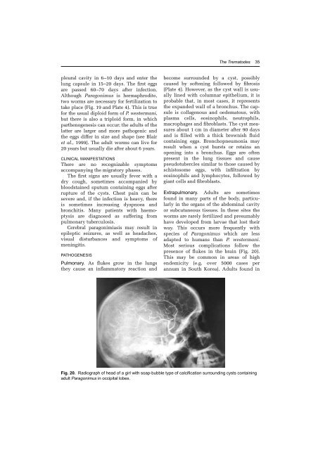
Most serious complications follow the<br />
presence of flukes in the brain (Fig. 20).<br />
This may be common in areas of high<br />
endemicity (e.g. over 5000 cases per<br />
annum in South Korea). Adults found in<br />
Fig. 20. Radiograph of head of a girl with soap-bubble type of calcification surrounding cysts containing<br />
adult Paragonimus in occipital lobes.