Effect of Glucantime on blood factors in patients with cutaneous ...
Effect of Glucantime on blood factors in patients with cutaneous ...
Effect of Glucantime on blood factors in patients with cutaneous ...
Create successful ePaper yourself
Turn your PDF publications into a flip-book with our unique Google optimized e-Paper software.
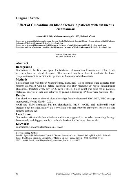
Orig<strong>in</strong>al Article<br />
<str<strong>on</strong>g>Effect</str<strong>on</strong>g> <str<strong>on</strong>g>of</str<strong>on</strong>g> <str<strong>on</strong>g>Glucantime</str<strong>on</strong>g> <strong>on</strong> <strong>blood</strong> <strong>factors</strong> <strong>in</strong> <strong>patients</strong> <strong>with</strong> <strong>cutaneous</strong><br />
leishmaniasis<br />
Ayatollahi.J 1 MD, Modares mosadegh.M 2 MD, Halvani.A 3 MD<br />
1-Associate pr<str<strong>on</strong>g>of</str<strong>on</strong>g>essor <str<strong>on</strong>g>of</str<strong>on</strong>g> <strong>in</strong>fectious and tropical diseases, Head <str<strong>on</strong>g>of</str<strong>on</strong>g> Infectious & Tropical Diseases Research Center, Shahid Sadoughi<br />
University <str<strong>on</strong>g>of</str<strong>on</strong>g> Medical Sciences and Health Services, Yazd, Iran<br />
2-Associate pr<str<strong>on</strong>g>of</str<strong>on</strong>g>essor <str<strong>on</strong>g>of</str<strong>on</strong>g> Pharmacology Shahid Sadoughi University <str<strong>on</strong>g>of</str<strong>on</strong>g> Medical Sciences and Health Services, Yazd, Iran<br />
3-Assistant pr<str<strong>on</strong>g>of</str<strong>on</strong>g>essor <str<strong>on</strong>g>of</str<strong>on</strong>g> pulm<strong>on</strong>ary Medic<strong>in</strong>e, Shahid Sadoughi University <str<strong>on</strong>g>of</str<strong>on</strong>g> Medical Sciences and Health Services, Yazd, Iran<br />
Received: 27 October 2010<br />
Accepted: 19 March 2011<br />
Abstract<br />
Background<br />
<str<strong>on</strong>g>Glucantime</str<strong>on</strong>g> is the first l<strong>in</strong>e agent for treatment <str<strong>on</strong>g>of</str<strong>on</strong>g> <strong>cutaneous</strong> leishmaniasis (CL). It has<br />
adverse effects <strong>on</strong> <strong>blood</strong> elements. This research has been d<strong>on</strong>e to evaluate the <strong>blood</strong><br />
complicati<strong>on</strong>s <str<strong>on</strong>g>of</str<strong>on</strong>g> this medic<strong>in</strong>e <strong>in</strong> <strong>patients</strong> <strong>with</strong> <strong>cutaneous</strong> leishmaniasis.<br />
Methods<br />
This cl<strong>in</strong>ical trial was d<strong>on</strong>e at Nikpour cl<strong>in</strong>ic, Yazd, Iran. Blood samples were collected from<br />
<strong>patients</strong> diagnosed <strong>with</strong> CL before treatment and after receiv<strong>in</strong>g 20 mg/kg <strong>in</strong>tramuscular<br />
glucantime. Injecti<strong>on</strong> every day for 20 days. Full cell <strong>blood</strong> count was d<strong>on</strong>e for all <strong>patients</strong>.<br />
Statistical analysis <str<strong>on</strong>g>of</str<strong>on</strong>g> data was achieved by paired-T-test us<strong>in</strong>g SPSS s<str<strong>on</strong>g>of</str<strong>on</strong>g>tware (versi<strong>on</strong> 13).<br />
Results<br />
The <strong>blood</strong> tests results showed glucantime significantly decreased RBC, PLT, WBC (except<br />
m<strong>on</strong>ocytes), Hb and Hct (P< 0.05).<br />
MCH and PMN decreased but not significantly. MCV, MCHC and eos<strong>in</strong>ophil count<br />
<strong>in</strong>creased but not significantly. No correlati<strong>on</strong> was seen between laboratory test results and<br />
<strong>patients</strong>' age and sex.<br />
C<strong>on</strong>clusi<strong>on</strong><br />
<str<strong>on</strong>g>Glucantime</str<strong>on</strong>g> affected the <strong>blood</strong> <strong>in</strong>dices and it was suggested to use other alternat<strong>in</strong>g therapy.<br />
Future study <strong>with</strong> bigger sample size should be d<strong>on</strong>e for the more clear results.<br />
Keywords<br />
<str<strong>on</strong>g>Glucantime</str<strong>on</strong>g>, Cutaneous leishmaniasis, Blood<br />
Corresp<strong>on</strong>d<strong>in</strong>g Author:<br />
Jamshid Ayatollahi, Infectious & Tropical Diseases Research Center, Shahid Sadoughi Hospital - Safaeieh<br />
Yazd –Iran,Shahid Sadoughi University <str<strong>on</strong>g>of</str<strong>on</strong>g> Medical Science, Yazd, Iran,Tel: 0351- 8224001-9,Tel:<br />
09133584891,Email: jamshidayatollahi@yahoo.com,Fax: 0351-8224100<br />
57<br />
Iranian Journal <str<strong>on</strong>g>of</str<strong>on</strong>g> Pediatric Hematology Oncology Vol1.No2.
Introducti<strong>on</strong><br />
Cutaneous leishmaniasis (CL) causes by Leishmania Tropica and is comm<strong>on</strong> <strong>in</strong> tropic and<br />
subtropic areas and <strong>in</strong> southern Europe (1). More than 90% <str<strong>on</strong>g>of</str<strong>on</strong>g> the world’s cases <str<strong>on</strong>g>of</str<strong>on</strong>g> <strong>cutaneous</strong><br />
leishmaniasis occur <strong>in</strong> Afghanistan, Algeria, Iran ,Iraq, Saudi Arabia, Syria, Brazil an Peru<br />
(2). In urban areas <str<strong>on</strong>g>of</str<strong>on</strong>g> Iran such as Mashhad, Neishapour, Shiraz, Kerman, Qom, Saveh,<br />
Isfahan, Kashan and Sabzevar, this disease is comm<strong>on</strong>. In some rural areas <str<strong>on</strong>g>of</str<strong>on</strong>g> Isfahan,<br />
Sarakhs, Torkaman Sahra, Lotfabad, Esphere<strong>in</strong>, Ahwaz, Desful, Shoosh, Soosangerd and<br />
Abadan also it is commom. Leishmania Tropica transmits to human by sand fly (3).<br />
Treatment <str<strong>on</strong>g>of</str<strong>on</strong>g> this disease is medical therapy, cryotherapy, heat therapy, surgery and<br />
cauterizati<strong>on</strong> (4-6). The drug <str<strong>on</strong>g>of</str<strong>on</strong>g> choice used <strong>in</strong> medical therapy are pentavalent antim<strong>on</strong>y<br />
compounds like pentostam and glucantime (7-8). These medic<strong>in</strong>es could cause not <strong>on</strong>ly<br />
changes <strong>in</strong> <strong>blood</strong> <strong>in</strong>dices also renal, hepatic, cardiac, gastro<strong>in</strong>test<strong>in</strong>al and pancreatic side<br />
effects (9). In order to <strong>in</strong>dicate the side effect <str<strong>on</strong>g>of</str<strong>on</strong>g> glucantime <strong>on</strong> <strong>blood</strong> <strong>in</strong>dexes, this study was<br />
c<strong>on</strong>ducted <strong>in</strong> <strong>patients</strong> referred to Nikpour cl<strong>in</strong>ic.<br />
Materials and Methods<br />
This cl<strong>in</strong>ical trial was d<strong>on</strong>e at Nikpour cl<strong>in</strong>ic, Yazd, Iran. Blood samples were collected from<br />
<strong>patients</strong> diagnosed <strong>with</strong> CL before and after treatment <strong>in</strong> order to compare any <strong>blood</strong> <strong>in</strong>dices<br />
changes. F<strong>in</strong>al diagnosis <str<strong>on</strong>g>of</str<strong>on</strong>g> Cl was d<strong>on</strong>e by histopathological f<strong>in</strong>d<strong>in</strong>g <str<strong>on</strong>g>of</str<strong>on</strong>g> the ulcer. Blood<br />
samples were collected from <strong>patients</strong> before treatment and each patient received 20 mg/kg<br />
<strong>in</strong>tramuscular <strong>in</strong>jecti<strong>on</strong> <str<strong>on</strong>g>of</str<strong>on</strong>g> the glucantime every day for 20 days. On the last day <str<strong>on</strong>g>of</str<strong>on</strong>g> therapy,<br />
<strong>blood</strong> test was d<strong>on</strong>e aga<strong>in</strong> and the result was compare <strong>with</strong> result <str<strong>on</strong>g>of</str<strong>on</strong>g> the test before treatment.<br />
The variables c<strong>on</strong>sidered <strong>in</strong> this research were age, sex and <strong>blood</strong> <strong>in</strong>dices. The exclusi<strong>on</strong><br />
criteria were <strong>patients</strong> <strong>with</strong> diabetes mellitus, other <strong>in</strong>fectious diseases, corticosteroid therapy<br />
and other known hematological diseases. Cell <strong>blood</strong> count and cell differentiati<strong>on</strong> were d<strong>on</strong>e<br />
before and after treatment for all <strong>patients</strong>, and they were compared by paired t-test.<br />
Results<br />
Out <str<strong>on</strong>g>of</str<strong>on</strong>g> 30 <strong>patients</strong>, 18(60%) were male and 12(40%) female <strong>with</strong> an the mean age <str<strong>on</strong>g>of</str<strong>on</strong>g> 26.72<br />
years old (9 to 42.5 years). The results <str<strong>on</strong>g>of</str<strong>on</strong>g> present study <strong>in</strong>dicated that the number <str<strong>on</strong>g>of</str<strong>on</strong>g> RBC,<br />
PLT, WBC, M<strong>on</strong>ocytes and PMN decreased. Hb, Hct and MCH also decreased. However,<br />
MCV and MCHC <strong>in</strong>creased and the number <str<strong>on</strong>g>of</str<strong>on</strong>g> Lymphocyte and eos<strong>in</strong>ophils also <strong>in</strong>creased<br />
.The mean changes <str<strong>on</strong>g>of</str<strong>on</strong>g> <strong>in</strong>dices for RBC, PLT, WBC, m<strong>on</strong>ocyte, HB, and HCT were<br />
significantly decreased (PV
Table 1: Changes <str<strong>on</strong>g>of</str<strong>on</strong>g> <strong>blood</strong> <strong>in</strong>dices after treatment <strong>with</strong> <str<strong>on</strong>g>Glucantime</str<strong>on</strong>g><br />
Stage<br />
Before<br />
treatment<br />
After treatment<br />
Changes<br />
Blood <strong>factors</strong><br />
SD<br />
X<br />
SD<br />
X<br />
CI95%<br />
Median<br />
P.value<br />
RBC10 6 /µl<br />
0.6<br />
5.1<br />
0.5<br />
4.9<br />
-0.13-0.24<br />
- 0.19<br />
0.000<br />
PLT10³/µl<br />
WBC10³/µl<br />
MONO%<br />
POLY %<br />
Hct%<br />
Hb g/dl<br />
MCH /pg<br />
MCHCg/dl<br />
MCV / fl<br />
EOS %<br />
LYMPH %<br />
33.1<br />
1.1<br />
1.1<br />
4.2<br />
4.9<br />
2.1<br />
1.8<br />
1.3<br />
7.6<br />
1.5<br />
4.9<br />
230.4<br />
7.3<br />
5.1<br />
59.2<br />
41.4<br />
14.65<br />
28.8<br />
33.2<br />
82<br />
3<br />
32.2<br />
33.4<br />
0.8<br />
1.5<br />
4.6<br />
2.5<br />
2<br />
1.7<br />
1.3<br />
4.7<br />
1.5<br />
3.9<br />
222<br />
6.5<br />
4.4<br />
57<br />
40.08<br />
13.99<br />
28.7<br />
33.2<br />
84<br />
3.1<br />
34.9<br />
-6.3- -10.4<br />
0.61-0.98<br />
0.49-0.97<br />
0.05- 0.71<br />
0.3-2.3<br />
0.53-0.78<br />
0.11-0.21<br />
0.23-0.06<br />
4.2-0.22<br />
0.4-0/13<br />
3.4-1.9<br />
- 8.4<br />
- 8.4<br />
-0.8<br />
- 0.33<br />
- 1.3<br />
- 0.6<br />
- 0.05<br />
- 0.08<br />
2<br />
0.1<br />
2.6<br />
0.000<br />
0.000<br />
0.000<br />
0.086<br />
0.013<br />
0.000<br />
0.528<br />
0.274<br />
0.076<br />
0.326<br />
0.000<br />
Table 2: Comparis<strong>on</strong> changes <str<strong>on</strong>g>of</str<strong>on</strong>g> <strong>blood</strong> <strong>in</strong>dices <strong>in</strong> two age groups<br />
Age group<br />
Blood <strong>in</strong>dices<br />
N=17 > 25 years<br />
SD M<br />
N=13 ≤ 25 years<br />
SD M<br />
P.value<br />
PLT10³/µl<br />
WBC10³/µl<br />
MONO%<br />
POLY %<br />
Hct%<br />
Hb g/dl<br />
MCH /pg<br />
MCV / fl<br />
MCHC g/dl<br />
EOS %<br />
LYMPH %<br />
4.8<br />
0.51<br />
- 0.77<br />
1.1<br />
2.7<br />
0.16<br />
0.53<br />
6.5<br />
0.48<br />
0.69<br />
1.3<br />
-8.7<br />
-0.76<br />
0.70<br />
- 0.29<br />
-1.04<br />
0.54<br />
-0.13<br />
2.9<br />
0.11<br />
0.11<br />
2.7<br />
6.3<br />
0.49<br />
0.42<br />
0.96<br />
2.8<br />
0.42<br />
0.19<br />
5.9<br />
0.28<br />
0.8<br />
2.3<br />
-8<br />
0.84-<br />
0.76<br />
0.38<br />
-1.6<br />
-o.8<br />
0.06<br />
0.81<br />
0.04<br />
0.15<br />
2.6<br />
0.713<br />
0.667<br />
0.793<br />
0.816<br />
0.553<br />
0.053<br />
0.177<br />
0.350<br />
0.651<br />
0.896<br />
0.896<br />
59<br />
Iranian Journal <str<strong>on</strong>g>of</str<strong>on</strong>g> Pediatric Hematology Oncology Vol1.No2.
Table 3: Comparis<strong>on</strong> changes <str<strong>on</strong>g>of</str<strong>on</strong>g> <strong>blood</strong> <strong>in</strong>dices <strong>in</strong> two sex group<br />
Sex<br />
N=18 male<br />
N=12 female<br />
Blood <strong>in</strong>dices<br />
SD<br />
M<br />
SD<br />
M<br />
P.value<br />
Diff-PLT10³/µl<br />
WBC10³/µl<br />
MONO%<br />
POLY %<br />
Hct%<br />
Hb g/dl<br />
MCH /pg<br />
MCV / fl<br />
MCHC g/dl<br />
EOS %<br />
LYMPH %<br />
6.1<br />
0.6<br />
0.59<br />
1.02<br />
2.8<br />
0.11<br />
0.45<br />
5.6<br />
0.32<br />
0.73<br />
1.1<br />
-8.8<br />
-0.86<br />
0.66<br />
- 0.11<br />
-1.01<br />
0.6<br />
-0.11<br />
1.6<br />
0.005<br />
0.22<br />
3<br />
4.4<br />
0.26<br />
0.71<br />
0.98<br />
2.5<br />
0.49<br />
0.38<br />
6.6<br />
0.5<br />
0.73<br />
2.5<br />
-7.8<br />
0.7-<br />
0.8<br />
0.66<br />
-1.8<br />
-o.74<br />
0.04<br />
2.5<br />
0.2<br />
0.00<br />
2.1<br />
0.631<br />
0.315<br />
0.494<br />
0.150<br />
0.451<br />
0.371<br />
0.348<br />
0.713<br />
0.256<br />
0.434<br />
0.227<br />
Discussi<strong>on</strong><br />
The relati<strong>on</strong> between age, sex and the changes <str<strong>on</strong>g>of</str<strong>on</strong>g> <strong>blood</strong> <strong>in</strong>dices were not statistically<br />
significant. In 1997, <strong>on</strong>e study by Talari <strong>in</strong> Kashan, Iran showed that <strong>in</strong> WBC, RBC, PLT,<br />
m<strong>on</strong>ocyte, Hb, Hct and neutrophile decreased (6). Variables that <strong>in</strong>creased were ESR, MCV,<br />
MCH, MCHC, and eos<strong>in</strong>ophil and basophil, lymphocyte.<br />
The decrease <strong>in</strong> Hb and Hct <strong>in</strong> this study compared to prior studies was statistically<br />
significant. Unlike the previous study, a decrease <strong>in</strong> PMN, and MCH was seen, yet both<br />
studies were unremarkable. In 1992, Alkhawaya and et al <strong>in</strong> Saudi Arabia (7) c<strong>on</strong>ducted a<br />
similar study <strong>in</strong> laboratory mice and they found out that the decrease <str<strong>on</strong>g>of</str<strong>on</strong>g> Hb ant Hct, yet there<br />
were no data show<strong>in</strong>g changes <strong>with</strong> <strong>in</strong> MCH, MCHC and RBC (7) .A year later, Soto et al. <strong>in</strong><br />
Spa<strong>in</strong> and <strong>in</strong> 1992 Berman et al. <strong>in</strong> England studies the side effect <str<strong>on</strong>g>of</str<strong>on</strong>g> the <str<strong>on</strong>g>Glucantime</str<strong>on</strong>g> <strong>on</strong><br />
leukocyte <str<strong>on</strong>g>of</str<strong>on</strong>g> the <strong>patients</strong> diagnosed <strong>cutaneous</strong> leishmaniasis. The result from these studies<br />
showed a decrease <strong>in</strong> some <strong>blood</strong> elements (10, 11). Mechanism <str<strong>on</strong>g>of</str<strong>on</strong>g> acti<strong>on</strong> <str<strong>on</strong>g>of</str<strong>on</strong>g> glucantime is<br />
<strong>in</strong>hibiti<strong>on</strong> <str<strong>on</strong>g>of</str<strong>on</strong>g> phosph<str<strong>on</strong>g>of</str<strong>on</strong>g>ructok<strong>in</strong>ase enzyme which has specific role <strong>in</strong> glycolytic activities <strong>in</strong><br />
neutrophils which stays active temperature <str<strong>on</strong>g>of</str<strong>on</strong>g> 37c (36+2). Blockage <str<strong>on</strong>g>of</str<strong>on</strong>g> this enzyme can result<br />
<strong>in</strong> decrease <str<strong>on</strong>g>of</str<strong>on</strong>g> ATP producti<strong>on</strong>, which lowers the WBC half-life and decrease the number<br />
WBC <strong>in</strong> the period <str<strong>on</strong>g>of</str<strong>on</strong>g> treatment <strong>with</strong> <str<strong>on</strong>g>Glucantime</str<strong>on</strong>g> (12). In additi<strong>on</strong>, the ma<strong>in</strong> cause <str<strong>on</strong>g>of</str<strong>on</strong>g><br />
leukocyte decrease can be due to the c<strong>on</strong>current decrease <str<strong>on</strong>g>of</str<strong>on</strong>g> m<strong>on</strong>ocyte <strong>with</strong><strong>in</strong> treatment.<br />
S<strong>in</strong>ce the role <str<strong>on</strong>g>of</str<strong>on</strong>g> m<strong>on</strong>ocyte plays a c<strong>on</strong>troll<strong>in</strong>g aspect <strong>with</strong><strong>in</strong> this disease, it is predictable that<br />
there will be an <strong>in</strong>crease prior to treatment and decrease after treatment. RBC decrease seems<br />
to be because <str<strong>on</strong>g>of</str<strong>on</strong>g> the phosph<str<strong>on</strong>g>of</str<strong>on</strong>g>ructok<strong>in</strong>ase <strong>in</strong>hibiti<strong>on</strong> result<strong>in</strong>g glycolytic metabolic disorder as<br />
a ATP ma<strong>in</strong> source <str<strong>on</strong>g>of</str<strong>on</strong>g> energy. The hereditary deficiency <str<strong>on</strong>g>of</str<strong>on</strong>g> this enzyme can cause hereditary<br />
n<strong>on</strong> spherocytic hemolytic anemia(6).<br />
In the study c<strong>on</strong>ducted <strong>in</strong> Kashan-Iran, the reas<strong>on</strong> why the was not an decrease <strong>in</strong> Hb and Hct<br />
can be due to the other mechanism such as RBC releas<strong>in</strong>g as reticulocytes(high MCH &<br />
MCV).However, our study showed a decrease <strong>in</strong> Hb & Hct, which was scientifically<br />
significant <strong>with</strong>out any reas<strong>on</strong>able mechanism . In the Kashan study, the <strong>in</strong>crease <strong>in</strong> ESR<br />
was not high enough to make a significant different <strong>in</strong> the f<strong>in</strong>al results <str<strong>on</strong>g>of</str<strong>on</strong>g> the study, and <strong>in</strong> the<br />
case <str<strong>on</strong>g>of</str<strong>on</strong>g> lymphocytes, there was an <strong>in</strong>crease but not as much where we can categorize it as<br />
lymphocytosis, where as <strong>in</strong> our study, lymphocyte <strong>in</strong>crease where at the level that it was<br />
significant <strong>with</strong> a unknown reas<strong>on</strong>.<br />
Iranian Journal <str<strong>on</strong>g>of</str<strong>on</strong>g> Pediatric Hematology Oncology Vol1.No2. 60
C<strong>on</strong>clusi<strong>on</strong><br />
Although we <strong>in</strong>cluded a limited number <str<strong>on</strong>g>of</str<strong>on</strong>g> samples <strong>in</strong> our study, it can be said that<br />
glucantime can effect the <strong>blood</strong> <strong>in</strong>dices dur<strong>in</strong>g the 20 days <str<strong>on</strong>g>of</str<strong>on</strong>g> therapeutic period and our<br />
recommendati<strong>on</strong> would be us<strong>in</strong>g different approaches such as cryotherpy, heat therapy and<br />
other alternat<strong>in</strong>g drugs for the <strong>patients</strong> <strong>with</strong> hemotologic underl<strong>in</strong>e disease.<br />
References<br />
1- Goto H, L<strong>in</strong>doso JA. Current diagnosis and treatment <str<strong>on</strong>g>of</str<strong>on</strong>g> <strong>cutaneous</strong> and muco<strong>cutaneous</strong> leishmaniasis.<br />
Expert Rev Anti Infect Ther. 2010 Apr; 8(4):419-33.<br />
2- David CV, Craft N. Cutaneous and muco<strong>cutaneous</strong> leishmaniasis. Dermatol Ther. 2009; 22(6):491-502.<br />
3- Lane RP. Phlebotom<strong>in</strong>e sandflies. In: Mans<strong>on</strong> P, Cook GC, Zumla A, eds. Mans<strong>on</strong>’s Tropical diseases. 21st<br />
ed. L<strong>on</strong>d<strong>on</strong>: Saunders. 2003: 1733-41.<br />
4- Alvajhi A.A. Cutaneous leishmaniasis <str<strong>on</strong>g>of</str<strong>on</strong>g> the Old World. Sk<strong>in</strong> Therapy. 2003; 8(2):1-4.<br />
5- Berman JD. Human Leishmaniasis: cl<strong>in</strong>ical, diagnostic, and chemotherapeutic developments <strong>in</strong> the last 10<br />
years. Cl<strong>in</strong> Infect Dis. 1997; 24: 684-703.<br />
6- Talari S. Toxicity <str<strong>on</strong>g>of</str<strong>on</strong>g> glucantime <strong>in</strong> <strong>blood</strong> element, <strong>in</strong> <strong>cutaneous</strong> leishmaniasis, Faze – Kashan university.<br />
1996; 10:18-21.<br />
7- Ar<strong>on</strong>s<strong>on</strong> NE, Wortmann GW, Johns<strong>on</strong> SC, Jacks<strong>on</strong> JE, Gasser RA Jr, Magill AJ, et al. Safety and efficacy<br />
<str<strong>on</strong>g>of</str<strong>on</strong>g> <strong>in</strong>travenous sodium stibogluc<strong>on</strong>ate <strong>in</strong> the treatment <str<strong>on</strong>g>of</str<strong>on</strong>g> leishmaniasis: recent U.S. military experience.<br />
Cl<strong>in</strong> <strong>in</strong>fect Dis. 1998; 1457-64.<br />
8- Jer<strong>on</strong>imo SMB, Sousa AQ ,Pears<strong>on</strong> RD. Leishmania Species. In : Mandell GL ,Douglas RG ,Bennet JE<br />
.Pr<strong>in</strong>ciples and Practice <str<strong>on</strong>g>of</str<strong>on</strong>g> <strong>in</strong>fectious Diseases USA ,Churchill Liv<strong>in</strong>gst<strong>on</strong>e. 2005;3145-3156.<br />
9- Franke ED, Wignall FS, Cruz ME, Rosales E, Tovar AA, Lucas CM, et al. Efficacy and toxicity <str<strong>on</strong>g>of</str<strong>on</strong>g> sodium<br />
stibogluc<strong>on</strong>ate for mucosal leishmaniasis. Ann Intern Med. 1990; 113(12):934-40.<br />
10- Soto J, Valda-Rodriquez L, Toledo J, Vera-Navarro L, Luz M, M<strong>on</strong>asterios-Torrico H, etal. Comparis<strong>on</strong> <str<strong>on</strong>g>of</str<strong>on</strong>g><br />
generic to branded pentavalent antim<strong>on</strong>y for treatment <str<strong>on</strong>g>of</str<strong>on</strong>g> new world <strong>cutaneous</strong> leishmaniasis. Am J Trop<br />
Med Hyg. 2004 Nov;71(5):577-81. Erratum <strong>in</strong>: Am J Trop Med Hyg. 2005; 72(3):359.<br />
11- Berman JD. Chemotherapy for leishmaniasis: biochemical mechanisms, cl<strong>in</strong>ical efficacy, and future<br />
strategies. Rev Infect Dis. 1988; 10(3): 560-86.<br />
12- Sharma S, Malhan P, Pujani M, Rath B. Acute erythroid toxicity <strong>in</strong> visceral leishmaniasis: a rare<br />
complicati<strong>on</strong> <str<strong>on</strong>g>of</str<strong>on</strong>g> antim<strong>on</strong>ial therapy. Indian J Pathol Microbiol. 2008 Oct-Dec;51(4):546-7.<br />
61<br />
Iranian Journal <str<strong>on</strong>g>of</str<strong>on</strong>g> Pediatric Hematology Oncology Vol1.No2.


