Diseases of Echinodermata. I. Agents ... - Inter Research
Diseases of Echinodermata. I. Agents ... - Inter Research
Diseases of Echinodermata. I. Agents ... - Inter Research

You also want an ePaper? Increase the reach of your titles
YUMPU automatically turns print PDFs into web optimized ePapers that Google loves.
150 Dis. aquat. Org. 2: 147-162, 1987<br />
reaction around the area concerned. There is a massive<br />
migration <strong>of</strong> coelomocytes, i.e, phagocytic cells and<br />
red spherule cells, around and within the affected area<br />
(Johnson 1971, Maes & Jangoux 1984, Gilles & Pearse<br />
1986, Maes et al. 1986).<br />
Bald-sea-urchin disease is communicable. Pieces <strong>of</strong><br />
necrotic tissues initiate the disease when painted on<br />
experimentally produced injuries <strong>of</strong> the outer body<br />
surface <strong>of</strong> healthy echinoids (Maes & Jangoux 1984,<br />
Gilles & Pearse 1986). Maes & Jangoux also showed<br />
that the disease- is not species-specific: it is easy to<br />
experimentally infect regular echinoids <strong>of</strong> several<br />
different species.<br />
The causative agent <strong>of</strong> bald-sea-urchin disease is <strong>of</strong><br />
bacterial nature (Maes & Jangoux 1985, Gilles &<br />
Pearse 1986). Gilles & Pearse isolated 14 different<br />
bacterial strains from lesions <strong>of</strong> diseased Strongylocen-<br />
trotus purpuratus. They demonstrated that only the<br />
isolates <strong>of</strong> Vibno anguillarum and Aeromonas sal-<br />
monicida - 2 well-known pathogenic marine bacteria<br />
- were able to initiate lesion formation in the labora-<br />
tory. Both Maes & Jangoux (1984) and Gilles & Pearse<br />
(1986) concluded from experimental infectivity tests<br />
that a stress such as physical injury is necessary for the<br />
formation <strong>of</strong> characteristic lesions. As suggested by<br />
Maes et al. (1986), lesions may possibly be caused by<br />
bacteria that are resistant to the antibacterial substan-<br />
ces naturally produced by the echinoids, namely the<br />
naphthoquinone pigments conveyed by the red<br />
spherule cells.<br />
Several authors reported mass mortalities <strong>of</strong><br />
echinoids, presumably due to bald-sea-urchin disease.<br />
Mass mortalities affected 60 to 95 % <strong>of</strong> Strongylocen-<br />
trotus franciscanus and 10 to 75 % <strong>of</strong> Paracentrotus<br />
lividus (Pearse et al. 1977, Boudouresque et al. 1980,<br />
1981, respectively). Azzolina (1983) noted that mortal-<br />
ity <strong>of</strong> P. Lividus is hlgher in shallow waters, and that<br />
diseased individuals are more numerous during sum-<br />
mer. However, as noted by Gilles & Pearse (1986),<br />
there are also reports <strong>of</strong> lesions on occasional individu-<br />
als in populations <strong>of</strong> otherwise healthy echinoids (e.g.<br />
Pearse et al. 1977, Maes & Jangoux 1984).<br />
Bacteria do not seem to be the causative agents <strong>of</strong><br />
the disease causing mass mortalities <strong>of</strong> Strongylocen-<br />
trotus droebachiensis along the Atlantic coast <strong>of</strong><br />
Canada (e.g. Miller & Colodey 1983). Although charac-<br />
teristic bacterial lesions do sometimes occur on these<br />
echinoids (Scheibling & Stephenson 1984), the bac-<br />
teria could have been a secondary factor associated<br />
with the mass mortality as suggested by Gilles &<br />
Pearse (1986). In such a case it may be supposed that<br />
bacteria developed on echinoids previously infested<br />
by a not yet identified pathogen that caused lysis <strong>of</strong><br />
tube feet and loss <strong>of</strong> spines (see Jones et al. 1985b) (see<br />
p. 159).<br />
Scattered information on other types <strong>of</strong> bacterial<br />
infection can be found in the literature. Olmsted (1917)<br />
noticed several small dark brown-green spherical<br />
masses <strong>of</strong> up to 1.5 mm in diameter in the body cavity<br />
<strong>of</strong> almost all individuals <strong>of</strong> the holothuroid Synaptula<br />
hydriformis examined. These masses consisted <strong>of</strong> 'bac-<br />
terial parasites' belonging to the genus Mycrocystus.<br />
Mortensen (1935) observed granular epidermal swel-<br />
lings on the echinoid Calvenosoma gracile. According<br />
to him these swellings were very probably <strong>of</strong> bacterial<br />
nature. Delavault & Leclerc (1969) briefly reported a<br />
bacterial disease affecting Astenna gibbosa under<br />
aquarium conditions. Diseased asteroids show patches<br />
<strong>of</strong> epidermal necrosis which progressively unite and<br />
finally cause death.<br />
<strong>Agents</strong>: Fungi<br />
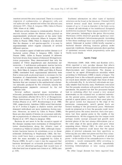
Mortensen (1909, 1928, 1936) and Koehler (1911,<br />
1912) reported a very peculiar disease that affects<br />
several species <strong>of</strong> Antarctic cidaroid echinoids (genera<br />
Rhynchocidans and Ctenocidaris). The disease is<br />
caused by an agent (Echinophyces mirabilis) which<br />
according to Mortensen (1909) is likely a fungus. The<br />
pathogen lives in the echinoid's primary spines which<br />
are much more slender and fragile than those <strong>of</strong> heal-<br />
thy echinoids (Fig. 3). As infected individuals are typi-<br />
cally smaller than healthy ones, Mortensen concluded<br />
that the parasite interferes with growth and dwarfs the<br />
specimens. He pointed out that the presumed fungus<br />
had no castrating effect but caused a very unusual<br />
abnormality on the echinoid test: in infected individu-<br />
als the genital pores, presumably together with the<br />
genital or apical plates, are not in their usual place in<br />
the apical system but displaced to the edge <strong>of</strong> the<br />
peristome; consequently, new(?) genital ducts are<br />
formed leading to the peristomeal pores. Mortensen<br />
made no proposal as to how the parasite could 'move'<br />
the genital pores. He noted, however, that a few speci-<br />
mens had their genital pores in the middle <strong>of</strong> the<br />
interambulacra. All this suggests that the fungus could<br />
perhaps modify normal echinoid test growth (new<br />
skeletal plates <strong>of</strong> the test always differentiate just at<br />
the outer edge <strong>of</strong> the apical plates and grow when<br />
migrating down; Markel 1981). In other words, the<br />
abnormalities observed could mean that the apical<br />
plates <strong>of</strong> infected echinoids lose their specificlty in<br />
behaving like any other test plates and that they conse-<br />
quently migrate downwards. The position <strong>of</strong> genital<br />
pores - in the middle <strong>of</strong> the interambulacra or near the<br />
peristome - would consequently differ according to<br />
whether the echinoid was infected immediately<br />
following metamorphosis or later during juvenile<br />
growth.<br />
No other fungal disease has been reported from