NEOSPORA CANINUM ELISA KIT
NEOSPORA CANINUM ELISA KIT
NEOSPORA CANINUM ELISA KIT

Create successful ePaper yourself
Turn your PDF publications into a flip-book with our unique Google optimized e-Paper software.
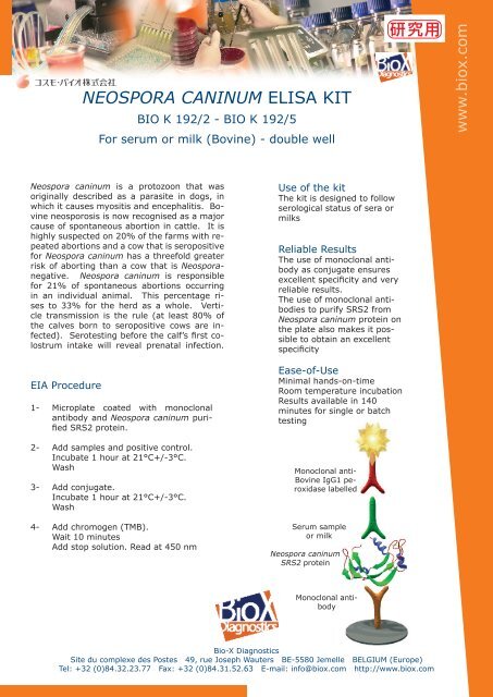
<strong>NEOSPORA</strong> <strong>CANINUM</strong> <strong>ELISA</strong> <strong>KIT</strong><br />
BIO K 192/2 - BIO K 192/5<br />
For serum or milk (Bovine) - double well<br />
Neospora caninum is a protozoon that was<br />
originally described as a parasite in dogs, in<br />
which it causes myositis and encephalitis. Bovine<br />
neosporosis is now recognised as a major<br />
cause of spontaneous abortion in cattle. It is<br />
highly suspected on 20% of the farms with repeated<br />
abortions and a cow that is seropositive<br />
for Neospora caninum has a threefold greater<br />
risk of aborting than a cow that is Neosporanegative.<br />
Neospora caninum is responsible<br />
for 21% of spontaneous abortions occurring<br />
in an individual animal. This percentage rises<br />
to 33% for the herd as a whole. Verticle<br />
transmission is the rule (at least 80% of<br />
the calves born to seropositive cows are infected).<br />
Serotesting before the calf’s first colostrum<br />
intake will reveal prenatal infection.<br />
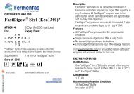
EIA Procedure<br />
1- Microplate coated with monoclonal<br />
antibody and Neospora caninum purified<br />
SRS2 protein.<br />
2- Add samples and positive control.<br />
Incubate 1 hour at 21°C+/-3°C.<br />
Wash<br />
3- Add conjugate.<br />
Incubate 1 hour at 21°C+/-3°C.<br />
Wash<br />
4- Add chromogen (TMB).<br />
Wait 10 minutes<br />
Add stop solution. Read at 450 nm<br />
Use of the kit<br />
The kit is designed to follow<br />
serological status of sera or<br />
milks<br />
Reliable Results<br />
The use of monoclonal antibody<br />
as conjugate ensures<br />
excellent specificity and very<br />
reliable results.<br />
The use of monoclonal antibodies<br />
to purify SRS2 from<br />
Neospora caninum protein on<br />
the plate also makes it possible<br />
to obtain an excellent<br />
specificity<br />
Ease-of-Use<br />
Minimal hands-on-time<br />
Room temperature incubation<br />
Results available in 140<br />
minutes for single or batch<br />
testing<br />
Monoclonal anti-<br />
Bovine IgG1 peroxidase<br />
labelled<br />
Serum sample<br />
or milk<br />
Neospora caninum<br />
SRS2 protein<br />
Monoclonal antibody<br />
Bio-X Diagnostics<br />
Site du complexe des Postes 49, rue Joseph Wauters BE-5580 Jemelle BELGIUM (Europe)<br />
Tel: +32 (0)84.32.23.77 Fax: +32 (0)84.31.52.63 E-mail: info@biox.com http://www.biox.com<br />
www.biox.com
BIO K 192<br />
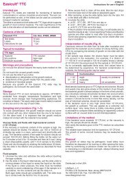
Kit performance<br />
The performance of the BIO K 192 kit (immunocapture test) was compared with BIO K 218<br />
<strong>ELISA</strong> kit (blocking test) test on 270 blood serum samples. The results of these comparisons<br />
are shown in Graph 1. 1 corresponds to the value obtained with the kit’s positive<br />
reference serum (S/P).<br />
BIO K 218 (Blocking test)<br />
(Pourcentage of inhibition)<br />
100<br />
90<br />
80<br />
70<br />
60<br />
50<br />
40<br />
30<br />
20<br />
10<br />
Threshold<br />
Graph 1<br />
0<br />
-0,1 0,1 0,3 0,5 0,7 0,9 1,1 1,3 1,5 1,7 1,9 2,1<br />
BIO K 192 (Immunocapture test)<br />
BIO K 218<br />
- +<br />
- 236 5 241<br />
+ 4 25 29<br />
240 30 270<br />
Concordance between the two tests: Kappa = 0.83<br />
The concordance between the two tests is considered excellent.<br />
Landis et Koch, The measurement of observer agreement for categorical data<br />
Biometrics 1977, 33, 159-74<br />
Threshold<br />
Bio-X Diagnostics<br />
Site du complexe des Postes 49, rue Joseph Wauters BE-5580 Jemelle BELGIUM (Europe)<br />
Tel: +32 (0)84.32.23.77 Fax: +32 (0)84.31.52.63 E-mail: info@biox.com http://www.biox.com<br />
S/P<br />
www.biox.com
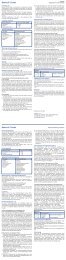
269 serum and 269 milk samples taken from the same animals were tested using the BIO K 192 kit.<br />
These samples came from twenty-seven Belgian farms. Their optical density readings were divided by<br />
the optimal density reading for the kit’s reference serum (S/P). Frequency histograms were then plotted<br />
for the blood sera (Graph 2) and milk samples (Graph 3).<br />
Frequency<br />
frequence<br />
Frequency<br />
frequency<br />
250<br />
200<br />
150<br />
100<br />
250<br />
200<br />
150<br />
100<br />
50<br />
50<br />
0<br />
0<br />
Serum serums<br />
-0,2 0 0,2 0,4 0,6 0,8 1 1,2 1,4 1,6 1,8 2<br />
Classes<br />
classes<br />
Milk Milks<br />
Graph 2<br />
Graph 3<br />
0 0,2 0,4 0,6 0,8 1 1,2 1,4 1,6 1,8 2<br />
Classes classes<br />
Bio-X Diagnostics<br />
Site du complexe des Postes 49, rue Joseph Wauters BE-5580 Jemelle BELGIUM (Europe)<br />
Tel: +32 (0)84.32.23.77 Fax: +32 (0)84.31.52.63 E-mail: info@biox.com http://www.biox.com<br />
www.biox.com
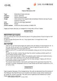
The frequency histograms generated for the 269 milk and 269 blood serum samples reveal that the thresholds<br />
must be set preferentially at 20% of the kit’s reference serum’s signal. Graph 4 shows the correlation<br />
between the serological findings yielded by the blood sera and those yielded by the milk samples.<br />
Milk milk<br />
2,2<br />
2,0<br />
1,8<br />
1,6<br />
1,4<br />
1,2<br />
1,0<br />
0,8<br />
0,6<br />
0,4<br />
0,2<br />
-0,2<br />
E/P<br />
Threshold<br />
Neospora (IC)<br />
y = 0,9226x - 0,0025<br />
R 2 y = 0,9226 - 0,0025<br />
R² = 0,8895<br />
= 0,8895<br />
0,0<br />
-0,2 0,0 0,2 0,4 0,6 0,8 1,0 1,2 1,4 1,6 1,8 2,0 2,2<br />
Sera<br />
Serum serum<br />
Milks<br />
- +<br />
- 239 1 240<br />
+ 6 23 29<br />
245 24 269<br />
Graph 4<br />
Threshold<br />
Concordance between the two tests: Kappa = 0.85<br />
The concordance between the two tests is considered excellent.<br />
Landis et Koch, The measurement of observer agreement for categorical data<br />
Biometrics 1977, 33, 159-74<br />
Bio-X Diagnostics<br />
Site du complexe des Postes 49, rue Joseph Wauters BE-5580 Jemelle BELGIUM (Europe)<br />
Tel: +32 (0)84.32.23.77 Fax: +32 (0)84.31.52.63 E-mail: info@biox.com http://www.biox.com<br />
E/P<br />
www.biox.com
Composition of the kit<br />
BIO-X <strong>NEOSPORA</strong> <strong>CANINUM</strong> <strong>ELISA</strong> <strong>KIT</strong> : BIO K 192<br />
BIO K 192/2 BIO K 192/5<br />
Microplates 2 (96 tests) 5 (240 tests)<br />
Washing solution 1 X 100 ml (20 X) 1 X 250 ml (20 X)<br />
Dilution buffer 1 X 50 ml (5 X) 1 X 100 ml (5 X)<br />
Conjugate 1 X 0.5 ml (50 X) 1 X 1.4 ml (50 X)<br />
Positive serum 1 X 0.5 ml (1 X) 1 X 0.5 ml (1 X)<br />
Single component TMB 1 X 25 ml (1 X) 1 X 55 ml (1 X)<br />
Stopping solution 1 X 15 ml (1 X) 1 X 30 ml (1 X)<br />
One year stability between +2°C and +8°C<br />
Bibliography<br />
Anti-Neospora caninum antibodies in milk in relation to production losses in dairy cattle.<br />
González-Warleta M, Castro-Hermida JA, Carro-Corral C, Mezo M.<br />
Prev Vet Med. 2011 Aug 1;101(1-2):58-64. Epub 2011 Jun 6.<br />
Bio-X Diagnostics<br />
Site du complexe des Postes 49, rue Joseph Wauters BE-5580 Jemelle BELGIUM (Europe)<br />
Tel: +32 (0)84.32.23.77 Fax: +32 (0)84.31.52.63 E-mail: info@biox.com http://www.biox.com<br />
www.biox.com
I - INTRODUCTION<br />
<strong>NEOSPORA</strong> <strong>CANINUM</strong> <strong>ELISA</strong> <strong>KIT</strong><br />
<strong>ELISA</strong> kit for serodiagnosis of Neospora caninum<br />
Indirect test for blood sera and milk<br />
Diagnostic test for cattle<br />
Neospora caninum is a protozoon that was originally described as a parasite in dogs, in which it causes myositis<br />
and encephalitis. Bovine neosporosis is now recognised as a major cause of spontaneous abortion in cattle. It is<br />
highly suspected on 20% of the farms with repeated abortions and a cow that is seropositive for Neospora<br />
caninum has a threefold greater risk of aborting than a cow that is Neospora-negative. Neospora is responsible<br />
for 21% of spontaneous abortions occurring in an individual animal. This percentage rises to 33% for the herd<br />
as a whole. Verticle transmission is the rule (at least 80% of the calves born to seropositive cows are infected).<br />
Serotesting before the calf’s first colostrum intake will reveal prenatal infection.<br />
II – PRINCIPLE OF THE TEST<br />
The test uses 96-well microtitration plates sensitised by a purified Neospora caninum protein. The plate's odd<br />
columns (1, 3, 5, 7, 9 and 11) contain the purified protein, whereas the even columns (2, 4, 6, 8, 10 and 12)<br />
contain a control antigen. We thus have a genuine negative control. Using such a control reduces the number of<br />
false positives considerably.<br />
The test blood sera or milks are diluted in the buffer for dilution. The plate is incubated and washed, then the<br />
conjugate, a peroxidase-labelled anti-bovine IgG1 monoclonal antibody, is added to the wells. The plate is then<br />
incubated a second time at 21°C +/- 3°C washed again and the chromogen tetramethylbenzidine (TMB) is added.<br />
This chromogen has the advantages of being more sensitive than the other peroxidase chromogens and not being<br />
carcinogenic. If specific anti-Neospora caninum immunoglobulins are present in the test sera or milks the<br />
conjugate remains bound to the microwell that contains the protozoon and the enzyme catalyses the<br />
transformation of the colorless chromogen into a pigmented compound. The intensity of the resulting blue colour<br />
is proportionate to the titre of specific antibody in the sample. The signal read off the negative control microwell<br />
is subtracted from that of the positive microwell sensitised by the protozoon protein.<br />
III - COMPOSITION OF THE <strong>KIT</strong><br />
- Microplates: 96-well microtitration plates (6 strips of 16 wells). The odd columns (1, 3, 5, 7, 9 and 11) are<br />
sensitised by purified protein from Neospora caninum and the even columns (2, 4, 6, 8, 10 and 12) by the<br />
control antigen.<br />
- Washing solution: One bottle of 20x concentrated washing solution. The solution crystallises spontaneously<br />
when cold. If only part of the solution is to be used, bring the bottle to 21°C +/- 3°C until all crystals have<br />
disappeared. Mix the solution well and remove the necessary volume. Dilute the buffer 1:20 with distilled or<br />
demineralised water.<br />
Bio-X Diagnostics – Site du Complexe des postes – 49, rue J. Wauters – 5580 Jemelle – Belgique<br />
Tél : 0032(0)84.32.23.77 - Fax : 0032(0)84.31.52.63 - E-mail : a.ginter@biox.com (21/03/2012)V2.0<br />
1
- Dilution buffer: One bottle of 5x concentrated buffer for diluting the blood sera, milks and conjugate. The<br />
bottle's content is to be diluted with distilled or demineralised water. If a deposit forms at the bottom of the<br />
receptacle filter the solution on Whatman filter paper.<br />
- Conjugate: 1 bottle of anti-bovine immunoglobulin-peroxidase conjugate (horseradish peroxidase-labelled<br />
anti-bovine IgG1 monoclonal antibody).<br />
- Positive serum: One bottle of positive serum. Reconstitute this serum with 0.5 ml of distilled or<br />
demineralised water. The reconstituted serum may be kept at -20°C. Divide the reconstituted serum into<br />
several portions before freezing in order to avoid repeated freezing and thawing. If these precautions are<br />
taken the reagent may be kept for several months.<br />
- Single component TMB: One bottle of the chromogen tetramethylbenzidine (TMB). Store between +2°C<br />
and +8°C protected from light. This solution is ready to use.<br />
- Stop solution: One bottle of the 1 M phosphoric acid stop solution.<br />
BIO K 192/2 BIO K 192/5<br />
Microplates 2 5<br />
Washing solution 1 X 100 ml (20 X) 1 x 250 ml (20x)<br />
Dilution buffer 1 X 50 ml (5 X) 1 x 100 ml (5 X)<br />
Conjugate 1 X 0,5 ml (50 X) 1 X 1,4 ml (50 X)<br />
Positive serum 1 X 0,5 ml (1 X) freeze-dried 1 X 0,5 ml (1 X) freeze-dried<br />
Single component TMB 1 X 25 ml (1 X) 1 x 55 ml (1 X)<br />
Stop solution 1 X 15 ml (1 X) 1 x 30 ml (1 X)<br />
IV - ADDITIONAL MATERIALS AND EQUIPMENT REQUIRED<br />
Distilled water, graduated cylinders, beakers, plastic tubes, tube rack, dispenser tips, reagent reservoir for<br />
multichannel pipettes, lid, adhesive for microplates, graduated automatic (mono- and multichannel) pipettes,<br />
microplate reader, and microplate washer and shaker (optional)<br />
V - PRECAUTIONS FOR USE<br />
- This test may be used for “in vitro” diagnosis only. It is strictly for veterinary use.<br />
- The reagents must be kept between +2°C and +8°C. The reagents cannot be guaranteed if the shelf-life dates<br />
have expired or if they have not been kept under the conditions described in this insert.<br />
- The concentrated wash solution and dilution buffer may be stored at room temperature. Once diluted, these<br />
solutions remain stable for six weeks if kept between +2°C and +8°C.<br />
- Unused strips must be stored immediately in the aluminium envelope, taking care to keep the desiccant dry<br />
and the envelope’s seal airtight. If these precautions are taken, the strips’ activity can be conserved up to the<br />
kit’s shelf-life date.<br />
- Do not use reagents from other kits.<br />
- The quality of the water used to prepare the various solutions is of the utmost importance. Do not use water<br />
that may contain oxidants (e.g., sodium hypochlorite) or heavy metal salts, as these substances can react with<br />
the chromogen.<br />
- Discard all solutions contaminated with bacteria or fungi.<br />
- The stop solution contains 1 M phosphoric acid. Handle it carefully.<br />
- All materials and disposable equipment that come in contact with the samples must be considered potentially<br />
infectious and be disposed of in compliance with the legislation in force in the country.<br />
- To guarantee the reliability of the results, one must follow the protocol to the letter. Special care must be<br />
taken in observing the incubation times and temperatures, as well as measuring the volumes and dilutions<br />
accurately.<br />
VI – PROCEDURE<br />
1- Bring all components to 21°C +/- 3°C before use. Remove the microplate from its wrapper.<br />
2- For blood serum samples, place 1 ml dilution buffer, prepared as instructed in the « Composition of the Kit »<br />
section, in 5- or 10-ml hemolysis tubes. Add 10 µl of the serum samples to each of these tubes and shake<br />
briefly on a mechanical agitator<br />
3- Prepare milks in the following way : centrifuge for 20 minutes at 4000 g. Through the superior layer of the<br />
cream, using a glass Pasteur pipette, collect the intermediate liquid, making sure you don’t touch the<br />
Bio-X Diagnostics – Site du Complexe des postes – 49, rue J. Wauters – 5580 Jemelle – Belgique<br />
Tél : 0032(0)84.32.23.77 - Fax : 0032(0)84.31.52.63 - E-mail : a.ginter@biox.com (21/03/2012)V2.0<br />
2
underlying cellular pellet. Dilute skimmed milks to ¼ in the dilution solution (1 part of milk + 3 parts of<br />
dilution solution).<br />
4- Dilute the positive serum of the kit to 1/100.<br />
5- Distribute 100 µl of the samples per well, respecting the following pattern : positive serum : wells A1 and<br />
A2, sample 1 : wells B1 and B2, sample 2 : wells C1 and C2 etc…<br />
Incubate the plate at 21°C +/- 3°C for one hour. Cover the plate with a lid.<br />
6- Rinse the plate with the washing solution, prepared as instructed in the "Composition of the Kit" section, as<br />
follows: empty the microplate of its contents by flipping it over sharply above a sink. Tap the microplate<br />
upside down against a piece of clean absorbent paper to remove all the liquid. Fill the used wells with the<br />
washing solution using a squeeze bottle or by plunging the plate in a vessel of the right dimensions, then<br />
empty the wells once more by turning the plate over above a sink. Repeat the entire operation two more<br />
times, taking care to avoid the formation of bubbles in the microwells. After the plate has been washed three<br />
times go on to the next step. An automatic plate washer may also be used, but in this case particular care<br />
must be taken to avoid any contact between the needles and the bottom of the wells to prevent any damage of<br />
the reagent layer.<br />
7- Dilute the conjugate 1:50 in the dilution buffer (for example, for one plate dilute 250 µl of the conjugate<br />
stock solution in 12.250 ml of diluent). Add 100 µl of the dilute conjugate solution to each well. Incubate for<br />
1 hour at 21°C +/- 3°C. Cover the plate with a lid.<br />
8- Wash the plate as described in step 6 above.<br />
9- Add 100 µl of the chromogen solution to each well on the plate. The chromogen solution must be absolutely<br />
colourless when it is pipetted into the wells. If a blue colour is visible, this means that the solution in the<br />
pipette has been contaminated.<br />
10- Incubate for 10 minutes at 21°C +/- 3°C protected from the light and uncovered. This time is given as a<br />
guideline only, for in some circumstances it may be useful to lengthen or shorten the incubation time.<br />
11- Add 50 µl of stop solution per microwell.<br />
12- Read the optical densities in the microwells using a plate reader and a 450 nm filtre. Results must be read<br />
fairly soon after the stopping solution has been added since the chromogen may cristallize in wells with<br />
strong signals and thereby distort the data.<br />
VII – INTERPRETING THE RESULTS<br />
Subtract from each value recorded for the odd columns the signal of the corresponding negative control well and<br />
write down the result. In performing this calculation, allow for any negative values that may exist. Carry out the<br />
same operations for the column corresponding to the positive control.<br />
The test can be validated only if the positive serum yields a difference in optical density at 10 minutes that is<br />
greater than 0,800<br />
Divide the signal read for each sample well by the corresponding positive control serum signal and multiply this<br />
result by 100 to express it as a percentage.<br />
Delta OD Sample * 100<br />
Val = _____________________<br />
Delta OD positive<br />
Using the following table, determine each serum or milk status (Positive, doubtful or negative).<br />
VIII – ORDERING INFORMATION<br />
0 +/- +<br />
Val < 10 % < Val < 15 % < Val<br />
BIO-X Neospora caninum <strong>ELISA</strong> <strong>KIT</strong>: 2x48 tests BIO K 192/2<br />
5x48 tests BIO K 192/5<br />
Bio-X Diagnostics – Site du Complexe des postes – 49, rue J. Wauters – 5580 Jemelle – Belgique<br />
Tél : 0032(0)84.32.23.77 - Fax : 0032(0)84.31.52.63 - E-mail : a.ginter@biox.com (21/03/2012)V2.0<br />
3
Bio-X Diagnostics – Site du Complexe des postes – 49, rue J. Wauters – 5580 Jemelle – Belgique<br />
Tél : 0032(0)84.32.23.77 - Fax : 0032(0)84.31.52.63 - E-mail : a.ginter@biox.com (21/03/2012)V2.0<br />
4