A Case of Meningitis Caused by Streptococcus pyogenes in a Child ...
A Case of Meningitis Caused by Streptococcus pyogenes in a Child ...
A Case of Meningitis Caused by Streptococcus pyogenes in a Child ...

You also want an ePaper? Increase the reach of your titles
YUMPU automatically turns print PDFs into web optimized ePapers that Google loves.
36<br />
Karaaslan et al.<br />
<strong>Streptococcus</strong> <strong>pyogenes</strong> <strong>Men<strong>in</strong>gitis</strong> <strong>in</strong> a <strong>Child</strong> J Pediatr Inf 2013; 7: 35-8<br />
cephalus (Figure 1). VP shunt was implanted after craniopharyngeoma<br />
surgery and bilateral ommaya reservoir<br />
implantation was carried out because <strong>of</strong> his hydrocephalus.<br />
The last ommaya reservoir was implanted one monthpreviously.<br />
This child was admitted to our cl<strong>in</strong>ic<br />
because <strong>of</strong> erythema, warmness and tenderness over the<br />
reservoir that has been implanted one monthpreviously.<br />
On physical exam<strong>in</strong>ation the child was drowsy with a<br />
poor general condition. The temperature was 39.9ºC and<br />
he had severe headache. His pulse rate was 94/m<strong>in</strong>,<br />
respiratory rate was 24/m<strong>in</strong> and blood pressure was<br />
118/77 mmHg. His pupils were unequal <strong>in</strong> size and<br />
slowly reactive to light. All the signs <strong>of</strong> men<strong>in</strong>geal irritation<br />
(neck rigidity, Kernig‘s and Brudz<strong>in</strong>ki‘s sign) were<br />
present. Exam<strong>in</strong>ation <strong>of</strong> the respiratory system, cardiovascular<br />
system and abdomen were found to be normal<br />
and no rashes were observed.<br />
Laboratory tests revealed a white blood cell count <strong>of</strong><br />
32.000 leukocytes/mL with 87% neutrophils. CRP<br />
(C-reactive prote<strong>in</strong>) was elevated to 327 mg/dL. (0-5 mg/<br />
dL.) Renal and liver function tests were with<strong>in</strong> normal<br />
limits. The cerebrosp<strong>in</strong>al fluid (CSF) was turbid, the CSF<br />
conta<strong>in</strong>ed abundant leukocytes (43% neutrophils), prote<strong>in</strong><br />
<strong>of</strong> 144 mg/dL, glucose <strong>of</strong> 70 mg/dL aga<strong>in</strong>st the blood<br />
glucose <strong>of</strong> 108 mg/dL.<br />
He went to surgery for the removal <strong>of</strong> the ommaya<br />
reservoir. In the operation, purulant discharge from the<br />
left ommaya reservoir was noticed so it was taken out.<br />
The culture from the surgically debrided tissue yielded<br />
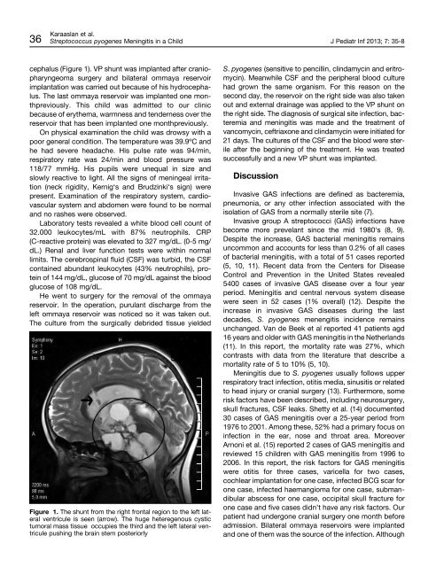
Figure 1. The shunt from the right frontal region to the left lateral<br />
ventricule is seen (arrow). The huge heteregenous cystic<br />
tumoral mass tissue occupies the third and the left lateral ventricule<br />
push<strong>in</strong>g the bra<strong>in</strong> stem posteriorly<br />
S. <strong>pyogenes</strong> (sensitive to pencill<strong>in</strong>, cl<strong>in</strong>damyc<strong>in</strong> and eritromyc<strong>in</strong>).<br />
Meanwhile CSF and the peripheral blood culture<br />
had grown the same organism. For this reason on the<br />
second day, the reservoir on the right side was also taken<br />
out and external dra<strong>in</strong>age was applied to the VP shunt on<br />
the right side. The diagnosis <strong>of</strong> surgical site <strong>in</strong>fection, bacteremia<br />
and men<strong>in</strong>gitis was made and the treatment <strong>of</strong><br />
vancomyc<strong>in</strong>, ceftriaxone and cl<strong>in</strong>damyc<strong>in</strong> were <strong>in</strong>itiated for<br />
21 days. The cultures <strong>of</strong> the CSF and the blood were sterile<br />
after the beg<strong>in</strong>n<strong>in</strong>g <strong>of</strong> the treatment. He was treated<br />
successfully and a new VP shunt was implanted.<br />
Discussion<br />
Invasive GAS <strong>in</strong>fections are def<strong>in</strong>ed as bacteremia,<br />
pneumonia, or any other <strong>in</strong>fection associated with the<br />
isolation <strong>of</strong> GAS from a normally sterile site (7).<br />
Invasive group A streptococci (GAS) <strong>in</strong>fections have<br />
become more prevelant s<strong>in</strong>ce the mid 1980‘s (8, 9).<br />
Despite the <strong>in</strong>crease, GAS bacterial men<strong>in</strong>gitis rema<strong>in</strong>s<br />
uncommon and accounts for less than 0.2% <strong>of</strong> all cases<br />
<strong>of</strong> bacterial men<strong>in</strong>gitis, with a total <strong>of</strong> 51 cases reported<br />
(5, 10, 11). Recent data from the Centers for Disease<br />
Control and Prevention <strong>in</strong> the United States revealed<br />
5400 cases <strong>of</strong> <strong>in</strong>vasive GAS disease over a four year<br />
period. <strong>Men<strong>in</strong>gitis</strong> and central nervous system disease<br />
were seen <strong>in</strong> 52 cases (1% overall) (12). Despite the<br />
<strong>in</strong>crease <strong>in</strong> <strong>in</strong>vasive GAS diseases dur<strong>in</strong>g the last<br />
decades, S. <strong>pyogenes</strong> menengitis <strong>in</strong>cidence rema<strong>in</strong>s<br />
unchanged. Van de Beek et al reported 41 patients agd<br />
16 years and older with GAS men<strong>in</strong>gitis <strong>in</strong> the Netherlands<br />
(11). In this report, the mortality rate was 27%, which<br />
contrasts with data from the literature that describe a<br />
mortality rate <strong>of</strong> 5 to 10% (5, 10).<br />
<strong>Men<strong>in</strong>gitis</strong> due to S. <strong>pyogenes</strong> usually follows upper<br />
respiratory tract <strong>in</strong>fection, otitis media, s<strong>in</strong>usitis or related<br />
to head <strong>in</strong>jury or cranial surgery (13). Furthermore, some<br />
risk factors have been described, <strong>in</strong>clud<strong>in</strong>g neurosurgery,<br />
skull fractures, CSF leaks. Shetty et al. (14) documented<br />
30 cases <strong>of</strong> GAS men<strong>in</strong>gitis over a 25-year period from<br />
1976 to 2001. Among these, 52% had a primary focus on<br />
<strong>in</strong>fection <strong>in</strong> the ear, nose and throat area. Moreover<br />
Arnoni et al. (15) reported 2 cases <strong>of</strong> GAS men<strong>in</strong>gitis and<br />
reviewed 15 children with GAS men<strong>in</strong>gitis from 1996 to<br />
2006. In this report, the risk factors for GAS men<strong>in</strong>gitis<br />
were otitis for three cases, varicella for two cases,<br />
cochlear implantation for one case, <strong>in</strong>fected BCG scar for<br />
one case, <strong>in</strong>fected haemangioma for one case, submandibular<br />
abscess for one case, occipital skull fracture for<br />
one case and five cases didn’t have any risk factors. Our<br />
patient had undergone cranial surgery one month before<br />
admission. Bilateral ommaya reservoirs were implanted<br />
and one <strong>of</strong> them was the source <strong>of</strong> the <strong>in</strong>fection. Although