A Case of Meningitis Caused by Streptococcus pyogenes in a Child ...
A Case of Meningitis Caused by Streptococcus pyogenes in a Child ...
A Case of Meningitis Caused by Streptococcus pyogenes in a Child ...

Create successful ePaper yourself
Turn your PDF publications into a flip-book with our unique Google optimized e-Paper software.
<strong>Case</strong> Report / Olgu Sunumu 35<br />
Received/Geliş Tarihi:<br />
28.05.2012<br />
Accepted/Kabul Tarihi:<br />
20.06.2012<br />
Correspondence<br />
Address<br />
Yazışma Adresi:<br />
Dr. Ayşe Karaaslan<br />
Department <strong>of</strong> Pediatric<br />
Infectious Diseases,<br />
Faculty <strong>of</strong> Medic<strong>in</strong>e,<br />
Marmara University,<br />
İstanbul, Turkey<br />
Phone: +90 216 657 06 06<br />
E-mail:<br />
akaraaslan78@gmail.com<br />
©Copyright 2013 <strong>by</strong><br />
Pediatric Infectious Diseases<br />
Society - Available onl<strong>in</strong>e at<br />
www.cocukenfeksiyon.com<br />
©Telif Hakkı 2013<br />
Çocuk Enfeksiyon Hastalıkları<br />
Derneği - Makale metn<strong>in</strong>e<br />
www.cocukenfeksiyon.com<br />
web sayfasından ulaşılabilir.<br />
doi:10.5152/ced.2013.08<br />
A <strong>Case</strong> <strong>of</strong> <strong>Men<strong>in</strong>gitis</strong> <strong>Caused</strong> <strong>by</strong> <strong>Streptococcus</strong><br />
<strong>pyogenes</strong> <strong>in</strong> a <strong>Child</strong> with Ventriculoperitoneal<br />
Shunt and Ommaya Reservoir<br />
Ventriküloperitoneal Şantı ve Ommaya Rezervuarı Olan Çocukta<br />
Streptokokkus <strong>pyogenes</strong> Menenjiti<br />
Ayşe Karaaslan 1, Ahmet Soysal 1, Canan Kuzdan 1, Eren Çağan 1, Yaşar Bayri 2, Mustafa Bakır 1<br />
1 Department <strong>of</strong> Pediatric Infectious Diseases, Faculty <strong>of</strong> Medic<strong>in</strong>e, Marmara University, İstanbul, Turkey<br />
2 Department <strong>of</strong> Bra<strong>in</strong> Surgery, Faculty <strong>of</strong> Medic<strong>in</strong>e, Marmara University, İstanbul, Turkey<br />
Abstract<br />
<strong>Streptococcus</strong> <strong>pyogenes</strong> is a well-known cause <strong>of</strong><br />
<strong>in</strong>vasive <strong>in</strong>fections. However, acute bacterial men<strong>in</strong>gitis<br />
caused <strong>by</strong> this pathogen is unusual. Here<strong>in</strong>, we<br />
presented a child with ventriculoperitoneal (VP) shunt<br />
and bilateral ommaya reservoir who developed group<br />
A streptococcal (GAS) men<strong>in</strong>gitis a month after the last<br />
neurosurgery operation. The patient was treated with<br />
removal <strong>of</strong> VP shunt and ommaya reservoir and antibiotic<br />
therapy <strong>in</strong>clud<strong>in</strong>g vancomyc<strong>in</strong>, ceftriaxone and<br />
cl<strong>in</strong>damyc<strong>in</strong> for 21 days. Although many predispos<strong>in</strong>g<br />
factors have been described for GAS men<strong>in</strong>gitis, this<br />
is the first report <strong>of</strong> an association between ommaya<br />
reservoir and VP shunt due to S. <strong>pyogenes</strong> men<strong>in</strong>gitis.<br />
Cl<strong>in</strong>icans should be aware that sporadic cases can<br />
occur and if it is treated promptly, the outcome tends<br />
to be favourable. (J Pediatr Inf 2013; 7: 35-8)<br />
Key words: <strong>Men<strong>in</strong>gitis</strong>, <strong>Streptococcus</strong> <strong>pyogenes</strong>,<br />
ventriculoperitoneal shunt, ommaya rezervoir<br />
Introduction<br />
Invasive <strong>in</strong>fections due to group A beta<br />
hemolytic streptococcus (GAS) <strong>in</strong>clude bacteremia,<br />
pneumonia, sk<strong>in</strong> and s<strong>of</strong>t tissue <strong>in</strong>fections,<br />
men<strong>in</strong>gitis and toxic shock syndrome. <strong>Men<strong>in</strong>gitis</strong><br />
represents 2% <strong>of</strong> all cases <strong>of</strong> GAS <strong>in</strong>vasive disease<br />
(1). GAS is usually not mentioned as the<br />
causative agent <strong>of</strong> bacterial men<strong>in</strong>gitis beyond<br />
the neonatal period (2-4). Although GAS has the<br />
ability to <strong>in</strong>vade s<strong>of</strong>t tissue, CNS <strong>in</strong>fections<br />
rema<strong>in</strong> a rare event and account for less than<br />
0.2% <strong>of</strong> all bacterial men<strong>in</strong>gitis (5, 6).<br />
In a MEDLINE search <strong>of</strong> English literature, we<br />
found only 12 cases <strong>of</strong> GAS men<strong>in</strong>gitis reported<br />
Özet<br />
<strong>Streptococcus</strong> <strong>pyogenes</strong> az rastlanan enfeksiyonların<br />
iyi bil<strong>in</strong>en bir nedenidir. Akut bakteriyel menenjit nedeni<br />
olarak bu patojen nadirdir. Biz bu makalede ventriküloperitoneal<br />
(VP) şantı ve bilateral Ommaya rezervuarı<br />
olan ve son ameliyatından sonra grup A streptokok<br />
(GAS) menenjiti gelişen bir çocuğu sunduk. Hasta VP<br />
şant ve Ommaya rezervuarlarının çıkarılması ve 21 gün<br />
süreyle vankomis<strong>in</strong>, seftriakson ve kl<strong>in</strong>damis<strong>in</strong> verilerek<br />
tedavi edildi. GAS menenjiti iç<strong>in</strong> birçok predispozan<br />
faktör rapor edilmiş olsa da bu Ommaya rezervuarı<br />
ve VP şant ilişkili ilk rapor edilen makaledir.<br />
Kl<strong>in</strong>isyenler sporadik vakaların olabileceği konusunda<br />
uyanık olmalıdır ve hızla tedavi uygulanması durumunda<br />
daha olumlu sonuçlar elde edilecektir.<br />
(J Pediatr Inf 2013; 7: 35-8)<br />
Anahtar kelimeler: Menenjit, Streptokok <strong>pyogenes</strong>,<br />
ventriküloperitoneal şant, ommaya rezervuar<br />
<strong>in</strong> the pediatric age group s<strong>in</strong>ce year 2000 but no<br />
cases <strong>of</strong> GAS men<strong>in</strong>gitis <strong>in</strong> a child with Ommaya<br />
reservoir. Here<strong>in</strong> we report the case <strong>of</strong> a child<br />
with ventriculoperitoneal shunt and bilateral<br />
Ommaya reservoirs who developed GAS reservoir<br />
poche abcess, men<strong>in</strong>gitis and bacteremia<br />
and was treated with removal <strong>of</strong> VP shunt-<br />
ommaya reservoir and antimicrobial therapy.<br />
<strong>Case</strong> Report<br />
A 8-year old boy was operated for multicystic<br />
craniopharyngeoma three years previously<br />
and after this operation he progressively lost<br />
vision and hear<strong>in</strong>g ability and developed hydro-
36<br />
Karaaslan et al.<br />
<strong>Streptococcus</strong> <strong>pyogenes</strong> <strong>Men<strong>in</strong>gitis</strong> <strong>in</strong> a <strong>Child</strong> J Pediatr Inf 2013; 7: 35-8<br />
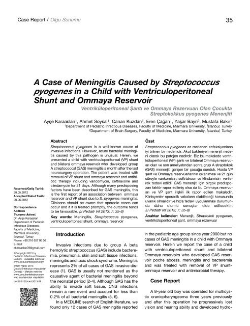
cephalus (Figure 1). VP shunt was implanted after craniopharyngeoma<br />
surgery and bilateral ommaya reservoir<br />
implantation was carried out because <strong>of</strong> his hydrocephalus.<br />
The last ommaya reservoir was implanted one monthpreviously.<br />
This child was admitted to our cl<strong>in</strong>ic<br />
because <strong>of</strong> erythema, warmness and tenderness over the<br />
reservoir that has been implanted one monthpreviously.<br />
On physical exam<strong>in</strong>ation the child was drowsy with a<br />
poor general condition. The temperature was 39.9ºC and<br />
he had severe headache. His pulse rate was 94/m<strong>in</strong>,<br />
respiratory rate was 24/m<strong>in</strong> and blood pressure was<br />
118/77 mmHg. His pupils were unequal <strong>in</strong> size and<br />
slowly reactive to light. All the signs <strong>of</strong> men<strong>in</strong>geal irritation<br />
(neck rigidity, Kernig‘s and Brudz<strong>in</strong>ki‘s sign) were<br />
present. Exam<strong>in</strong>ation <strong>of</strong> the respiratory system, cardiovascular<br />
system and abdomen were found to be normal<br />
and no rashes were observed.<br />
Laboratory tests revealed a white blood cell count <strong>of</strong><br />
32.000 leukocytes/mL with 87% neutrophils. CRP<br />
(C-reactive prote<strong>in</strong>) was elevated to 327 mg/dL. (0-5 mg/<br />
dL.) Renal and liver function tests were with<strong>in</strong> normal<br />
limits. The cerebrosp<strong>in</strong>al fluid (CSF) was turbid, the CSF<br />
conta<strong>in</strong>ed abundant leukocytes (43% neutrophils), prote<strong>in</strong><br />
<strong>of</strong> 144 mg/dL, glucose <strong>of</strong> 70 mg/dL aga<strong>in</strong>st the blood<br />
glucose <strong>of</strong> 108 mg/dL.<br />
He went to surgery for the removal <strong>of</strong> the ommaya<br />
reservoir. In the operation, purulant discharge from the<br />
left ommaya reservoir was noticed so it was taken out.<br />
The culture from the surgically debrided tissue yielded<br />
Figure 1. The shunt from the right frontal region to the left lateral<br />
ventricule is seen (arrow). The huge heteregenous cystic<br />
tumoral mass tissue occupies the third and the left lateral ventricule<br />
push<strong>in</strong>g the bra<strong>in</strong> stem posteriorly<br />
S. <strong>pyogenes</strong> (sensitive to pencill<strong>in</strong>, cl<strong>in</strong>damyc<strong>in</strong> and eritromyc<strong>in</strong>).<br />
Meanwhile CSF and the peripheral blood culture<br />
had grown the same organism. For this reason on the<br />
second day, the reservoir on the right side was also taken<br />
out and external dra<strong>in</strong>age was applied to the VP shunt on<br />
the right side. The diagnosis <strong>of</strong> surgical site <strong>in</strong>fection, bacteremia<br />
and men<strong>in</strong>gitis was made and the treatment <strong>of</strong><br />
vancomyc<strong>in</strong>, ceftriaxone and cl<strong>in</strong>damyc<strong>in</strong> were <strong>in</strong>itiated for<br />
21 days. The cultures <strong>of</strong> the CSF and the blood were sterile<br />
after the beg<strong>in</strong>n<strong>in</strong>g <strong>of</strong> the treatment. He was treated<br />
successfully and a new VP shunt was implanted.<br />
Discussion<br />
Invasive GAS <strong>in</strong>fections are def<strong>in</strong>ed as bacteremia,<br />
pneumonia, or any other <strong>in</strong>fection associated with the<br />
isolation <strong>of</strong> GAS from a normally sterile site (7).<br />
Invasive group A streptococci (GAS) <strong>in</strong>fections have<br />
become more prevelant s<strong>in</strong>ce the mid 1980‘s (8, 9).<br />
Despite the <strong>in</strong>crease, GAS bacterial men<strong>in</strong>gitis rema<strong>in</strong>s<br />
uncommon and accounts for less than 0.2% <strong>of</strong> all cases<br />
<strong>of</strong> bacterial men<strong>in</strong>gitis, with a total <strong>of</strong> 51 cases reported<br />
(5, 10, 11). Recent data from the Centers for Disease<br />
Control and Prevention <strong>in</strong> the United States revealed<br />
5400 cases <strong>of</strong> <strong>in</strong>vasive GAS disease over a four year<br />
period. <strong>Men<strong>in</strong>gitis</strong> and central nervous system disease<br />
were seen <strong>in</strong> 52 cases (1% overall) (12). Despite the<br />
<strong>in</strong>crease <strong>in</strong> <strong>in</strong>vasive GAS diseases dur<strong>in</strong>g the last<br />
decades, S. <strong>pyogenes</strong> menengitis <strong>in</strong>cidence rema<strong>in</strong>s<br />
unchanged. Van de Beek et al reported 41 patients agd<br />
16 years and older with GAS men<strong>in</strong>gitis <strong>in</strong> the Netherlands<br />
(11). In this report, the mortality rate was 27%, which<br />
contrasts with data from the literature that describe a<br />
mortality rate <strong>of</strong> 5 to 10% (5, 10).<br />
<strong>Men<strong>in</strong>gitis</strong> due to S. <strong>pyogenes</strong> usually follows upper<br />
respiratory tract <strong>in</strong>fection, otitis media, s<strong>in</strong>usitis or related<br />
to head <strong>in</strong>jury or cranial surgery (13). Furthermore, some<br />
risk factors have been described, <strong>in</strong>clud<strong>in</strong>g neurosurgery,<br />
skull fractures, CSF leaks. Shetty et al. (14) documented<br />
30 cases <strong>of</strong> GAS men<strong>in</strong>gitis over a 25-year period from<br />
1976 to 2001. Among these, 52% had a primary focus on<br />
<strong>in</strong>fection <strong>in</strong> the ear, nose and throat area. Moreover<br />
Arnoni et al. (15) reported 2 cases <strong>of</strong> GAS men<strong>in</strong>gitis and<br />
reviewed 15 children with GAS men<strong>in</strong>gitis from 1996 to<br />
2006. In this report, the risk factors for GAS men<strong>in</strong>gitis<br />
were otitis for three cases, varicella for two cases,<br />
cochlear implantation for one case, <strong>in</strong>fected BCG scar for<br />
one case, <strong>in</strong>fected haemangioma for one case, submandibular<br />
abscess for one case, occipital skull fracture for<br />
one case and five cases didn’t have any risk factors. Our<br />
patient had undergone cranial surgery one month before<br />
admission. Bilateral ommaya reservoirs were implanted<br />
and one <strong>of</strong> them was the source <strong>of</strong> the <strong>in</strong>fection. Although
J Pediatr Inf 2013; 7: 35-8<br />
GAS frequently colonizes the oropharynx, <strong>in</strong> our patient<br />
we th<strong>in</strong>k that sk<strong>in</strong> colonization over the ommaya reservoir<br />
was the source <strong>of</strong> the <strong>in</strong>fection or, s<strong>in</strong>ce <strong>in</strong>fection developed<br />
<strong>in</strong> 30 days after ommaya reservoir implantation it<br />
may have been due to surgical site <strong>in</strong>fection because <strong>of</strong><br />
culture from the ommaya reservoir yielded S. <strong>pyogenes</strong>.<br />
The organisms most frequently caus<strong>in</strong>g <strong>in</strong>fections <strong>of</strong><br />
<strong>in</strong>dwell<strong>in</strong>g CNS prostheses are the coagulase negative<br />
staphylococci. The second most frequent pathogen is<br />
Staphylococcus aureus (16-19). As far as we know there<br />
was no previous report concerned with ommaya reservoir<br />
related GAS men<strong>in</strong>gitis.<br />
Treatment <strong>of</strong> GAS men<strong>in</strong>gitis <strong>in</strong>cludes management <strong>of</strong><br />
the complications <strong>of</strong> sepsis, aggressive surgical debridement,<br />
if an appropriate site <strong>of</strong> <strong>in</strong>fection is identified and<br />
antibiotics for the underly<strong>in</strong>g GAS <strong>in</strong>fection. The antibiotic<br />
<strong>of</strong> choice for treatment is penicill<strong>in</strong> and there have<br />
been no reports <strong>of</strong> resistance <strong>of</strong> this agent to this drug<br />
(15). For patients who are allergic to penicill<strong>in</strong>, treatment<br />
with ceftriaxone may be an alternative (20).<br />
Animal models and a review <strong>of</strong> treatment <strong>of</strong> 56 children<br />
with GAS bacteremia favor comb<strong>in</strong>ation treatment<br />
with a beta-lactam plus c<strong>in</strong>damyc<strong>in</strong> (21, 22). Our patient<br />
was treated <strong>by</strong> <strong>in</strong>travenous vancomyc<strong>in</strong> (60 mg/kg/day<br />
divided every 6 hours), ceftriaxone (100 mg/kg/day divided<br />
every 12 hours) and cl<strong>in</strong>damyc<strong>in</strong> (40 mg/kg/day<br />
divided every 6 hours).<br />
Uncomplicated GAS men<strong>in</strong>gitis cases have been<br />
treated for 10-14 days (9, 13, 23, 24). Length <strong>of</strong> therapy<br />
depends on the cl<strong>in</strong>ical response to antibiotic treatment.<br />
Therapy is usually cont<strong>in</strong>ued for 14 days from the last<br />
positive culture obta<strong>in</strong>ed dur<strong>in</strong>g surgical debridement.<br />
Our patient was treated for 21 days.<br />
Group A streptococci men<strong>in</strong>gitis is associated with a<br />
low mortality if treated promptly. The case fatality rate <strong>in</strong><br />
the review <strong>of</strong> Barlodes et al. (25) was 12%. The most<br />
frequent complications are neurologic, although <strong>in</strong> pediatric<br />
patients, neuroendocr<strong>in</strong>ologic complications predom<strong>in</strong>ate<br />
(19, 20). Chow&Muder reported a global letality<br />
<strong>of</strong> 5% and sequelae <strong>in</strong> 46% <strong>of</strong> the cases <strong>in</strong> a review<br />
conducted between 1981-1991 (5). A case series <strong>of</strong> 51<br />
patients (mixed adult and pediatric patients) showed a<br />
44% prevalence rate <strong>of</strong> sequelae <strong>in</strong> pediatric patients<br />
(the majority be<strong>in</strong>g neurological) compared with approximatetly<br />
7% <strong>in</strong> adults (25).<br />
Conclusion<br />
Group A streptococcus is an uncommon cause <strong>of</strong><br />
men<strong>in</strong>gitis <strong>in</strong> children but has to be considered as a<br />
causative pathogen.<br />
Conflict <strong>of</strong> Interest<br />
No conflicts <strong>of</strong> <strong>in</strong>terest were declared <strong>by</strong> the authors.<br />
References<br />
Karaaslan et al.<br />
<strong>Streptococcus</strong> <strong>pyogenes</strong> <strong>Men<strong>in</strong>gitis</strong> <strong>in</strong> a <strong>Child</strong><br />
37<br />
1. Zurawski CA, Barsdley MS, Beall B, et al. Invasive group A<br />
streptococcal disease <strong>in</strong> metropolitan Atlanta: a populationbased<br />
assessment. Cl<strong>in</strong>ical Infectious Diseases 1998; 27: 150-<br />
7. [CrossRef]<br />
2. Feig<strong>in</strong> RD, Perlman E. Bacterial men<strong>in</strong>gitis beyond the neonatal<br />
period. In: Feig<strong>in</strong> RD, Cherry JD (eds) Textbook <strong>of</strong> pediatric<br />
<strong>in</strong>fectious diseases, 4th edn. Saunders, Philadelphia: 1998.<br />
pp.400-29.<br />
3. Prober CG. Infections <strong>of</strong> the central nervous system. In: Nelson<br />
WE (ed) Textbook <strong>of</strong> pediatrics, 15th edn. Saunders,<br />
Philadelphia: 1996.pp.707-16.<br />
4. Tunkel AR, Scheld WM. Acute men<strong>in</strong>gitis. In:Mandell GL,<br />
Bennett JE, Dol<strong>in</strong> R(eds) Pr<strong>in</strong>ciples and practice <strong>of</strong> <strong>in</strong>fectious<br />
diseases, 4 th edn. Churchill Liv<strong>in</strong>gstone, New York: 1995.<br />
pp.831-65.<br />
5. Chow JW, Muder RR. Group A Streptococcal men<strong>in</strong>gitidis. Cl<strong>in</strong><br />
Infect Dis 1992; 14; 418-21. [CrossRef]<br />
6. Schelech WF, Ward JI, Band JD, et al. Bacterial men<strong>in</strong>gitidis <strong>in</strong><br />
the United States, 1978 through 1981. The natioanal bacterial<br />
men<strong>in</strong>gitis surveillance study. J Am Med Assoc 1985; 253:<br />
1749-54. [CrossRef]<br />
7. Stevens DL. Invasive group A streptococcus Infectious. Cl<strong>in</strong><br />
Infect Dis 1992; 14: 2-13. [CrossRef]<br />
8. Davies HD, Mc Geer A, Schwatrz B. Invasive group A streptococcal<br />
<strong>in</strong>fectious <strong>in</strong> Ontario, Canada. N Engl J Med 1996; 335:<br />
547-54. [CrossRef]<br />
9. Moses A, Ziv A, Harari M, Rahav G, Shapira M, Engelhard D.<br />
Increased <strong>in</strong>cidence and severity <strong>of</strong> streptoccocus <strong>pyogenes</strong><br />
bacteremia <strong>in</strong> young children. Pediatr Infect Dis J 1995; 14:<br />
767-70. [CrossRef]<br />
10. Mathur P, Arora NK, Kapil A, Das BK. <strong>Streptococcus</strong> <strong>pyogenes</strong><br />
men<strong>in</strong>gitis. Indian J Pediatr 2004; 71: 423-6. [CrossRef]<br />
11. Van de Beek D, de Gans J, Spanjuard L, Sela S, Vermeulen M,<br />
Dankert J. Group A Streptococcal men<strong>in</strong>gitis <strong>in</strong> adults, reports<br />
<strong>of</strong> 41 cases and a review <strong>of</strong> the literature. Cl<strong>in</strong> Infect Dis 2002;<br />
34: 32-6. [CrossRef]<br />
12. O‘Loughl<strong>in</strong> RE, Roberson A, Cieslak PR, et al. Active Bacterial<br />
Care Surveillance Team. The epidemiology <strong>of</strong> <strong>in</strong>vasive group A<br />
Streptococcal <strong>in</strong>fection and potential vacc<strong>in</strong>e implications.<br />
United States. 2000-2004. Cl<strong>in</strong> Infect Dis 2007; 45: 853-62.<br />
[CrossRef]<br />
13. Asnis DS, Knez T. Group a Streptococcal men<strong>in</strong>gitis. Arch<br />
Intern Med 1998; 58: 810-4. [CrossRef]<br />
14. Shetty AK, Frankel LR, Maldarado Y, Falco DA, Lewis DB.<br />
Group A Streptococcal men<strong>in</strong>gitis: Report <strong>of</strong> a case and review<br />
<strong>of</strong> literature s<strong>in</strong>ce 1976. Pediatr Emerg Care 2001; 17: 430-4.<br />
[CrossRef]<br />
15. Arnoni MV, Berez<strong>in</strong> EN, Safadi MA, Almeida FJ, Lopes CR.<br />
<strong>Streptococcus</strong> <strong>pyogenes</strong> men<strong>in</strong>gitis <strong>in</strong> children: Report <strong>of</strong> Two<br />
<strong>Case</strong>s and Literature Review. Braz J Infect Dis 2007; 11: 375-7.<br />
[CrossRef]<br />
16. Bisno AL, Sternau L. Infections <strong>of</strong> central nervous system<br />
shunts. In:Bisno AL, Waldvagel FA, editor. Infections Associated<br />
with Indwell<strong>in</strong>g Medical Devices. American Society for<br />
Microbiology, Wash<strong>in</strong>gton; 1994.pp.91-109.<br />
17. Crnich CJ, Safdar N, Maki DG. Infections associated with<br />
implanted medical devices. In: F<strong>in</strong>ch RG,Greenwood D, Norr<strong>by</strong><br />
SR, Whitley RJ, editor. Antibiotic and Chemotherapy: Anti<strong>in</strong>fective<br />
Agents and Their Use <strong>in</strong> Therapy.8. Churchill<br />
Liv<strong>in</strong>gstone; 2003.pp.575-618.
38<br />
Karaaslan et al.<br />
<strong>Streptococcus</strong> <strong>pyogenes</strong> <strong>Men<strong>in</strong>gitis</strong> <strong>in</strong> a <strong>Child</strong> J Pediatr Inf 2013; 7: 35-8<br />
18. Naradzay JFX, Browne BJ, Rolnick MA, Doherty RJ. Cerebral<br />
ventricular shunts. J Emerg Med 1999; 17: 311-22. [CrossRef]<br />
19. Filka J, Huttova M, Tuharsky J, Sagat T, Kral<strong>in</strong>sky K, Kremery<br />
VJ. Nosocomial men<strong>in</strong>gitis <strong>in</strong> children after ventriculoperitoneal<br />
shunt <strong>in</strong>sertion. Acta Pediatr 1999; 88: 576-8. [CrossRef]<br />
20. L<strong>in</strong> HH, Liu YC, Chiou CC, Lheng DL. Group A Streptococcal<br />
men<strong>in</strong>gitis. J Formes Med Assoc 1996; 95: 802-3.<br />
21. Stevens DL, Gibbons AE, Bergstrom R, W<strong>in</strong>n V. The Eagle<br />
effect revisited: efficacy <strong>of</strong> cl<strong>in</strong>damyc<strong>in</strong>, erytromyc<strong>in</strong> and penicill<strong>in</strong><br />
<strong>in</strong> the treatment <strong>of</strong> streptococcal myositis. J Infect Dis<br />
1998; 158: 23-8. [CrossRef]<br />
22. Zimbelman J, Palmer A, Todd J. Improved outcome <strong>of</strong><br />
cl<strong>in</strong>damyc<strong>in</strong> compared with betalactam antibiotic treatment for<br />
<strong>in</strong>vasive streptococcus <strong>pyogenes</strong> <strong>in</strong>fection. Pediatr Infect Dis J<br />
1999; 18: 1096-100. [CrossRef]<br />
23. Jagdis F. Group A Streptococcal men<strong>in</strong>gitis and bra<strong>in</strong> abscess.<br />
Pediatr Infect Dis J 1988; 7: 885-6. [CrossRef]<br />
24. Campello MG, Miguel Diaz J. <strong>Men<strong>in</strong>gitis</strong> por <strong>Streptococcus</strong><br />
<strong>pyogenes</strong>, absceso cerebral y ependimitis. Enferm Infecc<br />
Microbiol Cl<strong>in</strong> 1995; 13: 68.<br />
25. Baraldes MA, Dom<strong>in</strong>go P, Mauri A, et al. Group A Streptococcal<br />
men<strong>in</strong>gitis <strong>in</strong> the antibiotic era. Eur J Cl<strong>in</strong> Microbial Infect Dis<br />
1999; 18: 572-8. [CrossRef]