The aorto-ventricular tunnels - Cambridge Journals
The aorto-ventricular tunnels - Cambridge Journals
The aorto-ventricular tunnels - Cambridge Journals

Create successful ePaper yourself
Turn your PDF publications into a flip-book with our unique Google optimized e-Paper software.
Continuing Medical Education<br />
<strong>The</strong> <strong>aorto</strong>-<strong>ventricular</strong> <strong>tunnels</strong><br />
Roxane McKay, 1 Robert H. Anderson, 2 Andrew C. Cook 2<br />
1 Department of Cardiology and Cardiovascular Surgery, Hamad Medical Corporation, Doha, Qatar;<br />
2 Cardiac Unit, Institute of Child Health, University College London, London, UK<br />
IT IS LEVY AND COLLEAGUES, IN 1963, WHO ARE<br />
generally credited with the first description of<br />
“aortico-left <strong>ventricular</strong> tunnel”. 1 Examples of<br />
the malformation, nonetheless, were illustrated initially<br />
by Burchell and Edwards in 1957, 2 and by<br />
Edwards in 1961. 3 <strong>The</strong> subsequent documentation<br />
of more than 130 cases has now elucidated many features<br />
of the so-called “<strong>tunnels</strong>,” including their clinical<br />
presentation and surgical management. While<br />
most of the abnormal channels extend between the<br />
aorta and the left ventricle, 4–79 it is now also recognized<br />
that some, alternatively, enter the right ventricle.<br />
80–91 <strong>The</strong> anatomic arrangement underscoring the<br />
malformations has been clarified by recent morphological<br />
studies, 92,93 while diagnosis during fetal life<br />
has established beyond any doubt that the lesions are<br />
congenital. 42,43 Although rare, the <strong>aorto</strong>-<strong>ventricular</strong><br />
tunnel is the foremost cause during infancy of regurgitant<br />
flow of blood from the aorta to one or the other<br />
of the ventricles. In this review, we will describe and<br />
illustrate the structure of the malformations, speculate<br />
upon their developmental basis, and discuss pertinent<br />
aspects of their diagnosis and treatment.<br />
Pathologic anatomy<br />
<strong>The</strong> <strong>aorto</strong>-<strong>ventricular</strong> tunnel is an abnormal channel<br />
that connects the lumen of the ascending aorta to the<br />
cavity of either the left or right ventricle. In its<br />
course, the tunnel forms a conduit that by-passes the<br />
sinutubular junction, this being the discrete ring<br />
Corresponding Author: Roxane McKay MD, FRCS, FRCSC, Hamad Medical<br />
Corporation, P.O. Box 3050, Doha, Qatar. Tel: 974 439 2584; Fax: 974 439<br />
2324; E-mail: rmck07@yahoo.com<br />
RHA is supported by the Joseph Levy Foundation together with the British<br />
Heart Foundation.<br />
ACC is supported by the British Heart Foundation.<br />
Manuscript received and accepted 12 August 2002<br />
Cardiol Young 2002; 12: 563–580<br />
© Greenwich Medical Media Ltd.<br />
ISSN 1047-9511<br />
that marks the junction of the aortic valvar sinuses<br />
with the tubular ascending aorta. At the same time,<br />
the tunnel by-passes the attachment of one of the leaflets<br />
of the aortic valve, which is tethered distally at<br />
the sinutubular junction. <strong>The</strong> abnormal pathway runs<br />
into the extra-cardiac tissues as it passes from its aortic<br />
origin to its <strong>ventricular</strong> termination. In the majority<br />
of cases, these extra-cardiac tissues are those that,<br />
normally, separate the subpulmonary infundibulum<br />
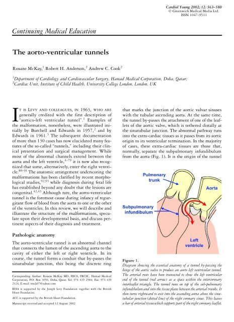
from the aorta (Fig. 1). It is the origin of the tunnel<br />
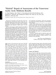
Figure 1.<br />
Diagram showing the essential anatomy of a tunnel by-passing the<br />
hinge of the aortic valve to produce an <strong>aorto</strong> left <strong>ventricular</strong> tunnel.<br />
<strong>The</strong> arterial roots have been transected to show the left <strong>ventricular</strong><br />
end of the tunnel (red arrow) as a space within the intercoronary<br />
interleaflet triangle. <strong>The</strong> tunnel runs on top of the sub-pulmonary<br />
infundibulum and into the tissue plane between the arterial trunks. It<br />
then turns rightward to exit into the ascending aorta above the sinutubular<br />
junction (dotted line) of the right coronary sinus. This leaves<br />
a bar of arterial tissue which supports part of the right coronary leaflet.
564 Cardiology in the Young December 2002<br />
from the tubular aorta that serves to differentiate it<br />
from rupture of an aneurysmal sinus of Valsalva. <strong>The</strong><br />
latter lesion also creates a communication between<br />
the aorta and a <strong>ventricular</strong> chamber, but one that originates<br />
below the sinutubular junction, and remains<br />
completely within the heart. <strong>The</strong> distinction from<br />
coronary-cameral fistula is less clear, because a coronary<br />
arterial orifice may arise above the sinutubular<br />
junction, and because the left, 21,84 circumflex, 85 anterior<br />
inter<strong>ventricular</strong> 78,91 and right 1,21,31,42,43,47,81,85<br />
coronary arteries have all been found arising within<br />
an <strong>aorto</strong>-<strong>ventricular</strong> tunnel. Fistulous connections of<br />
the coronary arteries, however, always pass through<br />
myocardium to reach the lumen of a cardiac chamber,<br />
and do not involve the hinge-point of an aortic<br />
valvar leaflet. As we will see, these features do not<br />
always serve to distinguish a fistula from a tunnel<br />
extending to open within the right ventricle, but<br />
they do contrast with most <strong>tunnels</strong> that open within<br />
the left ventricle. <strong>The</strong> frequent association of the<br />
<strong>tunnels</strong> with abnormalities of the coronary arteries,<br />
moreover, does suggest the possibility of a shared<br />
developmental origin.<br />
When considering the morphology of the <strong>tunnels</strong>,<br />
we will describe an aortic origin and a <strong>ventricular</strong><br />
termination. Since the pressures will be comparable<br />
throughout the length of the <strong>tunnels</strong>, and because<br />
flow may occur in either direction, this distinction is<br />
arbitrary. In our opinion, nonetheless, it helps in<br />
accounting for the anatomic features, which can otherwise<br />
be difficult to understand. <strong>The</strong> aortic origin<br />
of a tunnel that extends to open within the left ventricle<br />
may be situated anywhere above the left or right<br />
aortic sinuses, or above the junction between the two<br />
coronary aortic sinuses. In four-fifths of reported<br />
cases, the aortic end of the tunnel is positioned above<br />
the right coronary sinus (Fig. 2). In the much smaller<br />
number of hearts in which the tunnel opens into the<br />
Figure 2.<br />
Site of aortic opening in relation to the aortic valvar sinuses of<br />
Valsalva in 85 cases of <strong>aorto</strong>-<strong>ventricular</strong> tunnel. Circled numbers<br />
indicate the number of orifices found in each position. Abbreviations:<br />
LV: left ventricle; RV: right ventricle; L: Left coronary aortic sinus;<br />
R: right coronary aortic sinus; N: non-coronary aortic sinus.<br />
right ventricle, more than one-third are described with<br />
the aortic origin above the left sinus of Valsalva. In<br />
some cases, clockwise or counter-clockwise rotation<br />
of the aortic root relative to its supporting <strong>ventricular</strong><br />
attachments has been observed. 6–8 A single case<br />
has been described in which the aortic orifice was<br />
positioned above the junction between the right and<br />
non-coronary aortic sinuses. 87<br />
<strong>The</strong> size and shape of the aortic origin of the tunnel<br />
are extremely variable. In some hearts, the opening<br />
is a slit of only two to three millimeters width<br />
(Fig. 3). 72,80 At the other extreme, an oval orifice has<br />
been encountered measuring two by two-and-a-half<br />
centimeters (Fig. 4). 18,44,45 <strong>The</strong> size of the aortic<br />
opening shows no correlation with either the age or<br />
the size of the patient. By definition, the orifice lies<br />
above the sinutubular ridge which, itself, may be situated<br />
abnormally low, 85 and which can be considerably<br />
thickened. 1,5,31,40 Diffuse dilation of the entire<br />
ascending aorta, with further localized enlargement<br />
around the entrance of the tunnel, 17 may obscure the<br />
exact position of the orifice, particularly in relation<br />
to the origin of a coronary artery. 17,40 Aneurysms of<br />
any of the three aortic sinuses may coexist with an<br />
Figure 3.<br />
This tunnel has a small slit-like opening within the left ventricle,<br />
and a much larger aortic orifice which is separate from the origin of<br />
the right coronary artery. Reproduced by kind permission of Siew<br />
Yen Ho and Gaetano Thiene.
Vol. 12, No. 6 McKay et al: Aorto-<strong>ventricular</strong> <strong>tunnels</strong> 565<br />
<strong>aorto</strong>-<strong>ventricular</strong> tunnel 17,85 Being situated below<br />
the sinutubular ridge, however, such aneurysms are<br />
distinct from the orifice of the tunnel.<br />
Having taken origin from the aorta, the initial<br />
course of the tunnel is within extra-cardiac tissues<br />
Figure 4.<br />
<strong>The</strong> large aortic opening of a tunnel originating above the left coronary<br />
sinus of Valsalva that extended to open in the left ventricle as<br />
seen by the surgeon at operation. Reproduced from Litwin SB, Color<br />
Atlas of Congenital Heart Surgery. Mosby, St Louis 1996 with<br />
permission from Mosby – Yearbook.<br />
Figure 5.<br />
This tunnel, extending from above the right coronary aortic sinus to<br />
open within the fibrous triangle between the left and right coronary<br />
leaflets of the aortic valve, runs within the tissue plane that separates<br />
the sinuses of the aortic valve from the free-standing muscular subpulmonary<br />
infundibulum (See Fig. 7). Abbreviations: R: right<br />
coronary aortic leaflet; N: non-coronary aortic leaflet; L: left coronary<br />
aortic leaflet.<br />
(Fig. 5), specifically in the area that, in the normal<br />
heart, forms a discrete plane between the aortic<br />
sinuses and the muscular subpulmonary infundibulum<br />
(Fig. 6). 92 Those <strong>tunnels</strong> that originate above<br />
the right coronary aortic sinus produce a large tubular,<br />
or saccular, protuberance on the anterior aspect<br />
of the aortic root (Figs 7 and 8). 1,11,19,30,32 Tunnels<br />
having their aortic end above the left coronary aortic<br />
Figure 6.<br />
Sectioning the normal heart in simulated parasternal long axis plane<br />
shows the extracardiac tissue plane that normally separates the<br />
sinuses of the aorta which give rise to the coronary arteries from the<br />
free-standing muscular subpulmonary infundibulum. Abbreviations:<br />
R: right coronary aortic sinus; N: non-coronary aortic sinus.<br />
Figure 7.<br />
<strong>The</strong> heart shown is the same as illustrated in Figure 5. Note the<br />
bulge made by the tunnel (starred) between the aorta and the<br />
pulmonary trunk.
566 Cardiology in the Young December 2002<br />
Figure 8.<br />
Appearances of our most recent example of an <strong>aorto</strong>-left <strong>ventricular</strong> tunnel diagnosed at 17 weeks gestation. Examination of the arterial roots<br />
from the front (a) shows a small dilation in the region of the right aortic sinus, which is the aortic end of the tunnel (starred), as well as a<br />
prominent connection, or “arterial ligament”, connecting the wall of the tunnel with the right facing sinus of the pulmonary trunk (between<br />
arrowheads). <strong>The</strong>re was marked cardiomegaly, and left <strong>ventricular</strong> hypertrophy present (b). <strong>The</strong> <strong>ventricular</strong> opening of the tunnel was large,<br />
and located between the coronary aortic leaflets in the place occupied, in the normal heart, by the interleaflet fibrous triangle, which is distal to<br />
the normal anatomic ventriculo-arterial junction. In this abnormal heart, the ventriculo-arterial junction is part of the wall of the tunnel.<br />
Thus, the parasternal long axis section, taken in the same heart (c), shows that the junction between arterial wall and <strong>ventricular</strong> myocardium<br />
is still present (arrow), but has become detached from the hinge-point of the aortic valve (*). <strong>The</strong> junction is displaced towards the subpulmonary<br />
infundibulum, and forms the mid-part of the tunnel. <strong>The</strong> tunnel itself (red arrow), therefore, bypasses the attachment of the aortic valve and<br />
enters above aortic root above the level of the sinutubular junction. Abbreviations: L: left coronary aortic leaflet; R: right coronary aortic leaflet;<br />
N: non-coronary aortic leaflet.<br />
sinus, in contrast, lie behind the pulmonary trunk,<br />
originating within the transverse sinus of the pericardium.<br />
78,90,91 Unless they open into the right ventricle<br />
(Fig. 9), <strong>tunnels</strong> originating above the left<br />
coronary aortic sinus are less obvious when viewed<br />
externally from the front of the heart (Fig. 10). 20,44<br />
As it leaves the base of the heart, the subpulmonary<br />
infundibulum, together with the pulmonary trunk,<br />
spirals round the right and left coronary aortic sinuses<br />
of Valsalva. 93 <strong>The</strong> infundibulum, therefore, is readily<br />
displaced anteriorly by a tunnel that terminates in<br />
the left ventricle, with such an arrangement potentially<br />
producing subvalvar obstruction of the right<br />
<strong>ventricular</strong> outflow tract. 2,9,16,26,36,40
Vol. 12, No. 6 McKay et al: Aorto-<strong>ventricular</strong> <strong>tunnels</strong> 567<br />
Figure 9.<br />
<strong>The</strong> external location of a tunnel which extended from the aorta to<br />
open into the right ventricle is shown as seen in the operating room.<br />
Abbreviation: ARVT: <strong>aorto</strong>-right <strong>ventricular</strong> tunnel. Reproduced<br />
from Hruda J, Hazekamp MG, Sobotka-Plojhar MA, Ottenkamp.<br />
Repair of <strong>aorto</strong>-right <strong>ventricular</strong> tunnel with pulmonary steosis and<br />
an anomalous origin of the left coronary artery. Eur J Cardiothorac<br />
Surg 2002; 21: 1123–1125, with permission from Elsevier Science.<br />
Figure 10.<br />
As shown in this illustration, the extracardiac course of the tunnel<br />
already seen in Figure 4, which arises above the left coronary sinus<br />
of Valsalva, lies in the transverse sinus. Because of this, it is not<br />
obvious when viewed from the front of the heart. Reproduced from<br />
Litwan SB, Color Atlas of Congenital Heart Surgery. Mosby,<br />
St Louis 1996, with permission from Mosby – Yearbook<br />
Those <strong>tunnels</strong> which originate above the right<br />
coronary aortic sinus, and which communicate with<br />
the left ventricle, almost always do so within the triangular<br />
fibrous area demarcated by the hinge-points<br />
of the right and left coronary leaflets as they ascend<br />
Figure 11.<br />
In this heart, the left ventricle has been opened to show the termination<br />
in the fibrous triangle between the right and left coronary aortic<br />
leaflets of a tunnel that originated above the right aortic sinus. Note<br />
that the opening, although below the hinge of the valvar leaflet, is<br />
distal to the anatomic ventriculo-arterial junction. Note also the<br />
mucoid tissue in the aortic origin of the tunnel (*). Abbreviations:<br />
R: right coronary aortic leaflet; N: non-coronary aortic leaflet;<br />
L: left coronary aortic leaflet.<br />
to fuse with each other at the sinutubular junction<br />
(Fig. 11). 92 Although proximal to the sinutubular<br />
junction, this area is distal to the anatomic junction<br />
between the left <strong>ventricular</strong> myocardium and the arterial<br />
walls of the aortic root. Many <strong>tunnels</strong> have been<br />
described in surgical reports as being “immediately<br />
below the valve”. It is almost certainly the case that<br />
these open also within this interleaflet fibrous triangle.<br />
A large orifice can extend to a variable extent<br />
under the hinge of the right coronary aortic leaflet<br />
(Fig. 11). 11,42 Indeed, in the most recent tunnel we<br />
have investigated, the essence of the lesion was the<br />
divorce of the attachment of the right coronary aortic<br />
leaflet from its normal location within the aortic<br />
root (Fig. 8). <strong>The</strong> <strong>tunnels</strong> never extend, however, to<br />
open within the interleaflet triangle between the right<br />
and noncoronary sinuses. Thus, <strong>tunnels</strong> opening to<br />
the left ventricle are related neither to the membranous<br />
septum nor the <strong>ventricular</strong> conduction tissues. 94<br />
Possible exceptions to these generalizations are rare.<br />
One is a tunnel that was said to have two adjacent <strong>ventricular</strong><br />
orifices separated by fibrous tissue. 19 Another<br />
may be a tunnel noted as entering the left ventricle<br />
about ten millimeters below the aortic valve. 36 In a<br />
third, with associated aortic stenosis, the left <strong>ventricular</strong><br />
communication was described as being one centimeter<br />
below the right aortic leaflet. 32<br />
Less commonly, a tunnel terminating in the left<br />
ventricle originates from the aorta above the left sinus<br />
of Valsalva. <strong>The</strong>se <strong>tunnels</strong> have a much less consistent<br />
site of entry into the left ventricle. None have been<br />
documented as opening within the fibrous triangle<br />
separating the two coronary aortic valvar leaflets. Two
568 Cardiology in the Young December 2002<br />
Figure 12.<br />
<strong>The</strong> tunnel in this heart extended from above the left coronary aortic<br />
sinus (a) to open within the left ventricle proximal to the inter-coronary<br />
interleaflet triangle (b). Abbreviation: PT: pulmonary trunk.<br />
cases have been described as opening below, and several<br />
millimeters from, the left coronary aortic leaflet, 20,78<br />
another was described as opening within the crest of<br />
the muscular septum, 28 and still another, into the<br />
“body” of the left ventricle. 13 <strong>The</strong> one case within<br />
our archive had its opening below a fibrous fold, half<br />
a centimetre beneath the intercoronary interleaflet<br />
triangle (Fig. 12).<br />
When the <strong>tunnels</strong> extend from the aorta to open<br />
within the right ventricle, the location of the <strong>ventricular</strong><br />
orifice again shows some correlation with the<br />
position of the aortic opening. Those taking origin<br />
above the right aortic sinus of Valsalva enter the right<br />
<strong>ventricular</strong> infundibulum just proximal to the hingepoint<br />
of the pulmonary valve, 84,86,87 or else open within<br />
the supra<strong>ventricular</strong> crest below the subpulmonary<br />
infundibulum (Fig. 13). 80,83,85 <strong>The</strong> <strong>ventricular</strong> orifice<br />
(a)<br />
(b)<br />
Figure 13.<br />
A tunnel (starred) is shown that extends from the aorta above the<br />
right coronary aortic sinus (a) and opens into the right ventricle at<br />
the base of the subpulmonary infundibulum (b). Originally diagnosed<br />
as a coronary arterial fistula draining to the right ventricle,<br />
on re-examination we believe that the structure is an <strong>aorto</strong>-right<br />
<strong>ventricular</strong> tunnel.<br />
of those <strong>tunnels</strong> which take their origin from the aorta<br />
above its left coronary sinus is more variable. Single<br />
cases have been described entering the right ventricle<br />
just below the pulmonary valvar leaflets, 88 in the subpulmonary<br />
infundibulum, 84 and in the body of the<br />
ventricle. 89<br />
(a)<br />
(b)
Vol. 12, No. 6 McKay et al: Aorto-<strong>ventricular</strong> <strong>tunnels</strong> 569<br />
Brief reflection upon the interrelations of the<br />
right and left <strong>ventricular</strong> outflow tracts 92–95 will<br />
confirm that, irrespective of conventional wisdom, it<br />
is exceedingly rare for the <strong>tunnels</strong> to pass through<br />
the true septal structures. This is because, in the<br />
normal heart, the interleaflet triangle separating the<br />
diverging hinge-points of the right and left coronary<br />
aortic valvar leaflets lies distal to the anatomic<br />
ventriculo-arterial junction, despite the fact that it is<br />
below the haemodynamic ventriculo-arterial junction.<br />
As already emphasized, it is through this triangular<br />
area that most <strong>tunnels</strong> empty into the cavity of<br />
the left ventricle. As a consequence of abnormal<br />
development, the crest of the muscular septum then<br />
forms the wall of the proximal part of the tunnel,<br />
being continuous with the freestanding subpulmonary<br />
infundibulum of the right ventricle (Fig. 8). Those<br />
<strong>tunnels</strong> that open into the outflow of the right ventricle<br />
pass through muscle to reach the <strong>ventricular</strong><br />
cavity, but it is muscle that is part of the free-standing<br />
infundibulum, and not part of the muscular <strong>ventricular</strong><br />
septum. Even <strong>tunnels</strong> which appear to lie several<br />
millimeters below the body of an aortic valvar leaflet<br />
have been found, on careful dissection, to enter the<br />
left ventricle through an in-folding of fibrous tissue,<br />
rather than passing through the left <strong>ventricular</strong> myocardium.<br />
78 It is only those which end in the body of<br />
the ventricle 28,89 that may possibly transverse septal<br />
structures.<br />
Histologically, the wall of the tunnel differs at its<br />
two ends. <strong>The</strong> aortic origin consists of fibrous tissue,<br />
with smooth muscle cells and elastic fibers. 1,11,92<br />
This arrangement gives way to non-specific hyalinized<br />
collagen or musculature towards the <strong>ventricular</strong><br />
opening. In reality, the “walls” of the tunnel incorporate<br />
the cardiac structures between which it passes.<br />
Thus, the usual <strong>aorto</strong>-left <strong>ventricular</strong> tunnel is confined<br />
by the musculature of the <strong>ventricular</strong> septum<br />
and subpulmonary infundibulum in its floor, and by<br />
the fibrous wall of the aortic sinus giving rise to right<br />
coronary aortic leaflet at its roof (Figs 8 and 14). 92<br />
<strong>The</strong> tunnel by-passes the semilunar hinge of the<br />
right coronary aortic leaflet, which can lose completely<br />
its usual attachments to the aortic sinus and the supporting<br />
left <strong>ventricular</strong> musculature (Figs 8 and 11).<br />
Commonly, the histologic structure of the mid-portion<br />
of the tunnel is indistinct, but there can also be a<br />
clearly demarcated ventriculo-arterial junction within<br />
the body of the tunnel, lending further support to<br />
the notion that the abnormality represents separation<br />
between the attachment of the leaflet and the<br />
wall of the arterial valvar sinus (Fig. 8). Membranous<br />
or cystic structures similar to tissues of the valvar<br />
leaflets have been found within the lumen of the<br />
tunnel. 15,16,72,84,85 Such structures can alternatively<br />
be attached to the sinutubular ridge, 31,42,78 or lie<br />
Figure 14.<br />
Histological section showing how the tunnel rests on the musculature<br />
of the subpulmonary infundibulum, but that its aortic origin is of<br />
arterial phenotype.<br />
within an aortic sinus adjacent to the orifice of the<br />
tunnel (Fig. 11). 20 <strong>The</strong>y can produce obstruction<br />
within the tunnel. 72<br />
Aorto-<strong>ventricular</strong> <strong>tunnels</strong> have important relationships<br />
to the proximal portions of the coronary<br />
arteries. 96 <strong>The</strong> orifice of the right coronary artery has<br />
been found above, 19,21,40 below, 1,42 and to the side 86<br />
of <strong>tunnels</strong> situated above the right sinus of Valsalva,<br />
as well as within the tunnel itself. 1,21,31,42,43,47,81,85<br />
Absence of the orifice of the right coronary<br />
artery 6,21,31,41,49,87 has been observed and, in one<br />
heart, 85 the circumflex coronary artery arose from the<br />
tunnel. When the tunnel originates from the aorta<br />
above the left sinus of Valsalva, in more than half<br />
of the reported cases an abnormal orifice of the left<br />
coronary artery has been observed to be above the<br />
tunnel, 13,28 within the tunnel, 21,84 or else to be<br />
absent 78 . Origin of a stenotic anterior inter<strong>ventricular</strong><br />
branch from such a tunnel has also been observed. 78,91<br />
A left coronary artery originating to the right of the<br />
tunnel may have an intramural course within the<br />
posterior wall of the tunnel. 44 Occlusion of a coronary<br />
artery by the tunnel has also been described. 2,3<br />
Developmental considerations<br />
<strong>The</strong> described morphological findings can be difficult<br />
to appreciate in the intact, beating heart, particularly<br />
with superimposed changes from long-standing<br />
hemodynamic trauma. It can also be difficult to conceptualize<br />
the location of the <strong>tunnels</strong> relative to the<br />
subaortic and subpulmonary outflow tracts. With<br />
increasing experience, however, we are beginning to
570 Cardiology in the Young December 2002<br />
Figure 15.<br />
This section, in frontal plane, is from human embryo #13 from the<br />
Hamilton collection, estimated to be at Carnegie stage 12. It shows<br />
the undivided outflow tract, with its myocardial walls, extending<br />
from the roof of the developing right ventricle towards the aortic sac.<br />
Note the cushions extending throughout the cavity of the undivided<br />
tract, and the bend that divides it into proximal and distal portion.<br />
This bend will, eventually, become the sinutubular junction.<br />
Reproduced by kind permission of Prof Nigel Brown and Dr Sandra<br />
Webb, St George’s Hospital Medical School, London.<br />
appreciate the potential embryological origin of<br />
the abnormal channels, albeit that none have yet<br />
been identified during the stages of their formation.<br />
Initially during its development, the solitary outflow<br />
tract of the heart has discrete proximal and<br />
distal portions (Fig. 15). <strong>The</strong> junction between these<br />
parts will become the sinutubular junction. At first,<br />
the entire wall of the outflow tract is composed of<br />
myocardium 97,98 . As it is divided by the distal cushions<br />
to form the intrapericardial portion of the aorta<br />
and the pulmonary trunk, the walls of the distal<br />
outflow tract, along with the walls formed from the<br />
cushions themselves, transdifferentiate to become<br />
arterial structures. 97 <strong>The</strong> initial myocardial wall persists<br />
for a longer period around the proximal outflow<br />
tract. Within this part, proximal to the developing<br />
sinutubular junction, the distal ends of the cushions<br />
that are dividing the outflow tract (Fig. 16), along<br />
with the intercalated cushions, transform themselves<br />
into the developing arterial sinuses and valvar<br />
leaflets. Thus, the cushions that initially fused to<br />
septate the proximal ouflow tract give rise to the facing<br />
sinuses and valvar leaflets of both the aorta and<br />
Figure 16.<br />
This section, taken in short axis across the proximal outflow tract,<br />
is from human embryo #8 in the Hamilton collection, estimated to be<br />
at Carnegie stage 16. It shows the developing aortic and pulmonary<br />
valvar roots, at this stage encased within a continuous myocardial<br />
cuff. Note the muscular tissue growing into the “septal” endocardial<br />
cushions. Reproduced by kind permission of Prof Nigel Brown and<br />
Dr Sandra Webb, St George’s Hospital Medical School, London.<br />
the pulmonary trunk (Fig. 17). <strong>The</strong> proximal part of<br />
the fused cushions, however, hangs down proximal<br />
to the forming sinuses as a shelf within the right<br />
ventricle (Fig. 18). This most proximal part of the<br />
fused cushions then muscularises, initially producing<br />
an embryonic outlet septum within the right<br />
ventricle (Fig. 18b). With subsequent growth, however,<br />
the muscular structure widens to become the<br />
free-standing infundibulum of the right ventricle<br />
(Fig. 19). At the same time, the cushions lose their<br />
septal location, as a tissue plane is formed between<br />
the developing roots of the aortic and pulmonary<br />
valves (Fig. 17a). In normal development, as the<br />
cushions have become converted into the sinuses of<br />
the aortic and pulmonary valves, so the myocardial<br />
cuff surrounding them has regressed. <strong>The</strong> disappearance<br />
of the muscular cuff causes the tissue plane<br />
developing between the infundibulum and the aortic<br />
sinuses to lie in communication with the extracardiac<br />
space. It is within this tissue plane that abnormal<br />
development will produce the <strong>aorto</strong>-<strong>ventricular</strong> <strong>tunnels</strong>,<br />
which persist as anomalous channels joining the<br />
distal and proximal parts of the initial solitary outflow<br />
tract. <strong>The</strong> precise mechanisms of formation have<br />
yet to be clarified, but are probably related to the<br />
mechanism of formation of the triangles of fibrous<br />
tissue that separate the aortic sinuses beneath the leaflets<br />
of the aortic valve, along with abnormal formation<br />
of one of the leaflets of the aortic valve. It is also<br />
noteworthy that the coronary arteries were initially
Vol. 12, No. 6 McKay et al: Aorto-<strong>ventricular</strong> <strong>tunnels</strong> 571<br />
(a)<br />
(b)<br />
Figure 17.<br />
Human embryo #21 from the Hamilton collection is estimated to be<br />
at Carnegie stage 20. <strong>The</strong>se sections from the embryo show the developing<br />
aortic and pulmonary valvar sinuses and their supporting <strong>ventricular</strong><br />
roots. Figure (a) is cut obliquely across the sinutubular<br />
junction, showing the aortic wall, a small part of the left coronary<br />
aortic sinus, and the three leaflets of the developing pulmonary valve,<br />
the latter all still encased within a muscular cuff. Note the tissue<br />
plane developing between the walls of the aorta and pulmonary trunk.<br />
Figure (b) is taken more proximally, and shows how the cushions<br />
have fused and muscularised. <strong>The</strong> dotted line shows their plane of<br />
fusion. Again note the muscular cuff which still encases the developing<br />
aortic sinuses. Regression of the muscular cuff will place the tissue<br />
plane developing between the arterial roots in communication with<br />
extracardiac space. <strong>The</strong> <strong>tunnels</strong> form abnormally within this tissue<br />
plane. Reproduced by kind permission of Prof Nigel Brown and<br />
Dr Sandra Webb, St George’s Hospital Medical School, London.<br />
encased within the myocardial cuff that surrounded<br />
the developing sinuses 97 . <strong>The</strong> arteries pierce this cuff<br />
as they grow into the aortic sinuses. 99,100 It is easy to<br />
envisage, therefore, that abnormal development of this<br />
junctional region permits channels to form so as to<br />
(a)<br />
(b)<br />
Figure 18.<br />
A sagittal section, replicating the parasternal long axis echocardiographic<br />
plane, has been selected from human embryo #17 from the<br />
Hamilton collection, estimated to be at Carnegie stage 18. <strong>The</strong><br />
proximal part of the fused outflow cushions hang as a commashaped<br />
shelf within the cavity of the right ventricle (a). <strong>The</strong> arrow<br />
points to the closing embryonic inter<strong>ventricular</strong> communication. As<br />
shown in the enlargement (b), at this stage the muscularising cushions<br />
form a right <strong>ventricular</strong> outlet septum. A tissue plane will eventually<br />
form within the shelf to separate the subpulmonary infundibulum<br />
from the aortic valvar sinuses as indicated by the arrow, (see<br />
Fig. 6). Abnormal formation of this plane permits the development<br />
of the <strong>aorto</strong>-<strong>ventricular</strong> <strong>tunnels</strong>. Reproduced by kind permission<br />
of Prof Nigel Brown and Dr Sandra Webb, St George’s Hospital<br />
Medical School, London.<br />
connect the aorta with the outflow tract of the left<br />
ventricle, by-passing the attachment of the valvar<br />
leaflet to produce an <strong>aorto</strong>-left <strong>ventricular</strong> tunnel, or<br />
with the infundibulum, giving rise to the <strong>aorto</strong>-right<br />
<strong>ventricular</strong> tunnel. We presume that the abnormal<br />
development involves failure of the outflow cushions<br />
properly to form the arterial sinuses, the valvar leaflets,<br />
and the fibrous interleaflet triangles, coupled
572 Cardiology in the Young December 2002<br />
with abnormal separation of the distal outflow tract<br />
into the aorta and pulmonary trunk. At the same<br />
time, the muscularising proximal cushions become<br />
converted into the proximal myocardial wall of the<br />
tunnel. <strong>The</strong> associations of the <strong>tunnels</strong> with abnormalities<br />
of the aortic sinuses, the proximal coronary<br />
arteries, and the leaflets themselves, therefore, are<br />
entirely predictable. <strong>The</strong> end-result is one of the few<br />
cardiac malformations in which congenital lesions<br />
(a)<br />
(b)<br />
Figure 19.<br />
This section, again in sagittal plane replicating the parasternal<br />
long axis echocardiographic cut, is from another human embryo from<br />
the Hamilton collection, estimated to be at Carnegie stage 22, just<br />
after the completion of cardiac septation. <strong>The</strong> comma-shaped muscular<br />
shelf that, at stage 20, formed an intra<strong>ventricular</strong> muscular<br />
outlet septum, has now been transformed into the muscular subpulmonary<br />
infundibulum (a). Note the tissue plane already formed<br />
between the pulmonary trunk and the aorta (arrow). <strong>The</strong> enlargement<br />
(b) shows that the tissue plane has yet to extend to separate the<br />
forming wall of the right coronary aortic sinus from the infundibulum,<br />
itself formed by muscularistion of the proximal cushions which<br />
divided the outflow tract. It is precisely within the site of formation<br />
of this tissue plane (dotted line) that we find the abnormal <strong>aorto</strong><strong>ventricular</strong><br />
<strong>tunnels</strong>. Reproduced by kind permission of Prof Nigel<br />
Brown and Dr Sandra Webb, St George’s Hospital Medical School,<br />
London.<br />
of both aortic and pulmonary valvar leaflets may<br />
coexist. 1,26<br />
Clinical presentation<br />
<strong>The</strong> most consistent echocardiographic feature on antenatal<br />
examination between 18 and 33 weeks gestation<br />
is dilation and hypertrophy of the left ventricle, with<br />
severe and progressively reduced shortening fraction.<br />
Apparent aortic regurgitation, which is extremely<br />
uncommon during fetal life, and enlargement of<br />
the aortic root, further support a diagnosis of <strong>aorto</strong><strong>ventricular</strong><br />
tunnel, while flow of blood around the<br />
hinge of the valve has been imaged with color flow<br />
Doppler echocardiography. 43 Thickening, or severe<br />
dysplasia, of the aortic valvar leaflets was found in<br />
three of seven fetal cases, suggesting that this group<br />
may represent the more severe end of the pathological<br />
spectrum. Furthermore, an incidence of 0.46%<br />
among fetal cardiac malformations 42 is nearly five<br />
times greater than has been previously recognized<br />
after birth. 25<br />
At birth, or at the initial examination, there is<br />
invariably a loud “to-and-fro” murmur. This is usually<br />
accompanied by systolic and diastolic thrills, and is<br />
heard over the entire precordium. Bounding peripheral<br />
pulses are also a consistent finding. Enlargement<br />
of the heart, and uniform dilation of the ascending<br />
aorta, are usually obvious on chest X-ray, although<br />
the dilated aorta is not infrequently mistaken for the<br />
thymus gland. <strong>The</strong> electrocardiogram occasionally<br />
is normal, 27 but typically shows left or bi<strong>ventricular</strong><br />
hypertrophy, with a “strain pattern” of inverted<br />
T waves seen in the precordial leads. Although clinical<br />
signs in older patients closely mimic those of valvar<br />
aortic stenosis and incompetence, with a widened<br />
pulse pressure, and systolic and diastolic murmurs,<br />
the aortic component of the second heart sound is<br />
conserved, as is a dicrotic notch on the arterial pressure<br />
trace. 6 Cardiac enlargement is seen on the chest<br />
X-ray, which can also reveal marked enlargement of<br />
the entire ascending aorta. With the passage of time,<br />
the dilation can become extreme, and may appear disproportionate<br />
to other signs and symptoms of cardiac<br />
disease. In some patients, the tunnel itself can be seen<br />
as a leftward prominence of the aortic root in the area<br />
of the pulmonary trunk. 6<br />
<strong>The</strong> onset, and severity, of symptoms is highly<br />
variable, and probably reflects complex interactions<br />
and incompletely understood contributions from the<br />
lesion itself, the compromised coronary circulation,<br />
and any associated malformations. While occasional<br />
patients remain active and asymptomatic into adulthood,<br />
7,27,35,45 there are also reports of fetal death, 43<br />
as well as rapidly fatal congestive heart failure 23,31 and<br />
sudden death 40 in previously compensated children
Vol. 12, No. 6 McKay et al: Aorto-<strong>ventricular</strong> <strong>tunnels</strong> 573<br />
and adults. <strong>The</strong>se latter groups may have a higher<br />
incidence of coronary arterial compromise, with or<br />
without obstruction of the right <strong>ventricular</strong> outflow<br />
tract. <strong>The</strong> majority of patients suffer congestive heart<br />
failure within the first year of life, and many show<br />
this feature during the neonatal period. This is true<br />
whether the tunnel communicates with the left or<br />
with the right ventricle, although pulmonary stenosis<br />
in association with <strong>aorto</strong>-right <strong>ventricular</strong> tunnel<br />
may delay the onset of symptoms. 91 Attempts have<br />
been made to correlate the clinical course with morphology<br />
of the tunnel itself, 41,101 but information<br />
in the literature is probably inadequate presently to<br />
substantiate meaningful inferences. <strong>The</strong> association<br />
with severe dysplasia, stenosis, or atresia of the aortic<br />
valve, nonetheless, constitutes a particularly lethal<br />
combination of malformations. Of eleven such<br />
cases, 16,32,43,52,53,60,72,76,77,86 four died before birth or<br />
on the first day of life, with the remaining patients<br />
all developing congestive heart failure or low cardiac<br />
output early in the neonatal period.<br />
Investigation<br />
Echocardiography, with cross-sectional and color-<br />
Doppler imaging, constitutes the diagnostic investigation<br />
of choice. 14,15,28,34 <strong>The</strong> parasternal long-axis<br />
view shows the tunnel beside the aorta, from which it<br />
can be followed to its aortic and <strong>ventricular</strong> openings.<br />
As explained, when opening to the left ventricle,<br />
these are above and below the right or left coronary<br />
Figure 20.<br />
<strong>The</strong> cross-sectional echocardiogram in the parasternal long axis view<br />
shows a tunnel (T) between the aorta (AO) and the left ventricle<br />
(LV). Small arrows indicate the aortic and <strong>ventricular</strong> orifices of the<br />
tunnel. Reproduced from Sreeram N, Franks R, Walsh K. Aorticoleft<br />
<strong>ventricular</strong> tunnel: long-term outcome after surgical repair. J Am<br />
Coll Cardiol 1991; 17: 950–955, with permission from Elsevier<br />
Science. Abbreviations: R: right ventricle; LA: left atrium.<br />
aortic sinuses, respectively (Fig. 20). Color-flow<br />
imaging demonstrates blood passing through the<br />
abnormal channel from the left ventricle to the aorta<br />
during systole, and in the opposite direction in diastole<br />
(Fig. 21). <strong>The</strong> right <strong>ventricular</strong> outflow tract,<br />
and the pulmonary valve, are also seen. This permits<br />
quantification of any obstruction due to displacement<br />
of the subpulmonary infundibulum, and identification<br />
of the much rarer tunnel extending from the<br />
aorta to open in the right ventricle. 36,86,90 Parasternal<br />
short-axis views at the levels of the aortic valve and<br />
ascending aorta should show intact, albeit often<br />
enlarged, sinuses of Valsalva, <strong>The</strong>se views also reveal<br />
any thickening or dysplasia of the valvar leaflets, the<br />
coronary arterial origins, and typically show uniform<br />
enlargement of the ascending aorta, which may be<br />
twice its normal diameter. 21 <strong>The</strong> short axis views also<br />
demonstrate <strong>tunnels</strong> opening to the right ventricle<br />
(Fig. 22). 90 In the apical four-chamber view, left<br />
<strong>ventricular</strong> dilation and hypertrophy, with variable<br />
impairment of the shortening fraction, and otherwise<br />
conserved cardiac architecture, are characteristic.<br />
Of all these features, extensive and uniform dilation<br />
of the ascending aorta may be the best non-invasive<br />
clue to the diagnosis of <strong>aorto</strong>-<strong>ventricular</strong> tunnel, for<br />
this is hardly ever present early in life with other cardiac<br />
malformations. Only extremely rarely is enlargement<br />
of the aorta not present, specifically when there<br />
is critical obstruction both to the aortic valve and<br />
within the tunnel. 72 <strong>The</strong> most common diagnostic<br />
errors from echocardiography have been to confuse<br />
the <strong>ventricular</strong> end of the tunnel with a <strong>ventricular</strong><br />
septal defect, 1,26 or to mistake displacement of the<br />
Figure 21.<br />
Doppler colour-flow imaging shows diastolic flow through a tunnel<br />
from the aorta to the left ventricle. Abbreviations: LA: left atrium,<br />
LV: left ventricle, T: tunnel, AO: aorta. Reproduced from Sousa-<br />
Uva M, Touchot A, Fermont L, Piot D, Delezoide AL, Serraf A,<br />
Lacour-Gayet F, Roussin R, Bruniaux J, Planché C. Aortico-left<br />
<strong>ventricular</strong> tunnel in fetuses and infants. Ann Thorac Surg 1996;<br />
61: 1805–1810, with permission from Elsevier Science.
574 Cardiology in the Young December 2002<br />
Figure 22.<br />
A parasternal short axis echocardiogram with Doppler colour flow<br />
mapping shows a tunnel (arrow heads) extending from the aorta to<br />
open into the right ventricle in a ten-day-old neonate. <strong>The</strong> patient<br />
also had valvar pulmonary stenosis. <strong>The</strong> external appearances of this<br />
heart are shown in Figure 8. Abbreviations: AO: aorta; RV: right<br />
ventricle. Reproduced from Hruda J, Sobotka-Plojhar MA, van<br />
Rossum AC. Aortico-right <strong>ventricular</strong> tunnel with pulmonary stenosis<br />
in a neonate. Heart 2001; 86: 316, with permission from BMJ<br />
Publishing Group.<br />
subpulmonary infundibulum for Fallot’s tetralogy. 42<br />
Flow of blood through the tunnel has also been misinterpreted<br />
as valvar aortic regurgitation, 42 or a ruptured<br />
aneurysm of the sinus of Valsalva. 43<br />
Although previously considered essential in the<br />
investigation of suspected <strong>aorto</strong>-<strong>ventricular</strong> tunnel,<br />
the present role of cardiac catheterization is to clarify<br />
the coronary arterial anatomy when the proximal vessels<br />
cannot reliably be imaged by echocardiography,<br />
and possibly to elucidate some associated malformations.<br />
Hemodynamic studies routinely confirm normal<br />
pressures in the right heart, even in the presence of<br />
massive left <strong>ventricular</strong> enlargement, 6,29,36 unless the<br />
tunnel has compressed the right <strong>ventricular</strong> outflow<br />
tract. 1,31,36 Typical findings in the left heart are a<br />
widened aortic pulse pressure, with a normal or minimally<br />
elevated left <strong>ventricular</strong> end diastolic pressure.<br />
29 Although gradients have been documented<br />
across the left <strong>ventricular</strong> outflow tract, 1,6 flow through<br />
the tunnel itself may conceal significant aortic valvar<br />
obstruction. 30,72 <strong>The</strong> shape and extracardiac course<br />
of the tunnel can be demonstrated by angiography<br />
(Fig. 23), as can obstruction of the subpulmonary<br />
infundibulum (Fig. 24), but this information adds<br />
little to that obtained from high-quality sector scanning.<br />
Magnetic resonance imaging 34,90 also shows<br />
clearly the structure of the tunnel and its anatomical<br />
(a)<br />
(b)<br />
Figure 23.<br />
A left ventriculogram, profiled in the antero-posterior view (a), and an<br />
ascending <strong>aorto</strong>gram profiled in lateral projection (b), show a tunnel<br />
(arrow) extending from the aorta to the left ventricle in a neonate.<br />
relationships, but remains of limited availability in<br />
most clinical situations. Both resonance imaging and<br />
interventional catheterization, however, could, potentially<br />
have wider application for management of these<br />
patients, the former to characterize abnormal patterns<br />
of flow in the aortic root, and the latter to relieve<br />
valvar obstruction. 50,79,90<br />
Differential diagnosis and associated<br />
malformations<br />
A number of other cardiac malformations must be<br />
excluded from the differential diagnosis of patients<br />
with congestive heart failure and signs of aortic regurgitation<br />
(Table 1). In the neonate or young infant, a<br />
ruptured fistula of the sinus of Valsalva, <strong>ventricular</strong>
Vol. 12, No. 6 McKay et al: Aorto-<strong>ventricular</strong> <strong>tunnels</strong> 575<br />
Figure 24.<br />
In this patient, a right <strong>ventricular</strong> angiogram demonstrates compression<br />
of the subpulmonary infundibulum (arrow) by a tunnel extending<br />
from the aorta to the left ventricle in a five-year-old patient.<br />
Reproduced from Knott-Craig CJ, van der Merwe PL, Kalis NN,<br />
Hunter J. Repair of aortico-left <strong>ventricular</strong> tunnel associated with<br />
subpulmonary obstruction. Ann Thorac Surg 1992; 54: 557–559,<br />
with permission from Elsevier Science.<br />
Table 1. Differential diagnosis of <strong>aorto</strong>-<strong>ventricular</strong> tunnel.<br />
Sinus of valsalva fistula<br />
Common arterial trunk with valvar regurgitation<br />
Aorto-pulmonary window<br />
Ventricular septal defect with aortic regurgitation<br />
Persistent patency of the arterial duct<br />
Coronary-cameral fistula<br />
Tetralogy of Fallot with absent pulmonary valve<br />
Valvar aortic stenosis and regurgitation<br />
Cerebral arterio-venous malformation<br />
septal defect with aortic regurgitation, and valvar aortic<br />
incompetence are extremely uncommon. Furthermore,<br />
they do not generally have accompanying aortic<br />
dilation with left <strong>ventricular</strong> dysfunction. <strong>The</strong> murmurs<br />
of the persistently patent arterial duct, <strong>aorto</strong>pulmonary<br />
window, and coronary-cameral fistulas are<br />
more continuous than “to-and-fro” in nature, while<br />
that of a cerebral arterio-venous malformation tends<br />
to localize over the head. Echocardiography should<br />
readily distinguish cases of common arterial trunk<br />
and Fallot’s tetralogy. In older patients, the history of<br />
loud systolic and diastolic murmurs heard soon after<br />
birth favors a diagnosis of <strong>aorto</strong>-<strong>ventricular</strong> tunnel, as<br />
does disproportionate enlargement of the heart and<br />
ascending aorta.<br />
Table 2. Cardiac anomalies associated with <strong>aorto</strong>-<strong>ventricular</strong><br />
tunnel in 105 reported cases.<br />
Number<br />
Cardiac lesion of cases References<br />
Aortic valve<br />
2-leaflet 6 1, 3, 21, 26, 31, 40<br />
Obstructed 3-leaflet 5 6, 7, 41, 42, 59<br />
Dysplastic/atretic 10 1, 16, 30, 32, 43, 52,<br />
53, 60, 72, 76<br />
Perforation of leaflet 2 8, 9<br />
Unspecified regurgitation 2 21, 45<br />
Valvar pulmonary stenosis 7 1, 26, 50, 78, 83, 87,<br />
90, 91<br />
Dysplastic tricuspid valve 1 83<br />
Absent origin of coronary artery<br />
Right 6 6, 21, 31, 41, 49, 87<br />
Left 1 88<br />
Aneurysm of sinus of Valsalva 3 17, 30, 85<br />
Atrial septal defect/patent 8 31, 37, 38, 41, 72,<br />
oval foramen 83, 84, 85<br />
Ventricular septal defect 3 15, 21, 38<br />
Persistent patency of the 19 15, 16, 20, 21, 26,<br />
arterial duct 30, 31, 38, 41, 52,<br />
60, 72, 79, 83, 84, 89<br />
Absent left atrial appendage 1 40<br />
Associated anomalies, apart from those involving<br />
the aortic sinuses, the aortic and pulmonary valvar<br />
leaflets, and the coronary arteries, have been found<br />
infrequently among this group of patients (Table 2).<br />
No associations have been observed with any genetic<br />
syndromes or extracardiac defects, but reports of <strong>aorto</strong><strong>ventricular</strong><br />
tunnel are rare among patients of African,<br />
Oriental, or Asian descent. An approximate ratio of<br />
two males to one female has remained constant among<br />
reported cases<br />
Treatment<br />
While it has been proposed that congenital “aortic<br />
regurgitation” is better tolerated than that acquired<br />
later in life, 35 there is no evidence to support medical<br />
treatment for patients having an <strong>aorto</strong>-<strong>ventricular</strong><br />
tunnel. Without surgical intervention, most die<br />
early in life from congestive heart failure. 9,10,41 Only<br />
patients undergoing surgery before six months of age<br />
have later had documentation of normal left <strong>ventricular</strong><br />
size and function. 43 Moreover, lack of support<br />
for the right or left coronary aortic leaflet invariably<br />
results in progressive aortic regurgitation, 102 often<br />
necessitating repair or replacement of the valve as<br />
a primary 45 or secondary 7,8,57,58,73 procedure. Surgery,<br />
therefore, should be undertaken without delay, even<br />
in asymptomatic patients. 25,103<br />
<strong>The</strong> goals of surgery are complete closure of<br />
the abnormal communication, restoration of aortic<br />
valvar function, preservation of the coronary arterial
576 Cardiology in the Young December 2002<br />
Figure 25.<br />
<strong>The</strong>se diagrams illustrate the most-commonly employed technique for repair of an uncomplicated tunnel running from the aorta to the left ventricle<br />
(a,b,c) or the right ventricle (a,d). <strong>The</strong> aortic orifice (a) is closed through an <strong>aorto</strong>tomy by suturing a patch to the sinutubular ridge and the<br />
aortic wall, having identified the orifices of both coronary arteries. <strong>The</strong> tunnel itself is opened vertically (b). <strong>The</strong> <strong>ventricular</strong> orifice is closed using<br />
a second patch, which is sutured to the <strong>ventricular</strong> myocardium, and, in the case of <strong>tunnels</strong> ending within the left ventricle, also to the fibrous wall<br />
of the unsupported aortic sinus, and the bottom of the first patch. <strong>The</strong> walls of the tunnel are then approximated over the patches, and the <strong>aorto</strong>tomy<br />
is closed. Figure (c) shows the completed repair, with the aortic leaflet now supported by the patches. Figure (d) shows a completed repair for<br />
a tunnel terminating in the right ventricle. Drawn after Ho et al. 92<br />
circulation, and relief of any obstruction within the<br />
right or left <strong>ventricular</strong> outflow tracts. Transcatheter<br />
closure of a tunnel to the left ventricle with an<br />
Amplatzer duct occluder has been reported in a single<br />
patient. 52 Attempted coil closure of a tunnel to<br />
the right ventricle, however, was not successful. 90<br />
Considering the benefit of providing support for the<br />
aortic valvar leaflets, as well as the spectrum of associated<br />
coronary arterial anomalies, it seems likely that<br />
repair of the <strong>aorto</strong>-<strong>ventricular</strong> <strong>tunnels</strong> should remain<br />
largely, if not exclusively, within the surgical domain.<br />
Surgical repair of an uncomplicated tunnel to the<br />
left ventricle is performed on cardiopulmonary bypass,<br />
usually using a single right atrial cannula and moderate<br />
hypothermia. <strong>The</strong> tunnel is compressed externally<br />
immediately after commencement of perfusion,<br />
and during administration of cardioplegia, thus preventing<br />
<strong>ventricular</strong> distension and facilitating myocardial<br />
protection, respectively. Origin of a coronary<br />
artery deep within the tunnel could be an indication<br />
for retrograde delivery of cardioplegia through the<br />
coronary sinus.<br />
While simple suture of the aortic end of the tunnel,<br />
essentially approximating the sinutubular ridge to the<br />
aortic wall, has occasionally given good results, 13,17<br />
it is generally accepted that a patch should be used<br />
to avoid further distortion of the aortic valvar leaflet.<br />
10,20,21 This is done through a transverse or oblique<br />
<strong>aorto</strong>tomy, using a continuous monofilament suture<br />
to attach a Gore-Tex or pericardial patch to the sinutubular<br />
ridge and aortic wall (Fig. 25a).<br />
When the orifice of a coronary artery lies within<br />
the proximal part of the tunnel, the patch on the aortic<br />
wall is deviated distally to avoid its exclusion. In<br />
all cases, care is taken to avoid narrowing a coronary<br />
arterial orifice that lies close to the aortic orifice of<br />
the tunnel, or a branch that follows an intraluminal<br />
course in its wall. <strong>The</strong> <strong>ventricular</strong> end of a tunnel is<br />
closed with a second patch placed through a vertical<br />
incision into the tunnel itself (Fig. 25b). This both<br />
provides support the right aortic sinus, and prevents<br />
ongoing displacement of the right <strong>ventricular</strong> outflow<br />
tract by high-pressure and turbulent flow in a<br />
blind-ending pouch. 36 <strong>The</strong> bottom of this patch is
Vol. 12, No. 6 McKay et al: Aorto-<strong>ventricular</strong> <strong>tunnels</strong> 577<br />
Tunnel<br />
(a)<br />
Aorta<br />
Pulmonary trunk<br />
sutured to the <strong>ventricular</strong> myocardium, while the<br />
top, of necessity, must be joined to the first patch<br />
and the wall of the aortic sinus (Fig. 25c,d). In a single<br />
case, an elegant plastic repair (Fig. 26a–c) used<br />
the tunnel itself to achieve complete obliteration of<br />
the abnormal channel and stabilization of the aortic<br />
valve, avoiding the need to implant artificial material.<br />
47 <strong>The</strong> <strong>ventricular</strong> end of a tunnel opening to the<br />
right ventricle is also usually closed either with a<br />
patch, or by direct suture 78,80 through a right ventriculotomy.<br />
On occasion, both the aortic and <strong>ventricular</strong><br />
ends have been patched through the tunnel. 79 In<br />
theory, however, <strong>tunnels</strong> to the right ventricle should<br />
not jeopardize support for the aortic leaflet, and the<br />
<strong>ventricular</strong> end of the tunnel is at the same pressure<br />
as the subpulmonary infundibulum. It is questionable,<br />
therefore, whether closure of the <strong>ventricular</strong> orifice<br />
of a tunnel ending in the right ventricle is either<br />
useful or necessary.<br />
Should the right coronary artery originate distally<br />
within a tunnel arising above the right coronary aortic<br />
sinus, it is resected with a generous button of the<br />
surrounding wall of the tunnel and anastamosed to the<br />
ascending aorta. 20,21,47,49 While similar treatment of<br />
the anterior inter<strong>ventricular</strong> 78 or circumflex 85 branches<br />
of the left coronary artery would seem logical, this<br />
has not, as yet, been reported. A left coronary artery,<br />
or one of its major branches, taking origin distally<br />
from a tunnel that originates above the left sinus of<br />
Valsalva presents a more difficult problem. Owing<br />
to its position in the transverse sinus, and its distance<br />
from the aorta, transfer of the coronary artery<br />
to the aorta may be technically more complicated or<br />
impossible. This situation has been encountered, thus<br />
Right coronary<br />
artery<br />
(b)<br />
far, mainly in <strong>tunnels</strong> terminating in the right ventricle,<br />
where closure of the <strong>ventricular</strong>, rather than the<br />
aortic, end of the tunnel has successfully conserved<br />
coronary arterial perfusion. 91 Presence of such an<br />
arrangement with a tunnel entering the left ventricle<br />
might constitute an indication for leaving open the<br />
<strong>ventricular</strong> end of the tunnel. Intraoperative differentiation<br />
of absence of the left coronary arterial orifice 88<br />
from origin deep within the tunnel 84,91 may be impossible.<br />
This fact emphasizes the need to obtain accurate<br />
imaging of the coronary arterial origins preoperatively.<br />
A stenotic coronary arterial orifice, if recognized during<br />
life, could be an indication for grafting with the<br />
internal mammary artery.<br />
Valvar pulmonary stenosis in association with<br />
<strong>tunnels</strong> entering either the left 26,50 or the right ventricle<br />
86,90,91 has successfully been managed by open<br />
valvotomy 26,86,91 or preoperative percutaneous balloon<br />
valvoplasty, 50,57 although balloon dilation did not<br />
achieve satisfactory relief of the obstruction in one<br />
case. 90 Aortic valvar lesions are treated on their own<br />
merits. Commissurotomy for stenotic valves with two<br />
and three leaflets, 32,53,72 homograft replacement of<br />
the aortic root, 76 and, in a single case of aortic atresia,<br />
neonatal <strong>aorto</strong>ventriculoplasty 52 have all been employed<br />
in small infants, as well as valvar repair or<br />
replacement in older children and adults.<br />
Results<br />
Aortic orifice<br />
Ventricular orifice<br />
Figure 26.<br />
<strong>The</strong>se diagrams illustrate an alternative technique for repair of a tunnel opening into the left ventricle. <strong>The</strong> wall of the tunnel is opened vertically,<br />
in this instance leaving a generous margin of tissue around the right coronary artery, which arises distally from the tunnel. <strong>The</strong> orifice of the<br />
right coronary artery is excised with a surrounding button of tunnel wall (a), and the opposite flap of tunnel tissue is incised to the wall of the<br />
aorta. Both the aortic and <strong>ventricular</strong> openings can be seen from the tunnel (b). <strong>The</strong> wall of the tunnel is now sutured around each orifice as a<br />
patch, closing the aortic and <strong>ventricular</strong> ends as well as supporting the right coronary aortic leaflet and obliterating the lumen of the tunnel.<br />
<strong>The</strong> right coronary artery is reimplanted into the ascending aorta above the closed orifice of the tunnel (c). <strong>The</strong> dashed lines indicate surgical<br />
incisions. Drawn after Grünenfelder et al. 47<br />
Aorta<br />
Survival following surgical repair has improved<br />
steadily. Prior to 1983, there was a collective mortality<br />
of 20%. 10 In recent series, mortality has approached<br />
zero. 21 While initial reports indicated a very high<br />
(c)
578 Cardiology in the Young December 2002<br />
incidence of severe aortic valvar regurgitation occurring<br />
eight to twelve years after operation, 7,8 most<br />
of these patients had been repaired by direct suture<br />
of the aortic end of the tunnel, and came to surgery<br />
beyond five years of age. A shorter period of<br />
follow-up now suggests that patients in whom the<br />
aortic sinus is supported, and the tunnel closed in early<br />
infancy, should have little if any leakage through<br />
their aortic valve, as well as a good possibility of<br />
normal left <strong>ventricular</strong> function. 21,43 <strong>The</strong> subset of<br />
patients with associated dysplasia, stenosis, or atresia<br />
of the aortic valve, however, remains a significant<br />
challenge. Although aggressive relief of obstruction<br />
within the left <strong>ventricular</strong> outflow tract at the time of<br />
repair has achieved some success, overall surgical mortality<br />
remains close to 50% in this cohort, and most<br />
survivors require multiple replacements of the aortic<br />
valve. 52,76 <strong>The</strong> extremely rare origin of a left coronary<br />
artery within a tunnel arising above the left aortic<br />
sinus may also constitute a surgical risk factor, as<br />
this combination is not readily apparent at the time<br />
of operation, and closure of the aortic orifice of such<br />
a tunnel extending to the right ventricle has proved<br />
fatal. 84<br />
Acknowledgements<br />
We are indebted to Professor Nigel Brown and<br />
Dr Sandra Webb for permission to reproduce pictures<br />
of embryos from the Hamilton collection, kept at<br />
St George’s Hospital Medical School, London, and for<br />
permission also to cite as yet unpublished data from<br />
work concerning the development of the <strong>ventricular</strong><br />
outflow tract carried out in collaboration with Professors<br />
Wouter Lamers and Antoon Moorman of the<br />
University of Amsterdam. We thank Dr. Michael J.<br />
Tyrrell of the University of Saskatchewan for investigation<br />
and referral of the patient illustrated in<br />
Figure 23, and Mr. Conrad M. Samia of Hamad<br />
Medical Corporation for the preparation of illustrations.<br />
<strong>The</strong> assistance of BMJ Publishing Group with<br />
permission to reproduce Figure 22; Elsevier Science<br />
Inc., to reproduce Figures 9, 20, 21, and 24; and<br />
Mosby – Yearbook Inc., to reproduce Figures 4 and 10<br />
is gratefully acknowledged.<br />
References<br />
1. Levy MJ, Lillehei CW, Anderson RC, Amplatz K, Edwards JE.<br />
Aortico-left <strong>ventricular</strong> tunnel. Circulation 1963; 27: 841–853.<br />
2. Edwards JE, Burchell HB. <strong>The</strong> pathological anatomy of deficiencies<br />
between the aortic root and the heart, including aortic sinus<br />
aneurysms. Thorax 1957; 12: 125–139.<br />
3. Edwards JE. An Atlas of Acquired Diseases of the Heart and<br />
Great Vessels. W. B. Saunders Company, Philadelphia, 1961.<br />
4. Sreeram N, Franks R, Arnold R, Walsh K. Aortico-left <strong>ventricular</strong><br />
tunnel: long-term outcome after surgical repair. J Am Coll Cardiol<br />
1991; 17: 950–955.<br />
5. Bernhard WF, Plauth W, Fyler D. Unusual abnormalities of the<br />
aortic root or valve necessitating surgical correction in early childhood.<br />
New Engl J Med 1970; 282: 68–71.<br />
6. Somerville J, English T, Ross DN. Aorto-left <strong>ventricular</strong> tunnel.<br />
Clinical features and surgical management. Br Heart J 1974; 36:<br />
321–328.<br />
7. Serino W, Andrade JL, Ross D, de Leval M, Somerville J. Aortoleft<br />
<strong>ventricular</strong> communication after closure. Late postoperative<br />
problems. Br Heart J 1983; 49: 501–506.<br />
8. Meldrum-Hanna W, Schroff R, Ross D.N. Aortico-left <strong>ventricular</strong><br />
tunnel: late follow-up. Ann Thorac Surg 1986; 42: 304–306.<br />
9. Warnke H, Bartel J, Blumenthal-Barby Ch. Aortico-<strong>ventricular</strong><br />
tunnel. Thorac Cardiovasc Surg 1988; 36:86–88.<br />
10. Björk VO, Hongo T, Åberg B, Bjarke B. Surgical repair of<br />
aortico-left <strong>ventricular</strong> tunnel in a 7-day-old child. Scand J Thor<br />
Cardiovasc Surg 1983; 17: 185–189.<br />
11. Pérez-Martínez V, Quero M, Castro C, Moreno F, Brito JM,<br />
Merino G. Aortico-left <strong>ventricular</strong> tunnel. A clinical and pathologic<br />
review of this uncommon entity. Am Heart J 1973; 85:<br />
237–245.<br />
12. Nichols GM, Lees MH, Henken DP, Sunderland CO, Starr A.<br />
Aortico-left <strong>ventricular</strong> tunnel. Recognition and repair in infancy.<br />
Chest 1978; 70: 74–76.<br />
13. Norwicki ER, Aberdeen E, Friedman S, Rashkind WJ. Congenital<br />
left aortic sinus-left ventricle fistula and review of <strong>aorto</strong>cardiac<br />
fistulas. Ann Thorac Surg 1977; 23: 378–388.<br />
14. Perry JC, Nanda NC, Kicks DG, Harris JP. Two-dimensional<br />
echocardiographic identification of aortico-left <strong>ventricular</strong> tunnel.<br />
Am J Cardiol 1983; 52: 913–914.<br />
15. Bash SE, Huhta JC, Nihill MR, Vargo TA, Hallman GL. Aorticoleft<br />
<strong>ventricular</strong> tunnel with <strong>ventricular</strong> septal defect: twodimensional/Doppler<br />
echocardiographic diagnosis. J Am Coll<br />
Cardiol 1985; 5: 757–760.<br />
16. Morton P, Murtagh JG, O’Hara MD. Aneurysm of inter<strong>ventricular</strong><br />
septum with aortic valve malformation in an infant. Br Heart J<br />
1969; 31: 807–808.<br />
17. Spooner EW, Dunn JM, Behrendt DM. Aortico-left <strong>ventricular</strong><br />
tunnel and sinus of Valsalva aneurysm. Case report with operative<br />
repair. J Thorac Cardiovasc Surg 1978; 75: 232–236.<br />
18. Björk VO, Eklöf O, Wallgren G, Zetterqvist P. Successful surgical<br />
treatment of an aortico-left <strong>ventricular</strong> tunnel in a four-month-old<br />
infant. J Thorac Cardiovasc Surg 1979: 78: 35–38.<br />
19. Morgan RI, Mazur JH. Congenital aneurysm of aortic root with<br />
fistula to left ventricle. A case report with autopsy findings.<br />
Circulation 1963; 28: 589–594.<br />
20. Hucin B, Horvath P, Skovránek J, Reich O, Samánek M. Correction<br />
of aortico-left <strong>ventricular</strong> tunnel during the first day of life.<br />
Ann Thorac Surg 1989; 47: 254–256.<br />
21. Horváth P, Balaji S, Škovránek S, Hucin, de Leval MR, Stark J.<br />
Surgical treatment of aortico-left <strong>ventricular</strong> tunnel. Eur J<br />
Cardiothorac Surg 1991; 5: 113–117.<br />
22. Levy MJ, Schachner A, Blieden LC. Aortico-left <strong>ventricular</strong> tunnel.<br />
Collective review. J Thorac Cardiovasc Surg 1982; 84: 102–109.<br />
23. Palacio J, Perretta A, Sanchez B, Alperovich M. Intrapericardial<br />
congenital supravalvular aortic aneurysm communicating with<br />
the outflow-tract of the left ventricle. Hypoplasia of the aortic<br />
orifice and ascending aorta. J Cardiovasc Surg 1964 ;5: 401–407.<br />
24. Llorens R, Arcas R, Herreros J, De la Fuente A, Barriuso C,<br />
Casillas JA, Enriquez A. Aortico-left <strong>ventricular</strong> tunnel: a case<br />
report and review of the literature. Tex Heart Inst J 1982; 9:<br />
169–175.<br />
25. Okoroma EO, Perry LW, Scott LP III, McClenathan JE. Aorticoleft<br />
<strong>ventricular</strong> tunnel. Clinical profile, diagnostic features, and<br />
surgical considerations. J Thorac Cardiovasc Surg 1976; 71:<br />
238–244.<br />
26. Turley K, Silverman NH, Teitel D, Mavroudis C, Snider R,<br />
Rudolph A. Repair of aortico-left <strong>ventricular</strong> tunnel in the
Vol. 12, No. 6 McKay et al: Aorto-<strong>ventricular</strong> <strong>tunnels</strong> 579<br />
neonate: surgical, anatomic and echocardiographic considerations.<br />
Circulation 1982; 65: 1015–1020.<br />
27. Kafka H, Chan KL, Leach AJ. Asymptomatic aortico-left <strong>ventricular</strong><br />
tunnel in adulthood. Am J Cardiol 1989; 63: 1021–1022.<br />
28. Grant P, Abrams LD, De Giovanni JV, Shah KJ, Silove ED.<br />
Aortico-left <strong>ventricular</strong> tunnel arising from the left aortic sinus.<br />
Am J Cardiol 1985; 55: 1657–1658.<br />
29. Sung CS, Leachman RD, Zerpa F, Angelini P, Lufschanowski R.<br />
Aortico-left <strong>ventricular</strong> tunnel. Am Heart J 1979; 98: 87–93.<br />
30. Cooley RN, Harris LC, Rodin AE. Abnormal communication<br />
between the aorta and left ventricle. Aortico-left <strong>ventricular</strong><br />
tunnel. Circulation 1965; 31: 564–571.<br />
31. Bove KE, Schwartz DC. Aortico-left <strong>ventricular</strong> tunnel. A new<br />
concept. Am J Cardiol 1967; 19: 696–709.<br />
32. Villani M, Tiraboschi R, Marino A, De Tommasi M, Velitti F,<br />
Giani PC, Parenzan L. Aortico-left <strong>ventricular</strong> tunnel in infancy.<br />
Two surgical cases. Scand J Thor Cardiovasc Surg 1980; 14:<br />
169–175.<br />
33. Mair DD, Fulton RE, McGoon DC. Successful surgical repair of<br />
aortico-left <strong>ventricular</strong> tunnel in an infant. Mayo Clin Proc 1975;<br />
50: 691–696.<br />
34. Humes RA, Hagler DJ, Julsrud PR, Levy JM, Feldt RH,<br />
Schaff HV. Aortico-left <strong>ventricular</strong> tunnel: diagnosis based on<br />
two-dimensional echocardiography, color flow Doppler imaging,<br />
and magnetic resonance imaging. Mayo Clin Proc 1986; 61:<br />
901–907.<br />
35. Ribeiro P, Bun-Tan LB, Oakley CM. Management of aortic left<br />
<strong>ventricular</strong> tunnel. Br Heart J 1985; 54: 333–336.<br />
36. Knott-Craig CJ, van der Merwe PL, Kalis NN, Hunter J. Repair<br />
of aortico-left <strong>ventricular</strong> tunnel associated with subpulmonary<br />
obstruction. Ann Thorac Surg 1992; 54: 557–559.<br />
37. Fripp RR, Werner JC, Whitman V, Nordenberg A,<br />
Wladhausen JA. Pulsed Doppler and two-dimensional echocardiographic<br />
findings in aortico-left <strong>ventricular</strong> tunnel. J Am<br />
Coll Cardiol 1984; 4: 1012–1014.<br />
38. Giardina ACV, Levin AR, Engle MA. Aortico-left <strong>ventricular</strong> tunnel<br />
with natal cardiac failure. South Med J 1977; 70: 1351–1354.<br />
39. Fishbone G, De Leuchtenberg N, Stansel HC Jr. Aortico-left <strong>ventricular</strong><br />
tunnel. Radiology 1971; 98: 579–580.<br />
40. Roberts WC, Morrow AG. Aortico-left <strong>ventricular</strong> tunnel. A cause<br />
of massive aortic regurgitation and of intracardiac aneurysm. Am<br />
J Med 1965; 39: 662–667.<br />
41. Hovaguimian H, Cobanoglu A, Starr A. Aortico-left <strong>ventricular</strong><br />
tunnel: a clinical review and new surgical classification. Ann<br />
Thorac Surg 1988; 45: 106–112.<br />
42. Cook AC, Fagg NLK, Ho SY, Groves AMM, Sharland GK,<br />
Anderson RH, Allen LD. Echocardiographic-anatomical correlations<br />
in <strong>aorto</strong>-left <strong>ventricular</strong> tunnel. Br Heart J 1995; 74:<br />
443–448.<br />
43. Sousa-Uva M, Touchot A, Fermont L, Piot D, Delezoide AL,<br />
Serraf A, Lacour-Gayet F, Roussin R, Bruniaux J, Planché C.<br />
Aortico-left <strong>ventricular</strong> tunnel in fetuses and infants. Ann Thorac<br />
Surg 1996; 61: 1805–1810.<br />
44. Litwin SB. Color Atlas of Congenital Heart Surgery. Mosby,<br />
St Louis 1996.<br />
45. Akalin H, Erol Ç, Oral D, Çorapçioglu T, Uçanok K, Özyurda Ü,<br />
Ulusoy V. Aortico-left <strong>ventricular</strong> tunnel: successful diagnostic<br />
and surgical approach to the oldest patient in the literature.<br />
J Thorac Cardiovasc Surg 1989; 97: 804–805.<br />
46. Reis M, Singer H, Hofbeck M, Buheitel G, von der Emde J.<br />
Aorto-left <strong>ventricular</strong> tunnel with origin in the left sinus of<br />
Valsalva: a rare cause of congenital aortic insufficiency. Z Kardiol<br />
1994; 83: 519–524.<br />
47. Grünenfelder J, Zünd G, Prêtre R, Schmidli J, Vogt PR, Turina<br />
MI. Right coronary artery from <strong>aorto</strong>-left <strong>ventricular</strong> tunnel: case<br />
report of a new surgical approach. J Thorac Cardiovasc Surg 1998;<br />
116: 363–365.<br />
48. Rosenkranz ER, Murphy DJ Jr. Aortico-left <strong>ventricular</strong> tunnel in<br />
a neonate. Cleve Clin J Med 1992; 59: 87–90.<br />
49. Rauzier JM, Bonnet D, Zniber L, Sidi D, Aggoun Y, Acar P,<br />
Kachaner J, Vouhe P. Aortic-<strong>ventricular</strong> tunnel with right coronary<br />
artery atresia. Arch Mal Coeur Vaiss 1997; 90: 725–727.<br />
50. Martin Jimenez J, Gonzalez Dieguez CC, Quero Jimenez C, Rico<br />
Gomez F, Bermudez Canete R, Quero Jimenez M. Aortico-left<br />
<strong>ventricular</strong> tunnel associated with pulmonary valve stenosis. Rev<br />
Esp Cardiol 1996; 49: 921–924.<br />
51. Szaroszyk W, Rydlewska-Sadowska W. Echocardiographic diagnosis<br />
of the aortic-left <strong>ventricular</strong> tunnel. A case report. Kardiol<br />
Pol 1989; 32 Suppl 2: 62–66.<br />
52. Guyton RA, Michalik RE, McIntyre AB, Plauth WH Jr,<br />
Nugent EW, Hatcher CR Jr, Williams WH. Aortic atresia and<br />
aortico-left <strong>ventricular</strong> tunnel: successful surgical management by<br />
Konno <strong>aorto</strong>ventriculoplasty in a neonate. J Thorac Cardiovasc<br />
Surg 1986; 92: 1099–1105.<br />
53. Diamant S, Luber JM Jr , Gootman N. Successful repair of aorticoleft<br />
<strong>ventricular</strong> tunnel associated with severe aortic stenosis in a<br />
newborn. Pediatr Cardiol 1985; 6: 171–173.<br />
54. Lindberg H, Ovrum E, Bjornstad PG, Stake G, Pedersen T.<br />
Surgical repair of aortico-left <strong>ventricular</strong> tunnel (ALVT). Scand<br />
J Thorac Cardiovasc Surg 1988; 22: 285–287.<br />
55. Oalla Antolin JJ, Martin Duran R, Bardaji Mayor JL, Trujeda<br />
Padilla A, Berrazueta Fernandez JR, Carrion Alvarez M. Left<br />
<strong>aorto</strong>-<strong>ventricular</strong> tunnel: apropos of a case and review of the literature.<br />
Rev Esp Cardiol 1986; 39: 238–241.<br />
56. Bastos P, Moreira J, Cunha D, Sousa AR, Torres JP, Gomes MR.<br />
Aorto-left <strong>ventricular</strong> tunnel arising from the left sinus of<br />
Valvalva. Rev Port Cardiol 1990; 9: 703–706.<br />
57. Lukacs L, Richter T, Kadar K. Aortico-left <strong>ventricular</strong> tunnel:<br />
late repoerations. Cardiovasc Surg 1997; 5: 439–442.<br />
58. Duveau D, Baron O, Michaud JL, Lefevre M, Laboux L, Dupon H.<br />
Aortico-left <strong>ventricular</strong> tunnel. Long-term follow-up, theraputic<br />
implications. Arch Mal Coeur Vaiss 1989; 82: 785–789.<br />
59. Michielon G, Sorbara C, Casarotto DC. Repair of aorftico-left<br />
<strong>ventricular</strong> tunnel originating from the left aortic sinus. Ann<br />
Thorac Surg 1998; 65: 1780–1783.<br />
60. Bitar FF, Smith FC, Kavey R-EW, Kveselis DA, Byrum CJ,<br />
Brandt B, Gaum WE. Aortico-left <strong>ventricular</strong> tunnel with aortic<br />
atresia in the newborn. Am Heart J 1993; 126: 1480–1482.<br />
61. Sutlic Z, Kokos Z, Fabecic-Sabadi V, Biocina B, Protrka N,<br />
Sokolic J. Aortico-left <strong>ventricular</strong> tunnel. Lijec Vjesn 1993; 115:<br />
230–233.<br />
62. Sudoh Y, Takahara Y, Murayama H, Sezaki T, Nakamura T.<br />
Surgical repair of aortico-left <strong>ventricular</strong> tunnel, a case report.<br />
Nippon Kyobu Geka Gakkai Zasshi 1993; 41: 694–698.<br />
63. Santalla Rando A, Rodriguez Bailon I, Cutillas M, Calleja<br />
Hernandez M, Perez de la Cruz JM, Ramirez JA, Lara Torrano J,<br />
Moreno Herrero T. Aortico-left <strong>ventricular</strong> tunnel. Clinical and<br />
surgical considerations. Rev Esp Cardiol 1993; 46: 116–118.<br />
64. Zannini L, Gargiulo G, Albanese SB, Bonvicini M, Santorelli MC,<br />
Frascaroli G, Pierangeli A. Successful surgical repair of an aorticoleft<br />
<strong>ventricular</strong> tunnel in a two day old child. J Cardiovasc Surg<br />
(Torino) 1992; 33: 295–297.<br />
65. Moron A, Maitre Azcarate MJ, Rico Gomez F, Brito Perez JM,<br />
Quero Jimenez M. Aortico-left <strong>ventricular</strong> tunnel. Presentation<br />
of 2 new cases. Usefulness of non-invasive study. Rev Esp Cardiol<br />
1990; 43: 114–118.<br />
66. Wu J-R, Huang T-Y, Chen Y-F, Lin Y-T, Roan H-R. Aortico-left<br />
<strong>ventricular</strong> tunnel: two-dimensional echocardiographic and<br />
angiocardiographic features. Am Heart J 1989; 11: 697–699.<br />
67. Guo DW. Aortico-left <strong>ventricular</strong> tunnel: diagnosis by cine<br />
angiocardiography. AJR Am J Roentgenol 1989; 152: 345–346.<br />
68. Deuvaert FE, Goffin Y, Wellens F, De Paepe J, Primo G. Aorticoleft<br />
<strong>ventricular</strong> tunnel (ALVT). A diagnostic and surgical “must.”<br />
Acta Cardiol 1986; 41: 53–62.
580 Cardiology in the Young December 2002<br />
69. Lang D, Hofstetter R, Kupferschmid C, Quintenz R, Messmer BJ,<br />
Bernuth G. Aortico-left <strong>ventricular</strong> tunnel. Report of 3 cases and<br />
review of the literature. Z Kardiol 1982; 71: 695–704.<br />
70. Ruschewski W, de Vivie ER, Kirchhoff PG. Aortico-left <strong>ventricular</strong><br />
tunnel. Thorac Cardiovasc Surg 1981; 29: 282–286.<br />
71. Saylam A, Ozme S, Aslamaci S, Olga R, Aytac A. Aortico-left<br />
<strong>ventricular</strong> tunnel. Thorac Cardiovasc Surg 1981; 29: 259–261.<br />
72. Webber S, Johnston B, LeBlanc J, Patterson M. Aortic-left<br />
<strong>ventricular</strong> tunnel associated with critical aortic stenosis in the<br />
newborn. Pediatr Cardiol 1991; 12: 237–240.<br />
73. Kakadekar AP, Sandor GGS, Patterson MW, LeBlanc JG. Role of<br />
transesophageal echocardiography in the management of aorticleft<br />
<strong>ventricular</strong> tunnel. Pediatr Cardiol 1995; 16: 137–140.<br />
74. Duveau D, Baron O, Michaud JL, Lefèvre M, Laboux, Dupon H.<br />
Tunnel <strong>aorto</strong>-ventriculaire. Suivi à long terme, implications<br />
thèrapeutiques. Arch Mal Coeur 1989; 82: 785–789.<br />
75. Chen YF, Chiu CC, Wu JR. Correction of aortico-left <strong>ventricular</strong><br />
tunnel in a small Oriental infant: a brief clinical review. J Cardiovasc<br />
Surg (Torino) 1994; 35: 71–73.<br />
76. Weldner P, Dhillon R, Taylor JF, de Leval MR. An alternative<br />
method for repair of aortico-left <strong>ventricular</strong> tunnel associated with<br />
severe aortic stensosis presenting in a newborn. Eur J Cardiothorac<br />
Surg 1996; 10: 380–382.<br />
77. Konzag I, Wagner G, Zerkowski H-R. Aorto-left <strong>ventricular</strong><br />
tunnel. Heart 1999; 82: 225.<br />
78. Cook AC. Unpublished observations 2001<br />
79. Chessa M, Chaudhari M, De Giovanni JV. Aorto-left <strong>ventricular</strong><br />
tunnel: transcatheter closure using an amplatzer duct occluder<br />
device. Am J Cardiol 2000; 86: 253–254.<br />
80. van Son JAM, Hambsch J, Schneider P, Mohr FW. Repair of<br />
aortico-right <strong>ventricular</strong> tunnel. Eur J Cardiothorac Surg 1998;<br />
14: 214–217.<br />
81. Talwar S, Choudhary UK, Kothari SS, Airan B. Aortico-right<br />
<strong>ventricular</strong> tunnel. Int J Cardiol 1999; 31: 201–205.<br />
82. Freedom RM. Congenital valvular regurgitation. In: Freedom RM,<br />
Benson N, Smallhorn J (eds.). Neonatal Heart Disease. Springer-<br />
Verlag, London, 1992, pp 679–682.<br />
83. Jureidini SB, de Mello D, Nouri S, Kanter K. Aortico-right<br />
<strong>ventricular</strong> tunnel and critical pulmonary stenosis: diagnosis by<br />
two-dimensional and Doppler echocardiography and angiography.<br />
Pediatr Cardiol 1989; 10: 99–103.<br />
84. Kleikamp G, Minami K, Thies W-R, Dohmann R, Raute-<br />
Kreinsen U, Meyer H, Körfer R. Aorta-right <strong>ventricular</strong> tunnel<br />
with a rudimentary valve and an anomalous origin of the left coronary<br />
artery. J Thorac Cardiovasc Surg 1992; 104: 1759–1760.<br />
85. Bharati S, Lev M, Cassels DE. Aortico-right <strong>ventricular</strong> tunnel.<br />
Chest 1973; 63: 198–202.<br />
86. Westaby S, Archer N. Aortico-right <strong>ventricular</strong> tunnel. Ann<br />
Thorac Surg 1992; 53: 1107–1109.<br />
87. Rosengart TK, Redel DA, Stark JF. Surgical repair of <strong>aorto</strong>-right<br />
<strong>ventricular</strong> tunnel in an infant. Ann Thorac Surg 1993; 55:<br />
520–522.<br />
88. Saylam A, Tuncali T, Ikizler C, Aytaç A. Aorto-right <strong>ventricular</strong><br />
tunnel. A new concept in congenital cardiac malformations. Ann<br />
Thorac Surg 1974;18: 634–637.<br />
89. Vargas FJ, Molina A, Martinez JC, Ranzini ME, Vazquez JC.<br />
Aortico-right <strong>ventricular</strong> tunnel. Ann Thorac Surg 1998; 66:<br />
1793–1795.<br />
90. Hruda J, Sobotka-Plojhar MA, van Rossum AC. Aortico-right<br />
<strong>ventricular</strong> tunnel with pulmonary stenosis in a neonate. Heart<br />
2001; 86: 316.<br />
91. Hruda J, Hazekamp MG, Sobotka-Plojhar MA, Ottenkamp J.<br />
Repair of <strong>aorto</strong>-right <strong>ventricular</strong> tunnel with pulmonary stenosis<br />
and an anomalous origin of the left coronary artery. Eur J<br />
Cardiothorac Surg 2002; 21: 1123–1125.<br />
92. Ho SY, Muriago M, Cook AC, Thiene G, Anderson RH. Surgical<br />
anatomy of <strong>aorto</strong>-left <strong>ventricular</strong> tunnel. Ann Thorac Surg 1998;<br />
65: 509–514.<br />
93. Merrick AF, Yacoub MH, Ho SY, Anderson RH. Anatomy of the<br />
muscular subpulmonary infundibulum with regard to the Ross<br />
procedure. Ann Thorac Surg 2000; 69: 556–561.<br />
94. Anderson RH. Surgical treatment of <strong>aorto</strong>-left <strong>ventricular</strong> tunnel.<br />
Eur J Cardio-thorac Surg 1991; 5: 443–444.<br />
95. Anderson RH. Clinical anatomy of the aortic root. Heart 2000;<br />
84: 670–673.<br />
96. McKay R. Invited commentary. Ann Thorac Surg 1992; 54: 559.<br />
97. Ya J, van den Hoff MJ, de Boer PA, Tesink-Taekema S, Franco D,<br />
Moorman AF, Lamers WH. Normal development of the outflow<br />
tract in the rat. Circ Res 1998; 82: 464–472.<br />
98. Anderson, RH, Brown, NA, Cook, AC, Webb S. Development and<br />
Structure of the Subpulmonary Infundibulum http://www.acc.org/<br />
community/pediatric/index_ped.htm<br />
99. Bogers AJJC, Gittenberger-de Groot AC, Dubbledam JA,<br />
Huysmans HA. <strong>The</strong> inadequacy of existing theories on development<br />
of the proximal coronary arteries and their connexions with<br />
the arterial trunks. Int J Cardiol 1998; 20: 117–123.<br />
100. Bernanke DH, Velkey JM. Development of the coronary blood<br />
supply : Changing concepts and current ideas. Anat Rec (New<br />
Anat) 2002; 269; 198–208.<br />
101. Myers JL, Mehta SM. Congenital heart surgery nomenclature and<br />
database project: aortico-left <strong>ventricular</strong> tunnel. Ann Thorac Surg<br />
2000; 69: S164–169.<br />
102. Tuna IC, Edwards JE. Aortico-left <strong>ventricular</strong> tunnel and aortic<br />
insufficiency. Ann Thorac Surg 1988; 45: 5–6.<br />
103. Edwards JE. Aortico-left <strong>ventricular</strong> tunnel. <strong>The</strong> case for early<br />
treatment. Chest 1976; 70: 5–6.