Diagnosis and management of early- and late-onset cerebellar ataxia
Diagnosis and management of early- and late-onset cerebellar ataxia
Diagnosis and management of early- and late-onset cerebellar ataxia
You also want an ePaper? Increase the reach of your titles
YUMPU automatically turns print PDFs into web optimized ePapers that Google loves.
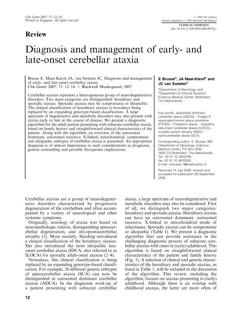
Clin Genet 2007: 71: 12–24<br />
Printed in Singapore. All rights reserved<br />
Review<br />
# 2006 The Authors<br />
Journal compilation # 2006 Blackwell Munksgaard<br />
CLINICAL GENETICS<br />
doi: 10.1111/j.1399-0004.2006.00722.x<br />
<strong>Diagnosis</strong> <strong>and</strong> <strong>management</strong> <strong>of</strong> <strong>early</strong>- <strong>and</strong><br />
<strong>late</strong>-<strong>onset</strong> <strong>cerebellar</strong> <strong>ataxia</strong><br />
Brusse E, Maat-Kievit JA, van Swieten JC. <strong>Diagnosis</strong> <strong>and</strong> <strong>management</strong><br />
<strong>of</strong> <strong>early</strong>- <strong>and</strong> <strong>late</strong>-<strong>onset</strong> <strong>cerebellar</strong> <strong>ataxia</strong>.<br />
Clin Genet 2007: 71: 12–14. # Blackwell Munksgaard, 2007<br />
Cerebellar <strong>ataxia</strong>s represent a heterogeneous group <strong>of</strong> neurodegenerative<br />
disorders. Two main categories are distinguished: hereditary <strong>and</strong><br />
sporadic <strong>ataxia</strong>s. Sporadic <strong>ataxia</strong>s may be symptomatic or idiopathic.<br />
The clinical classification <strong>of</strong> hereditary <strong>ataxia</strong>s is nowadays being<br />
replaced by an exp<strong>and</strong>ing genotype-based classification. A large<br />
spectrum <strong>of</strong> degenerative <strong>and</strong> metabolic disorders may also present with<br />
<strong>ataxia</strong> <strong>early</strong> or <strong>late</strong> in the course <strong>of</strong> disease. We present a diagnostic<br />
algorithm for the adult patient presenting with subacute <strong>cerebellar</strong> <strong>ataxia</strong>,<br />
based on family history <strong>and</strong> straightforward clinical characteristics <strong>of</strong> the<br />
patient. Along with the algorithm, an overview <strong>of</strong> the autosomal<br />
dominant, autosomal recessive, X-linked, mitochondrial, symptomatic<br />
<strong>and</strong> idiopathic subtypes <strong>of</strong> <strong>cerebellar</strong> <strong>ataxia</strong> is presented. An appropriate<br />
diagnosis is <strong>of</strong> utmost importance to such considerations as prognosis,<br />
genetic counselling <strong>and</strong> possible therapeutic implications.<br />
Cerebellar <strong>ataxia</strong>s are a group <strong>of</strong> neurodegenerative<br />
disorders characterized by progressive<br />
degeneration <strong>of</strong> the cerebellum <strong>and</strong> <strong>of</strong>ten accompanied<br />
by a variety <strong>of</strong> neurological <strong>and</strong> other<br />
systemic symptoms.<br />
Originally, nosology <strong>of</strong> <strong>ataxia</strong> was based on<br />
neuropathologic criteria, distinguishing spino<strong>cerebellar</strong><br />
degeneration, <strong>and</strong> olivoponto<strong>cerebellar</strong><br />
atrophy (1). More recently, Harding introduced<br />
a clinical classification <strong>of</strong> the hereditary <strong>ataxia</strong>s.<br />
She also introduced the term idiopathic <strong>late</strong><strong>onset</strong><br />
<strong>cerebellar</strong> <strong>ataxia</strong> (IDCA, also referred to as<br />
ILOCA) for sporadic adult-<strong>onset</strong> <strong>ataxia</strong>s (2–4).<br />
Nowadays, this clinical classification is being<br />
replaced by an exp<strong>and</strong>ing genotype-based classification.<br />
For example, 26 different genetic subtypes<br />
<strong>of</strong> spino<strong>cerebellar</strong> <strong>ataxia</strong> (SCA) can now be<br />
distinguished in autosomal dominant <strong>cerebellar</strong><br />
<strong>ataxia</strong>s (ADCA). In the diagnostic work-up <strong>of</strong><br />
a patient presenting with subacute <strong>cerebellar</strong><br />
12<br />
E Brusse a , JA Maat-Kievit b <strong>and</strong><br />
JC van Swieten a<br />
a Department <strong>of</strong> Neurology, <strong>and</strong><br />
b Department <strong>of</strong> Clinical Genetics,<br />
Erasmus Medical Center, Rotterdam,<br />
The Netherl<strong>and</strong>s<br />
Key words: autosomal dominant<br />
<strong>cerebellar</strong> <strong>ataxia</strong> (ADCA) – Fragile-Xassociated<br />
tremor <strong>ataxia</strong> syndrome<br />
(FXTAS) – Friedreich <strong>ataxia</strong> – idiopathic<br />
<strong>late</strong>-<strong>onset</strong> <strong>cerebellar</strong> <strong>ataxia</strong> (ILOCA) –<br />
multiple system atrophy (MSA) –<br />
spino<strong>cerebellar</strong> <strong>ataxia</strong> (SCA)<br />
Corresponding author: E. Brusse, MD.<br />
Department <strong>of</strong> Neurology, Erasmus<br />
Medical Centre, P.O Box 2040,<br />
3000 CA Rotterdam, The Netherl<strong>and</strong>s.<br />
Tel.: 00 31 10 4633780;<br />
fax: 00 31 10 4633208;<br />
e-mail: e.brusse.1@erasmusmc.nl<br />
Received 14 July 2006, revised <strong>and</strong><br />
accepted for publication 28 September<br />
2006<br />
<strong>ataxia</strong>, a large spectrum <strong>of</strong> neurodegenerative <strong>and</strong><br />
metabolic disorders may also be considered. First<br />
<strong>of</strong> all, we distinguish two major categories:<br />
hereditary <strong>and</strong> sporadic <strong>ataxia</strong>s. Hereditary <strong>ataxia</strong>s<br />
can have an autosomal dominant, autosomal<br />
recessive, X-linked or mitochondrial mode <strong>of</strong><br />
inheritance. Sporadic <strong>ataxia</strong>s can be symptomatic<br />
or idiopathic (Table 1). We present a diagnostic<br />
algorithm that can provide assistance in the<br />
challenging diagnostic process <strong>of</strong> subacute <strong>cerebellar</strong><br />
<strong>ataxia</strong>s with <strong>onset</strong> in (<strong>early</strong>) adulthood. This<br />
algorithm is based on straightforward clinical<br />
characteristics <strong>of</strong> the patient <strong>and</strong> family history<br />
(Fig. 1). A selection <strong>of</strong> clinical <strong>and</strong> genetic characteristics<br />
<strong>of</strong> the hereditary <strong>and</strong> sporadic <strong>ataxia</strong>s, as<br />
listed in Table 1, will be included in the discussion<br />
<strong>of</strong> the algorithm. This review, including the<br />
algorithm, focuses on <strong>ataxia</strong>s presenting in (<strong>early</strong>)<br />
adulthood. Although there is an overlap with<br />
childhood <strong>ataxia</strong>s, the latter are more <strong>of</strong>ten <strong>of</strong>
Table 1. Classification <strong>of</strong> <strong>cerebellar</strong> <strong>ataxia</strong>s<br />
I. Hereditary <strong>cerebellar</strong> <strong>ataxia</strong>s<br />
a. Autosomal dominant <strong>cerebellar</strong> <strong>ataxia</strong>s (ADCA)<br />
i. Episodic <strong>ataxia</strong>s (types 1–6)<br />
ii. Spino<strong>cerebellar</strong> <strong>ataxia</strong> (SCA) subtypes 1–28<br />
iii. Dentatorubral-pallidoluysian atrophy (DRPLA)<br />
b. Autosomal recessive <strong>cerebellar</strong> <strong>ataxia</strong>s<br />
i. With identified gene defect<br />
ii. With identified gene locus<br />
iii. As part <strong>of</strong> metabolic disorder, extended disease<br />
iv. Other metabolic <strong>and</strong> degenerative disease<br />
with congenital or childhood <strong>onset</strong><br />
c. X-linked <strong>cerebellar</strong> <strong>ataxia</strong><br />
i. Adrenoleukodystrophy<br />
ii. Fragile-X tremor <strong>ataxia</strong> syndrome (FXTAS)<br />
iii. Hereditary sideroblastic anemia <strong>and</strong> <strong>ataxia</strong><br />
iv. Other X-linked congenital <strong>and</strong> childhood <strong>ataxia</strong>s<br />
d. Mitochondrial <strong>cerebellar</strong> <strong>ataxia</strong><br />
II. Sporadic <strong>cerebellar</strong> <strong>ataxia</strong><br />
a. Symptomatic <strong>cerebellar</strong> <strong>ataxia</strong><br />
i. Structural lesions, malformations<br />
ii. Toxic<br />
d Alcohol<br />
d Drugs<br />
a. Antiepileptic drugs<br />
b. Benzodiazepines<br />
c. Lithium<br />
d. Antineoplastics<br />
e. Others (see text)<br />
d Others<br />
a. Heavy metals (mercury, lead)<br />
b. Chemicals (solvents, pesticides)<br />
iii. Endocrine<br />
d Hypothyroidism<br />
iv. Malabsorption<br />
d Celiac disease (gluten <strong>ataxia</strong>)<br />
d Vitamin deficiency<br />
v. Miscellaneous<br />
d Paraneoplastic syndromes<br />
d Demyelinating disorders<br />
vi. Inflammatory<br />
d Whipple disease<br />
d Post-viral/immune-mediated <strong>ataxia</strong><br />
b. Idiopathic<br />
1. Multiple system atrophy (MSA)<br />
2. Idiopathic <strong>late</strong>-<strong>onset</strong> <strong>cerebellar</strong> <strong>ataxia</strong> (ILOSCA)<br />
congenital, metabolic or syndrome-associated origin.<br />
This requires other diagnostic considerations,<br />
which are beyond the scope <strong>of</strong> this review.<br />
The diagnostic algorithm<br />
Magnetic resonance imaging <strong>of</strong> the brain<br />
The diagnostic process starts with magnetic<br />
resonance imaging (MRI) <strong>of</strong> the brain. This<br />
may identify structural lesions that may be the<br />
cause <strong>of</strong> symptomatic <strong>ataxia</strong>s. Cerebellar atrophy<br />
is a nonspecific feature. Other features may be<br />
more specific, such as white matter lesions in the<br />
<strong>cerebellar</strong> pedunculi, seen in fragile-X-associated<br />
tremor <strong>ataxia</strong> syndrome (FXTAS).<br />
<strong>Diagnosis</strong> <strong>and</strong> <strong>management</strong> <strong>of</strong> <strong>cerebellar</strong> <strong>ataxia</strong><br />
Family history<br />
In patients with a positive family history,<br />
a detailed genealogy will <strong>of</strong>ten reveal an apparent<br />
mode <strong>of</strong> inheritance. The presence <strong>of</strong> <strong>ataxia</strong> in<br />
consecutive generations or the presence <strong>of</strong> maleto-male<br />
inheritance is suggestive <strong>of</strong> an autosomal<br />
dominant inheritance. The presence <strong>of</strong> multiple<br />
affected sibs in a single generation, or consanguinity<br />
in the parents, supports the idea <strong>of</strong> an<br />
autosomal recessive mode <strong>of</strong> inheritance. In<br />
a disorder affecting only males in one or more<br />
generations in the maternal line, X-linked inheritance<br />
can be considered. It is important to realize<br />
that an ADCA may present as an apparently<br />
sporadic or recessive <strong>ataxia</strong>: relatives who carry<br />
the mutation may be clinically unaffected due to<br />
anticipation with <strong>late</strong>r <strong>onset</strong> <strong>and</strong> milder phenotype<br />
in elderly generations, or due to reduced<br />
penetrance. Furthermore, a negative family<br />
history cannot rule out a hereditary <strong>ataxia</strong>, for<br />
example in relation with a de novo mutation. A<br />
genetic defect may be identified in 2–19% <strong>of</strong> the<br />
sporadic <strong>ataxia</strong>s, most frequently representing<br />
Friedreich <strong>ataxia</strong> (FA) or SCA6 (5–7).<br />
Mitochondrial diseases may represent different<br />
modes <strong>of</strong> inheritance because mutations may<br />
originate in nuclear or mitochondrial DNA<br />
(mtDNA). Furthermore, mtDNA mutations<br />
may mimic autosomal dominant, recessive or<br />
X-linked inheritance due to phenomena such as<br />
heteroplasmy <strong>and</strong> threshold effects for biochemical<br />
mutation expression. Moreover, deletions<br />
<strong>and</strong> insertions in mtDNA are <strong>of</strong>ten sporadic (8).<br />
Consequently, the algorithm does not provide<br />
a diagnostic tool for mitochondrial disease based<br />
on family history. Specific phenotypic features <strong>of</strong><br />
the patient (<strong>and</strong> family) may suggest mitochondrial<br />
<strong>ataxia</strong>, <strong>of</strong>ten associated with typical mitochondriopathies,<br />
as described <strong>late</strong>r.<br />
Apparently dominant <strong>ataxia</strong>s<br />
Autosomal dominant <strong>cerebellar</strong> <strong>ataxia</strong>s<br />
SCA constitutes the main group in the genetic<br />
classification <strong>of</strong> ADCA, with the current identification<br />
<strong>of</strong> 26 subtypes. Furthermore, a clinically<br />
complex form (dentatorubral-pallidoluysian<br />
atrophy, DRPLA) <strong>and</strong> six episodic <strong>ataxia</strong>s (EA)<br />
are distinguished (Tables 1 <strong>and</strong> 2) (9–11). Other<br />
rare autosomal dominant disorders, like hereditary<br />
spastic <strong>ataxia</strong>s, sensory motor neuropathy<br />
with <strong>ataxia</strong> <strong>and</strong> adult-<strong>onset</strong> leukodystrophy, may<br />
also present with <strong>ataxia</strong>, but these heterogeneous,<br />
rare disorders will not be considered in this<br />
overview (11, 12). Although most patients with<br />
EA2 develop interictal <strong>cerebellar</strong> signs, the<br />
13
Brusse et al.<br />
yes<br />
EA<br />
DNA: EA 2<br />
yes<br />
MRI cerebrum<br />
Apparently<br />
dominant <strong>ataxia</strong><br />
(more generations<br />
affected) 1<br />
Episodic<br />
SCA<br />
no<br />
DNA: SCA 3<br />
Subacute <strong>cerebellar</strong> <strong>ataxia</strong>, <strong>onset</strong> (<strong>early</strong>) adulthood<br />
Structural<br />
lesions, other<br />
than atrophy<br />
episodic occurrence <strong>of</strong> symptoms differentiates<br />
EA from SCA (13).<br />
Episodic <strong>ataxia</strong>s. EA1, EA2 <strong>and</strong> EA5 are chanelopathies<br />
associated with mutations in genes<br />
encoding voltage-dependent potassium (EA1) <strong>and</strong><br />
calcium (EA2 <strong>and</strong> 5) channel subunits. In EA6,<br />
mutations in SLC1A3 have been found, a sodiumdependent<br />
transporter molecule regulating neurotransmitter<br />
concentrations (14). The genetic<br />
locus in EA3 has been found, whereas the genetic<br />
defect in EA4 still awaits identification (Table 2).<br />
EA1 <strong>and</strong> EA2 can be distinguished on clinical<br />
grounds, EA5 has an EA2 phenotype <strong>and</strong> EA6 is<br />
associated with seizures (13, 14).<br />
Movement-induced or the so-called kinesigenic<br />
attacks <strong>of</strong> <strong>ataxia</strong>, being provoked by exercise,<br />
startle <strong>and</strong> change <strong>of</strong> position, are characteristic<br />
for EA1, whereas caffeine, alcohol <strong>and</strong> phenytoin<br />
<strong>of</strong>ten trigger attacks in the nonkinesigenic EA2.<br />
In both subtypes, stress may also induce attacks.<br />
Typically, <strong>onset</strong> <strong>of</strong> EA occurs in childhood or<br />
<strong>early</strong> adulthood, although the <strong>onset</strong> <strong>of</strong> EA2 may<br />
be up to the fifth decade. Attacks in EA2 can last<br />
hours to days, whereas the attacks in EA1 are<br />
shorter (,15 min) but more frequent (up to 15<br />
times a day). Tremor, muscle cramps <strong>and</strong><br />
stiffening <strong>of</strong> extremities <strong>of</strong>ten accompany <strong>ataxia</strong><br />
in EA1. In EA2, <strong>ataxia</strong> is <strong>of</strong>ten associated with<br />
nausea, migrainous headache <strong>and</strong> (hemi)paresis<br />
<strong>and</strong> sometimes also with dystonia, diplopia <strong>and</strong><br />
no<br />
Family history<br />
positive<br />
yes<br />
Onset<br />
< 25 y<br />
neg.<br />
Apparently recessive<br />
<strong>ataxia</strong> (sibs affected<br />
<strong>and</strong>/or consanguinity) 1<br />
yes<br />
neg.<br />
Complete screening<br />
for recessive ataxic<br />
syndromes <strong>and</strong><br />
metabolic disorders 4<br />
DNA: FRDA<br />
neg.<br />
no<br />
Onset<br />
> 25 y<br />
Limited screening for<br />
<strong>late</strong> <strong>onset</strong> recessive<br />
ataxic syndromes <strong>and</strong><br />
metabolic disorders 5<br />
Apparently Xlinked<br />
<strong>ataxia</strong><br />
(affected males) 1<br />
yes<br />
no<br />
yes<br />
pos.<br />
Onset<br />
< 50y<br />
yes<br />
male, <strong>onset</strong> >50 y,<br />
X-linked or<br />
sporadic<br />
DNA:FMR1premutation<br />
Screening for<br />
X-ALD 6<br />
yes<br />
yes:<br />
consider as apparently recessive or (in males) X-linked <strong>ataxia</strong><br />
Fig. 1. Diagnostic algorithm for patients with (<strong>early</strong>) adult <strong>onset</strong> subacute <strong>cerebellar</strong> <strong>ataxia</strong>.<br />
14<br />
FXTAS<br />
no<br />
no<br />
no<br />
Apparently sporadic<br />
<strong>ataxia</strong> 1<br />
tinnitus. Interictal myokymia <strong>of</strong> the face, arms<br />
<strong>and</strong> legs is seen in EA1, whereas permanent<br />
interictal <strong>cerebellar</strong> signs (nystagmus, limb<br />
<strong>ataxia</strong>) develop during the course <strong>of</strong> EA2.<br />
Acetazolamide is an effective therapy in most<br />
patients with EA2 <strong>and</strong> in half <strong>of</strong> the patients with<br />
EA1; phenytoin is an alternative therapy in EA1<br />
(Fig. 1, note 2) (13, 15).<br />
Apart from EA2, mutations in the CACNA1A<br />
gene are also associated with familial hemiplegic<br />
migraine (FHM) <strong>and</strong> SCA6. Evident clinical<br />
overlap exists between EA2, FHM <strong>and</strong> SCA6,<br />
even within families (13, 15).<br />
Spino<strong>cerebellar</strong> <strong>ataxia</strong>s <strong>and</strong> dentatorubral-pallidoluysian<br />
atrophy. The relative prevalence <strong>of</strong><br />
various SCA subtypes varies considerably<br />
between different countries due to founder<br />
effects. However, worldwide SCA1, 2, 3, 6 <strong>and</strong><br />
7 explain 50–65% <strong>of</strong> all cases <strong>of</strong> autosomal<br />
dominant <strong>ataxia</strong>, with SCA3 as the most<br />
common subtype. Other known SCA genotypes<br />
are rare (,1%), whereas in the remaining 35–<br />
50% <strong>of</strong> SCA families, the genotype is unknown<br />
(9–11, 16).<br />
Cerebellar dysfunction in SCA is most <strong>of</strong>ten<br />
associated with a variety <strong>of</strong> other clinical<br />
features. Involvement <strong>of</strong> the long spinal tracts<br />
is common, with signs <strong>of</strong> diminished vibratory<br />
sense <strong>of</strong> the legs <strong>and</strong> hyperreflexia. Extrapyramidal<br />
features, spasticity, cognitive impairment,<br />
yes<br />
Screening for toxic <strong>ataxia</strong> (alcohol, drugs),<br />
endocrine disorders or gluten <strong>ataxia</strong> 7<br />
Onset<br />
> 50y<br />
neg.<br />
Meets criteria<br />
for MSA 8<br />
Progressive<br />
course within<br />
6 months<br />
Diarrhea/<br />
weight loss/<br />
abdominal pain<br />
Onset<br />
< 50y<br />
yes<br />
yes<br />
yes<br />
MSA<br />
Screening for<br />
paraneoplastic<br />
antibodies or<br />
inflammatory/immunemediated<br />
disorders 9<br />
Screening for Whipple/<br />
malabsorption 10
Table 2. The autosomal dominant <strong>cerebellar</strong> <strong>ataxia</strong>s<br />
Name Gene/gene product Locus<br />
polyneuropathy, ophthalmoplegia <strong>and</strong> epilepsy<br />
are also seen. Although there is a considerable<br />
overlap in clinical features between different<br />
SCA genotypes, some distinctive clinical features<br />
may be useful in the genetic screening <strong>of</strong> a specific<br />
patient. The presence <strong>of</strong> pigmentary retinopathy<br />
is almost always associated with SCA7. A pure<br />
<strong>cerebellar</strong>, <strong>late</strong>-<strong>onset</strong> subtype is most <strong>of</strong>ten<br />
described in SCA6, 10, 11 <strong>and</strong> 12 (Fig. 1, note<br />
3) (9–12). The mean age <strong>of</strong> <strong>onset</strong> in SCA is in the<br />
third or fourth decade, but the <strong>onset</strong> varies<br />
considerably between the SCA genotypes <strong>and</strong><br />
also between patients <strong>of</strong> the same SCA subtype<br />
or same family (Table 2). This is partly due to the<br />
<strong>Diagnosis</strong> <strong>and</strong> <strong>management</strong> <strong>of</strong> <strong>cerebellar</strong> <strong>ataxia</strong><br />
Mean age at <strong>onset</strong><br />
(range)<br />
SCA1 ATXN1/ataxin-1 6p23 37 (,10 to .70) 1<br />
SCA2 ATXN2/ataxin-2 12q24 32 (,10 to .60) 1<br />
SCA3 ATXN3/ataxin-3; Machado<br />
Joseph disease (MJD) protein 1<br />
14q24.3-q31 36 (,10 to .60) 1<br />
SCA4 Q9H7K4/puratrophin-1 16q22.1 ? (19–72) 1<br />
SCA5 SPTBN2/spectrin beta chain, brain 2 11q13 30 (10–68) 1<br />
SCA6 CACNA1A/voltage-dependent<br />
P/Q-type calcium channel<br />
alpha-1A subunit<br />
19p13 52 (20 to .70) 1<br />
SCA7 ATXN7/ataxin-7 3p21.1-p12 35 (,1 to .60) 1<br />
SCA8 KLHL1AS 13q21 40 (1 to .60) 1<br />
SCA9 Reserved<br />
SCA10 ATXN10/ataxin-10/E46L 22q13 36 (26–45) 1<br />
SCA11 — 15q14-q21.3 25 (15 to .60)<br />
SCA12 PPP2R2B/brain-specific regulatory<br />
subunit <strong>of</strong> protein phosphatase 2A<br />
(serine threonine phosphatase)<br />
5q31-q33 35 (8–55) 1<br />
SCA13 19q13.3-q13.4 Childhood (,1 to ,45)<br />
SCA14 PRKCG/protein kinase C,<br />
gamma subtype<br />
19q13.4 27 (12–42) 1<br />
SCA15 3p26.1-p25.3 26 (10–50)<br />
SCA16 8q22.1-q24.1 40 (20–66)<br />
SCA17 TBP/TATA-box binding-protein 6q27 33 (6 to .40) 1<br />
SCA18 7q22-q32 15 (12–25)<br />
SCA19 1p21-q21 34 (11–45)<br />
SCA20 11p13-q11 46 (19–64)<br />
SCA21 7p21.3-p15.1 18 (7–30)<br />
SCA22 1p21-q21 ? (10–46)<br />
SCA23 20p13-12.3 ? (40–60)<br />
SCA24 Reserved<br />
SCA25 2p21-p13 ? (1–39)<br />
SCA26 19p13.3<br />
SCA27 FGF14/fibroblast growth factor 14 13q34 11 (7–20) 1<br />
SCA28 18p11.22-q11.2<br />
DRPLA DRPLA/atrophin-1-re<strong>late</strong>d protein 12p13.31 30 (0 to .60) 1<br />
EA1 KCNA1/potassium voltage-gated<br />
channel component<br />
12p13 ? (2–15) 1<br />
EA2 CACNA1A/voltage-dependent P/Q-type<br />
calcium channel alpha-1A subunit<br />
19p13 ? (3 to .50) 1<br />
EA3 — 1q42 Variable<br />
EA4 — — 30–60<br />
EA5 CACNB4/voltage-dependent L-type<br />
calcium channel beta-4 subunit<br />
2q22-q23 ?<br />
EA6 SCL1A3/sodium-dependent<br />
gluatamate transporter (EAAT1)<br />
5p13 ?<br />
Clinical testing<br />
available<br />
phenomenon <strong>of</strong> anticipation, described below. A<br />
childhood <strong>onset</strong> with severe phenotype due to<br />
extreme anticipation is indicative <strong>of</strong> SCA2, SCA7<br />
<strong>and</strong> SCA17 (9, 11), while a childhood <strong>onset</strong> with<br />
tremor is characteristic <strong>of</strong> SCA27 (17). In all<br />
SCA subtypes, MRI <strong>of</strong> the brain shows progressive<br />
<strong>cerebellar</strong> atrophy in the course <strong>of</strong> the<br />
disease, sometimes combined with atrophy <strong>of</strong> the<br />
brain stem or spinal cord (9).<br />
The underlying gene defect is known in 13 <strong>of</strong><br />
the 26 SCA genotypes <strong>and</strong> DRPLA (Table 2).<br />
Most <strong>of</strong> the known SCA mutations give rise to<br />
CAG trinucleotide repeat expansions. There is<br />
evident correlation between the size <strong>of</strong> this<br />
15
Brusse et al.<br />
expansion <strong>and</strong> age at <strong>onset</strong> <strong>and</strong> severity <strong>of</strong> the<br />
disease. This explains the phenomenon <strong>of</strong> anticipation,<br />
where instability <strong>of</strong> larger repeats gives<br />
rise to further expansions in subsequent generations,<br />
resulting in earlier <strong>onset</strong> <strong>and</strong> a more<br />
severe phenotype. Repeat instability is more<br />
obvious in paternal transmission (9, 11). SCA1,<br />
2, 3, 6, 7, 17 <strong>and</strong> DRPLA are caused by CAG<br />
repeat expansions in coding regions, resulting in<br />
polyglutamine tracts. SCA12 gives rise to CAG<br />
repeat expansion in a promotor region <strong>of</strong> the<br />
PP2R2B gene, not encoding a polyglutamine<br />
tract (9, 11). In SCA10, an ATTCT pentanucleotide<br />
repeat expansion has been identified in<br />
intron 9 <strong>of</strong> the ATXN10 gene. The functional<br />
effect <strong>of</strong> this expansion is still under investigation<br />
(18). In SCA8, it is still questioned whether the<br />
CTG trinucleotide expansion in the noncoding 3#<br />
untrans<strong>late</strong>d region <strong>of</strong> the KLHL1AS gene is<br />
causative <strong>of</strong> or only associative to the disorder<br />
because there is no relationship between repeat<br />
size <strong>and</strong> age at <strong>onset</strong> or disease severity (9, 10).<br />
Four other SCA subtypes are associated with<br />
point mutations or deletions (Table 2) (17, 19–21).<br />
The unravelling <strong>of</strong> the complex genetic background<br />
<strong>of</strong> SCA has not yet resulted in finding<br />
a final common pathogenic mechanism. The<br />
CAG trinucleotide repeat variants give rise to<br />
polyglutamine tracts, aggregating in cells. Many<br />
other proteins colocalize with intranuclear inclusions<br />
or interact with the mutant polyglutamine<br />
stretches. These proteins may be<br />
pathogenic but may also block the toxic properties<br />
<strong>of</strong> polyglutamine. The ubiquitin–proteasome<br />
pathway is presumed to play a protective role<br />
against the toxicity <strong>of</strong> mutant polyglutamine<br />
proteins by sequestering them into inclusions (9,<br />
22). Furthermore, an RNA-mediated pathophysiological<br />
mechanism, like the toxic gain <strong>of</strong><br />
function described in myotonic dystrophia <strong>and</strong><br />
FXTAS, has been suggested (23). In SCA<br />
subtypes associated with point mutations, even<br />
less is known about pathogenesis.<br />
Apparently recessive <strong>cerebellar</strong> <strong>ataxia</strong>s<br />
The autosomal recessive <strong>ataxia</strong>s represent a heterogeneous<br />
group <strong>of</strong> rare neurodegenerative<br />
disorders. Harding’s original clinical classification<br />
is gradually replaced by a genetic classification<br />
(Table 3) (2). Congenital <strong>and</strong> childhood <strong>ataxia</strong>s<br />
are reviewed in several recent publications <strong>and</strong><br />
will not be discussed in this review (24, 25).<br />
FA <strong>and</strong> <strong>ataxia</strong> telangiectasia (AT) are the<br />
most frequent subtypes <strong>of</strong> recessive <strong>ataxia</strong>, with<br />
an estimated prevalence <strong>of</strong> 1 in 50,000 <strong>and</strong> 1 in<br />
16<br />
100,000, respectively. Some subtypes <strong>of</strong> recessive<br />
<strong>ataxia</strong> show a regional distribution: for example,<br />
<strong>ataxia</strong> oculomotor apraxia type I (AOA1) is most<br />
frequent in Portugal <strong>and</strong> Japan (26, 27). In the<br />
algorithm for recessive <strong>ataxia</strong>, FA, being the<br />
most frequent cause, is excluded first. The age at<br />
<strong>onset</strong> determines the next step in the screening<br />
process. The <strong>onset</strong> age <strong>of</strong> 25 years is an arbitrary<br />
but reasonable cut<strong>of</strong>f point because most recessive<br />
<strong>ataxia</strong>s <strong>and</strong> metabolic neurodegenerative<br />
disorders have a childhood <strong>onset</strong>. A minority <strong>of</strong><br />
recessive <strong>ataxia</strong>s <strong>and</strong> metabolic disorders can<br />
have an adult <strong>onset</strong>, <strong>of</strong>ten associated with milder<br />
phenotypes due to residual enzyme activity, <strong>and</strong><br />
these disorders should be excluded patients older<br />
than 25 years (Fig. 1, note 5).<br />
When screening indicates a recessive <strong>ataxia</strong> or<br />
metabolic disorder, it is advisable to check the<br />
local availability <strong>of</strong> DNA screening for this<br />
disorder (9, 11, 12). Ultimately, screening for<br />
SCA mutations is advised in all patients with<br />
apparently recessive <strong>ataxia</strong> <strong>and</strong> a negative screening<br />
for recessive disorders because an autosomal<br />
dominant inheritance cannot be ruled out (5–7).<br />
Friedreich <strong>ataxia</strong><br />
Ataxia, dysarthria, absent deep tendon reflexes,<br />
pyramidal signs <strong>and</strong> an <strong>early</strong>-<strong>onset</strong> (,25 years)<br />
are the classical clinical features <strong>of</strong> FA. Cardiomyopathy<br />
is <strong>of</strong>ten present, <strong>and</strong> scoliosis, distal<br />
muscle atrophy, sensorineural deafness, optic<br />
atrophy <strong>and</strong> diabetes are common features (26).<br />
Since the introduction <strong>of</strong> genetic testing, the<br />
phenotype has appeared to be more variable, <strong>and</strong><br />
up to 25% does not meet the original diagnostic<br />
criteria for FA (28). A <strong>late</strong>-<strong>onset</strong> variant <strong>of</strong> FA,<br />
presenting at the age <strong>of</strong> 25–48 years, with<br />
a milder, slowly progressive phenotype, <strong>of</strong>ten<br />
without cardiomyopathy, has been described <strong>and</strong><br />
also a phenotype with spastic paraplegia but<br />
without <strong>ataxia</strong> or neuropathy (26, 28, 29). This<br />
variation in phenotype is re<strong>late</strong>d to the size <strong>of</strong> the<br />
GAA trinucleotide repeat expansion in the<br />
FRDA1 gene, varying from 90 to 1300 repeats<br />
(normal range 6–33 repeats). The expansion size<br />
is inversely re<strong>late</strong>d to age at <strong>onset</strong> <strong>and</strong> impairment<br />
<strong>of</strong> mobility <strong>and</strong> directly re<strong>late</strong>d to the<br />
incidence <strong>of</strong> cardiomyopathy (26, 28). The<br />
FRDA1 gene encodes frataxin, a mitochondrial<br />
protein, <strong>and</strong> FA is believed to be a disease <strong>of</strong><br />
mitochondrial dysfunction (26).<br />
Other recessive <strong>ataxia</strong>s<br />
The algorithm indicates an age-dependent<br />
screening for recessive ataxic syndromes <strong>and</strong><br />
metabolic diseases. Obviously, specific clinical
Table 3. The autosomal recessive <strong>cerebellar</strong> <strong>ataxia</strong>s a<br />
Name Gene/locus Protein Age at <strong>onset</strong><br />
With identified gene defect<br />
Friedreich <strong>ataxia</strong> FRDA1/9q13-q21 Frataxin 2–48<br />
Ataxia telangiectasia ATM/11q22-q23 Phosphoinositol-3kinase-type<br />
enzyme<br />
2–22<br />
Ataxia with oculomotor apraxia type 1 APTX/9p13 Aprataxin 2–16<br />
Ataxia with oculomotor apraxia type 2 SETX/9q34 Senataxin 10–22<br />
Spino<strong>cerebellar</strong> <strong>ataxia</strong> with<br />
TDP1/14q31 Tyrosyl DNA<br />
Teenage<br />
axonal neuropathy<br />
phosphodiesterase 1<br />
Ataxia with vitamin E deficiency a -TTP/8q13 a-tocopherol<br />
transfer protein<br />
2–52<br />
Abetalipoproteinemia MPT/4q22-24 Microsomal triglyceride<br />
transfer protein MPT<br />
Second decade<br />
Spastic <strong>ataxia</strong> Charlevoix-Saguenay SACS/13q12 Sacsin First decade<br />
Infantile <strong>onset</strong> spino<strong>cerebellar</strong> <strong>ataxia</strong> C10orf2/10q24 Mitochondrial proteins<br />
Twinkle <strong>and</strong> Twinky<br />
9–18 months<br />
Autosomal recessive mitochondrial<br />
POLG (DNA<br />
Juvenile or adult<br />
ataxic syndrome<br />
Polymerase g)<br />
Marinesco-Sjögren syndrome SIL1/5q31 Infancy<br />
Cayman <strong>ataxia</strong> ATCAY/19p13.3 Caytaxin Childhood<br />
Nonprogressive <strong>cerebellar</strong> <strong>ataxia</strong>,<br />
VLDLR (very<br />
mental retardation <strong>and</strong> cerebral<br />
low-density<br />
gyral simplification<br />
With identified gene locus<br />
lipoprotein<br />
receptor) gene<br />
Childhood <strong>onset</strong> slow progressive <strong>ataxia</strong> 11p15 Childhood<br />
Early-<strong>onset</strong> <strong>ataxia</strong> with hearing impairment<br />
<strong>and</strong> optic atrophy<br />
6p21-23<br />
Early-<strong>onset</strong> <strong>ataxia</strong> with developmental<br />
delay <strong>and</strong> failure to thrive<br />
22q11 Infancy<br />
Congenital <strong>ataxia</strong> with mental retardation,<br />
optic atrophy <strong>and</strong> skin abnormalities<br />
15q24-q26 Congenital<br />
Nonprogressive infantile <strong>ataxia</strong><br />
As part <strong>of</strong> metabolic disorder,<br />
extended disease<br />
20q11-q13 Infancy<br />
Metachromatic leucodystrophy ARSA/22q13 Arylsulfatase A Infancy to adulthood<br />
Krabbe GALC/14q31 Galactosylceramidase Infancy to fifth decade<br />
Cerebrotendinous xanthomatosis CYP27A1/2q33-ter Sterol 27-hydroxylase Infancy to juvenile<br />
Niemann-Pick C (sphingomyelin<br />
storage disorder)<br />
NPC1/18q11-121 NPC1 protein Childhood/adulthood<br />
GM1 gangliosidosis GLB1/3p21.33 Beta-galactosidase Childhood<br />
Tay-Sachs disease (GM2-gangliosidosis,<br />
hexosaminidase deficiency<br />
HEXA/15q23-24 Hexosaminidase A Childhood/adulthood<br />
Wilson’s disease ATP7B/13q14-21 ATPase Cu transporting<br />
b-polypeptide<br />
3–50<br />
Aceruloplasminemia CP Ceruloplasmin 25–60<br />
Refsum’s disease PHYH,PEX7/<br />
Phytanoyl-CoA hydroxylase, Infancy to 28(.50)<br />
10p11-pter, 6q22-24 peroxin 7<br />
Sialidosis Neu1/6p21.3 Neuraminidase Childhood<br />
Chorea-acanthocytosis CHAC/9q21 Chorein 23–59<br />
Gamma-glutamyl cysteine synthetase GCLC/6p21 Gamma-glutamyl<br />
cysteine synthetase<br />
Leucoencephalopathy with<br />
EIF2B1, B2, B3, Translocation initiation Variable<br />
vanishing white matter<br />
B4, B5/12, 14q24,<br />
1, 2p23, 3q27<br />
factor EIF2B 5 subunits<br />
findings may help differentiate the recessive<br />
<strong>cerebellar</strong> <strong>ataxia</strong>s as classified in Table 3 <strong>and</strong><br />
may indicate a selective use <strong>of</strong> diagnostic tests.<br />
Therefore, the following part presents a brief<br />
overview <strong>of</strong> most recessive <strong>ataxia</strong>s, based on<br />
prominent clinical characteristics apart from<br />
<strong>ataxia</strong>, to determine first-line testing. In the<br />
<strong>Diagnosis</strong> <strong>and</strong> <strong>management</strong> <strong>of</strong> <strong>cerebellar</strong> <strong>ataxia</strong><br />
a Other metabolic <strong>and</strong> degenerative disease <strong>of</strong> childhood <strong>onset</strong> <strong>and</strong> congenital <strong>ataxia</strong>s are given in references 11, 24 <strong>and</strong> 25.<br />
absence <strong>of</strong> such characteristics, a complete<br />
screening is advised (Fig. 1, notes 4 <strong>and</strong> 5).<br />
Oculomotor apraxia. Oculomotor apraxia is a<br />
common sign in AT <strong>and</strong> AOA1 <strong>and</strong> 2. AT is further<br />
characterized by <strong>ataxia</strong> <strong>and</strong> oculocutaneous<br />
17
Brusse et al.<br />
telangiectases <strong>and</strong> <strong>of</strong>ten choreoathetosis <strong>and</strong><br />
dystonia are present. Immunodeficiency, hypersensitivity<br />
to ionizing radiation (IR) <strong>and</strong> predisposition<br />
to malignancy are also specific for AT.<br />
In AOA1 <strong>and</strong> 2, polyneuropathy is also seen. In<br />
AOA1, mild mental retardation may be present.<br />
Onset <strong>of</strong> AT is usually in <strong>early</strong> childhood, patients<br />
are wheelchair bound from their <strong>early</strong> teenage<br />
years <strong>and</strong> die between 20 <strong>and</strong> 30 years. Onset <strong>of</strong><br />
AOA is mostly in childhood or <strong>early</strong> teens. An<br />
elevated a-fetoprotein level is found in AT <strong>and</strong><br />
AOA2 (26, 30, 31).<br />
AT is associated with mutations in the AT<br />
mutated (ATM) gene, which encodes the phosphatidylinositol<br />
3-kinase protein, which is re<strong>late</strong>d<br />
to processes <strong>of</strong> DNA repair. The clinical<br />
variation in AT is partly re<strong>late</strong>d to the relative<br />
preservation <strong>of</strong> ATM protein expression in<br />
specific ATM mutations, leading to milder<br />
phenotypes with <strong>late</strong>r <strong>onset</strong>, slower progression<br />
<strong>and</strong> no sensitivity to IR (32, 33). AOA1 is<br />
associated with the recently identified aprataxin<br />
gene (APTX); the senataxin (SETX) gene is<br />
involved in AOA2. Both genes are believed to<br />
play a role in the DNA repair pathway (31, 32,<br />
34). The frequency <strong>of</strong> AOA2 in non-Friedreich<br />
autosomal recessive <strong>cerebellar</strong> <strong>ataxia</strong> (ARCA) is<br />
estimated at 8%, suggesting AOA2 to be a frequent<br />
cause <strong>of</strong> ARCA (31).<br />
Polyneuropathy. Areflexia <strong>and</strong> loss <strong>of</strong> vibration<br />
sense <strong>of</strong> the lower limbs, <strong>of</strong>ten combined with<br />
pyramidal signs, are common in FA <strong>and</strong> also in<br />
<strong>ataxia</strong> with vitamin E deficiency (AVED).<br />
However, in AVED, cardiomyopathy <strong>and</strong> diabetes<br />
are uncommon. The <strong>onset</strong> is usually before<br />
the age <strong>of</strong> 20 years, but this may range from 2 to<br />
52 years (26). The defect in the gene coding for<br />
a-tocopherol transfer protein, associated with<br />
AVED, causes impairment <strong>of</strong> incorporation <strong>of</strong><br />
dietary vitamin E (a-tocopherol) into very lowdensity<br />
lipoproteins, leading to a reduction <strong>of</strong><br />
plasma vitamin E (24, 26).<br />
Peripheral neuropathy with sensory loss, distal<br />
muscle atrophy <strong>and</strong> areflexia is also seen in<br />
abetalipoproteinemia. This disorder starts in<br />
childhood or <strong>early</strong> teens with intestinal celiaclike<br />
symptoms, followed by <strong>ataxia</strong>. Retinopathy<br />
is common, probably caused by vitamin A<br />
deficiency. On fresh blood smears, acanthocytes<br />
are <strong>of</strong>ten present (24, 26, 35). The disorder is<br />
caused by mutations in the microsomal triglyceride<br />
transfer protein (MTP) gene, ultimately<br />
leading to fat malabsorption with vitamin A, E<br />
<strong>and</strong> K deficiency.<br />
SCA with axonal neuropathy (SCAN1), caused<br />
by a mutation in the DNA repair protein tyrosyl<br />
18<br />
DNA phosphodiesterase 1, is a rare variant,<br />
overlapping the AT phenotype, with <strong>onset</strong> in<br />
teenage years but without oculomotor apraxia (32).<br />
Late-<strong>onset</strong> hexosaminidase A deficiency or<br />
GM2 gangliosidosis may present as a FA-like<br />
phenotype. Motor neuron disease with muscular<br />
atrophy <strong>of</strong>ten occurs <strong>and</strong> dementia or psychiatric<br />
features may develop <strong>late</strong>r (35, 36).<br />
Ophthalmologic signs. A Kaiser-Fleischer ring<br />
<strong>of</strong> the cornea is characteristic <strong>of</strong> Wilson disease,<br />
a disorder <strong>of</strong> copper metabolism, which may also<br />
show <strong>ataxia</strong>, chorea, dystonia <strong>and</strong> hepatic <strong>and</strong><br />
psychiatric symptoms (37). Retinal degeneration<br />
represents one <strong>of</strong> the characteristic signs <strong>of</strong><br />
aceruloplasminemia, another disorder <strong>of</strong> copper<br />
metabolism, in a triad with diabetes mellitus <strong>and</strong><br />
neurological symptoms, especially <strong>ataxia</strong> <strong>and</strong><br />
dementia. Heterozygotes <strong>of</strong> ceruloplasmin gene<br />
mutations may be symptomatic, showing hypoceruloplasminaemia<br />
<strong>and</strong> mild <strong>ataxia</strong> (38).<br />
Retinitis pigmentosa, combined with anosmia,<br />
polyneuropathy, <strong>cerebellar</strong> <strong>ataxia</strong> <strong>and</strong> <strong>of</strong>ten also<br />
deafness <strong>and</strong> ichthyosis, is characteristic for<br />
Refsum’s disease. Retinitis pigmentosa always<br />
develops before the age <strong>of</strong> 28 years, whereas<br />
other symptoms develop gradually over decades<br />
up to the fifth decade. Deficiency <strong>of</strong> phytanoyl-<br />
CoA hydroxylase causes accumulation <strong>of</strong> phytanic<br />
acid (26). Juvenile cataracts are a clinical<br />
hallmark in cerebrotendinous xanthomatosis<br />
(CTX). CTX is one <strong>of</strong> the leukodystrophies, also<br />
characterized by white matter lesions on MRI<br />
(see below) (39).<br />
Cerebral white matter lesions on MRI. Apart<br />
from (recessive) mitochondrial disorders, white<br />
matter lesions on MRI <strong>of</strong> the brain or myelum<br />
are also seen in all the leukodystrophies (36).<br />
Subtypes with adult <strong>onset</strong> are well described in<br />
metachromatic leukodystrophy (MLD), Xlinked<br />
adrenoleukodystrophy (X-ALD) <strong>and</strong> also<br />
in Krabbe disease, which may occasionally<br />
present just in the fifth decade, with symptoms<br />
<strong>of</strong> weakness, paresthesias <strong>of</strong> the extremities <strong>and</strong><br />
<strong>ataxia</strong>. Mental deterioration is <strong>of</strong>ten seen in <strong>late</strong><strong>onset</strong><br />
forms <strong>of</strong> MLD <strong>and</strong> Krabbe disease <strong>and</strong><br />
also in CTX. Clinical hallmarks <strong>of</strong> CTX are<br />
juvenile cataract, tendon xanthomas, chronic<br />
diarrhoea <strong>and</strong> progressive neurological symptoms<br />
<strong>of</strong> <strong>ataxia</strong>, pyramidal signs, dementia,<br />
epilepsia <strong>and</strong> axonal polyneuropathy. CTX is<br />
caused by a deficiency <strong>of</strong> the enzyme sterol 27hydroxylase,<br />
MLD is associated with defects<br />
in the arylsulfatase gene <strong>and</strong> Krabbe disease<br />
is caused by galactocerebrosidase deficiency<br />
(36, 39).
Other indicative neurological characteristics.<br />
Manifestations <strong>of</strong> mitochondrial disorders, with a<br />
combination <strong>of</strong> sensory axonal polyneuropathy,<br />
progressive external ophthalmoplegia (PEO), optic<br />
atrophy <strong>and</strong> seizures, are seen in infantile-<strong>onset</strong><br />
spino<strong>cerebellar</strong> <strong>ataxia</strong> (IOSCA), a rare disease,<br />
described in Finnish patients (40). Ataxia with<br />
a variable combination <strong>of</strong> migraine, epilepsy,<br />
myoclonus, neuropathy, <strong>late</strong>-<strong>onset</strong> PEO <strong>and</strong><br />
cognitive decline is seen in autosomal recessive<br />
mitochondrial ataxic syndrome (MIRAS). Both<br />
MIRAS <strong>and</strong> IOSCA are associated with nuclear<br />
gene mutations leading to mitochondrial dysfunction<br />
(Table 3) (41–43).<br />
Dementia or psychiatric features are <strong>of</strong>ten<br />
associated with adult-<strong>onset</strong> subtypes <strong>of</strong> Niemann-<br />
Pick C, a lipid storage disorder, which may present<br />
with <strong>ataxia</strong> (44).<br />
Spasticity, combined with <strong>early</strong>-<strong>onset</strong> <strong>cerebellar</strong><br />
<strong>ataxia</strong> <strong>and</strong> peripheral neuropathy, is characteristic<br />
<strong>of</strong> autosomal recessive spastic <strong>ataxia</strong> <strong>of</strong><br />
Charlevoix-Saguenay, associated with the sacsin<br />
(SACS) gene. Recently, a phenotype without<br />
spasticity was described in Japan, associated with<br />
a novel SACS mutation (26, 45).<br />
Dystonia is the most prominent feature in the<br />
adult-<strong>onset</strong> variant <strong>of</strong> GM1 gangliosidosis,<br />
which may present with abnormal gait <strong>and</strong><br />
dysarthria (46).<br />
Apparently X-linked <strong>cerebellar</strong> <strong>ataxia</strong><br />
X-linked <strong>ataxia</strong>, <strong>onset</strong> ,50 years<br />
X-linked congenital <strong>and</strong> childhood ataxic<br />
syndromes as X-linked <strong>cerebellar</strong> <strong>ataxia</strong> <strong>and</strong><br />
sideroblastic anemia, usually nonprogressive disorders,<br />
are beyond the scope <strong>of</strong> the algorithm<br />
<strong>and</strong> will not be discussed (25, 47).<br />
Adrenoleukodystrophy. Cerebellar <strong>ataxia</strong> is present<br />
in 5–10% <strong>of</strong> adult <strong>and</strong> childhood patients with<br />
X-ALD <strong>and</strong> may even be the presenting symptom.<br />
Impaired adrenocortical function <strong>and</strong> sometimes<br />
cognitive decline are present. White matter lesions,<br />
especially in the parietooccipital region, are well<br />
observed on MRI <strong>of</strong> the brain. An increased level<br />
<strong>of</strong> very long chain fatty acids in plasma is diagnostic.<br />
An adrenomyeloneuropathy subtype can<br />
be present in adult males, characterized by a progressive<br />
spastic paraparesis with sphincter <strong>and</strong><br />
sexual dysfunction. X-ALD is associated with<br />
over 400 different mutations in the ATP-binding<br />
cassette, subfamily D (ABCD)1 gene (48).<br />
X-linked <strong>ataxia</strong>, <strong>onset</strong> .50 years<br />
Fragile-X-associated tremor <strong>ataxia</strong> syndrome.<br />
Progressive intention tremor, gait <strong>ataxia</strong>, parkin-<br />
<strong>Diagnosis</strong> <strong>and</strong> <strong>management</strong> <strong>of</strong> <strong>cerebellar</strong> <strong>ataxia</strong><br />
sonism <strong>and</strong> autonomic dysfunction are characteristic<br />
features in FXTAS. Polyneuropathy <strong>and</strong><br />
dementia may be associated features. It has been<br />
described in elderly male carriers <strong>of</strong> premutation<br />
alleles in the fragile-X mental retardation 1<br />
(FMR1) gene, resulting in a CGG repeat expansion<br />
<strong>of</strong> 55–200 repeats. A full mutation in this<br />
FMR1 gene (over 200 repeats) causes fragile-X<br />
syndrome, a relatively frequent cause <strong>of</strong> mental<br />
retardation in boys (49). Diagnostic criteria for<br />
FXTAS are based on clinical signs (<strong>ataxia</strong>,<br />
tremor, parkinsonism), radiological findings<br />
(symmetric hyperintense lesions in the middle<br />
<strong>cerebellar</strong> peduncles visible on T2-weighted MRI)<br />
<strong>and</strong> pathological findings (specific neuronal <strong>and</strong><br />
astrocytic intranuclear inclusions) (49). FXTAS is<br />
estimated to form a considerable contribution to<br />
the causes <strong>of</strong> sporadic <strong>ataxia</strong> in older adult males:<br />
it is seen in 30% <strong>of</strong> male FMR1 premutation<br />
carriers over 50 years <strong>and</strong> even in 75% <strong>of</strong> male<br />
carriers over 80 years. Moreover, the premutation<br />
carrier frequency is relatively high (1:259 females<br />
<strong>and</strong> 1:810 males) (49). Therefore, the algorithm<br />
suggests the screening <strong>of</strong> all males with <strong>onset</strong> <strong>of</strong><br />
<strong>ataxia</strong> above 50 years for FXTAS. In several<br />
studies in older male populations with SCA, the<br />
prevalence <strong>of</strong> FMR1 premutation was reported to<br />
be around 5%, although lower numbers were also<br />
found (49, 50). A few female premutation carriers<br />
with FXTAS have been described, but screening <strong>of</strong><br />
females for FXTAS is not considered necessary (49).<br />
Increased FMR1 messenger RNA (mRNA)<br />
levels are found in individuals with the FMR1<br />
gene premutation. A toxic gain <strong>of</strong> function <strong>of</strong><br />
these mRNA levels is suspected in the pathogenesis<br />
<strong>of</strong> FXTAS, discrepant with the loss <strong>of</strong><br />
function <strong>of</strong> the FMR1 gene in the full-mutation<br />
fragile-X syndrome (51).<br />
Mitochondrial <strong>cerebellar</strong> <strong>ataxia</strong><br />
Cerebellar <strong>ataxia</strong> may also be associated with<br />
maternally transmitted mutations in the<br />
mtDNA. On clinical grounds, different subtypes<br />
<strong>of</strong> mitochondriopathies are distinguished, <strong>of</strong>ten<br />
presenting as multisystem disorders, with<br />
involvement <strong>of</strong> the peripheral nervous system,<br />
endocrinium, heart, eyes, ears, guts, kidney <strong>and</strong><br />
bone marrow. Ataxia is associated with Kearns<br />
Sayre syndrome (chronic PEO or CPEO, pigmentary<br />
retinopathy, cardiac conduction defects,<br />
<strong>ataxia</strong>), MELAS (myopathy, encephalopathy,<br />
lactic acidosis <strong>and</strong> stroke-like episodes), MERFF<br />
(myoclonic epilepsy <strong>and</strong> ragged red fibers, with<br />
symptoms <strong>of</strong> myopathy, <strong>ataxia</strong>, dementia,<br />
CPEO, deafness, epilepsy), Leigh syndrome<br />
(developmental delay, seizures, optic atrophy,<br />
retinitis pigmentosa, CPEO, lactic acidosis,<br />
19
Brusse et al.<br />
hypotonia), NARP (neuropathy, <strong>ataxia</strong>, retinitis<br />
pigmentosa) <strong>and</strong> May-White syndrome (myoclonus,<br />
<strong>ataxia</strong>, deafness) (8). Although rare, <strong>cerebellar</strong><br />
<strong>ataxia</strong> has been described as the presenting<br />
symptom in these mitochondriopathies, for<br />
example in MELAS (52).<br />
Apparently sporadic <strong>ataxia</strong><br />
According to the algorithm, after exclusion <strong>of</strong><br />
symptomatic <strong>cerebellar</strong> <strong>ataxia</strong>, a hereditary<br />
<strong>ataxia</strong> should be excluded in patients younger<br />
than 50 years because a negative family history<br />
cannot rule out a hereditary cause <strong>of</strong> <strong>ataxia</strong>.<br />
First, it is advised to screen for recessive <strong>ataxia</strong>s,<br />
in male patients combined with screening for Xlinked<br />
<strong>ataxia</strong>. Then, screening for autosomal<br />
dominant SCA mutations is also advised.<br />
Although the yield <strong>of</strong> this screening may be<br />
relatively low (2–19%, see above) (5–7) the<br />
finding <strong>of</strong> a gene mutation is important for<br />
genetic counselling <strong>of</strong> patients <strong>and</strong> their relatives.<br />
Furthermore, this may indicate preventive<br />
screening, as for instance with cardiomyopathy<br />
<strong>and</strong> diabetes in patients with FA. When all<br />
diagnostic tests are negative, the descriptive<br />
acronym ILOCA can be used.<br />
Symptomatic <strong>cerebellar</strong> <strong>ataxia</strong><br />
Ataxia may be caused by toxic agents including<br />
alcohol, mercury, lead, solvents <strong>and</strong> pesticides.<br />
Specific drugs may induce <strong>cerebellar</strong><br />
symptoms as side effects: antiepileptic drugs like<br />
carbamazepine, phenytoin, valproic acid; chemotherapeutic<br />
agents; sedative drugs; lithium;<br />
antidepressants, amiodarone, cyclosporine, isoniazid,<br />
metronidazole, nitr<strong>of</strong>urantoin <strong>and</strong> procainamide<br />
(53). Endocrine disorders such as<br />
hypothyroidism <strong>and</strong> diabetes are treatable causes<br />
<strong>of</strong> <strong>ataxia</strong>. Vitamin deficiencies, resulting from<br />
malabsorption syndromes may cause neurotoxic<br />
effects, as was described before in hereditary<br />
vitamin E deficiencies <strong>and</strong> abetalipoproteinemia.<br />
Combined spinal cord degeneration may follow<br />
vitamin B12 avitaminosis. Careful history <strong>and</strong><br />
simple laboratory tests will exclude the most<br />
common symptomatic <strong>ataxia</strong>s (Fig. 1, note 7).<br />
Sometimes it is helpful to discontinue specific<br />
medication <strong>and</strong> evaluate the effect on <strong>ataxia</strong>.<br />
Gluten <strong>ataxia</strong> may manifest in patients with<br />
gluten sensitivity or celiac disease <strong>and</strong> is believed<br />
to display an organ-specific manifestation <strong>of</strong><br />
gluten sensitivity, rather than a secondary symptom<br />
due to malabsorption (54). Patients with<br />
gluten <strong>ataxia</strong> <strong>of</strong>ten do not have gastrointestinal<br />
symptoms. Antigliadin antibodies (a diagnostic<br />
test for celiac disease) are found in 30–40% <strong>of</strong><br />
20<br />
patients with sporadic idiopathic <strong>cerebellar</strong><br />
<strong>ataxia</strong>, compared with 12% in the normal<br />
population (54). However, the concept <strong>of</strong> gluten<br />
<strong>ataxia</strong> is still under discussion. Other studies<br />
could not confirm the higher prevalence <strong>of</strong><br />
antigliadin antibodies in idiopathic <strong>cerebellar</strong><br />
<strong>ataxia</strong>. Also, a high prevalence <strong>of</strong> antigliadin<br />
antibodies was found in hereditary <strong>ataxia</strong> (SCA)<br />
<strong>and</strong> Huntington disease. Gluten sensitivity may<br />
therefore be an epiphenomenon in neurodegenerative<br />
disorders rather than a pathogenic factor<br />
(55). Cerebellar <strong>ataxia</strong> is a well-known paraneoplastic<br />
phenomenon, <strong>and</strong> it should be considered<br />
in patients developing a progressive ataxic<br />
syndrome in less than 6 months (Fig. 1, note 9)<br />
(56). Demyelinating diseases <strong>of</strong>ten display<br />
a relapsing remitting course, <strong>and</strong> MRI <strong>of</strong> the<br />
brain can support the diagnosis. Cerebellar<br />
<strong>ataxia</strong> may occur as a manifestation <strong>of</strong> whipple<br />
disease, caused by the bacillus Tropheryma<br />
whippelii. Systemic manifestations include<br />
diarrhoea, weight loss <strong>and</strong> abdominal pain.<br />
Central nervous system involvement further includes<br />
supranuclear ophthalmoplegia, movement<br />
disorders, memory loss <strong>and</strong> sleep disturbances<br />
(57). Post-viral <strong>ataxia</strong> can present as a rather acute<br />
<strong>ataxia</strong> in children, with a benign course. Ataxia<br />
can also occur in Bickerstaff’s brain stem encephalitis,<br />
a post-viral inflammatory disease with<br />
a clinical spectrum overlapping Miller-Fisher<br />
syndrome <strong>and</strong> Guillain Barre syndrome.<br />
Multiple system atrophy<br />
Multiple system atrophy (MSA) is the most<br />
common cause (30%) <strong>of</strong> iso<strong>late</strong>d <strong>late</strong>-<strong>onset</strong><br />
<strong>cerebellar</strong> <strong>ataxia</strong> (58). The prevalence <strong>of</strong> MSA<br />
has been estimated as 1.9–4.9 cases per 100,000<br />
people. Clinical hallmarks are autonomic <strong>and</strong><br />
urinary dysfunction, parkinsonism, <strong>cerebellar</strong><br />
dysfunction <strong>and</strong> corticospinal tract dysfunction.<br />
The diagnosis <strong>of</strong> possible, probable or definite<br />
MSA is established on consensus diagnostic<br />
criteria regarding clinical symptoms <strong>and</strong> signs.<br />
The diagnostic criteria also provide exclusion<br />
criteria, based on history, physical examination<br />
<strong>and</strong> laboratory investigation (Fig. 1, note 8) (58).<br />
MSA usually starts in the sixth decade with rapid<br />
progression <strong>and</strong> a mean survival <strong>of</strong> 6–9 years.<br />
Patients who initially do not meet this criteria<br />
may still develop MSA in a <strong>late</strong>r phase <strong>of</strong> the<br />
disease. Two major subtypes can be distinguished:<br />
MSA-P, with predominating parkinsonian<br />
features, <strong>and</strong> MSA-C, in which <strong>cerebellar</strong><br />
<strong>ataxia</strong> is the main motor feature. MRI <strong>of</strong> the<br />
brain may display aspecific olivoponto<strong>cerebellar</strong><br />
<strong>and</strong> putaminal atrophy, but also signal hyperintensities<br />
<strong>of</strong> the pons <strong>and</strong> middle <strong>cerebellar</strong>
peduncles on T2-weighted images. Increased<br />
putaminal hypointensities on T2-weighted gradient<br />
echo imaging are more common in MSA than<br />
in Parkinson disease, <strong>and</strong> combination with<br />
a hyperintense slit-like b<strong>and</strong> <strong>late</strong>ral to the putamen<br />
may be specific for MSA. Pathologically,<br />
MSA is an a-synucleinopathy, with glial cytoplasmic<br />
inclusions (58).<br />
Therapeutic strategies<br />
Treatment <strong>of</strong> the underlying disorder is the first<br />
step in symptomatic <strong>ataxia</strong>. Intoxications, vitamin<br />
deficiencies <strong>and</strong> endocrine disorders, for<br />
example, are treatable causes <strong>of</strong> <strong>ataxia</strong>. Treatment<br />
<strong>of</strong> underlying malignancy may improve or<br />
cure paraneoplastic syndromes. Although there<br />
is an increasing insight into genetic <strong>and</strong> pathophysiological<br />
mechanisms underlying hereditary<br />
<strong>ataxia</strong>s, therapeutic options modifying neurodegeneration<br />
are still very limited (59). This does<br />
not, however, diminish the importance <strong>of</strong> an<br />
adequate diagnosis for the identification <strong>of</strong><br />
potentially treatable disorders. In autosomal<br />
dominant EA, acetazolamide is very effective,<br />
especially in EA2. Acetazolamide <strong>and</strong> also<br />
gabapentin have shown some improvement in<br />
<strong>cerebellar</strong> signs in SCA in open trials, but these<br />
effects have to be confirmed in further studies<br />
(59). In the autosomal recessive <strong>ataxia</strong> subtypes,<br />
daily supplementation <strong>of</strong> vitamin E in AVED<br />
prevents further neurodegeneration (26). Dietary<br />
treatment in Refsum’s disease, the restriction <strong>of</strong><br />
intake <strong>of</strong> phytanic acid, may prevent <strong>onset</strong> <strong>of</strong><br />
symptoms (26). Treatment <strong>of</strong> CTX with chenodeoxycholic<br />
acid stabilizes or lessens the symptoms<br />
(39). In all patients with <strong>cerebellar</strong> <strong>ataxia</strong>,<br />
symptomatic therapy may relieve symptoms.<br />
Spasmolytic drugs such as bacl<strong>of</strong>en may be used<br />
to treat spasticity, <strong>and</strong> in selected patients<br />
botulinum toxin may be considered, which is<br />
also effective in treatment <strong>of</strong> dystonia. Dystonia,<br />
tremor or bradykinesia may be an indication for<br />
dopaminergic <strong>and</strong> anticholinergic therapy. Anticholinergic<br />
drugs are also successful in treatment<br />
<strong>of</strong> hypersalivation due to swallowing difficulties.<br />
Intention tremor has been treated with betablockers,<br />
benzodiazepines or even thalamic<br />
stimulation (59). Muscle cramps can be relieved<br />
with for instance benzodiazepines (clonazepam).<br />
Conclusive remarks<br />
This review illustrates the broad clinical spectrum<br />
<strong>of</strong> disorders associated with <strong>cerebellar</strong> <strong>ataxia</strong>.<br />
For the individual patients <strong>and</strong> their family, it is<br />
very important to come to an appropriate<br />
<strong>Diagnosis</strong> <strong>and</strong> <strong>management</strong> <strong>of</strong> <strong>cerebellar</strong> <strong>ataxia</strong><br />
diagnosis. This provides insights into prognosis,<br />
enables adequate genetic counselling in hereditary<br />
<strong>ataxia</strong>s <strong>and</strong> may have implications for therapy or<br />
preventive screening, as described above.<br />
We developed a diagnostic algorithm to facilitate<br />
the diagnostic process. We do not suggest<br />
this algorithm to be absolute or applicable for<br />
every patient. Obviously, specific diagnostic clues<br />
should lead to the specific use <strong>of</strong> diagnostic tests.<br />
The cut<strong>of</strong>f point for ages at <strong>onset</strong> in the algorithm<br />
needs flexible interpretation. For example,<br />
some SCA subtypes, especially SCA6, may have<br />
an <strong>onset</strong> at age above 50 years. Of course, genetic<br />
testing is dependent on local availability. Furthermore,<br />
frequent updating is essential with the<br />
ongoing identification <strong>of</strong> genes associated with<br />
<strong>ataxia</strong>. In this context, it should be mentioned<br />
that identifying families with undiagnosed hereditary<br />
<strong>ataxia</strong> may be valuable for linkage studies.<br />
It is reasonable to expect that the increasing insight<br />
into the genetic background <strong>of</strong> <strong>ataxia</strong>s will<br />
eventually lead to considerable therapeutic options<br />
in the future. Currently, optimal treatment <strong>of</strong> patients<br />
with <strong>ataxia</strong> requires the multidisciplinary<br />
expertise <strong>of</strong> neurologist, pr<strong>of</strong>essionals in the field<br />
<strong>of</strong> revalidation <strong>and</strong> clinical geneticist. In addition,<br />
foundations <strong>of</strong> patients with (hereditary) <strong>ataxia</strong>s<br />
may be very useful as a source <strong>of</strong> information<br />
<strong>and</strong> support for patients <strong>and</strong> their families.<br />
Notes<br />
Supplementary information for Figure 1<br />
1. To obtain further phenotypic clues, consider<br />
for example electromyography, ophthalmologic<br />
or cardiologic screening or consultation <strong>of</strong><br />
clinical geneticist.<br />
2. EA1 vs EA2:<br />
EA1 EA2<br />
KCNA1 gene CACNA1A gene<br />
Kinesigenic EA Nonkinesiogenic EA<br />
Brief attacks (,15 min) Variable attacks: minutes to<br />
hours (days)<br />
Interictal myokemia Eventually interictal<br />
<strong>cerebellar</strong> signs<br />
Acetazolamide may<br />
be effective<br />
(nystagmus, <strong>ataxia</strong>)<br />
Acetazolamide very<br />
effective<br />
3. SCA with retinopathy: start with SCA7;<br />
otherwise start with SCA1, 2, 3, 6, <strong>and</strong> 7;<br />
second-line testing: DRPLA, SCA(8), 12, 14,<br />
17, 27 (adjust for local situation, depending on<br />
regional prevalences <strong>of</strong> SCA subtypes <strong>and</strong><br />
availability <strong>of</strong> gene analysis).<br />
21
Brusse et al.<br />
4. Laboratory studies for recessive <strong>ataxia</strong>, <strong>onset</strong><br />
,25 years:<br />
Study Indicative <strong>of</strong><br />
Vitamin E, acanthocytes AVED, abetalipoproteinemia,<br />
chorea-acanthocytosis<br />
Cholesterol, triglycerides,<br />
low-density lipoprotein, very<br />
low-density lipoprotein<br />
abetalipoproteinemia<br />
Lactate, pyruvate mitochondrial disease<br />
Protein, albumin, immuno- <strong>ataxia</strong> telangiectasia,<br />
globulins IgA, IgG, IgE, afetoprotein<br />
AOA1/2<br />
24-h urine sample: meta- general screening <strong>and</strong><br />
bolic screening <strong>and</strong> bile cerebrotendinous<br />
alcohols<br />
xanthomatosis<br />
Plasma: phytanic acid; cop- Refsum; Wilson,<br />
per, ceruloplasmin<br />
aceruloplasminemia.<br />
Heparin: beta-hexosamini- GM2 gangliosidosis<br />
dase; beta-galactosidase; (hexosaminidase<br />
arylsulfatase-A; beta-galac- deficiency); GM1<br />
tocerebrosidase;neuramini- gangliosidosis; MLD;<br />
dase<br />
Krabbe; sialidosis<br />
Skin biopsy Niemann-Pick type C<br />
When metabolic screening is indicative, check<br />
the availability <strong>of</strong> DNA screening for this disorder<br />
with a clinical geneticist or on the Internet.<br />
5. Limited screening for recessive <strong>ataxia</strong>, <strong>onset</strong><br />
.25 years:<br />
Study Indicative <strong>of</strong><br />
Vitamin E, acanthocytes AVED, chorea-acanthocytosis<br />
Lactate, pyruvate mitochondrial disease<br />
Protein, albumin, immunoglobulins<br />
IgA, IgG, IgE, a-fetoprotein<br />
<strong>ataxia</strong> telangiectasia,<br />
Plasma: phytanic acid; cop- Refsum; Wilson, aceruloplasper,<br />
ceruloplasmin<br />
minaemia.<br />
Heparin: beta-hexosamini- GM2 gangliosidosis (hexosadase;<br />
arylsulfatase-A; betaminidase deficiency); GM1<br />
galactocerebrosidase. gangliosidosis MLD, Krabbe<br />
Skin biopsy Niemann-Pick type C<br />
6. Plasma: VLCFA (very long chain fatty acids)<br />
(screening leukodystrophies only when white<br />
matter lesions on brain MRI).<br />
7. Screening for toxic <strong>ataxia</strong>, endocrine disorders<br />
or gluten <strong>ataxia</strong>:<br />
Study Indicative <strong>of</strong><br />
Hb, Ht, electrolytes, liver<br />
tests (blood levels <strong>of</strong><br />
antiepileptic drugs)<br />
Glucose, thyroid stimulating<br />
hormone (TSH)<br />
Antigliadin antibodies,<br />
antiendomysial antibodies<br />
22<br />
intoxications (alcohol,<br />
drugs)<br />
endocrine disorders<br />
celiac disease, gluten <strong>ataxia</strong><br />
8. Criteria for MSA: (see ref. 58)<br />
I. Possible MSA One criterion plus two features<br />
from separate other domains<br />
II. Probable MSA Criterion for autonomic<br />
failure or urinary dysfunction<br />
plus poorly levodoparesponsive<br />
parkinsonism or<br />
<strong>cerebellar</strong> dysfunction<br />
III Definite MSA Pathologically confirmed<br />
Clinical domains, features <strong>and</strong> criteria, used in<br />
the diagnosis <strong>of</strong> MSA (see ref 58)<br />
Domain Features Criteria<br />
I. Autonomic <strong>and</strong><br />
urinary dysfunction<br />
1. Orthostatic<br />
hypotension<br />
2. Urinary<br />
incontinence or<br />
incomplete<br />
bladder emptying<br />
II. Parkinsonism 1. Bradykinesia<br />
2. Rigidity<br />
3. Postural instability<br />
(not caused<br />
by visual, vestibular<br />
<strong>cerebellar</strong> or<br />
proprioceptive<br />
dysfunction)<br />
4. Tremor<br />
(postural, resting<br />
III. Cerebellar<br />
dysfunction<br />
IV Corticospinal<br />
tract dysfunction<br />
or both)<br />
1. Gait <strong>ataxia</strong><br />
2. Ataxic<br />
dysarthria<br />
3. Limb <strong>ataxia</strong><br />
4. Sustained gaze-<br />
evoked nystagmus<br />
1. extensor plantar<br />
responses with<br />
hyperreflexia<br />
Exclusion criteria for MSA<br />
Orthostatic fall in<br />
blood pressure<br />
(by 30 mmHg<br />
systolic or 15<br />
mmHg diastolic)<br />
or urinary incontinence<br />
(persistent,<br />
involuntary partial<br />
or total bladder<br />
emptying, accompanied<br />
by erectile<br />
dysfunction in<br />
men) or both.<br />
Bradykinesia plus<br />
at least one <strong>of</strong><br />
features 2–4<br />
Gait <strong>ataxia</strong> plus at<br />
least one <strong>of</strong><br />
features 2–4<br />
No corticospinal<br />
tract features are<br />
used in defining the<br />
diagnosis <strong>of</strong> MSA<br />
I. History Onset ,30 years, positive<br />
family history, other identifiable<br />
(systemic) causes,<br />
hallucinations unre<strong>late</strong>d to<br />
medication<br />
II. Physical examination diagnostic <strong>and</strong> statistical<br />
manual <strong>of</strong> mental disorders<br />
(DSM) IV criteria for dementia,<br />
prominent slowing <strong>of</strong> vertical<br />
saccades or supranuclear<br />
gaze palsy, evidence <strong>of</strong> focal<br />
cortical dysfunction such as<br />
aphasia, alien limb syndrome<br />
III Laboratory investigation Metabolic, molecular genetic<br />
<strong>and</strong> imaging evidence <strong>of</strong> an<br />
alternative cause or features
9. Paraneoplastic antibodies:<br />
Antibody<br />
Coexisting<br />
symptoms (apart<br />
from <strong>ataxia</strong>) Associated cancers<br />
Anti-Yo — Gynaecological,<br />
breast<br />
Anti-Tr — Hodgkin lymphoma<br />
Anti-mGluR1-a — Hodgkin lymphoma<br />
Anti-Zic4 — Small cell lung<br />
Anti-Hu Encephalomyelitis,<br />
limbic encephalitis,<br />
sensory neuronopathy,<br />
autonomic<br />
dysfunction<br />
Anti-Ri Opsoclonus-myoclonus,<br />
brain stem<br />
encephalitis<br />
Anti-Ma Limbic <strong>and</strong> brain<br />
stem encephalitis,<br />
Opsoclonus-myoclonus<br />
carcinoma (SCLC)<br />
SCLC, neuroblastoma,<br />
sarcoma,<br />
other<br />
Breast, gynaecological,<br />
SCLC<br />
Breast<br />
Anti-PCA2 Encephalomyelitis SCLC<br />
Anti-CRMP Encephalomyelitis,<br />
chorea, polyneur-<br />
opathy<br />
Anti-VGCC Autonomic dysfunction,<br />
Lambert<br />
Eaton myastenic<br />
syndrome<br />
SCLC, thymoma,<br />
germ cell tumours<br />
<strong>of</strong> testis<br />
SCLC<br />
Lumbar puncture in screening for inflammatory/immune-mediated<br />
disorders:<br />
Study Indicative <strong>of</strong><br />
Mononuclear <strong>and</strong> polynu- infections, immuneclear<br />
lymphocytes, glucose mediated disorders<br />
Protein, IgG index (immune inflammation, immune-<br />
electrophoresis)<br />
mediated disorders,<br />
demyelinization<br />
Consider: lactate, pyruvate mitochondrial disorders<br />
Consider: viral/infectious infection by borrelia, crypto-<br />
screening<br />
coccus, herpes, Epstein-<br />
Barr virus (EBV),<br />
Coxsackie, echo, human<br />
immunodeficiency virus<br />
(HIV), human T-cell lymphotropic<br />
virus (HTLV1), or<br />
toxoplasmosis, leptospirosis,<br />
tuberculosis<br />
10. Laboratory studies for whipple <strong>and</strong> malabsorption:<br />
Study Indicative <strong>of</strong><br />
Vitamin E, B1, B6, B12,<br />
cholesterol triglycerides,<br />
low-density lipoprotein, very<br />
low-density lipoprotein<br />
malabsorption, vitamin<br />
deficiency<br />
PCR Tropheryma whippelii whipple disease<br />
<strong>Diagnosis</strong> <strong>and</strong> <strong>management</strong> <strong>of</strong> <strong>cerebellar</strong> <strong>ataxia</strong><br />
References<br />
1. Holmes G. An attempt to classify <strong>cerebellar</strong> disease, with<br />
a note on Marie’s hereditary <strong>cerebellar</strong> <strong>ataxia</strong>. Brain 1907:<br />
30: 545–567.<br />
2. Harding AE. Clinical features <strong>and</strong> classification <strong>of</strong> inherited<br />
<strong>ataxia</strong>s. Adv Neurol 1993: 61: 1–14.<br />
3. Harding AE. Classification <strong>of</strong> the hereditary <strong>ataxia</strong>s <strong>and</strong><br />
paraplegias. Lancet 1983: 21: 1151–1155.<br />
4. Harding AE. ‘‘Idiopathic’’ <strong>late</strong> <strong>onset</strong> <strong>cerebellar</strong> <strong>ataxia</strong>. A<br />
clinical <strong>and</strong> genetic study <strong>of</strong> 36 cases. J Neurol Sci 1981: 51:<br />
259–271.<br />
5. Kerber KA, Jen JC, Perlman S et al. Late-<strong>onset</strong> pure<br />
<strong>cerebellar</strong> <strong>ataxia</strong>: differentiating those with <strong>and</strong> without<br />
identifiable mutations. J Neurol Sci 2005: 238: 41–45.<br />
6. Abele M, Bu¨ rk K, Scho¨ ls L et al. The aetiology <strong>of</strong> sporadic<br />
adult-<strong>onset</strong> <strong>ataxia</strong>. Brain 2002: 125: 961–968.<br />
7. Scho¨ ls L, Szymanski S, Peters S et al. Genetic background<br />
<strong>of</strong> apparently idiopathic sporadic <strong>cerebellar</strong> <strong>ataxia</strong>. Hum<br />
Genet 2000: 107: 132–137.<br />
8. Finsterer J. Mitochondriopathies. Eur J Neurol 2004: 11:<br />
163–186.<br />
9. Scho¨ ls L, Bauer P, Schmidt T et al. Autosomal dominant<br />
<strong>cerebellar</strong> <strong>ataxia</strong>s: clinical features, genetics <strong>and</strong> pathogenesis.<br />
Lancet Neurol 2004: 3: 291–304.<br />
10. Perlman SL. Autosomal dominant <strong>ataxia</strong>s. In: Lynch DR,<br />
ed. Neurogenetics, scientific <strong>and</strong> clinical advances, 1st<br />
edn. Taylor <strong>and</strong> Francis Group, New York, 2006:<br />
311–355.<br />
11. Bird TD. Hereditary <strong>ataxia</strong> overview. http://www.gene<br />
clinics.org/servlet/access?db=geneclinics&site=gt&id=<br />
8888891&key=Fkx-A35oEHZ2G&gry=&fcn=y&fw=g8bn<br />
&filename=/pr<strong>of</strong>iles/<strong>ataxia</strong>s/index.html (accessed 30<br />
March 2006).<br />
12. Pestronk A. http://www.neuro.wustl.edu/neuromuscular/<br />
<strong>ataxia</strong>/aindex.html (accessed 30 March 2006).<br />
13. Spacey S. Episodic <strong>ataxia</strong> type 2. http://www.geneclinics.org/<br />
servlet/access?db=geneclinics&site=gt&id=8888891&key=<br />
06iI-vAoT3w2W&gry=&fcn=y&fw=XK5l&filename=/<br />
pr<strong>of</strong>iles/ea2/index.html (accessed 9 April 2006).<br />
14. Jen JC, Wan J, Palos TP et al. Mutation in the glutamate<br />
transporter EAAT1 causes episodic <strong>ataxia</strong>, hemiplegia <strong>and</strong><br />
seizures. Neurology 2005: 65: 529–534.<br />
15. Baloh RW, Jen JC. Genetics <strong>of</strong> familial episodic vertigo<br />
<strong>and</strong> <strong>ataxia</strong>. Ann NY Acad Sci 2002: 956: 338–345.<br />
16. Van de Warrenburg BP, Sinke RJ, Verschuuren-Bemelmans<br />
CC et al. Spino<strong>cerebellar</strong> <strong>ataxia</strong>s in the Netherl<strong>and</strong>s:<br />
prevalence <strong>and</strong> age at <strong>onset</strong> variance analysis. Neurology<br />
2002: 58: 702–708.<br />
17. Van Swieten JC, Brusse E, De Graaf B et al. A mutation in<br />
the fibroblast growth factor 14 gene is associated with<br />
autosomal dominant <strong>cerebellar</strong> <strong>ataxia</strong> (corrected). Am J<br />
Hum Genet 2003: 72: 191–199.<br />
18. Lin X, Ashizawa T. Recent progress in spino<strong>cerebellar</strong><br />
<strong>ataxia</strong> type-10 (SCA10). Cerebellum 2005: 4: 37–42.<br />
19. Ishiwaka K, Toru S, Tsunemi T et al. An autosomal<br />
dominant <strong>cerebellar</strong> <strong>ataxia</strong> linked to chromosome<br />
16q22.1 is associated with a single-nucleotide substitution<br />
in the 5# untrans<strong>late</strong>d region <strong>of</strong> the gene encoding a<br />
protein with spectrin repeat <strong>and</strong> rho guanine-nucleotide<br />
exchange-factor domains. Am J Hum Genet 2005: 77:<br />
280–296.<br />
20. Ikeda Y, Dick KA, Weatherspoon MR et al. Spectrin<br />
mutations cause spino<strong>cerebellar</strong> <strong>ataxia</strong> type 5. Nat Genet<br />
2006: 38: 184–190.<br />
21. Dalski A, Atici J, Kreuz FR et al. Mutation analysis in the<br />
fibroblast growth factor 14 gene: frameshift mutation <strong>and</strong><br />
23
Brusse et al.<br />
polymorphisms in patients with inherited <strong>ataxia</strong>s. Eur J<br />
Hum Genet 2005: 13: 118–120.<br />
22. Koeppen AH. The pathogenesis <strong>of</strong> spino<strong>cerebellar</strong> <strong>ataxia</strong>.<br />
Cerebellum 2005: 4: 62–73.<br />
23. Michlewski G, Krzyzosiak WJ. Pathogenesis <strong>of</strong> spino<strong>cerebellar</strong><br />
<strong>ataxia</strong> viewed from the RNA perspective. Cerebellum<br />
2005: 4: 19–24.<br />
24. Koenig M. Rare forms <strong>of</strong> autosomal recessive neurodegenerative<br />
<strong>ataxia</strong>. Semin Pediatr Neurol 2003: 10: 183–192.<br />
25. Breedveld GJ, van Wetten B, te Raa GD et al. A new locus<br />
for a childhood <strong>onset</strong>, slowly progressive autosomal recessive<br />
spino<strong>cerebellar</strong> <strong>ataxia</strong> maps to chromosome 11p15. J. Med<br />
Genet 2004: 41: 858–866.<br />
26. Di Donato S, Gellera C, Mariotti C. The complex clinical<br />
<strong>and</strong> genetic classification <strong>of</strong> inherited <strong>ataxia</strong>s. II. Autosomal<br />
recessive <strong>ataxia</strong>s. Neurol Sci 2001: 22: 219–228.<br />
27. Mariotti C, Fancellu R, Di Donato S. An overview <strong>of</strong> the<br />
patient with <strong>ataxia</strong>. J Neurol 2005: 252: 511–518.<br />
28. Bhidayasiri R, Perlman SL, Pulst SM et al. Late-<strong>onset</strong><br />
Friedreich <strong>ataxia</strong>. Phenotypic analysis, magnetic resonance<br />
imaging findings <strong>and</strong> review <strong>of</strong> the literature. Arch Neurol<br />
2005: 62: 1865–1869.<br />
29. Castelnovo G, Biolsi B, Barbaud A et al. Iso<strong>late</strong>d spastic<br />
paraparesis leading to diagnosis <strong>of</strong> Friedreich’s <strong>ataxia</strong>. J<br />
Neurol Neurosurg Psychiatry 2000: 69: 693.<br />
30. Date H, Onodera O, Tanaka H et al. Early-<strong>onset</strong> <strong>ataxia</strong><br />
with oculomotor apraxia <strong>and</strong> hypoalbuminemia is caused<br />
by mutations in a new HIT superfamily gene. Nat Genet<br />
2001: 21: 184–188.<br />
31. Le Ber I, Bouslam N, Rivaud-Péchoux S et al. Frequency<br />
<strong>and</strong> phenotypic spectrum <strong>of</strong> <strong>ataxia</strong> with oculomotor<br />
apraxia 2: a clinical <strong>and</strong> genetic study in 18 patients. Brain<br />
2004: 127: 759–767.<br />
32. Taylor AMR, Byrd PJ. Molecular pathology <strong>of</strong> <strong>ataxia</strong><br />
telangiectasia. J Clin Pathol 2005: 58: 1009–1015.<br />
33. Sutton IJ, Last JIK, Ritchie S et al. Adult-<strong>onset</strong> <strong>ataxia</strong><br />
teleangiectasia due to ATM 5762ins137 mutation homozygosity.<br />
Ann Neurol 2004: 55: 891–895.<br />
34. Moreia MC, Klur S, Watanabe M et al. Senataxin, the<br />
ortholog <strong>of</strong> a yeast RNA helicase is mutant in <strong>ataxia</strong>oculomotor<br />
apraxia 2. Nat Genet 2004: 36: 225–227.<br />
35. Pulst SM, Perlman S. Hereditary <strong>ataxia</strong>s. In: Pulst SM, ed.<br />
Neurogenetics, 1st edn. Oxford University Press, New York,<br />
2000: 231–263.<br />
36. Wenger, DA, Coppola S. Krabbe disease. http://www.<br />
geneclinics.org/servlet/access?db=geneclinics&site=gt<br />
&id=8888891&key=RFszZyMxhU2Vy&gry=&fcn=<br />
y&fw=6Z2O&filename=/pr<strong>of</strong>iles/krabbe/index.html<br />
(accessed 25 April 2006).<br />
37. Kitzberger R, Madl C, Ferenci P. Wilson disease. Metab<br />
Brain Dis 2005: 20: 295–302.<br />
38. Miyajima H, Kono S, Takahashi T et al. Cerebellar <strong>ataxia</strong><br />
associated with heteroallelic ceruloplasmin gene mutation.<br />
Neurology 2001: 57: 2205–2210.<br />
39. Verrips A. Cerebrotendinous xanthomatosis. Ned Tijdschr<br />
Geneeskd 2001: 145: 1673–1677.<br />
40. Nikali K, Suomalainen A, Saharinen J et al. Infantile <strong>onset</strong><br />
spino<strong>cerebellar</strong> <strong>ataxia</strong> is caused by recessive mutations in<br />
mitochondrial proteins Twinkle <strong>and</strong> Twinky. Hum Mol<br />
Genet 2005: 14: 2981–2990.<br />
24<br />
41. Van Goethem G, Luoma P, Rantama¨ ki M et al. POLG<br />
mutations in neurodegenerative disorders with <strong>ataxia</strong> but<br />
no muscle involvement. Neurology 2004: 63: 1251–1257.<br />
42. Winterthun S, Ferrari G, He L et al. Autosomal recessive<br />
mitochondrial ataxic syndromes due to mitochondrial<br />
polymerase g mutations. Neurology 2005: 64: 1204–<br />
1208.<br />
43. Longley MJ, Graziewicz MA, Bienstock RJ. Consequences<br />
<strong>of</strong> mutations in human DNA polymerase g. Gene 2005:<br />
354: 125–131.<br />
44. Patterson M. Niemann-Pick disease type C. http://www.<br />
geneclinics.org/servlet/access?db=geneclinics&site=gt<br />
&id=8888891&key=RFszZyMxhU2Vy&gry=&fcn=<br />
y&fw=2Jwi&filename=/pr<strong>of</strong>iles/npc/index.html (accessed<br />
25 April 2006).<br />
45. Shimazaki H, Takiyama Y, Sakoe K et al. A phenotype<br />
without spasticity in sacsin-re<strong>late</strong>d <strong>ataxia</strong>. Neurology 2005:<br />
64: 2129–2131.<br />
46. Muthane U, Chickabasaviah Y, Kaneski C et al. Clinical<br />
features <strong>of</strong> adult GM1 gangliosidosis: report <strong>of</strong> three<br />
Indian patients <strong>and</strong> review <strong>of</strong> 40 cases. Mov. Disord 2004:<br />
19: 1334–1341.<br />
47. Maguire A, Hellier K, Hammans S, May A. X-linked<br />
<strong>cerebellar</strong> <strong>ataxia</strong> <strong>and</strong> sideroblastic anaemia associated with<br />
a missense mutation in the ABC7 gene predicting V411L.<br />
Br J Haematol 2001: 115: 910–917.<br />
48. Moser HW, Moser AB, Steinberg SJ. Adrenoleukodystrophy.<br />
http://www.geneclinics.org/servlet/access?db=geneclinics<br />
&site=gt&id=8888891&key=7x5UfdPTe1Qud&gry=&fcn=y&fw=WHAn&filename=/pr<strong>of</strong>iles/x-ald/<br />
index.html (accessed 1 May 2006).<br />
49. Hagerman PJ, Hagerman RJ. The fragile-X premutation: a<br />
maturing perspective. Am J Hum Genet 2004: 74: 805–816.<br />
50. Milunsky JM, Maher TA. Fragile X carrier screening <strong>and</strong><br />
spino<strong>cerebellar</strong> <strong>ataxia</strong> in older males. Am J Med Genet<br />
2004: 125A: 320.<br />
51. Oostra BE, Willemsen R. A fragile balance: FMR1<br />
expression levels. Hum Mol Genet 2003: 12: R249–R257.<br />
52. Petruzella V. Cerebellar <strong>ataxia</strong> as atypical manifestation<br />
<strong>of</strong> the 3243A.G MELAS mutation. Clin Genet 2004: 65:<br />
64–65.<br />
53. Jain KK. Drug-induced <strong>cerebellar</strong> disorders. In: Jain KK,<br />
ed. Drug-induced neurological disorders, 1st edn. Hogrefe<br />
<strong>and</strong> Huber Publishers, Seattle, 1996: 275–282.<br />
54. Hadjivassiliou M, Gru¨ newald R, Sharrack B et al. Gluten<br />
<strong>ataxia</strong> in perspective: epidemiology, genetic susceptibility<br />
<strong>and</strong> clinical characteristics. Brain 2003: 126: 685–691.<br />
55. Bushara KO. Neurologic presentation <strong>of</strong> celiac disease.<br />
Gastroenterology 2005: 128: 592–597.<br />
56. Bataller L, Dalmau JO. Paraneoplastic disorders <strong>of</strong> the<br />
central nervous system: update on diagnostic criteria <strong>and</strong><br />
treatment. Semin Neurol 2004: 24: 461–471.<br />
57. Matthews BR, Jones LK, Saad DA et al. Cerebellar <strong>ataxia</strong><br />
<strong>and</strong> central nervous system whipple disease. Arch Neurol<br />
2005: 62: 618–620.<br />
58. Wenning GK, Colosimo C, Geser F et al. Multiple system<br />
atrophy. Lancet Neurol 2004: 3: 93–103.<br />
59. Matilla Duen˜ as A, Goold R, Giunti P. Molecular<br />
pathogenesis <strong>of</strong> spino<strong>cerebellar</strong> <strong>ataxia</strong>s. Brain 2006: 129:<br />
1357–1370.


