Evidence based green synthesis of nanoparticles - Advanced ...
Evidence based green synthesis of nanoparticles - Advanced ...
Evidence based green synthesis of nanoparticles - Advanced ...

Create successful ePaper yourself
Turn your PDF publications into a flip-book with our unique Google optimized e-Paper software.
SPR bands depending on the particle shape [31]. In the<br />
present study, a reaction mixture confirms a single SPR<br />
band disclosing spherical shape <strong>of</strong> AgNPs. The reduction<br />
<strong>of</strong> Ag+ ions was validated by performing qualitative<br />
analysis for free Ag + ions presence with NaCl in the<br />
supernatant obtained after centrifugation <strong>of</strong> the reaction<br />
mixture. The AgNPs obtained from the reaction mixture<br />
consisting <strong>of</strong> 1mM AgNO3 and leaf extract, were purified<br />
and further examined. The AgNPs were found to be<br />
amazingly stable even after 6 months.<br />
Rapid <strong>synthesis</strong> <strong>of</strong> steady AgNPs using leaf broth (20<br />
g <strong>of</strong> leaf biomass) and 1 mM aqueous AgNO3 have been<br />
reported by Sastry et al. [10] Shivshankar et al. [11] have<br />
reported rapid <strong>synthesis</strong> <strong>of</strong> stable gold, silver and bimetallic<br />
Ag/Au core shell <strong>nanoparticles</strong> using 20 g <strong>of</strong> leaf<br />
biomass <strong>of</strong> 1mM aqueous AgNO3. Similarly, Pratap et al.<br />
[29] reported <strong>synthesis</strong> <strong>of</strong> gold and silver <strong>nanoparticles</strong><br />
with leaf extract using ammonia as a speed-up agent for<br />
<strong>synthesis</strong> <strong>of</strong> silver. All the above research papers used leaf<br />
broth made by boiling finely chopped fresh leaves. This<br />
procedure involves incessant agitation <strong>of</strong> the broth after<br />
addition <strong>of</strong> salt solution.<br />
The reduction <strong>of</strong> the silver ions is moderately rapid at<br />
ambient conditions. This is innovative and interesting to<br />
the field <strong>of</strong> material science as, the evaluated leaf biomass<br />
was found to have the capability to reduce metal ions at<br />
ambient circumstances. Furthermore, the biomass handling<br />
and processing is less rigid since, it does not involve<br />
boiling for long hours or successive treatment.<br />
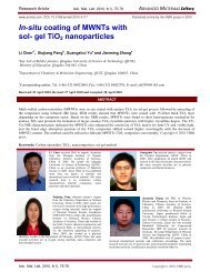
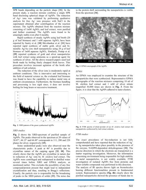
Fig. 2. XRD pattern <strong>of</strong> the <strong>green</strong> synthesized AgNPs.<br />
XRD studies<br />
Fig. 2 shows the XRD-spectrum <strong>of</strong> purified sample <strong>of</strong><br />
AgNPs. The peaks observed in the spectrum at 2θ values <strong>of</strong><br />
38.07°, 44.15° and 64.49° corresponds to 111, 200 and 220<br />
planes for silver, respectively [15].<br />
Some unidentified peaks were also observed near the<br />
characteristic peaks. A peak at 46º is possibly due to<br />
crystalline nature <strong>of</strong> the capping agent [32, 10]. This<br />
clearly shows that the AgNPs are crystalline in nature due<br />
to reduction <strong>of</strong> Ag + ions by M. oleifera leaf extract. The<br />
AgNPs were centrifuged and redispersed in distilled water<br />
several times before XRD and EPMA analysis, as<br />
mentioned earlier. This excludes the possibility <strong>of</strong> any free<br />
compound/protein present that might lead to independent<br />
crystallization and thus, resulting in Bragg’s reflections.<br />
Usually, the particle size is responsible for the broadening<br />
<strong>of</strong> peaks in the XRD pattern <strong>of</strong> solids [33]. The noise due<br />
Mubayi et al.<br />
to the protein shell surrounding the <strong>nanoparticles</strong> is visible<br />
from the spectrum [34].<br />
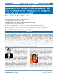
Fig. 3. EPMA micrograph <strong>of</strong> the AgNPs<br />
EPMA<br />
An EPMA was employed to examine the structure <strong>of</strong> the<br />
<strong>nanoparticles</strong> that were synthesized. Representative EPMA<br />
micrographs <strong>of</strong> the reaction mixtures comprising 10 ml <strong>of</strong><br />
M. oleifera leaf extract and 1 mM <strong>of</strong> silver nitrate<br />
magnified 30,000 times are shown in Fig. 3. From the<br />
figure, it is clear that the AgNPs adhered to nano-clusters.<br />
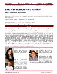
Fig. 4. FTIR spectra <strong>of</strong> dried powder <strong>of</strong> (a) M. oleifera leaf extract (b)<br />
<strong>nanoparticles</strong> synthesized by leaf extract solution.<br />
FTIR studies<br />
The exact procedures <strong>of</strong> bio-reduction is not fully<br />
comprehended and have reported that the reduction <strong>of</strong> Ag +<br />
to Ag <strong>nanoparticles</strong> takes place possibly in the presence <strong>of</strong><br />
the enzyme, NADPH-dependent dehydrogenase [32]. The<br />
precise direction in which the electrons are transported is a<br />
matter requiring investigation. Moreover, the information<br />
concerning environment being responsible for high stability<br />
<strong>of</strong> metal <strong>nanoparticles</strong>, is not widely available. FTIR<br />
investigation <strong>of</strong> isolated AgNPs free from proteins and<br />
water-soluble compounds was performed in this direction.<br />
The analysis <strong>of</strong> IR spectra throws light on biomolecules<br />
bearing different functionalities present in fundamental<br />
system. Representative spectra (Fig. 4b) clearly show the<br />
purified <strong>nanoparticles</strong> showed the presence <strong>of</strong> bands due to<br />
Adv. Mat. Lett. 2012, 3(6), 519-525 Copyright © 2012 VBRI press 522