Plant Mediated Synthesis of Gold Nanoparticles Using Fruit Extracts ...
Plant Mediated Synthesis of Gold Nanoparticles Using Fruit Extracts ...
Plant Mediated Synthesis of Gold Nanoparticles Using Fruit Extracts ...

You also want an ePaper? Increase the reach of your titles
YUMPU automatically turns print PDFs into web optimized ePapers that Google loves.
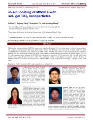
Research Article Adv. Mat. Lett. 2013, 4(5), 332-337 ADVANCED MATERIALS Letters<br />
alcohols/phenols. The weak bands that appeared at 995 and<br />
1132 cm –1 can be ascribed to C – O – C or C – O stretching<br />
modes in phenolic compound or phenolic derivatives (Fig.<br />
1). The absorption appearing at 1262 and 1641 cm -1 can be<br />
assigned to the amide III and amide I bands <strong>of</strong> proteins and<br />
the band at 1386 cm -1 corresponds to C – N stretching<br />
vibrations <strong>of</strong> aromatic amines. The band at 2936 cm -1 is<br />
characteristic <strong>of</strong> the secondary amines. The intense broad<br />
absorbance at 3374 cm -1 corresponds to the hydroxyl<br />
functional group in alcohols and phenolic compounds [20-<br />
24].<br />
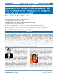
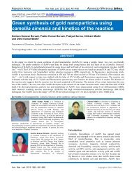
Fig. 3. EDX analysis illustrating the formation <strong>of</strong> gold nanoparticles<br />
biologically synthesized by reduction <strong>of</strong> AuCl4- ions using fruit extract <strong>of</strong><br />
Ananas comosus.<br />
Table 1. Antimicrobial activity <strong>of</strong> the gold nanoparticles synthesized<br />
from Pineapple.<br />
Diameter <strong>of</strong> zone <strong>of</strong> inhibition (mm)<br />
Organisms<br />
Antibiotics Aspergillus<br />
flavus<br />
Aspergillus niger E.coli Streptobacillus<br />
A A+P A A+P A A+P A A+P<br />
Amphicilin - - - - 13.63±0<br />
.45 18.05±0 19.49± 20.33±0<br />
.42 0.38 .29<br />
Penicillin - - - - 14.75±0<br />
.66 17.55±0 15.67± 19.52±0<br />
.43 0.40 .40<br />
Bavistin - - 14.45±0 15.67±0 - - - -<br />
.48 .398<br />
The BSE-SEM images, elemental analysis with EDX<br />
confirmed the presence <strong>of</strong> gold nanoparticles in tested<br />
sample – bright points visible with help <strong>of</strong> Back Scattered<br />
Electrons (BSE) detector (Fig. 2, 3). As shown in Fig. 2C,<br />
the obtained nanoparticles have sphere shapes. During the<br />
separation the gold nanoparticles form aggregate in a large<br />
cluster shown in Fig. 2C-D. However, the distribution <strong>of</strong><br />
agglomerates on the surface was almost uniform as shown<br />
in Fig. 2B.<br />
The EDX quantitative analysis confirmed that the gold<br />
content has the highest elementary composition, while<br />
chlorine has a minor content together with only a trace <strong>of</strong><br />
potassium and calcium (Fig. 3 S2).<br />
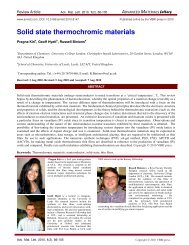
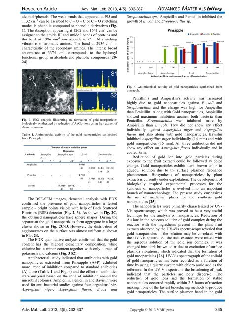
Anti bacterial study indicated that antibiotics with gold<br />
nanoparticles extracted from Pineapple (A+P) exhibited<br />
more zone <strong>of</strong> inhibition compared to standard antibiotics<br />
(A) alone (Table 1 and Fig. 4) and the effect <strong>of</strong> antibiotics<br />
were analysed based on the zone <strong>of</strong> inhibition around the<br />
microbial colonies. Ampicillin, Penicillin and Bavistin were<br />
used for anti bacterial studies against four organisms’ viz.<br />
Aspergillus niger, Aspergillus flavus, E.coli and<br />
Streptobacillus sps. Ampicillin and Penicillin inhibited the<br />
growth <strong>of</strong> E. coli and Streptobacillus sp.<br />
Fig. 4. Antimicrobial activity <strong>of</strong> gold nanoparticles synthesized from<br />
pineapple.<br />
Pencillin’s and Ampicillin’s activity was increased<br />
highly due to gold nanoparticles against E. coli and<br />
Streptobacillus and the change was high for Ampicillin<br />
than Penicillin. Along with <strong>Gold</strong> nanoparticles, Ampicillin<br />
showed maximum inhibition against both bacteria than<br />
Penicillin. Streptobacillus was inhibited more by<br />
Ampicillin than E. coli. They did not show any effect<br />
individually against Aspergillus niger and Aspergillus<br />
flavus and also along with gold nanoparticles. Bavistin<br />
inhibited Aspergillus niger individually (14 mm) and with<br />
gold nanoparticles (15 mm). All three antibiotics did not<br />
show any effect on Aspergillus flavus indivdually and in<br />
coated form.<br />
Reduction <strong>of</strong> gold ion into gold particles during<br />
exposure to the fruit extracts could be followed by color<br />
change. <strong>Gold</strong> nanoparticles exhibit dark brown color in<br />
aqueous solution due to the surface plasmon resonance<br />
phenomenon. Biosynthesis <strong>of</strong> nanoparticles by plant<br />
extracts is currently under exploitation. The development <strong>of</strong><br />
biologically inspired experimental processes for the<br />
synthesis <strong>of</strong> nanoparticles is evolved into an important<br />
branch <strong>of</strong> nanotechnology. The present study emphasizes<br />
the use <strong>of</strong> medicinal plants for the synthesis gold<br />
nanoparticles [25].<br />
The nanoparticles were primarily characterized by UV-<br />
Vis spectroscopy, which was proved to be a very useful<br />
technique for the analysis <strong>of</strong> nanoparticles. Reduction <strong>of</strong><br />
Au ions in the aqueous solution <strong>of</strong> gold complex during the<br />
reaction with the ingredients present in the plant fruit<br />
extracts observed by the UV-Vis spectroscopy revealed that<br />
gold nanoparticles in the solution may be correlated with<br />
the UV-Vis spectra. As the fruit extracts were mixed with<br />
the aqueous solution <strong>of</strong> the gold ion complex, it was<br />
changed into dark brown color due to excitation <strong>of</strong> surface<br />
plasmon vibrations, which indicated that the formation <strong>of</strong><br />
gold nanoparticles [26]. UV-Vis spectrograph <strong>of</strong> the colloid<br />
<strong>of</strong> gold nanoparticles has been recorded as a function <strong>of</strong><br />
time by using a quartz cuvette with chloro auric acid as the<br />
reference. In the UV-Vis spectrum, the broadening <strong>of</strong> peak<br />
indicated that the particles are poly dispersed. The<br />
reduction <strong>of</strong> gold ions and the formation <strong>of</strong> stable<br />
nanoparticles occurred rapidly within 2-3 hours <strong>of</strong> reaction<br />
making it one <strong>of</strong> the fastest bioreducing methods to produce<br />
gold nanoparticles. The surface plasmon band in the gold<br />
Adv. Mat. Lett. 2013, 4(5), 332-337 Copyright © 2013 VBRI press 335