Obstetric dimensions of the true pelvis in a Medieval ... - Anthropology
Obstetric dimensions of the true pelvis in a Medieval ... - Anthropology
Obstetric dimensions of the true pelvis in a Medieval ... - Anthropology

Create successful ePaper yourself
Turn your PDF publications into a flip-book with our unique Google optimized e-Paper software.
AMERICAN JOURNAL. OF PHYSICAL ANTHROPOLOGY 89:421430 (1992)<br />
<strong>Obstetric</strong> Dimensions <strong>of</strong> <strong>the</strong> True Pelvis <strong>in</strong> a <strong>Medieval</strong> Population<br />
From Sudanese Nubia<br />
LYNN M. SIBLEY, GEORGE J. ARMELAGOS, AND<br />
DENNIS P. VAN GERVEN<br />
Universitv <strong>of</strong> Colorado. Boulder. CO 80309 (L.M.S.. D.P.V.G. J and<br />
Departm&t'<strong>of</strong> <strong>Anthropology</strong>, Emory Uniuerszty, Atlanta, Georgia 30322<br />
(G. J.A.)<br />
KEY WORDS Pelvis, Nubia, <strong>Obstetric</strong>s<br />
ABSTRACT Functional analysis <strong>of</strong> <strong>the</strong> <strong>true</strong> <strong>pelvis</strong> (def<strong>in</strong>ed as that por-<br />
tion ly<strong>in</strong>g below and <strong>in</strong>clud<strong>in</strong>g <strong>the</strong> pelvic brim) was undertaken on a sample <strong>of</strong><br />
36 females from <strong>the</strong> <strong>Medieval</strong> site <strong>of</strong> Kulubnarti <strong>in</strong> Sudanese Nubia. Standard<br />
obstetric measurements were taken and compared to four additional prehis-<br />
toric skeletal samples and to modern American standards for <strong>the</strong> same obstet-<br />
ric <strong>dimensions</strong>. Relative to <strong>the</strong> o<strong>the</strong>r prehistoric populations, <strong>the</strong> Kulubnarti<br />
pelves are smaller <strong>in</strong> most <strong>dimensions</strong> and, when compared to modern Amer-<br />
ican standards, from one-third to one-half would be diagnosed as contracted<br />
<strong>in</strong> one or more planes.<br />
Given <strong>the</strong> meager, fluctuat<strong>in</strong>g resources <strong>of</strong> <strong>the</strong>se <strong>Medieval</strong> Nubians' harsh<br />
desert environment, pelvic size reduction is a likely result <strong>of</strong> body size reduc-<br />
tion as one biological response to nutritional stress (Mittler and Van Gerven,<br />
1989; Moore et al., 1986; Van Gerven et al., 1981). It is argued, however, that<br />
size reduction created a high potential for ei<strong>the</strong>r maternal-neonatal morbidity<br />
and mortality due to fetopelvic disproportion or neonatal loss due to low birth<br />
weight. In ei<strong>the</strong>r case, it is suggested that <strong>the</strong> Kulubnarti population paid a<br />
significant biological price for this aspect <strong>of</strong> size reduction.<br />
0 1992 Wiley-Liss, Inc.<br />
Functional analysis has been an essential<br />
perspective <strong>of</strong> skeletal biology. The fact that<br />
form follows function provides <strong>the</strong> basis for<br />
understand<strong>in</strong>g and <strong>in</strong>terpret<strong>in</strong>g much <strong>of</strong> <strong>the</strong><br />
variation found <strong>in</strong> skeletal morphology. Be-<br />
cause <strong>of</strong> this, complex skeletal features such<br />
as <strong>the</strong> human <strong>pelvis</strong> have been an important<br />
focus <strong>of</strong> <strong>in</strong>vestigation. The <strong>pelvis</strong> has been <strong>of</strong><br />
particular <strong>in</strong>terest to anthropologists be-<br />
cause <strong>of</strong> its role <strong>in</strong> human bipedalism. How-<br />
ever, <strong>in</strong> addition to study<strong>in</strong>g <strong>the</strong> evolution-<br />
ary response <strong>of</strong> <strong>the</strong> <strong>pelvis</strong> to bipedal<br />
strid<strong>in</strong>g, <strong>the</strong>re has been a great deal <strong>of</strong> <strong>in</strong>ter-<br />
est <strong>in</strong> how <strong>the</strong> female <strong>pelvis</strong> is able to accom-<br />
modate upright posture and ma<strong>in</strong>ta<strong>in</strong><br />
enough space to allow for delivery <strong>of</strong> a viable<br />
term-size fetus.<br />
The study <strong>of</strong> sexual dimorphism <strong>of</strong> <strong>the</strong> pel-<br />
vis, however, has generated <strong>the</strong> greatest <strong>in</strong>-<br />
terest <strong>in</strong> functional skeletal morphology. In-<br />
0 1992 WILEY-LISS, INC.<br />
terest<strong>in</strong>gly, a paradox becomes apparent<br />
when we evaluate how skeletal biologists<br />
have applied a functional paradigm to <strong>the</strong><br />
study <strong>of</strong> sexual dimorphism. They have used<br />
obvious male-female differences such as <strong>the</strong><br />
subpubic arch, subpubic angle, or sacrosci-<br />
atic notch, each functionally related to ob-<br />
stetrics, to diagnose sex. Yet, <strong>the</strong>y have gen-<br />
erally failed to evaluate <strong>the</strong>se features with<br />
respect to <strong>the</strong>ir functional-obstetric signifi-<br />
cance. This has been particularly <strong>true</strong> for<br />
<strong>the</strong> study <strong>of</strong> anatomically modern popula-<br />
tions.<br />
Several factors may have contributed to<br />
<strong>the</strong> lack <strong>of</strong> a functional approach to <strong>the</strong> ob-<br />
Received June 23, 1989; accepted March 9,1992<br />
Address correspondence to Dennis P. Van Gerven, Department<br />
<strong>of</strong> <strong>Anthropology</strong>, University <strong>of</strong> Colorado, Boulder, CO 80309
422<br />
stetric <strong>pelvis</strong>. First, such analysis requires<br />
articulation <strong>of</strong> <strong>the</strong> hip bones with <strong>the</strong><br />
sacrum. The fragmentary nature <strong>of</strong> most ar-<br />
chaeological rema<strong>in</strong>s <strong>of</strong>ten precludes this<br />
procedure. Second, <strong>in</strong>terest <strong>in</strong> sexual dimor-<br />
phism has focused more on sexual diagnosis<br />
than functional <strong>in</strong>terpretation. Both factors<br />
are unfortunate given <strong>the</strong> importance <strong>of</strong><br />
such variation to our understand<strong>in</strong>g <strong>of</strong><br />
growth and nutrition <strong>in</strong> <strong>the</strong> atta<strong>in</strong>ment <strong>of</strong><br />
adult pelvic form as well as <strong>the</strong> impact <strong>of</strong> <strong>the</strong><br />
adult female form on maternal and neonatal<br />
health.<br />
For example, J. L. Angel (1975, 1978,<br />
1982) argued that flatten<strong>in</strong>g <strong>of</strong> <strong>the</strong> pelvic<br />
<strong>in</strong>let is a response to nutritional stress. Cook<br />
(1984) reported that low status Hopewell fe-<br />
males from <strong>the</strong> lower Ill<strong>in</strong>ois Valley had<br />
flatter pelvic <strong>in</strong>lets than did higher status<br />
females. Cook argued that this may be direct<br />
evidence for nutritional stress.<br />
<strong>Obstetric</strong> practitioners, work<strong>in</strong>g with liv-<br />
<strong>in</strong>g populations, have likewise associated<br />
nutritional deficiency with contracture <strong>of</strong><br />
<strong>the</strong> <strong>pelvis</strong> (Baird, 1945,1969; Thomas, 1947,<br />
1956). The modern cl<strong>in</strong>ical focus, however,<br />
has not been on female pelvic morphology<br />
per se but on parturition and fetopelvic dis-<br />
proportion as <strong>the</strong>se affect maternal-neona-<br />
tal morbidity and mortality (Caldwell and<br />
Moloy, 1933; Friedman, 1978; Pritchard et<br />
al., 1985; Thoms, 1956; Thoms et al., 1939).<br />
In this paper we analyze <strong>the</strong> obstetric di-<br />
mensions <strong>of</strong> female pelves from an ancient<br />
Sudanese Nubian population. Our purpose<br />
is to determ<strong>in</strong>e <strong>the</strong> extent to which size re-<br />
duction and contracture <strong>in</strong> <strong>the</strong> <strong>true</strong> <strong>pelvis</strong><br />
had occurred and presented important re-<br />
productive challenges, 1) from <strong>the</strong> stand-<br />
po<strong>in</strong>t <strong>of</strong> obstetric difficulty, and 2) from <strong>the</strong><br />
standpo<strong>in</strong>t <strong>of</strong> neonatal size.<br />
MATERIALS AND METHODS<br />
Sample selection<br />
L. M. SIBLEY ET AL<br />
Materials for <strong>the</strong> present study were excavated<br />
by one <strong>of</strong> us (D.V.G.) <strong>in</strong> 1979 at <strong>the</strong><br />
<strong>Medieval</strong> site <strong>of</strong> Kulubnarti <strong>in</strong> Sudanese<br />
Nubia. The site is located some 80 miles<br />
south <strong>of</strong> Wadi Halfa <strong>in</strong> <strong>the</strong> Republic <strong>of</strong><br />
Sudan. A total <strong>of</strong> 406 <strong>in</strong>dividuals were dis<strong>in</strong>terred<br />
from two cemeteries spann<strong>in</strong>g <strong>the</strong><br />
Due to <strong>the</strong> desert climate <strong>of</strong> Sudanese Nu-<br />
bia, <strong>the</strong> preservation <strong>of</strong> both s<strong>of</strong>t and skele-<br />
tal tissues is exceptional. One female was<br />
discovered buried on a mat with a fetal skel-<br />
etal <strong>in</strong> <strong>the</strong> breech position. It is reasonable<br />
to assume that her death resulted from pro-<br />
longed obstructed labor.<br />
All adult females (ages 1944) with com-<br />
plete pelvic rema<strong>in</strong>s, lack<strong>in</strong>g gross morpho-<br />
logical abnormalities and without adher<strong>in</strong>g<br />
s<strong>of</strong>t tissues that would impede measure-<br />
ment, were <strong>in</strong>cluded <strong>in</strong> <strong>the</strong> analysis<br />
(n = 36). Age at death was determ<strong>in</strong>ed by<br />
observations <strong>of</strong> epiphyseal union, morphol-<br />
ogy <strong>of</strong> <strong>the</strong> 0s pubis, dental wear, and degen-<br />
erative changes <strong>of</strong> articular surfaces (Van<br />
Gerven et al., 1981). Mean age at death for<br />
<strong>the</strong> sample was 30.5 2 6.9 years.<br />
Of <strong>the</strong> 36 <strong>in</strong>dividuals analyzed, 10 were<br />
sexed positively on <strong>the</strong> basis <strong>of</strong> s<strong>of</strong>t tissues<br />
(breasts and genitalia) and <strong>the</strong> rema<strong>in</strong>der<br />
were sexed accord<strong>in</strong>g to morphology <strong>of</strong> <strong>the</strong><br />
<strong>pelvis</strong>, <strong>in</strong>clud<strong>in</strong>g sciatic notch morphology,<br />
elevation <strong>of</strong> <strong>the</strong> auricular surface, subpubic<br />
angle, pubiclischial <strong>in</strong>dex, and presence <strong>of</strong> a<br />
pre-auricular sulcus. It should be noted that<br />
such estimation <strong>of</strong> sex is unlikely to con-<br />
found <strong>the</strong> present analysis through an <strong>in</strong>ad-<br />
vertent <strong>in</strong>clusion <strong>of</strong> mis-sexed male pelves.<br />
Earlier analysis <strong>of</strong> both males and females<br />
<strong>of</strong> known sex produced non-overlapp<strong>in</strong>g<br />
male-female morphological distributions,<br />
and <strong>the</strong> <strong>in</strong>dividuals utilized <strong>in</strong> <strong>the</strong> present<br />
sample fall so well with<strong>in</strong> <strong>the</strong> female pattern<br />
that <strong>the</strong> likelihood <strong>of</strong> misdiagnosis is re-<br />
mote.<br />
Procedures and <strong>in</strong>struments<br />
Anthropometric measurements were<br />
taken <strong>of</strong> each <strong>true</strong> obstetric <strong>pelvis</strong>, def<strong>in</strong>ed<br />
as that portion <strong>of</strong> <strong>the</strong> <strong>pelvis</strong> ly<strong>in</strong>g below and<br />
<strong>in</strong>clud<strong>in</strong>g <strong>the</strong> pelvic brim. This anatomical<br />
area has obstetric significance s<strong>in</strong>ce it is <strong>the</strong><br />
bony canal through which <strong>the</strong> fetus must<br />
pass dur<strong>in</strong>g parturition. To facilitate de-<br />
scription and analysis, <strong>the</strong> <strong>true</strong> <strong>pelvis</strong> was<br />
divided <strong>in</strong>to three functional planes: <strong>the</strong> <strong>in</strong>-<br />
let, mid<strong>pelvis</strong>, and <strong>the</strong> outlet.<br />
The plane <strong>of</strong> <strong>the</strong> <strong>in</strong>let is bounded anteri-<br />
orly by <strong>the</strong> posterior superior marg<strong>in</strong> <strong>of</strong> <strong>the</strong><br />
pubic symphysis, laterally by <strong>the</strong> l<strong>in</strong>ea ter-<br />
m<strong>in</strong>alis, and posteriorly by <strong>the</strong> promontory<br />
current era from 550 to c. 1550.- and alae <strong>of</strong> <strong>the</strong> sacrum (Oxorn and Foote,
OBSTETRIC DIMENSIONS OF NUBIAN PELVIS 423<br />
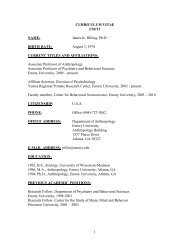
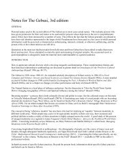
A. Anteroposterior view.<br />
6. Sagittal section.<br />
1980). The follow<strong>in</strong>g <strong>in</strong>let diameters were<br />
measured (Fig. 1):<br />
a. anteroposterior (obstetric conjugate&<br />
extend<strong>in</strong>g from <strong>the</strong> posterior superior mar-<br />
g<strong>in</strong> <strong>of</strong> <strong>the</strong> pubic symphysis to <strong>the</strong> middle <strong>of</strong><br />
<strong>the</strong> sacral promontory;<br />
b. transverse-extend<strong>in</strong>g from <strong>the</strong> widest<br />
distance across <strong>the</strong> brim at <strong>the</strong> level <strong>of</strong> <strong>the</strong><br />
iliopect<strong>in</strong>eal l<strong>in</strong>e;<br />
c. obliqu-xtend<strong>in</strong>g from each sacroiliac<br />
jo<strong>in</strong>t, right and left, to <strong>the</strong> correspond<strong>in</strong>g ilio-<br />
pect<strong>in</strong>eal em<strong>in</strong>ence.<br />
‘antero posterior<br />
Fig. 1. Schematic <strong>of</strong> measurements taken <strong>of</strong> <strong>the</strong> pelvic <strong>in</strong>let.<br />
conjugate<br />
conj u ga te<br />
conjugate<br />
The plane <strong>of</strong> <strong>the</strong> mid<strong>pelvis</strong>, which con-<br />
ta<strong>in</strong>s <strong>the</strong> narrowest obstetric dimension, is<br />
bounded anteriorly by <strong>the</strong> <strong>in</strong>ferior border <strong>of</strong><br />
<strong>the</strong> pubic symphysis, laterally by <strong>the</strong> ischial<br />
sp<strong>in</strong>es, and posteriorly by <strong>the</strong> sacrum at or<br />
near <strong>the</strong> junction <strong>of</strong> <strong>the</strong> fourth and fifth sac-<br />
ral vertebrae (Oxorn and Foote, 1980). The<br />
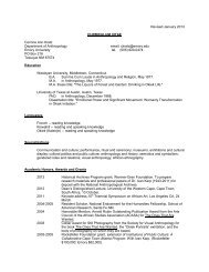
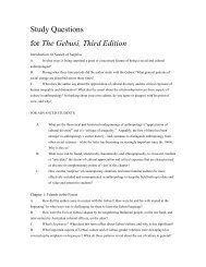
follow<strong>in</strong>g diameters were measured (Fig. 2):<br />
a. anteroposterior-extend<strong>in</strong>g from <strong>the</strong><br />
lower border <strong>of</strong> <strong>the</strong> pubic symphysis to <strong>the</strong><br />
junction <strong>of</strong> <strong>the</strong> fourth and fifth sacral verte-<br />
brae;
424<br />
L. M. SIBLEY ET AL<br />
A. Anteroposterior view show<strong>in</strong>g <strong>the</strong> anteroposterior<br />
and transverse diameters.<br />
6. Sagittal section show<strong>in</strong>g <strong>the</strong> anteroposterior<br />
b. transverse-extend<strong>in</strong>g from <strong>the</strong> tip <strong>of</strong><br />
one ischial sp<strong>in</strong>e to <strong>the</strong> tip <strong>of</strong> <strong>the</strong> o<strong>the</strong>r;<br />
c. posterior-sagittal-extend<strong>in</strong>g from <strong>the</strong><br />
midpo<strong>in</strong>t <strong>of</strong> <strong>the</strong> biischial sp<strong>in</strong>ous diameter to<br />
<strong>the</strong> junction <strong>of</strong> <strong>the</strong> fourth and fifth sacral<br />
vertebrae.<br />
The outlet consists <strong>of</strong> two approximately<br />
triangular areas not <strong>in</strong> <strong>the</strong> same plane but<br />
shar<strong>in</strong>g <strong>the</strong> bituberous diameter as a com-<br />
mon base. Rni<strong>in</strong>daries <strong>of</strong> <strong>the</strong> anterior trian-<br />
gle <strong>in</strong>clude <strong>the</strong> apex <strong>of</strong> <strong>the</strong> subpubic angle,<br />
Fig. 2. Diameter measurements <strong>of</strong> <strong>the</strong> mid<strong>pelvis</strong>.<br />
diameter.<br />
<strong>the</strong> pubic rami, and <strong>the</strong> ischial tuberosities.<br />
The posterior triangle is bounded by <strong>the</strong> l<strong>in</strong>e<br />
<strong>of</strong> <strong>the</strong> sacrotuberous ligaments spann<strong>in</strong>g <strong>the</strong><br />
sacrosciatic notch and <strong>the</strong> sacrococcygeal<br />
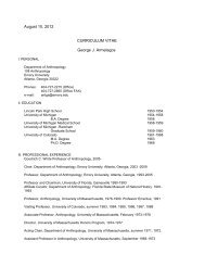
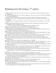
jo<strong>in</strong>t (Oxorn and Foote, 1980). The subpubic<br />
angle as well as <strong>the</strong> follow<strong>in</strong>g diameters<br />
were measured (Fig. 3):<br />
a. anteroposterior-extend<strong>in</strong>g from <strong>the</strong><br />
<strong>in</strong>ferior marg<strong>in</strong> <strong>of</strong> <strong>the</strong> pubic symphysis to<br />
<strong>the</strong> sacrococcygeal jo<strong>in</strong>t;
A. Inferior view.<br />
B. Sagittal section.<br />
b. transverse-extend<strong>in</strong>g from <strong>the</strong> <strong>in</strong>ner<br />
surface <strong>of</strong> one ischial tuberosity to <strong>the</strong> <strong>in</strong>ner<br />
surface <strong>of</strong> <strong>the</strong> o<strong>the</strong>r.<br />
Each <strong>pelvis</strong> was manually articulated and<br />
secured with an elastic band. Dimensions<br />
were taken to <strong>the</strong> nearest 0.1 mm us<strong>in</strong>g a<br />
Helios slid<strong>in</strong>g caliper. Exceptions to this<br />
were <strong>the</strong> anteroposterior and posterior sag-<br />
ittal diameters <strong>of</strong> <strong>the</strong> mid<strong>pelvis</strong>. These were<br />
OBSTETRIC DIMENSIONS OF NUBIAN PELVIS 425<br />
Fig. 3. Diameter measurements <strong>of</strong> <strong>the</strong> pelvic outlet.<br />
.riangle<br />
taken to <strong>the</strong> nearest 0.5 mm us<strong>in</strong>g a ruler.<br />
The subpubic angle was measured with a<br />
protractor to <strong>the</strong> nearest degree. Follow<strong>in</strong>g<br />
data collection, all l<strong>in</strong>ear measurements<br />
were converted to centimeters and rounded<br />
to <strong>the</strong> nearest 0.1 cm.<br />
After measurement, all pelves were classi-<br />
fied accord<strong>in</strong>g to <strong>the</strong> system <strong>of</strong> Thoms et al.<br />
(1939), based on <strong>the</strong> relative lengths <strong>of</strong> <strong>the</strong><br />
<strong>in</strong>let diameters, as follows:
426<br />
a. dolicopellic (long oval)-<strong>the</strong> anteropos-<br />
terior diameter exceeds <strong>the</strong> transverse di-<br />
ameter.<br />
b. mesatipellic (roundt<strong>the</strong> anteroposte-<br />
rior and transverse diameters are <strong>of</strong> equal<br />
length, or <strong>the</strong> former exceeds <strong>the</strong> latter by<br />
not more than 1 cm.<br />
c. brachypellic (transverse ovalt<strong>the</strong><br />
transverse diameter exceeds <strong>the</strong> anteropos-<br />
terior diameter by more than 1 cm but less<br />
than 3 cm.<br />
d. platypellic (flatr<strong>the</strong> transverse diam-<br />
eter exceeds <strong>the</strong> anteroposterior diameter<br />
by 3 cm or more.<br />
In addition, certa<strong>in</strong> obstetrically impor-<br />
tant morphologic features, not associated<br />
with measurement, were observed. These<br />
<strong>in</strong>cluded l<strong>in</strong>earity <strong>of</strong> <strong>the</strong> pelvic sidewalls;<br />
breadth, curvature, and <strong>in</strong>cl<strong>in</strong>ation <strong>of</strong> <strong>the</strong><br />
sacrum; shape and breadth <strong>of</strong> <strong>the</strong> sacrosci-<br />
atic notch; prom<strong>in</strong>ence <strong>of</strong> <strong>the</strong> ischial sp<strong>in</strong>es;<br />
and curvature <strong>of</strong> <strong>the</strong> pubic rami.<br />
Statistics<br />
Age at death and <strong>dimensions</strong> <strong>of</strong> <strong>the</strong> <strong>pelvis</strong><br />
were described <strong>in</strong> terms <strong>of</strong> means, standard<br />
deviations, and range values. In order to de-<br />
term<strong>in</strong>e whe<strong>the</strong>r a differential size effect<br />
was operat<strong>in</strong>g with<strong>in</strong> <strong>the</strong> sample, all pelvic<br />
<strong>dimensions</strong> were correlated with femur<br />
length. No correlations were significant at<br />
P < 0.05 and, <strong>the</strong>refore, no fur<strong>the</strong>r attempt<br />
to statistically remove a body size effect was<br />
made. Comparison to o<strong>the</strong>r archaeological<br />
populations were made us<strong>in</strong>g Student's t<br />
test.<br />
RESULTS<br />
General description<br />
From <strong>the</strong> standpo<strong>in</strong>t <strong>of</strong> shape, 58% <strong>of</strong> <strong>the</strong><br />
pelves were classifiable as brachypellic<br />
(transverse oval), 28% mesatipellic (round),<br />
11% dolichopellic (long oval), and 3% as<br />
platypellic (flat). Shape <strong>of</strong> <strong>the</strong> mid<strong>pelvis</strong> is<br />
most <strong>of</strong>ten characterized by slightly conver-<br />
gent sidewalls (<strong>in</strong>dicated by greater <strong>in</strong>let<br />
than outlet transverse dimension) associ-<br />
ated with well-rounded anterior and poste-<br />
rior segments; well-curved neutrally <strong>in</strong>-<br />
cl<strong>in</strong>ed sacra; and well-rounded and wide<br />
sacrosciatic notches associated with ischial<br />
L. M. SIBLEY ET AL.<br />
TABLE I. <strong>Obstetric</strong> dzmensions <strong>of</strong> <strong>the</strong> Kulubnarti Delves<br />
Pelvic Inlet<br />
Anteroposterior<br />
Transverse<br />
Oblique Left<br />
Oblique Right<br />
Pelvic Midplane<br />
Anteroposterior<br />
Transverse<br />
Posterior Sagittal<br />
Pelvic Outlet<br />
Anteroposterior<br />
Transverse<br />
Subpubic Angle'<br />
36<br />
36<br />
36<br />
36<br />
36<br />
36<br />
36<br />
36<br />
36<br />
36<br />
10.3<br />
11.6<br />
11.3<br />
11.1<br />
11.5<br />
9.1<br />
4.3<br />
11.1<br />
9.7<br />
82.0<br />
0.75<br />
0.74<br />
0.55<br />
0.52<br />
0.70<br />
0.73<br />
0.70<br />
0.71<br />
0.87<br />
7.80<br />
8.6-1 2.0<br />
10.5-14.1<br />
10.2-12.9<br />
10.1-12.2<br />
10.4-13.2<br />
7.8-1 0.5<br />
3.1-5.9<br />
9.8-12.1<br />
8.4-12.1<br />
64.0-97.0<br />
TABLE 2. Kulubnarti pelves compared with American<br />
obstetric standard.s<br />
%<br />
US US Kulubnarti<br />
<strong>Obstetric</strong> Dimensions Normal Contracted Contracted<br />
Pelvic Inlet<br />
Anteroposterior 10.5 40.0 33%<br />
Transverse 13.5 42.0 67%<br />
Oblique Left 12.5 - -<br />
Oblique Right<br />
Pelvic Midplane<br />
12.5 - -<br />
Anteroposterior 11.5 - -<br />
Transverse (a) 10.5
Dimensions _ ~ _<br />
Inlet<br />
Ant-Post.<br />
Trans.<br />
Midplane<br />
Ant. Post<br />
Trans<br />
Post-Sag.<br />
Outlet<br />
Ant-Post<br />
-<br />
X<br />
S.D.<br />
- N<br />
X<br />
S.D.<br />
N<br />
~<br />
X<br />
S.D.<br />
N<br />
fz<br />
S.D.<br />
3<br />
X<br />
S.D.<br />
N<br />
-<br />
X<br />
S.D.<br />
N<br />
OBSTETRIC DIMENSIONS OF NUBIAN PELVIS 427<br />
TABLE 3. Kulubnarti Delves comuared to North American Groups<br />
Kulubnarti<br />
10.3<br />
0.8<br />
36<br />
11.6<br />
0.7<br />
36<br />
11.5<br />
0.7<br />
36<br />
9.1<br />
0.7<br />
36:<br />
4.3<br />
0.7<br />
36<br />
11.0<br />
0.7<br />
36<br />
'Amer<strong>in</strong>dian data from R G Tague, 1986.<br />
*Student's t test comparison to Kulubnarti P < 0.05<br />
-, data not available.<br />
nosed as contracted <strong>in</strong> one or more planes.<br />
For example, 33% <strong>of</strong> <strong>the</strong> pelves have antero-<br />
posterior diameters <strong>of</strong> <strong>the</strong> <strong>in</strong>let <strong>of</strong> C10.0 cm<br />
and 67% have transverse diameters <strong>of</strong> <strong>the</strong><br />
<strong>in</strong>let
428<br />
to <strong>dimensions</strong> considered pathological by<br />
modern cl<strong>in</strong>ical standards.<br />
DISCUSSION<br />
A small mesatipellic or brachypellic <strong>pelvis</strong><br />
is most commonly observed <strong>in</strong> <strong>the</strong> Kulub-<br />
narti sample. Indeed <strong>the</strong> distribution <strong>of</strong> pel-<br />
vic types is similar to that observed for mod-<br />
ern North American females (Caldwell and<br />
Moloy, 1933; Thoms et al., 1939). The re-<br />
duced size is <strong>of</strong> <strong>in</strong>terest <strong>in</strong> <strong>the</strong> context <strong>of</strong><br />
previous research on <strong>the</strong> Kulubnarti re-<br />
ma<strong>in</strong>s. For example, Hummert and Van<br />
Gerven (1983) and Moore et al. (1986) ob-<br />
served evidence for growth retardation.<br />
There is also abundant evidence for nutri-<br />
tional stress <strong>in</strong>ferred from high frequencies<br />
<strong>of</strong> porotic hyperostosis (Van Gerven et al.,<br />
1981) and enamel hypoplasia (Van Gerven<br />
et al., 1988). The population also experi-<br />
enced extremely high <strong>in</strong>fant mortality with<br />
a modal age at death <strong>of</strong> birth (Van Gerven et<br />
al., 1981).<br />
Beyond <strong>the</strong> suggested relationship to<br />
growth and nutritional stress, <strong>the</strong> obstetric<br />
significance <strong>of</strong> <strong>the</strong> smaller Kulubnarti pel-<br />
ves is difficult to assess. The capacity <strong>of</strong> <strong>the</strong><br />
female <strong>pelvis</strong> for childbear<strong>in</strong>g is pr<strong>of</strong>oundly<br />
<strong>in</strong>fluenced by size and shape relative to that<br />
<strong>of</strong> <strong>the</strong> fetus. These factors <strong>of</strong>ten complement<br />
one ao<strong>the</strong>r; a poorly shaped <strong>pelvis</strong> may be<br />
compensated for by large dimension. and a<br />
small <strong>pelvis</strong> by well-formed contours, More-<br />
over, <strong>the</strong> force <strong>of</strong> labor is one <strong>of</strong> <strong>the</strong> most<br />
important factors <strong>in</strong>fluenc<strong>in</strong>g outcome. A<br />
small mesatipellic or brachypellic <strong>pelvis</strong> <strong>of</strong>-<br />
ten occurs <strong>in</strong> women <strong>of</strong> small stature who, as<br />
a rule, have babies that are proportionately<br />
small. Labor <strong>in</strong> <strong>the</strong>se women generally re-<br />
solves <strong>in</strong> a spontaneous delivery, because <strong>of</strong><br />
<strong>the</strong> relative amount <strong>of</strong> space <strong>in</strong> <strong>the</strong> <strong>pelvis</strong>.<br />
However, if any <strong>of</strong> <strong>the</strong> obstetric <strong>dimensions</strong><br />
are reduced beyond <strong>the</strong> capacity <strong>of</strong> <strong>the</strong> fetus<br />
to adapt itself by means <strong>of</strong> favorable presen-<br />
tation, position, attitude, andor cranial<br />
mold<strong>in</strong>g, difficulties occur <strong>in</strong> <strong>the</strong> form <strong>of</strong> pro-<br />
tracted or arrested labor (Baird, 1969; Cald-<br />
well and Moloy, 1933; Friedman, 1978; Ox-<br />
orn and Foote, 1980; Pritchard et al., 1985;<br />
Thoms, 1956; Thoms et al., 1939). Vary<strong>in</strong>g<br />
k<strong>in</strong>ds and degrees <strong>of</strong> maternal-neonatal<br />
morbidity or mortality may <strong>the</strong>n result.<br />
L. M. SIBLEY ET AL.<br />
That <strong>the</strong> Kulubnarti pelves are smaller<br />
than <strong>the</strong> contemporary North American<br />
standard does not <strong>in</strong> itself establish a high<br />
rate <strong>of</strong> obstetric difficulty. The Nubian females<br />
are shorter <strong>in</strong> stature with an average<br />
height <strong>of</strong> 62 <strong>in</strong>ches (Hummert and Van Gerven,<br />
1983). What may be <strong>of</strong> greater obstetric<br />
importance is <strong>the</strong> substantial number <strong>of</strong> <strong>the</strong><br />
Kulubnarti pelves that are cl<strong>in</strong>ically classifiable<br />
as contracted. Of <strong>the</strong> 36 analyzed,<br />
33% had moderate <strong>in</strong>let contracture<br />
(~9.0 cm). For <strong>the</strong> moderately contracted<br />
<strong>the</strong> prognosis for successful deliveiy ul<br />
term size fetus is borderl<strong>in</strong>e, and for <strong>the</strong> severely<br />
contracted <strong>the</strong> prognosis is considered<br />
nearly hopeless (Pritchard et al., 1985).<br />
It is likely, <strong>of</strong> course, that <strong>the</strong> Nubian neonates<br />
were also <strong>of</strong> reduced size and while<br />
this may reduce obstetric complications, it<br />
<strong>in</strong>creased <strong>the</strong> risk <strong>of</strong> neonatal morbidity and<br />
mortality. As previously discussed, <strong>the</strong><br />
modal age at death at Kulubnarti is birth<br />
(Van Gerven et al., 1981). We suggest that<br />
fetopelvic disporportion comb<strong>in</strong>ed with reduced<br />
neonatal size may have been important<br />
factors contribut<strong>in</strong>g to this high death<br />
rate.<br />
Certa<strong>in</strong>ly, fetopelvic disproportion and obstetric<br />
tragedy existed <strong>in</strong> ancient Nubia as<br />
<strong>in</strong> modern populations. The possible breach<br />
delivery at Kulubnarti bears silent witness<br />
to this grim fact. It is possible that <strong>in</strong>dividuals<br />
S-224, R-85, and R-99, or o<strong>the</strong>rs like<br />
<strong>the</strong>m with markedly reduced pelvic <strong>dimensions</strong>,<br />
suffered untold reproductive morbidity<br />
and, perhaps, mortality.<br />
CONCLUSIONS<br />
The size and shape <strong>of</strong> <strong>the</strong> adult female<br />
<strong>pelvis</strong> reflects a complex history <strong>of</strong> heredity<br />
and environmental <strong>in</strong>teractions. It has been<br />
noted <strong>in</strong> temperate, <strong>in</strong>dustrialized popula-<br />
tions that women <strong>of</strong> greater socioeconomic<br />
means are generally healthier, taller, have<br />
roomier pelves, and better reproductive his-<br />
tories than those less fortunate (Baird,<br />
1945, 1969; Thoms et al., 1939; Thoms,<br />
1956). It cannot be assumed, however, that<br />
<strong>the</strong> same holds <strong>true</strong> for women liv<strong>in</strong>g under<br />
different biocultural conditions, past or<br />
present (Tanner, 1978). For example, Geb-<br />
bie (19811, <strong>in</strong> an excellent survey <strong>of</strong> fetopel-<br />
vic disproportion <strong>in</strong> several populations <strong>of</strong>
sub-Saharan Africa, has l<strong>in</strong>ked undernutri-<br />
tion dur<strong>in</strong>g <strong>the</strong> formative years to develop-<br />
ment <strong>of</strong> small non-pathological pelves and to<br />
subsequent widespread reproductive mor-<br />
bidity and mortality. Arriaza et al. (1988)<br />
have estimated that one <strong>in</strong> four females<br />
from an early agricultural period <strong>in</strong> Chile<br />
died <strong>of</strong> complications related to childbirth.<br />
While all <strong>of</strong> <strong>the</strong> affected females had normal<br />
pelves relative to modern obstetric stan-<br />
dards, we would suggest that overall size<br />
and nutrition played some role <strong>in</strong> this high<br />
rate. On <strong>the</strong> o<strong>the</strong>r hand, Frisancho and rol-<br />
leagues (1973) and St<strong>in</strong>i (1972), study<strong>in</strong>g<br />
undernourished South African populations,<br />
have observed that small maternal body size<br />
(and presumably small pelvic size) is associ-<br />
ated with heightened reproductive success.<br />
On this basis, St<strong>in</strong>i concluded:<br />
S<strong>in</strong>ce smaller <strong>in</strong>fants are generally born by smaller<br />
mo<strong>the</strong>rs who have experienced a relatively smaller<br />
weight ga<strong>in</strong> dur<strong>in</strong>g pregnancy, <strong>the</strong> survival <strong>of</strong> such<br />
<strong>in</strong>fants <strong>in</strong> areas <strong>of</strong> poor nutrition would add a ge-<br />
netic factor to <strong>the</strong> development and behavioral <strong>in</strong>-<br />
fluences associated with reduced stature (St<strong>in</strong>i,<br />
1972).<br />
It is not with<strong>in</strong> <strong>the</strong> scope <strong>of</strong> this paper to<br />
debate <strong>the</strong> issue <strong>of</strong> <strong>the</strong> adaptive advantages<br />
<strong>of</strong> small body size under such life circum-<br />
stances. However, it is important to under-<br />
stand that regardless <strong>of</strong> one’s position on <strong>the</strong><br />
issue, <strong>the</strong> potential for maternal-neonatal<br />
morbidity and mortality due to fetopelvic<br />
disproportion and reduced neonatal birth<br />
weight rema<strong>in</strong>s (Wells, 1975; Wells and<br />
Hanke, 1975).<br />
In conclusion, one can envision small body<br />
size as one biological response <strong>of</strong> this Medi-<br />
eval population to <strong>the</strong> meager, fluctuat<strong>in</strong>g<br />
resources <strong>of</strong> a harsh desert land (Hummert<br />
& Van Gerven, 1983). It is likely, however,<br />
that this response had associated biological<br />
costs <strong>in</strong> terms <strong>of</strong> obstetric-related morbidity<br />
and mortality.<br />
ACKNOWLEDGMENT<br />
We thank Robert G. Tague for <strong>the</strong> use <strong>of</strong><br />
data from his <strong>the</strong>sis and his comments on<br />
<strong>the</strong> manuscript.<br />
LITERATURE CITED<br />
Angel JL (1975) Paleoecology, paleodemography, and<br />
health. In S Polgar (ed): Populations, Ecology and So-<br />
cial Behavior. The Hague: Mouton Press.<br />
OBSTETRIC DIMENSION S OF NUBIAN PELVIS 429<br />
Angel JL (1978) Pelvic <strong>in</strong>let form: A neglected <strong>in</strong>dex <strong>of</strong><br />
nutritional status. Am. J. Phys. Anthropol. 48,378<br />
(Abstr).<br />
Angel JL (1982) A new measure <strong>of</strong> growth efficiency:<br />
Skull base height. Am. J. Phys. Anthropol. 58,297-<br />
305.<br />
Arriaza B, Allison M, and Gerszten E (1988) Maternal<br />
mortality <strong>in</strong> Pre-Columbian Indians <strong>of</strong> Arica, Chile.<br />
Am. J. Anthrop. 77,3541.<br />
Baird D (1945) The <strong>in</strong>fluence <strong>of</strong> social and economic<br />
factors on still births and neonatal deaths. J Obstet.<br />
Gynaecol. Br. Emp. 52t217-234.<br />
Baird D (1969) Comb<strong>in</strong>ed Textbook on <strong>Obstetric</strong>s and<br />
Gynaecology (8th Ed). Ed<strong>in</strong>burgh and London: E. and<br />
S. Liv<strong>in</strong>gstone.<br />
Caldwell WE, and Moloy HL (1933) Anatomical variations<br />
<strong>in</strong> <strong>the</strong> female <strong>pelvis</strong> and <strong>the</strong>ir effect on labor with<br />
a suggested classification. Am. J. Obstet. Gynecol.<br />
26:479.<br />
Cook DC (1984) Subsistence and health <strong>in</strong> <strong>the</strong> lower<br />
Ill<strong>in</strong>ois Valley: Osteological evidence. In MN Cohen<br />
and GJ Armelagos (eds): Paleopathology at <strong>the</strong> Orig<strong>in</strong>s<br />
<strong>of</strong> Agriculture. Orlando: Academic Press.<br />
Friedman EA (1978) Labor: Cl<strong>in</strong>ical Evaluation and<br />
Management (2nd Ed). New York: Appleton-Century-<br />
Cr<strong>of</strong>ts.<br />
Frisancho AR, Sanchez J, Pollardel D, and Yanez L<br />
(1973) Adaptive significance <strong>of</strong> small body size under<br />
poor socio-economic conditions <strong>in</strong> sou<strong>the</strong>rn Peru. Am.<br />
J. Phys. Anthropol. 39.255-262.<br />
Gebbie D (1981) Reproductive <strong>Anthropology</strong>: Decent<br />
Through Woman. New York: Wiley and Sons.<br />
Hummert JR (1983) Childhood Growth and Morbidity <strong>in</strong><br />
a <strong>Medieval</strong> Population from Kulubnarti <strong>in</strong> <strong>the</strong> Rato el<br />
Hajar <strong>of</strong> Sudanese Nubia. Ph.D. Thesis, University <strong>of</strong><br />
Colorado, Boulder.<br />
Hummert JR, and Van Gerven DP (1983) Skeletal<br />
growth <strong>in</strong> a <strong>Medieval</strong> population from Sudanese Nubia.<br />
Am. J. Phys. Anthropol. 60t471-478.<br />
Jordaan HVF (1976) Newborn adult bra<strong>in</strong> ratios <strong>in</strong> hom<strong>in</strong>id<br />
evolution. Am. J. Phvs. Anthropol. 44t271-278.<br />
Leutenegger W (1980) Encephalization and obstetrics <strong>in</strong><br />
primates with particular reference to human evolution.<br />
In E Armstrong and D Falk (eds): Primate Bra<strong>in</strong><br />
Evolution: Methods and Concepts. New York: Plenum<br />
Press.<br />
Mittler DM, and Van Gerven DP (1989) Developmental<br />
and demographic patterns <strong>of</strong> cribra orbitalia <strong>in</strong> a <strong>Medieval</strong><br />
Christian population from Sudanese Nubia.<br />
Am. J. Phys. Anthropol. 78,273-274.<br />
Moore KP, Thorp S, and Van Gerven DP (1986) Patterns<br />
<strong>of</strong> dental eruption, skeletal maturation and stress <strong>in</strong> a<br />
<strong>Medieval</strong> population from Sudanese Nuhia. Hum.<br />
Evol. 1t325-330.<br />
Oxorn H, and Foote WR (1980) Human Labor and Birth<br />
(4th Ed). New York: Appleton-Century-Cr<strong>of</strong>ts.<br />
Pritchard JA, MacDonald PC, and Gant NF (1985)<br />
Williams <strong>Obstetric</strong>s (17th Ed). New York: Appleton-<br />
Century-Cr<strong>of</strong>ts.<br />
Stewart DB (1984a) The <strong>pelvis</strong> as a passageway. I. Evolution<br />
and adaptations. British Journal <strong>of</strong> <strong>Obstetric</strong>s<br />
and Gynaecology 91t611-617.<br />
Stewart DB (1984h) The <strong>pelvis</strong> as a passageway. 11. The
430<br />
modern human <strong>pelvis</strong>. Br. J. Obstet. Gynaecol.<br />
91t618-623.<br />
St<strong>in</strong>i WA (1972) Malnutrition, body size and proportion.<br />
Ecology <strong>of</strong> Food and Nutrition 1:121-126.<br />
Tague RG (1986) <strong>Obstetric</strong> Adaptations <strong>of</strong> <strong>the</strong> Human<br />
Bony Pelvis. Ph.D. Thesis, Kent State University,<br />
Ohio.<br />
Tanner JM (1978) Fetus Into Man: Physical Growth<br />
from Conception to Maturity. Cambridge, MA: Har-<br />
vard University Press.<br />
Thoms H (1947) The role <strong>of</strong> nutrition <strong>in</strong> pelvic variation.<br />
Am. J. Obstet. Gynecol. 54t62-73.<br />
Thoms H (1956) Pelvimetry. London: Cassel and Co.<br />
Thorn6 H; Fuotc WR, and Friedman I (1 939) The cl<strong>in</strong>ical<br />
L. M. SIBLEY ET AL.<br />
significance <strong>of</strong> pelvic variations. Am. J. Obstet. Gyne-<br />
col. 38t634-642.<br />
Van Gerven DP, Sandford MK, and Hummert JR (1981)<br />
Mortality and culture change <strong>in</strong> Nubia’s Bato el<br />
Hajar. J. Hum. Evol. 10:395408.<br />
Van Gerven DP, Beck R, and Hummert JR (1990) Pat-<br />
terns <strong>of</strong> enamel hypoplasia <strong>in</strong> two <strong>Medieval</strong> popula-<br />
tions. Am. J. Phys. Anthropol. 82t412-420.<br />
Wells C (1975) Ancient obstetric hazards and female<br />
mortality. Bullet<strong>in</strong> <strong>of</strong> <strong>the</strong> New York Academy <strong>of</strong> Med-<br />
ic<strong>in</strong>e 51:1235-1249.<br />
Wells C, and Hawkes CS (1975) An obstetric calamity<br />
from K<strong>in</strong>gsworthy, Hampshire. Med. Biol. Ill. 25:47-<br />
51.