English - Comunidad Virtual CIENCIAS DEL DEPORTE - RedIRIS
English - Comunidad Virtual CIENCIAS DEL DEPORTE - RedIRIS
English - Comunidad Virtual CIENCIAS DEL DEPORTE - RedIRIS

You also want an ePaper? Increase the reach of your titles
YUMPU automatically turns print PDFs into web optimized ePapers that Google loves.
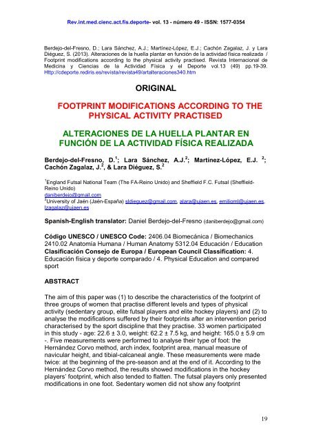
Rev.int.med.cienc.act.fís.deporte- vol. 13 - número 49 - ISSN: 1577-0354<br />
Berdejo-del-Fresno, D.; Lara Sánchez, A.J.; Martínez-López, E.J.; Cachón Zagalaz, J. y Lara<br />
Diéguez, S. (2013). Alteraciones de la huella plantar en función de la actividad física realizada /<br />
Footprint modifications according to the physical activity practised. Revista Internacional de<br />
Medicina y Ciencias de la Actividad Física y el Deporte vol.13 (49) pp.19-39.<br />
Http://cdeporte.rediris.es/revista/revista49/artalteraciones340.htm<br />
ORIGINAL<br />
FOOTPRINT MODIFICATIONS ACCORDING TO THE<br />
PHYSICAL ACTIVITY PRACTISED<br />
ALTERACIONES DE LA HUELLA PLANTAR EN<br />
FUNCIÓN DE LA ACTIVIDAD FÍSICA REALIZADA<br />
Berdejo-del-Fresno, D. 1 ; Lara Sánchez, A.J. 2 ; Martínez-López, E.J. 2 ;<br />
Cachón Zagalaz, J. 2 , & Lara Diéguez, S. 2<br />
1<br />
England Futsal National Team (The FA-Reino Unido) and Sheffield F.C. Futsal (Sheffield-<br />
Reino Unido)<br />
daniberdejo@gmail.com<br />
2<br />
University of Jaén (Jaén-España) sldieguez@gmail.com, alara@ujaen.es, emilioml@ujaen.es,<br />
lzagalaz@ujaen.es<br />
Spanish-<strong>English</strong> translator: Daniel Berdejo-del-Fresno (daniberdejo@gmail.com)<br />
Código UNESCO / UNESCO Code: 2406.04 Biomecánica / Biomechanics<br />
2410.02 Anatomía Humana / Human Anatomy 5312.04 Educación / Education<br />
Clasificación Consejo de Europa / European Council Classification: 4.<br />
Educación física y deporte comparado / 4. Physical Education and compared<br />
sport<br />
ABSTRACT<br />
The aim of this paper was (1) to describe the characteristics of the footprint of<br />
three groups of women that practise different levels and types of physical<br />
activity (sedentary group, elite futsal players and elite hockey players) and (2) to<br />
analyse the modifications suffered by their footprints after an intervention period<br />
characterised by the sport discipline that they practise. 33 women participated<br />
in this study - age: 22.6 ± 3.0, weight: 62.2 ± 7.5 kg, and height: 165.0 ± 5.9 cm<br />
-. Five measurements were performed to analyse their type of foot: the<br />
Hernández Corvo method, arch index, footprint area, manual measure of<br />
navicular height, and tibial-calcaneal angle. These measurements were made<br />
twice: at the beginning of the pre-season and at the end of it. According to the<br />
Hernández Corvo method, the results showed modifications in the hockey<br />
players’ footprint, which also tended to flatten. The futsal players only presented<br />
modifications in one foot. Sedentary women did not show any footprint<br />
19
Rev.int.med.cienc.act.fís.deporte- vol. 13 - número 49 - ISSN: 1577-0354<br />
modifications. Therefore, this study has proved that continued physical activity<br />
causes footprint modifications in those who practise it.<br />
KEY WORDS: photopodogram, arch index, Hernández Corvo method, women<br />
futsal, women hockey.<br />
RESUMEN<br />
El objetivo de este trabajo ha sido describir las características de la huella<br />
plantar en tres grupos de mujeres con distintos niveles y tipo de actividad física<br />
(sedentarias, jugadoras de élite de fútbol sala y hockey hierba) y analizar la<br />
evolución del morfotipo de pie tras un período de intervención marcado por la<br />
disciplina deportiva que practiquen. Han participado 33 mujeres con unas<br />
medias de edad, masa y estatura de 22,6 ± 3,0 años, 62,2 ± 7,5 kg y 165,0 ±<br />
5,9 cm. Se realizaron cinco mediciones para analizar el tipo de pie: método de<br />
Hernández Corvo, índice del arco, determinación de la superficie de la huella,<br />
medida manual de la altura del escafoides y del ángulo tibio-calcáneo. Estas<br />
medidas se tomaron en dos momentos, al inicio y final de la pretemporada. Los<br />
resultados demostraron modificaciones en las huellas de las jugadoras de<br />
hockey, según el método de Hernández Corvo, con tendencia a aplanarse. En<br />
las de fútbol sala sólo se observaron diferencias en un pie. Por el contrario, las<br />
sedentarias no presentaron modificación de la huella plantar. Por lo tanto, la<br />
actividad deportiva continuada ha provocado modificaciones en la huella<br />
plantar de las jugadoras analizadas en este trabajo.<br />
PALABRAS CLAVE: fotopodograma, índice del arco, método Hernández<br />
Corvo, fútbol sala femenino, hockey hierba femenino.<br />
INTRODUCTION<br />
The foot, as locomotor structure, is the basis of our body as it is the only contact<br />
we have with the surface, it requires special mention. Thus many authors who<br />
have studied it. Viladot (2000) states that "the foot is a three dimensional<br />
structure variable, antigravity base and is a key position for bipodal and human<br />
progress."<br />
Lippert (2005) comments that the human foot is the result of the transformation<br />
of the prehensile foot of the monkeys in static foot support. The foot has a<br />
function on both static and dynamic (Escobar, 2007; Torrijos et al. 2009). The<br />
functionality of the human foot is clearly influenced by its structure (McCrory et<br />
al., 1997; Shiang et al. 1998; Menz and Munteanu, 2005) and thanks to the<br />
cupular shape (Hernández Corvo, 1989; Kapandji, 1998; Viladot , 2000) of the<br />
plantar arch and heel support points and metatarsals, is capable of supporting<br />
the entire body weight without sinking. Besides the internal longitudinal arch<br />
height influences other body structures, such as the back (Hernández Corvo,<br />
1989; Gómez, 2003; Menz and Munteanu, 2005). The morphological<br />
characteristics in the human foot varies somewhat with age (Scott, Menz and<br />
20
Rev.int.med.cienc.act.fís.deporte- vol. 13 - número 49 - ISSN: 1577-0354<br />
Newcombe, 2007) and between individuals (Cowan, Jones and Robinson,<br />
1993; Shiang et al., 1998; Mayorga-Vega et al., 201 2). These variations are<br />
beyond the visual assessment. That is why we have to analyse each individual's<br />
feet in detail. For this there are a lot of techniques both direct (x-rays...) and<br />
indirect (anthropometry, photopodorgam...). Based on these analyses the type<br />
of the height of the navicular to the ground, the Chipaux index, the angle of<br />
Clarke (1933) or Feiss line (López et al., 2005) could be some of them. A simple<br />
and valid way to analyse the foot is by obtaining the footprint (Shiang et al.,<br />
1998). As well as structural variations due to the evolution marked by age, feet<br />
exhibit variations in structure due to many factors, such as: the age when you<br />
start wearing shoes (Sachithanandam and Joseph, 1995), the age when you<br />
begin to play a sport with medium or high dedication (Aydog et al., 2005a;<br />
Martin, 2008, Zahínos, González and Salinero, 2010), overweight<br />
(Sachithanandam and Joseph, 1995; Hills et al., 2001; Masaun,<br />
Dhakshinamoorthy and Parihar, 2009; Vidal et al., 2010), the own repetition of a<br />
sport technique, the possible fatigue (Abián et al., 2005), or the fact of playing a<br />
sport or even a specific discipline (Sirgo and Aguado, 1991; Sirgo et al., 1997;<br />
Aydog et al., 2005a; López et al., 2005, López et al., 2006, Cain et al., 2007;<br />
Elvira et al., 2008; Antolinos and Martínez, 2010; González Pérez and Floría,<br />
2012).<br />
The objectives of this study has been: 1) to describe the characteristics of the<br />
footprint of three groups of women with different levels and types of physical<br />
activity, 2) to analyse the modifications suffered by their footprints after an<br />
intervention period characterised by the sport discipline that they practise and 3)<br />
to compare several methods of evaluating the footprint described in the<br />
literature by Hernández Corvo method (HC), arch index (AI), the height of the<br />
navicular bone (AE) and the measurement of the footprint surface (SH) with<br />
Autocad 2007.<br />
METHOD<br />
Participants<br />
In this longitudinal study (two measurements: at the beginning and at the end of<br />
the preseason) 33 young women have participated, with a mean of age, mass<br />
and height of about 23 years (22.6 ± 3), 62.23 ± 7.55 kg and 165 ± 5.9 cm,<br />
respectively (Table 1), belonging to three groups, depending on their<br />
characteristics:<br />
The first group consisted of 11 players of futsal (FS), belonging to an<br />
elite team (Division de Honor – Women First Division).<br />
The second group made up by 12 hockey players (HH), members of an<br />
elite team (Division de Honor – Women First Division).<br />
The third group consisted of 10 female sedentary (S) voluntarily<br />
participated in the study.<br />
21
Rev.int.med.cienc.act.fís.deporte- vol. 13 - número 49 - ISSN: 1577-0354<br />
All the participants in this study were informed about the nature of it and<br />
volunteered to be part of the research.<br />
Table 1. Characteristics of the participants (FS: futsal; HH: hockey; S: sedentary women)<br />
FS HH S TOTALS<br />
N 11 12 10 33<br />
AGE 23.2 (±3.28) 21.9 (±3.75) 22.8 (±1.32) 22.6 (±3)<br />
WEIGHT 59.464 (±4.699) 62.5 (±6.32) 64.98 (±10.55) 62.239(±7.55)<br />
HEIGHT 162.4 (±6.36) 166.3 (±5.754) 168 (±4.163) 165.485(±5.87)<br />
Inclusion criterion for the HH and FS group were: be federated in their<br />
corresponding sports federation and have been practicing their sport<br />
competitively for at least two years (Elvira et al., 2008a). Finally, it was<br />
considered necessary a regularity in attendance to training during the<br />
intervention period (Lopez et al., 2005). Meanwhile, the inclusion criterion for<br />
the inclusion in the sedentary group have been: to be woman aged 18 to 28<br />
years old who have not done any training programme continuously in the<br />
previous three months, nor practiced any physical activity more than one day<br />
per week (Abián, 2008). Moreover, they can not be part of these groups and will<br />
cause exclusion of those women who had any surgical intervention in the lower<br />
limbs (Macrory et al., 1997) or structural deformities as: Genu valgum or varum,<br />
or coxa vara or valga (Escobar, 2007); or foot deformities as congenital valgus<br />
flat foot, flat foot paralytic or spastic equinus foot. Also, flat foot or cavus due to<br />
bone disorders or neuromuscular disorders (Viladot, 2000), to a serious injury or<br />
fracture in the past six months (Masaun, Dhakshinamoorthy and Parihar, 2009)<br />
or current pain in the feet (Scott, Menz and Newcombe, 2007).<br />
Procedure<br />
Footprints of both feet by photopodogram were recorded (Viladot, 2000) and<br />
analysed following the HC method (Hernández Corvo, 1989) and the IA<br />
(Cavanagh and Rodgers, 1987). We also measured the ATC (tibio-calcaneal<br />
angle) (Stacpoole-Shea et al., 1998; Viladot, 2000; Hertel, Gay and Deny, 2002,<br />
Lopez et al., 2005, Albert, 2009), the SH with the software "Autocad 2007"<br />
(Gómez, 2003; Billis et al., 2006; Nikolaidou and Boudolos, 2006) and manually<br />
the AE (Cowan, Jones and Robinson, 19 93; Chu et al., 1995, Williams, McClay<br />
and Hamill, 2001; Menz and Munteanu, 2005). Definitions and descriptions of<br />
each of these methods of footprint analysis is as follows:<br />
- HC Method: This method involves typing stand as measures carried out<br />
according to the footprint. A good accuracy has been chosen, both in<br />
performance and in the classification of foot type, ranging from flat feet to end<br />
cavus (Sirgo and Aguado, 1991; Sirgo et al., 1997; Abián et al ., 2005, López et<br />
al., 2006; Zurita, Martínez and Zurita, 2007; Abián, 2008). The procedure is as<br />
follows (Figure 1): two points are marked in the innermost prominences of the<br />
22
Rev.int.med.cienc.act.fís.deporte- vol. 13 - número 49 - ISSN: 1577-0354<br />
footprint (1 and 1'), the "initial stroke" is made to connect the two points. After<br />
one point is marked on the anterior part of the footprint (including fingers) and<br />
other point in the rear part (2 and 2'). Perpendicular lines to the latter points<br />
from the initial stroke are drawn. The distance between this line and the point 1<br />
is the "fundamental measure" and it must be transferred as many times as it fits<br />
in the initial stroke (3, 4 and 5). A perpendicular line to 3 is drawn, passing<br />
through the outermost part of the footprint, and another perpendicular to 4 and<br />
another to 5 as well as through the outer part (6, 7 and 8 respectively). The<br />
distance between the initial stroke and 6 is X (metatarsal width), the distance<br />
between 9 and 7 is Y (external arch, midfoot support surface).<br />
Figure 1. Assesment of the footprints according to the protocol of<br />
Hernández Corvo (1989).<br />
With the obtained measures before and using the Equation 1 we will get the<br />
type of foot by the Hernández Corvo method (1989).<br />
%X = (X-Y) * 100/X<br />
Equation 1. Equation of Hernández Corvo (1989) to assess the type of foot. 0-34%: Flat<br />
foot; 35-39%: Flat/normal foot; 40-54%: Normal foot; 55-59%: Cavus/normal foot; 60-74%:<br />
Cavus foot; 75-84%: High cavus foot; 85-100%: Extreme cavus foot.<br />
- IA: It is defined as the ratio of the contact areas of the different parts of the<br />
footprint excluding the fingers. To divide the foot into three equal parts, the first<br />
axis of the foot must be taken, which is a line from the center of the heel to the<br />
top of the second toe. The IA is measured as the ratio of the area of the midfoot<br />
between the total area of the foot excluding the toes (Equation 2).<br />
23
Rev.int.med.cienc.act.fís.deporte- vol. 13 - número 49 - ISSN: 1577-0354<br />
B<br />
= IA<br />
A + B + C<br />
Ecuation 2: Equation to assess the type of foot regarding the (If IA ≤ 0,21: cavus foot;<br />
normal foot: 0,21 < IA< 0.26; flat foot: IA ≥ 0.26).<br />
- SH: The footprints were scanned with the software Autocad 2007 (Figure 2) to<br />
measure their (Gómez, 2003; Yu, Lo and Chiou, 2003; Hurtado, 2006;<br />
Nikolaidou and Boudolos, 2006; Paiva et al., 2009).<br />
Figure 2. Assessment of the footprints surface with the software<br />
Autocad 2007.<br />
- ATC: It is the angle formed the heel of the foot with the rest of the leg (Figure<br />
3). According to Helbing line, the vertical has to go through the center of the<br />
popliteal fossa and the centre of the heel (Viladot, 2000; Hertel,Gay, and<br />
Denegar, 2002; Redmond, Crane and Menz, 2008; Albert, 2009). There is a<br />
degree of physiological valgus of about 5° to 10° according to Viladot (2000)<br />
and Albert (2009) and up to 7º according to Ricard (2001) in healthy men under<br />
18. This line should be done with the foot relaxed, after putting the foot on the<br />
ground distributing the weight between both feet and then measured with a<br />
goniometer. Also measurements can be taken according to the horizontal with<br />
the ground (Sell et al. 1994; Elvira et al. 2008a; Elvira, Vera-García and Meana,<br />
2008b), but in this study it has been done according to the first method of<br />
measurement. It was measured with a manual goniometer. According to Elvira<br />
et al. (2008a), the eversion and consequently the calcaneal valgus, is<br />
considered negative and the inversion or calcaneal varus considered positive<br />
when recording data. The following protocol was followed:<br />
o Barefoot, in bipodal support stand on a smooth surface.<br />
o It was marked with a pen the vertical line passing through the center of<br />
the popliteal fossa, up to the Achilles tendon.<br />
24
Rev.int.med.cienc.act.fís.deporte- vol. 13 - número 49 - ISSN: 1577-0354<br />
o Other side is marked by the bisector of the calcaneus by reference to the<br />
posterior bulge, instead of the Achilles tendon insertion.<br />
o Finally, the angle formed by two lines is measured with the goniometer.<br />
Figure 3. Assessment of the angle formed by the heel and the leg.<br />
- AE: The manually measurement of the navicular bone was used, taking as<br />
reference the navicular protuberance and measuring the distance from it to the<br />
floor. There are also more sophisticated methods to measure this variable, such<br />
as x-rays. Cowan, Jones and Robinson (1993) collect the navicular height<br />
measured manually and its correspondence with the type of foot that was<br />
observed in their study (measures taken in centimeters): Flat: 2.72-4.08;<br />
Normal: 4.09-5.08; Cavus: 5.09-6.05.<br />
The procederes were as follows:<br />
o Barefoot and with your weight distributed between both legs, draw a line<br />
through the tuberosity of the navicular by the lower part.<br />
o Then, with one arm of the goniometer with a precision of 1 mm, placed in<br />
contact with the ground up to the line marked on the skin.<br />
The method of obtaining the footprint has been the photopodogram (Figure 4)<br />
that, according to Viladot (2000), is the most useful for obtaining the footprint.<br />
Images were digitalised to make certain measurements, such as IA or the SH<br />
(Chu et al., 1995; Michelson, Durant and McFarland, 2002; Gómez, 2003;<br />
López e t al., 2006; Billis et al., 2006; Scott, Menz and Newcombe, 2007; Elvira,<br />
Vera-García and Meana, 2008b; Aydog et al., 2005a and 2005b; Menz and<br />
Munteanu, 2005 and Murley, Menz and Landorf, 2009). To obtain the<br />
photopodogram, the protocol described by Aguado, Izquierdo and González<br />
(1997) has been used. This protocol consists on brushing the sole with<br />
photodeveloper liquid photodeveloper and place the foot on photografic paper<br />
previously light veiled. Then, the support the foot on the paper is maintained for<br />
45 s. After this time the foot is lifted. The paper with the footprint is put on a tray<br />
25
Rev.int.med.cienc.act.fís.deporte- vol. 13 - número 49 - ISSN: 1577-0354<br />
with fixer fluid. Finally, paper is taken off the liquid, washed very well with water<br />
and let it dry.<br />
The measurements of FS and HH groups have been performed prior to the<br />
completion of exercise, i.e., before training (Cowan, Jones and Robinson,<br />
1993). Similarly, the footprints were taken in bipodal position distributing the<br />
weight between both feet (Chu et al., 1995; Viladot, Cohí and Clavell, 1997,<br />
Shiang et al., 1998; Michelson, Durant and McFarland, 2002).<br />
Variables<br />
As dependent variables included in this study would be all measurements that<br />
were made at both times. So they would be the footprint analysis using the HC<br />
method, the IA, the delimitation of the SH, the ATC and the masurement of the<br />
AE measured manually. The intervention to the subjects would be the<br />
independient variable. This intervention has been the activity of the pre-season<br />
training in each of the sports (futsall and hockey). The duration of this<br />
intervention in both cases has been six weeks with a training load of three<br />
sessions per week and a duration of 2 hours per session. In the sedentary<br />
group the same time has been considered, but considering they had no change<br />
in their activity, i.e., they remained their sedentary condition.<br />
Material<br />
Figure 4. Photopodogram of left and right feet done by the potocol described<br />
by Aguado, Izquierdo and González (1997).<br />
To describe the anthropometric characteristics of the population a SECA<br />
stadiometer (SECA Ltd, Germany) and a SECA floor scale (SECA Ltd,<br />
Germany) were used. For printing of the footprints by photopodogram, ILFORD<br />
photographic paper, developer and fixer liquid were used. Meanwhile, for the<br />
26
Rev.int.med.cienc.act.fís.deporte- vol. 13 - número 49 - ISSN: 1577-0354<br />
physiotherapic evaluation has been necessary to use a demographic pencil and<br />
a plastic standard manual goniometer, scaling by 2º in 2º, with a maximum error<br />
of 1, used by some specific studies to measure the angle of the rearfoot<br />
(Stacpoole-Shea et al., 1998, Kaufman et al., 1999; Hertel, Gay and Denegar,<br />
2002). Also, an assessment template or proposed clinical examination<br />
assessing the sagittal alignment of the legs when standing (varus or valgus<br />
hindfoot and leg), the pelvic and possible slope d eformidad of the tibia<br />
(Salazar, 2007) was used. This template included also the grades of the tibiocalcaneus<br />
angle and the centimeters of the navicular height above the ground.<br />
For scanning and analysis of the images the software "Autocad 2007"<br />
(Autodesk) was used for the delimitation of the surface of the scanned footprints<br />
and obtaining the IA.<br />
Statistical Analysis<br />
Statistical analysis of the data was done using SPSS v.15.0 for Windows.<br />
Descriptive statistics have been conducted. Analysis was performed using<br />
repeated measures ANOVA, post hoc analysis used as the process of Scheffé.<br />
In the inferential tests the statistical criteria of significance of p
ATC<br />
(º)<br />
HC<br />
(%)<br />
AE<br />
(cm)<br />
SH<br />
(cm 2 )<br />
IA<br />
Rev.int.med.cienc.act.fís.deporte- vol. 13 - número 49 - ISSN: 1577-0354<br />
Table 2: Differences in the right foot between the beginnng of the pre-season and the<br />
season.<br />
FS pre FS seas DIF HH pre HH seas DIF S pre S seas DIF<br />
-7.1 -8.7<br />
-7.3 ± -7.1<br />
- 6.7 - 7.0<br />
± 3.9<br />
66.07<br />
± 3.98<br />
-<br />
87.9<br />
± 15.2<br />
0.23<br />
± 0.06<br />
± 5.4<br />
59.39<br />
± 9.29<br />
4.4<br />
± 0.6<br />
90.1<br />
± 14.4<br />
0.23<br />
± 0.04<br />
-<br />
*<br />
-<br />
*<br />
-<br />
2.8<br />
62.94<br />
± 4.17<br />
4.0<br />
± 0.5<br />
88.26<br />
± 8.14<br />
0.23<br />
± 0.02<br />
± 1.2<br />
61.08<br />
± 4.77<br />
3.9<br />
± 0.4<br />
90.0<br />
± 9.5<br />
0.24<br />
± 0.03<br />
Results obtained in the right foot assesment at the beginning of the preaseason and the season<br />
( x = mean; σ = standard desviation; FS = futsal group; HH = hockey group; S = sedentary<br />
group; pre = measurement performed before preseason began; seas = measurement<br />
performed before season starteed (end preseason); ATC = tibiocalcanal angle; HC = type of<br />
foot following Hernández Corvo; AE = navicular height; SH = footprint surface; IA = arch index).<br />
ATC<br />
(º)<br />
HC<br />
(%)<br />
AE<br />
(cm)<br />
SH<br />
(cm 2 )<br />
IA<br />
Results obtained in the left foot assesment at the beginning of the preaseason and the season<br />
( x = mean; σ = standard desviation; FS = futsal group; HH = hockey group; S = sedentary<br />
group; pre = measurement performed before preseason began; seas = measurement<br />
performed before season starteed (end preseason); ATC = tibiocalcanal angle; HC = type of<br />
foot following Hernández Corvo; AE = navicular height; SH = footprint surface; IA = arch index).<br />
When performing statistical analysis of the variables it has been observed that<br />
there are no significant differences in any of the variables between the groups<br />
studied (FS, HH and S). Moreover, when comparing each group separately at<br />
different times of the preseason some significant differences in certain variables<br />
have been found. Thus, the FS group showed significant differences in the HC<br />
variable on the right foot only (not left foot) (Figure 5). Also, the HH group<br />
showed differences in this variable in both feet (Figure 6). Finally, the S group<br />
has not presented differences in either foot in this variable.<br />
-<br />
***<br />
-<br />
-<br />
-<br />
± 4.2<br />
66.44<br />
± 15.48<br />
3.9<br />
± 0.4<br />
89.52<br />
± 12.33<br />
0.22<br />
± 0.04<br />
± 4.1<br />
65.31<br />
± 14.56<br />
3.9<br />
± 0.4<br />
93.5<br />
± 13.45<br />
0.23<br />
± 0.04<br />
Table 3: Differences in the left foot between the beginnng of the pre-season and the season.<br />
FS pre FS seas DIF HH pre HH seas DIF S pre S seas DIF<br />
-6.8<br />
± 4.5<br />
-8.1<br />
± 4.9 -<br />
-8.3<br />
± 4.5<br />
-7.6<br />
± 2.9 -<br />
-6.6<br />
± 3.5<br />
-7.2<br />
± 3.2 -<br />
65.75<br />
± 8.22<br />
65.23<br />
± 14.84 -<br />
63.96<br />
± 7.20<br />
60.88<br />
± 6.00 *<br />
74.11<br />
± 19.66<br />
74.95<br />
± 19.53 -<br />
-<br />
4.3<br />
± 0.4 -<br />
4.1<br />
± 0.4<br />
4.0<br />
±0.3 -<br />
4.0<br />
± 0.3<br />
4.0<br />
± 0.4 -<br />
87.8<br />
± 11.1<br />
88.7<br />
±12.8 -<br />
87.5<br />
± 8.7<br />
89.1<br />
± 8.3 *<br />
84.5<br />
± 13.8<br />
88.0<br />
± 15.2 **<br />
0.23<br />
± 0.03<br />
0.22<br />
± 0.04 -<br />
0.23<br />
± 0.03<br />
0.23<br />
± 0.03 -<br />
0.20<br />
± 0.05<br />
0.21<br />
± 0.05 -<br />
28<br />
-<br />
-<br />
-<br />
**<br />
-
HC<br />
80<br />
70<br />
60<br />
50<br />
40<br />
30<br />
20<br />
10<br />
0<br />
Rev.int.med.cienc.act.fís.deporte- vol. 13 - número 49 - ISSN: 1577-0354<br />
FÚTBOL SALA<br />
Pretemporada Temporada<br />
Pretemporada Temporada<br />
Pre-season Season Pre-season Season<br />
Pie Derecho Pie Izquierdo<br />
HC<br />
80<br />
70<br />
60<br />
50<br />
40<br />
30<br />
20<br />
10<br />
0<br />
HOCKEY HIERBA<br />
Pie Derecho Pie Izquierdo<br />
Figures 5 and 6. Differences in the footprint assessment in the Futsal and Hockey groups with<br />
the Hernández Corvo method in several points of the pre-season and season.<br />
Furthermore, no significant differences were found in any of the groups in the IA<br />
variable. However, the SH variable has presented some differences, different in<br />
each of the groups. Thus, in the S group differences were observed in both feet,<br />
while in the FS group were observed only in the right foot and in the HH group<br />
were observed only in the left foot.<br />
DISCUSSION<br />
FUTSAL HOCKEY<br />
Right Foot Left Foot<br />
Right Foot<br />
Differences between assessment techniques of the footprint<br />
Left Foot<br />
By comparing the results obtained with the methods used in this study to<br />
evaluate the footprint, IA and HC, we observe a high variability exists between<br />
them. Thus, using the HC method it could be considered that one foot is cavus,<br />
while evaluating the same foot by IA, we get a normal foot, in most cases. On<br />
the other hand, we must also consider that both methods use different<br />
classification ones. So the IA only considers three morphotypes of foot (flat,<br />
normal and cavus), while the HC method considers six (flat, normal, normalcavus,<br />
cavus, high cavus and extreme cavus). In this regard, we believe that<br />
the HC method provides a broader classification, so a priori, it will make a more<br />
accurate differentiation between the different types of feet. Similarly, Wearing et<br />
al. (2004) concluded that the IA was closely related to the percentage of body<br />
mass and would be influenced by this parameter. The most significant limitation<br />
proposed by the authors of this study is that a measurement of medial arch<br />
height (taking only the footprint with a pressure platform, and digitised image t)<br />
is not incorporated. However, they still suggest that special care must be taken<br />
in measurements based on photopodogram footprint, such as the IA, in groups<br />
with higher percentages of lean mass. Nevertheless, Gómez (2003) suggests<br />
that there is a significant relationship between mass and footprint. According to<br />
29
Rev.int.med.cienc.act.fís.deporte- vol. 13 - número 49 - ISSN: 1577-0354<br />
him, under normal conditions a person weighing less than 60 kg can be normal<br />
footprint and be interpreted as a cavus according to this index.<br />
Unlike these authors, Shiang et al. (1998) conclude that the parameters that<br />
take two or three dimensions, such as the IA, are better predictors of the arch<br />
than measures that are taken in one dimension, for example, the HC method.<br />
Similarly, authors like Abián (2008) also claim that the IA is a more effective<br />
method than the HC, as it may determine any error in any type of foot, as false<br />
cavus foot or not detect well the planes of first grade.<br />
Regarding the AE, we must mention that one of the limitations, as claimed by<br />
Menz and Munteanu (2005), is that there are no values to classify certain types<br />
of foot and is difficult to make an assessment of their morphology. Therefore, it<br />
cannot be determined whether a foot is normal or abnormal. Thus, these<br />
authors showed a manually mean AE measured from the floor of 26.5 mm. In<br />
radiographs the average of AE is 31.1 mm. This value is lower than that<br />
obtained by Williams, McClay and Hamill (2001), whose size was 37 mm or by<br />
Cowan, Jones and Robinson (1993), whose measure was even higher, 46 mm.<br />
The fact that Menz and Munteanu (2005) obtained lower measures could be<br />
due to the number of subjects with less than 20 mm high navicular who<br />
participated in the study. Meanwhile, Nawoczenski, Saltzman and Cook (1998)<br />
obtained a mean of 40.2 mm in the height of the navicular tuberosity from the<br />
ground measured in RX. In our study AE values very similar to those obtained<br />
by Nawoczenski, Saltzman and Cook (1998),have been reported, which are<br />
around 40.6 mm.<br />
Furthermore, Macrory et al. (1997) found a correlation of -0.71 between the IA<br />
and the standard AE and Williams, McClay and Hamill (2001) found a<br />
correlation between the AE manually measured with the X-rays of r = 0.87.<br />
However, these authors suggest that the measure of the navicular bone, by<br />
itself, does not provide a representation of the arch and to make it more valid,<br />
the standard AE (AE / foot length) should be used. Meanwhile, Murley, Menz<br />
and Landorf (2009) found a significant correlation between the clinical<br />
measures used (IA and normalized AE) and the measurements obtained with<br />
radiography, especially lateral (p
Rev.int.med.cienc.act.fís.deporte- vol. 13 - número 49 - ISSN: 1577-0354<br />
SH Considerations<br />
Depending on the results of this study, it would be possible to think that the<br />
variable SH, by itself, is ineffective in predicting the type of foot. There cannot<br />
be considered a measure of morphotype foot. This variable only provides the<br />
printed and digitised footprint. The SH is one more mesure that must be<br />
performed to obtain and classify the foot according to the IA method. In this<br />
respect, an equation determined in which the midfoot area is divided by the total<br />
area is used. Therefore, it can be considered only as a tool to determine the IA.<br />
It must be said that there have been differences in the SH measurements in<br />
different study groups. However, these differences have not caused a change in<br />
the type of foot through the IA method therefore cannot be considered as a<br />
good indicator to evaluate the type of foot.<br />
In this sense, it is noteworthy that no author propose as the only measure of<br />
foot type assessment. Only Gómez (2003) uses this parameter to see the<br />
differences on the surface of the footprint before and after receiving a<br />
manipulation of the spine (specifically a posterior ilium).<br />
Differences in terms of physical activity and sport<br />
Sirgo and Aguado (1991) demonstrated acute adaptations that occurred in the<br />
foot as a result of a sporting situation (a volleyball game). These authors<br />
observed an increase in the footprint both longitudinally and transverse existing<br />
variations in terms of body composition (between ectomorphic and<br />
mesomorphic). According to these authors, it is expected that these acute<br />
changes become chronic over time in an individual athlete bowing the feet to a<br />
heavy load and effort. In our study, an increased percentage of the footprint by<br />
the method HC after a period of intervention has been found. Likewise, the type<br />
of sport practiced will have some influence (Sirgo et al. 1997). Thus, Sirgo et al.<br />
(1997) found differences in the footprint and support between players and<br />
swimmers.<br />
Meanwhile, Lopez et al. (2005) found the same type of foot in football players<br />
from different levels. The results indicated that the majority of subjects had a<br />
normal foot and more valgus foot in the ATC in the right foot than the left one.<br />
These results are the same with those arising from the type of foot found in our<br />
study subjects using the IA method. In contrast, when using the HC method the<br />
morphotype foot results are closer to the cavus. In addition, López et al. (2005)<br />
found a cavus foot associated to calcaneal valgus, similarly to what has been<br />
found in our study. Volkov (1977), meanwhile, found a flattening of the plantar<br />
arch in young athletes practising ski and gymnastics. Unlike these, Elvira et al.<br />
(2008a) found that the racewalking did not cause asymmetry or specific<br />
adaptations to the foot type. However, significant differences between feet were<br />
found. These authors explain that this fact was caused by the circuit direction.<br />
31
Rev.int.med.cienc.act.fís.deporte- vol. 13 - número 49 - ISSN: 1577-0354<br />
Furthermore, Aydog et al. (2005a and 2005b) found lower values of AI in<br />
dancers than in sedentary healthy subjects, in contrast to our study, where<br />
higher values were found in the groups of athletes than in the sedentary women<br />
group. Therefore, we consider that depending on the activity undertaken a<br />
different morphotype foot can be found..<br />
Regarding gender, Igbigbi and Msamati (2002), found that men have higher AI<br />
values than women, and that through this method it had more probability of<br />
getting flat foot (24.26%) than using other methods. Comparing with the results<br />
of our study, it shows, as mentioned, that by HC method the three groups have<br />
a normal/cavus foot, with sometimes a tendency to cavus foot, while the IA<br />
method have a normal foot.<br />
As stated by López et al. (2006), the usual tendency is toward an expansion of<br />
the footprint in all its parts (forefoot, midfoot and hindfoot), although regarding<br />
the training and sports activities, some areas will be more affected than others.<br />
In our study we observed the same in the two groups of athletes, the foot tends<br />
to flatten after the intervention period.<br />
Kulthanan, Techakampuch and Bed (2004) found differences in the footprints of<br />
athletes and non-athletes. However, these authors used another type of<br />
measurements that have not been used in this study, such as the Flat Indext or<br />
the metatarsal distance. Thus, in our study differences between the footprints of<br />
the groups of athletes compared to the sedentary group were not found.<br />
However, differences in the sedentary group between the left foot and right foot<br />
were found.<br />
These differences have not been found between the groups of athletes,<br />
coinciding with different authors (Sirgo and Aguado, 1991; Aydog et al., 2005a<br />
and 2005b; Cain et al., 2007) explaining that the sport causes certain changes<br />
in the foot type and that these variations occur evenly.<br />
Differences between groups of athletes and the effects of the intervention<br />
As seen from the results, no differences were found between the groups studied<br />
in this studies. This fact, could be due either to the number of subjects studied<br />
was not too high or because the intervention period was not sufficient to show<br />
the differences between the types of feet of different groups.<br />
In our paper, we have taken measures of two feet at both times, before and<br />
after the intervention. There are some studies that have been conducted only<br />
measurements in the dominant foot (Hamilton et al., 1989; Cowan, Jones and<br />
Robinson, 1993; Elvira, Vera-García and Meana, 2008b). However, as we have<br />
seen, there are variations between feet, so that it becomes necessary to<br />
perform the evaluation of the two. Moreover, most authors use measurements<br />
of both feet (Sirgo and Aguado, 1991; Sirgo et al., 1997, López et al. 2005;<br />
Aydog et al., 2005a and 2005b; López et al., 2006, Elvira et al., 2008a).<br />
32
Rev.int.med.cienc.act.fís.deporte- vol. 13 - número 49 - ISSN: 1577-0354<br />
Regarding the FS group, significant differences were found in the right foot after<br />
the intervention period, these differences were not found in the left foot. The<br />
differences showed a direction toward the flattening of the foot after the<br />
intervention period. These results may be due to the different functions that the<br />
feet perform in this sport. Authors such as Aydog et al. (2005b) obtained similar<br />
results in football players. These authors explained this fact by explaining the<br />
use of dominant foot in football. The foot that performs more functions (the<br />
dominant) will be modified by different morphology muscle and ligament strains<br />
and end up giving higher values in the different measurement methods. These<br />
authors also suggest that being a high-level athlete and be subjected to intense<br />
training, can also cause these structural changes in the musculoskeletal and<br />
ligamentous system. In the FS group of this study, all the players were rightfooted,<br />
so they used it to dribble, control, pass and shoot. Therefore, the left<br />
foot is used only to support. Furthermore, according to Hamill et al. (1989), the<br />
differences between feet can be due to the increased muscle activity in the flat<br />
foot. Thus, the foot that has more activity or function, in this case the right foot,<br />
would show a tendency to flatten out. Therefore, it could also consider the<br />
technical gesture of stepping on the ball and stop the ball with the plantar<br />
forefoot, gestures widely used in football, can casue an upward displacement of<br />
the metatarsal head and consequently a stretching of the plantar fascia, which<br />
can give a flat foot tendency as observed.<br />
On the contrary, in the HH Group both feet are used only for the locomotion<br />
function, having both the same rol, as the ball is hit with a stick and not with the<br />
lower limbs. In this case, because both feet are doing the same function in this<br />
HH group, significant differences have been found in both ones after the<br />
intervetion period.<br />
Therefore, the differences found in the variable HC between th FS and HH<br />
groups may be due to the sport technique, since in the FS group, one foot is<br />
used as a pivot or support and the other to dribble, pass, control the ball and<br />
shoot, while in the HH group, both feet have the same role.<br />
CONCLUSSIONS<br />
It cannot be stated that there is a dertermined foot morphotype associated to<br />
either the physical activity or the sport discipline practised in the groups<br />
analysed.<br />
The continued practice of physical activity and sport has caused different<br />
modifications in the studied players’ footprints according to the discipline<br />
practised. These modifications have been provoked to the different techniques<br />
used in each sport.<br />
There is variability between the different methods that have been used to<br />
characterise the type of foot in the 3 groups participating in this study.<br />
Consequently, the Hernández Corvo method has been a method more<br />
33
Rev.int.med.cienc.act.fís.deporte- vol. 13 - número 49 - ISSN: 1577-0354<br />
appropriate than the Arch Index to classify the type of foot studied.<br />
Furthermore, the footprint’s surface has not been a proper method itself to<br />
classify the type of foot.<br />
The differences in the technique between football and hockey have provoked<br />
that the differences casued by the continued sport practised in the footprint in<br />
the participant analysed are different. Therefore, the futsal players have only<br />
had modifications in the dominant foot, while the hockey players have had<br />
them in both feet. In the sedentary group, there have not been found any<br />
differences.<br />
A higher heterogeneity in the foot morphotype in the analysed sedentary<br />
group has been found. The continued practice of physical activity and sport<br />
causes that the players participating in this study have showed a higher<br />
homogeneity in the their footprint.<br />
34
REFERENCES<br />
Rev.int.med.cienc.act.fís.deporte- vol. 13 - número 49 - ISSN: 1577-0354<br />
Abián, J. (2008). Biomecánica del vendaje funcional preventivo de tobillo:<br />
elástico vs no elástico. Tesis Doctoral Inédita. Facultad de Ciencias del<br />
Deporte de la Universidad de Castilla la Mancha. Toledo.<br />
Abián, J., Alegre, L. M., Lara, A. J., Jiménez, L. y Aguado, X. (2005). Fuerzas<br />
de reacción del suelo en pies planos y cavos. Archivos de Medicina del<br />
Deporte, 108, 285-292.<br />
Aguado X., Izquierdo, M. y González, J. L. (1997). Biomecánica fuera y dentro<br />
del laboratorio. León: Universidad de León.<br />
Albert, C. (2009). Pie plano infantil. Tratamiento conservador y postquirúrgico.<br />
III Documento: Sociedad Española de Fisioterapia en Pediatría.<br />
Antolinos, P.J. y Martínez, N.M. (2010). Estudio del esguince de tobillo en el<br />
jugador de baloncesto. Trances, 2 (5), 454-478.<br />
Aydog, S. T., Özçakar, L., Tetik, O., Demirel, H. A., Hasçelik, Z. y Doral, M. N.<br />
(2005a). Relation between foot arch index and ankle strength in elite<br />
gymnasts: a preliminary study. British Journal of Sports Medicine, 39, 13.<br />
Disponible en http://www.bjsportmed.com/cgi/content/full/39/3/e13<br />
Aydog, S. T., Tetik, O., Demirel, H. A. y Doral, M. N. (2005b). Differences in<br />
sole arch indices in various sports. British Journal of Sport Medicine, 39, e5.<br />
Disponible en http://www.bjsportmed.com/cgi/content/full/39/2/e5<br />
Billis, E., Katsakiori, E., Kapodistrias, C. y Kapreli, E. (2006). Assessment of<br />
foot posture: correlation between different clinical techniques. The Foot, 17,<br />
65-72.<br />
Cain, L. E., Nicholson, L. L., Adams, R. D. y Burns, J. (2007). Foot morphology<br />
and foot/ankle injury in indoor football. Journal of Science and Medicine in<br />
Sport, 10, 311-319.<br />
Cavanagh, P. R. y Rodgers, M. M. (1987). The arch index: a useful measure<br />
from footprints. Journal of Biomechanics, 20, 547-551.<br />
Chu, W. C., Lee, S. H., Chu, W., Wang, T. J. y Lee, M. C. (1995). The use of<br />
arch index to characterize arch height: a digital image processing approach.<br />
IEEE Transactions on Biomedical Engineering, 42 (11), 1088-1092.<br />
Clarke, H. (1933). An objective method of measuring the height of the<br />
longitudinal arch in foot examinations. Research Quarterly, 4, 99-107.<br />
Cowan, D. N., Jones, B. H. y Robinson, J. R. (1993). Foot Morphologic<br />
Characteristics and Risk of Exercise-Relatedn Injury. Archives of Family<br />
Medicine, 2, 773-777.<br />
Elvira, J. L. L., Vera-García, F. J., Meana, M. y García, J. A. (2008a). Análisis<br />
Biomecánico del apoyo plantar en la marcha atlética. Relación entre la<br />
huella plantar, ángulos de la articulación subastragalina y presiones<br />
plantares. European Journal of Human Movement, 20, 41-60.<br />
Elvira, J. L. L., Vera-García, F. J. y Meana, M. (2008b). Subtalar joint kinematic<br />
correlations with footprint arch index in race walkers. Journal of Sports<br />
Medicine and Physical Fitness, 48 (2), 225-234.<br />
Escobar, C. (2007). Análisis comparativo de la cinemática entre hombre y la<br />
mujer. http://www.efisioterapia.net/certamen2007/imprimir.php?id_texto=258<br />
35
Rev.int.med.cienc.act.fís.deporte- vol. 13 - número 49 - ISSN: 1577-0354<br />
Gómez, A. (2003). Repercusión de la manipulación de una disfunción<br />
osteopática de iliaco posterior sobre la morfología de la huella plantar. Tesis<br />
para la obtención del Diploma en Osteopatía. Escuela de Osteopatía de<br />
Madrid. Madrid.<br />
González Jurado, J. A., Pérez Amate, M. M. y Floría Martín, P. (2012).<br />
Diferencias en parámetros cinemáticos del golpeo en fútbol entre hombres y<br />
mujeres. Revista Internacional de Medicina y Ciencias de la Actividad Física<br />
y el Deporte, 12 (47), 431-443.<br />
Hamill, J., Bates, B. T., Knutzen, K. M. y Kirpatrick, G. M. (1989). Relationship<br />
between selected static and dynamic lower extremity measures. Clinical<br />
Biomechanics, 4, 217-225.<br />
Hawes, M. R., Nachbauer, W., Sovak, D. y Nigg, B. M. (1992). Footprint<br />
parameters as a measure of arch height. Foot & Ankle, 13, 22-26.<br />
Hernández Corvo, R. (1989). Morfología funcional deportiva. México D.F:<br />
Editorial Paidotribo.<br />
Hertel, J., Gay, M. R. y Denegar, C. R. (2002). Differences in postural control<br />
during single-leg stance among healthy individuals with different foot types.<br />
Journal of athletic training, 37(2), 129-132.<br />
Hills, A. P., Hennig, E. M., McDonald, M. y Bar-O, O. (2001). Plantar pressure<br />
differences between obese and non-obese adults: a biomechanical analysis.<br />
International Journal of Obesity, 25, 1674-1679.<br />
Hurtado, A. (2006). Uso de la baropodometría. Disponible en<br />
www.medigraphic.com/pdfs/orthotips/ot-2006/ot064d.pdf<br />
Igbigbi, P. S. y Msamati, B. C. (2002). The footprint ratio as a predictor of pes<br />
planus: a study of indigenous malawians. The Journal of Foot & Ankle<br />
Surgery, 41 (6), 394-397.<br />
Kapandji, I.A. (1998). Fisiología articular (Tomo 2). Madrid: Editorial<br />
Panamericana.<br />
Kaufman, K., Brodine, S. K., Shaffer, R. A., Johnson, C. W. y Cullison, T. R.<br />
(1999). The Effect of Foot Structure and Range of Motion on<br />
Musculoskeletal Overuse Injuries. Amercian Journal of Sports Medicine, 27,<br />
585. Disponible en http://ajs.sagepub.com/content/27/5/585<br />
Kulthanan, T., Techakampuch, S. y Bed, N. D. (2004). A study of footprints in<br />
athletes and non-atheltic people. Journal of the Medical Association of<br />
Thailand, 87 (7), 788-793.<br />
Lippert, H. (2005). Anatomía con orientación clínica.: Madrid: Marván.<br />
López, J. L., Meana, M., Vera, F. J. y García, J. A. (2006). Respuestas,<br />
adaptaciones y simetría de la huella plantar producidas por la practica de la<br />
marcha atlética. Cultura, ciencia y deporte, 3 (2), 21-26.<br />
López, N., Alburquerque, F., Santos, M., Sánchez, M. y Domínguez, R. (2005).<br />
Evaluation and analysis of the footprint of young individuals. A comparative<br />
study between football players and non-players. European Journal of<br />
Anatomy, 9 (3), 135-142.<br />
Martín, J. A., Fernández, L., Pérez, T. y Portillo, J. (2008). Beneficios de la<br />
actividad física sobre aspectos mecánicos óseos: estudio preliminar.<br />
Fisioterapia, 30 (1), 16-23.<br />
36
Rev.int.med.cienc.act.fís.deporte- vol. 13 - número 49 - ISSN: 1577-0354<br />
Masaun, M., Dhakshinamoorthy, P. y Parihar, R. S. (2009). Comparison of<br />
calcaneal eversion, gastrocnemius extensibility and angle of toe-out<br />
between normal and overweight females. The Foot and Ankle Online<br />
Journa,l 2 (8). Disponible en<br />
http://faoj.files.wordpress.com/2009/08/comparison_calc_eversion_.pdf<br />
Mayorga-Vega, D., Brenes Podadera, A., Rodríguez Teero, M. y Merino<br />
Marban, R. (2012). Asociaton of BMI and physical level among elementary<br />
school students. Journal of Sport and Health Research, 4 (3), 299-310.<br />
McCrory, J. L., Young, M. J., Boulton, A. J. M. y Cavanagh, P. R. (1997). Arch<br />
index as a predictor of arch height. The Foot, 7, 79-81.<br />
Menz, H. B. y Munteanu, S. E. (2005). Validity of 3 clinical techniques for the<br />
measurement of static foot posture in older people. Journal of Orthopaedic<br />
and Sports Physical Therapy, 35, 479-486.<br />
Michelson, J. D., Durant, D. M. y McFarland, E. (2002). The injury risk<br />
associated with pes planus in athletes. Foot Ankle International, 23 (7), 629-<br />
633.<br />
Murley, G. S., Menz, H. B. y Landorf, K. B. (2009). A protocol for classifying<br />
normal and flat-arched foot posture for research studies using clinical and<br />
radiographic measurements. Journal of Foot and Ankle Research, 2, 22.<br />
Disponible en http://www.jfootankleres.com/content/2/1/22<br />
Nawoczenski, D. A., Saltzman, C. L. y Cook, T. M. (1998). The effect of foot<br />
structure on the three-dimensional kinematic coupling behavior of the leg<br />
and rear foot. Physical Therapy, 78 (4), 404-416.<br />
Nikolaidou, M. E. y Boudolos, K. D. (2006). A footprint-based approach for the<br />
rational classification of foot types in young schoolchildren. The Foot, 16, 82-<br />
90.<br />
Paiva, A., Rubens, J., Rabiatti, T. y Greve, P. (2009). The influence of arthritis<br />
on the anthropometic parameters of the feet of older women. Archives of<br />
Gerontology and Geriatrics (en prensa).<br />
Redmond, A. C., Crane, Y. Z. y Menz, H. B. (2008). Normative values for the<br />
Foot Posture Index. Journal of Foot and Ankle research, 1 (6). Disponible en<br />
http://www.jfootankleres.com/content/1/1/6<br />
Ricard, F. (2001). Cuadernos de Estudios Nº 4, 2º Nivel, Tomo II. Rodilla.<br />
Tobillo y pie. Alcalá de Henares: Escuela de Osteopatía de Madrid.<br />
Sachithanandam, V. y Joseph, B. (1995). The influence of footwear on the<br />
prevalence of flat foot: a survey of 1846 skeletally mature persons. Journal<br />
of Bone Joint Surgery, 77-B, 254-257.<br />
Salazar, C. (2007). Pie plano como origen de alteraciones biomecánicas en<br />
cadena ascendente. Fisioterapia, 29 (2), 80-89.<br />
Scott, G., Menz, H. B. y Newcombe, L. (2007). Age-related differences in foot<br />
structure and function. Gait & Posture, 26, 68-75.<br />
Sell K., Verity, T. M., Worrel, T. W., Pease, B. J. y Wigglesworth, J. (1994). Two<br />
measurement techniques for assessing subtalar joint position: a reliability<br />
study. Journal of Orthopaedic and Sport Physical Therapy, 19 (3), 162-167.<br />
Shiang, T. Y., Lee, S. H., Lee, S. J. y Chu, W. C. (1998). Evaluating different<br />
footprint parameters as a predictor of arch height. IEEE Engineering in<br />
Medicine and Biology Magazine, 17 (6), 62-66.<br />
37
Rev.int.med.cienc.act.fís.deporte- vol. 13 - número 49 - ISSN: 1577-0354<br />
Sirgo, G. y Aguado, X. (1991). Estudio del comportamiento de la huella plantar<br />
en jugadores de voleibol después del esfuerzo considerando su<br />
composición corporal y somatotipo. Apuntes Medicina del deporte, 18, 207-<br />
212.<br />
Sirgo, G. y Méndez, B., Egocheaga, J., Maestro, A. y Del Valle, M. (1997).<br />
Problemática en la clínica diaria en relación a varios métodos de análisis de<br />
la huella plantar. Archivos de medicina del Deporte, 14 (61), 381-387.<br />
Stacpoole-Shea, S., Shea, G., Otago, L. y Payne, W. (1998). Instrumentation<br />
considerations of a clinical and a computerized technique for the<br />
measurement of foot angles. The Journal of Foot & Ankle Surgery, 37 (5),<br />
410-415.<br />
Torrijos, A.; Abián-Vicen, J.; Abián, P.; Abián, M. (2009). Plantar fascitis<br />
treatment. Journal of Sport and Health Research, 1 (2), 123-131.<br />
Vidal, J., Borràs, P.A., Cantallops, J., Ponseti, X. y Palou, P. (2010). Propuesta<br />
de intervención para la prevención del dolor de espalda en ámbito escolar.<br />
Trances, 2 (6), 536-551<br />
Viladot, A. (2000). Quince lecciones sobre patología del pie. (2ª Edición).<br />
Barcelona: Springer.<br />
Viladot, R., Cohí, O. y Clavell, S. (1997). Órtesis y prótesis del aparato<br />
locomotor. Barcelona: Masson.<br />
Volkov, B. M. (1977). Influence of considerable athletic training on the foot<br />
condition of young athletes at a boarding school with a cross-section of<br />
sports. Arkhiv anatomii, gistologii i émbriologii, 72(6), 32-34.<br />
Williams, D. S., McClay, I. S. y Hamill, J. (2001). Arch structure and injury<br />
patterns in runners. Clinical Biomechanics, 16, 341-347.<br />
Wearing, S. C., Hills, A. P., Byrne, N. M., Hennig, E. M. y McDonald, M. (2004).<br />
The arch index: a measure of flat or fat-feet? Foot Ankle International, 25,<br />
575-581.<br />
Yu, C. Y., Lo, Y. H. y Chiou, W. K. (2003). The 3D scanner for measuring body<br />
surface area: a simplified calculation in the Chinese adult. Applied<br />
Ergonomics, 34, 273-278.<br />
Zahínos, J.I., González, C. y Salinero, J. (2010). Epidemiological study of the<br />
injuries, the processes of readaptation and prevention of the injury of<br />
anterior cruciate ligament in the professional football. Journal of Sport and<br />
Health Research, 2 (2), 139-150.<br />
Zurita, F., Martínez, A. y Zurita, A. (2007). Influencia de la tipología del pie en la<br />
actividad físicodeportiva. Fisioterapia, 29 (2), 74-79.<br />
Referencias totales / Total references: 61 (100%)<br />
Referencias propias de la revista / Journal's own references: 1<br />
Rev.int.med.cienc.act.fís.deporte- vol. 13 - número 49 - ISSN: 1577-0354<br />
38