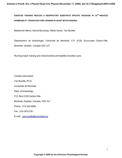
1 Mariannick Marcil, Karine Bourduas, Alexis Ascah, Yan Burelle ...
1 Mariannick Marcil, Karine Bourduas, Alexis Ascah, Yan Burelle ...
1 Mariannick Marcil, Karine Bourduas, Alexis Ascah, Yan Burelle ...

You also want an ePaper? Increase the reach of your titles
YUMPU automatically turns print PDFs into web optimized ePapers that Google loves.
Articles in PresS. Am J Physiol Heart Circ Physiol (November 11, 2005). doi:10.1152/ajpheart.00913.2005<br />
EXERCISE TRAINING INDUCES A RESPIRATORY SUBSTRATE SPECIFIC INCREASE IN CA 2+ -INDUCED<br />
PERMEABILITY TRANSITION PORE OPENING IN HEART MITOCHONDRIA.<br />
<strong>Mariannick</strong> <strong>Marcil</strong>, <strong>Karine</strong> <strong>Bourduas</strong>, <strong>Alexis</strong> <strong>Ascah</strong>, <strong>Yan</strong> <strong>Burelle</strong><br />
Département de kinésiologie, Université de Montréal, C.P. 6128, Succursale Centre-Ville,<br />
Montréal, Québec, Canada H3C 3J7<br />
Running head: training and mitochondrial permeability transition pore<br />
Contact information:<br />
<strong>Yan</strong> <strong>Burelle</strong>, Ph.D<br />
Université de Montréal<br />
Dept. of kinesiology<br />
P.O. Box 6128 Centre-Ville<br />
Montreal, Quebec, Canada, H3C 3J7<br />
Phone : 514-343-6083<br />
Fax : 514-343-2181<br />
E-mail : yan.burelle@umontreal.ca<br />
Copyright © 2005 by the American Physiological Society.<br />
1
ABSTRACT<br />
The purpose of this study was to determine if regular exercise (treadmill running, 10 weeks)<br />
alters the susceptibility of rat isolated heart mitochondria to Ca 2+ -induced PTP opening and if this<br />
could be associated with changes in the modulation of PTP opening by selected physiological<br />
effectors. Basal leak-driven and ADP-stimulated respiration in the presence of substrates for<br />
complex I, II and IV were not affected by training. Fluorimetric studies revealed that in the control<br />
(C) and exercise-trained (T) groups, the amount of Ca 2+ required to trigger PTP opening was<br />
greater in the presence of complex II vs I substrates (230 ± 12 vs 134 ± 7 nmol Ca 2+ .mg prot. -1<br />
respectively, P < 0.01, pooled average of C and T groups). In addition, with a substrate feeding<br />
the complex II, training increased by 45 % (P < 0.01) the amount of Ca 2+ required to trigger PTP<br />
opening, both in the presence and absence of the PTP inhibitor cyclosporin A. However, ,<br />
ROS production, NAD(P)H ratio and Ca 2+ uptake kinetics were not different in mitochondria from<br />
both groups. Taken together, these results suggest the existence of a substrate-specific<br />
regulation of the PTP in heart mitochondria and that regular exercise results in a reduced<br />
sensitivity to Ca 2+ -induced PTP opening in presence of complex II substrates.<br />
Key words: Exercise, heart, mitochondrial function, Ca 2+ stress<br />
2
INTRODUCTION<br />
Mitochondria play a pivotal role in controlling cell death through their capacity to trigger<br />
both necrosis and apoptosis (16, 18, 25, 39). A number of studies have shown that an increased<br />
permeability of the mitochondrial membranes is a key event in these processes (37, 41, 59).<br />
Although several mechanisms of membrane permeation have been suggested, one of the best<br />
documented involves the opening of the permeability transition pore (PTP).<br />
The mPTP is a high conductance non-specific pore presumably formed by a<br />
supramolecular complex spanning the double membrane system of the mitochondria mainly at<br />
contact sites. Although it is increasingly recognized the molecular composition of the mPTP is<br />
probably variable (61), the prevailing hypothesis is that it includes the adenylate translocator<br />
(ANT), the porin pore (VDAC) and the matrix protein foldase cyclophilin-D. Opening of the PTP<br />
induces the loss of mitochondrial membrane potential ( ), uncoupling of oxidative<br />
phosphorylation, high amplitude swelling of the matrix and the release of several pro-apoptotic<br />
factors that are normally sequestered in mitochondria such as cytochrome c, AIF, Smac/Diablo,<br />
endonuclease G and Omi/HtrA2 (5, 25).<br />
In the heart, PTP opening was shown to occur during reperfusion following ischemia and<br />
to be involved in contractile dysfunction and tissue injury (19, 23, 24, 31, 32, 35). This<br />
phenomenon can be explained by the fact that many of the conditions required to open the PTP<br />
in vitro prevail in cardiac cells early during reperfusion. On the other hand, ischemic pre-<br />
conditioning was shown to decrease the sensitivity to PTP opening in isolated mitochondria (1)<br />
and intact cardiomyocytes (28) and perfused hearts (27, 31). However whether exercise training,<br />
another physiological stress capable of inducing a cardio-protective phenotype (9, 14, 42, 51),<br />
can beneficially alter the regulatory properties of the PTP remains largely unknown.<br />
3
Calcium concentration in the matrix is the most important determinant of PTP gating, with<br />
high [Ca 2+ ] favouring the open conformation (60). In addition, a variety of factors modulate the<br />
sensitivity of the PTP to Ca 2+ , including variations in the , redox state of pyridine nucleotides<br />
(PNs), ROS production and matrix pH as well as Pi and adenylates content (see (60) for review).<br />
In skeletal muscle mitochondria the Ca 2+ sensitivity of the PTP was also shown to depend on the<br />
type of respiratory substrate oxidized, with complex I donors acting as sensitizers compared to<br />
complex II donors (22).<br />
In the present study we therefore determined if exercise training is associated with<br />
changes in the sensitivity to Ca 2+ -induced PTP opening in isolated heart mitochondria. We also<br />
determined if the type of substrate used for energization influences Ca 2+ -induced PTP opening in<br />
this organ and whether exercise training elicits changes in selected physiological modulators of<br />
PTP gating.<br />
4
METHODS<br />
Animal care<br />
All experiments were conducted according to the directives of the Canadian Council on<br />
Animal Care. Female Sprague-Dawley Rats (Charles River, St-Constant, PQ, Canada)<br />
weighing approximately 250 g were housed by pair and kept in a temperature, humidity, and<br />
light controlled (12:12 h light-dark cycle) environment. The animals had free access to standard<br />
rat chow and water.<br />
Exercise protocol<br />
Following a week of habituation, animals were divided into control (C) and exercise-<br />
trained (T) groups. Trained rats were run on a motor-driven rodent treadmill (Quinton<br />
Instruments, Seattle, WA) at 25 m/min, 16 % slope, 4 days/wk for 10 wk. Running time was set<br />
at 30 min during the 1 st wk, 60 min during the 2 nd wk and 90 min during the 3 rd wk. Running time<br />
was then maintained to at 90 min for the remaining 7 wk.<br />
Materials<br />
All chemicals were purchased from Sigma (St-Louis, MO, USA), with the exception of<br />
Cyclosporin A (Tocris, Ellisville, MO, USA), and CaGreen-5N (Molecular Probes, Eugene, OR,<br />
USA).<br />
Mitochondrial isolation<br />
Heart mitochondria were prepared as described by Fontaine et al. (22) with slight modifications.<br />
Animals were anesthetized (sodium pentobarbital 50 mg/kg ip) 48h after the last training<br />
session. Hearts were rapidly excised and immersed into ice-cold isolation medium (buffer A, in<br />
mM: 300 sucrose, 10 Tris (hydroxymethyl) aminomethane Hydrochloride (Tris-HCl),<br />
1 Ethyleneglycol-bis(M-aminoethyl)-N,N,NN,NN-tetraacetic Acid (EGTA), pH 7.3) and weighed.<br />
5
Ventricular tissue was minced with scissors in 5 ml of buffer A supplemented with 0.2 % fatty<br />
acid free bovine serum albumin (BSA) and homogenized using a Polytron tissue tearer (~3 sec<br />
at a setting of 3). The homogenate was then incubated with the protease Nagarse (1.5 mg/g) for<br />
5 min and further homogenized at the same settings. The homogenate volume was completed to<br />
30 ml with Buffer A + 0.2 % BSA and centrifuged at 800 x g for 10 min. The pellet was discarded<br />
and the supernatant was decanted and centrifuged at 10 000 x g for 10 min. The pellet obtained<br />
was re-suspended in buffer B (in mM: 300 sucrose, 0.5 EGTA, 10 Tris-HCl, pH 7.3) and<br />
centrifuged at 10 000 x g for 10 min. After repeating this washing step twice, the final<br />
mitochondrial pellet was re-suspended in 0.3 ml of buffer B to a protein concentration of ~20<br />
mg/ml. All procedures were carried out at 4 o C. Protein determinations were performed using<br />
the bicinchoninic acid method (Pierce, Rockford, IL, USA), with bovine serum albumin as a<br />
standard.<br />
Mitochondrial respiration<br />
Mitochondrial oxygen consumption was measured polarographically at 22 °C, using Clark-type<br />
electrodes (Oxygraph, Hansatech Instruments, Kings Lynn, England). Experiments were started<br />
with the addition of 0.30 mg mitochondria in 1ml of buffer C (in mM: 125 KCl, 10 KH2PO4, 0.05<br />
EGTA, 10 Tris (hydroxymethyl)-aminomethane - 3-(N-morpholino)propanesulfonic acid (Tris-<br />
MOPS), 2.5 MgCl2). Respiratory substrates feeding complex I (glutamate-malate 5:2.5 mM),<br />
complex II (succinate 5 mM) or complex IV (TMPD-Ascorbate 0.1:1 mM) were added in the<br />
incubation medium. All substrates were free acids buffered to pH 7.3 with Tris. Experiments for<br />
the complex II were made in presence or absence of the complex I inhibitor rotenone (1QM)<br />
(Figure 1). The medium was then supplemented with 0.25 mM ADP to measure maximal rate of<br />
oxidative phosphorylation (VADP). When respiration reached state 4 following complete<br />
phosphorylation of ADP, 0.5 QM oligomycin was added in order to measure oligomycin-<br />
insensitive respiration (Voligo), which eliminates the contribution of slow turnover of adenylates to<br />
6
asal respiration due to the presence of residual ATPase activity in the mitochondrial<br />
preparation. Respiratory control ratio (RCR) was calculated as the ratio VADP / Voligo while P/O<br />
was calculated from the amount of ADP added and that of O2 consumed before state 4 was<br />
reached.<br />
Calcium challenge:<br />
Mitochondria (0.3 mg/ml) were incubated at 22 o C in 2 ml of buffer D (in mM: 250<br />
sucrose, 10 MOPS, 0.05 EGTA, 10 KH2PO4, pH 7.2) containing glutamate-malate (5:2.5 mM) or<br />
succinate (5 mM) in the presence or absence of 1 QM rotenone. Changes in extra-mitochondrial<br />
calcium concentration were monitored fluorimetrically (Hitachi, F4500 spectrofluorometer) using<br />
Calcium-green 5N (1 QM, excitation-emission: 505-535 nm) as described by Ichas et al. (30).<br />
Residual calcium concentration was adjusted to the same level at the beginning of every<br />
experiment by adding a small amount of EGTA. Unless stated otherwise, calcium pulses (42<br />
nmol/mg prot) were then added at 2 min intervals until a Ca 2+ -induced mitochondrial Ca 2+<br />
release was observed. Calcium retention capacity (CRC) was taken as the total amount of Ca 2+<br />
accumulated by mitochondria prior to the Ca 2+ pulse triggering Ca 2+ release.<br />
In some experiments, mitochondrial membrane potential ( ) was measured under the<br />
same experimental conditions. For this purpose, Calcium-Green 5N was replaced by Rhodamine<br />
123 (0.2 QM: excitation-emission, 503-525 nm) and measurements were performed as described<br />
by Emaus et al. (21). Mitochondrial release of Rhodamine 123 following uncoupling with 100 nM<br />
carbonyl cyanide m-chlorophenylhydrazone (CCCP) was taken as an index of membrane potential.<br />
Mitochondrial swelling in response to Ca 2+ pulses was measured under the same experimental<br />
conditions using light diffraction at a setting of 545-545 nm (29). Each experiment was<br />
performed either in the presence or absence of 1 QM cyclosporin A (CsA). This concentration is<br />
commonly used (4, 22, 45) and is several fold higher than that required for full inhibition of PTP<br />
7
in liver (~0.15 QM: (47)) and heart (~0.3 QM: (15, 17) mitochondria under standardized<br />
conditions of Ca 2+ loading.<br />
ROS production<br />
Mitochondrial H2O2 production was measured fluorimetrically, as described in Servais et<br />
al. (53) (excitation-emission, 319-420 nm). Mitochondria (0.1 mg/ml) were incubated in 2 ml<br />
buffer D containing 5 U.mL -1 of horseradish peroxidase (HRP) and 0.1 mM homovanilic acid<br />
(HVA). At the end of each test 2 Qmoles of H2O2 were added as an internal standard to allow<br />
calculation of endogenous ROS production by the respiratory chain.<br />
Pyridine nucleotide oxidation-reduction status<br />
The oxidation-reduction status of the mitochondrial pyridine nucleotide (PN) pool was<br />
evaluated based on endogenous NAD(P)H fluorescence (excitation-emission, 340-460 nm).<br />
Mitochondria (0.3 mg/ml) were incubated in 2 ml buffer D and redox state of PN’s was measured<br />
in the presence of glutamate-malate (5:2.5 mM) or succinate (5 mM) ± rotenone (1 QM). Redox<br />
state of PN’s was calculated as described by Arieli et al. (2).<br />
NAD(P)H ratio = (Fs – Fmin) / (Fmax – Fmin)<br />
Where Fs is the fluorescence measured following addition of respiratory substrates, Fmin is the<br />
baseline fluorescence of mitochondria measured in the absence of respiratory substrates, which<br />
represents PNs in their fully oxidized form, and Fmax is the fluorescence recorded after addition<br />
of 2 QM Antimycin A, which represents PNs in their fully reduced form. Preliminary experiments<br />
indicated that Fmin values measured using this method were similar to those obtained when<br />
mitochondria were uncoupled by the addition of 100 nM CCCP.<br />
8
Mitochondrial Adenylate Content<br />
Endogenous ATP and ADP contents were measured in neutralized perchloric acid<br />
extracts using a luciferin/luciferase assay described by Drew & Leeuwenburgh (20) with<br />
modifications. Briefly, ATP content was evaluated based on the light production (hv) from the<br />
following reactions:<br />
Luciferin + ATP V luciferyl adenylate + PPi<br />
Luciferyl adenylate + O2 V oxyluciferin + AMP + hv.<br />
In an aliquot of the same sample, ATP + ADP content was measured in a similar way<br />
after endogenous ADP was converted to ATP using excess amounts of pyruvate kinase (20<br />
U.ml -1 ) and phosphoenolpyruvate (PEP: 5 mM) added directly in the cuvette. ADP content was<br />
calculated by subtracting the bioluminescence decay curves obtained in both samples. For every<br />
assay, baseline light emission (hv) prior to the addition of the sample was subtracted from the<br />
luminescent decay. In addition, it was verified that the presence of PK or PEP did not affect the<br />
relationship between [ATP] and hv.<br />
Statistical analyses<br />
Results are expressed as means ± S.E.M. Two-tailed Student’s t-tests were performed to<br />
assess statistical significance. When multiple comparisons were made, differences were<br />
compared using ANOVA and Tuckey Post-hoc tests were performed to identify the location of<br />
significant differences. The Bonferonni correction was applied to the P value obtained to correct<br />
for multiple comparisons. A corrected P value < 0.05 was considered significant.<br />
9
RESULTS<br />
Morphometric parameters:<br />
In line with our previous work (14), the exercise training regimen used in the present<br />
study resulted in myocardial hypertrophy, as indicated by a significant increase in heart weight,<br />
ventricular weight (+11 %, P < 0.05) and heart to body weight ratio (+10 %, P < 0.05). No<br />
significant changes in body weight and daily food intake were observed between both<br />
experimental groups. Mitochondrial yields were similar in hearts from C and T rats (~16 mg/g of<br />
ventricular tissue) (Table 1).<br />
Mitochondrial respiration:<br />
Table 2 shows the results of experiments aimed at characterizing the effect of exercise<br />
training on basic respiratory parameters. In mitochondria respiring with substrates for complex I,<br />
II and IV, training was not associated with any changes in maximal ADP-stimulated and<br />
oligomycin-insensitive leak-driven respiration, RCR and P/O ratio. These data are consistent<br />
with previous reports showing that exercise training does not significantly affect oxidative<br />
capacity (43).<br />
Ca 2+ challenge:<br />
Figure 2 shows the typical response of mitochondria energized with the complex I<br />
substrates glutamate-malate (GM) to series of Ca 2+ pulses. In these conditions, mitochondria<br />
accumulated 134 ± 7 nmol Ca 2+ /mg prot before abrupt release of accumulated Ca 2+ occurred.<br />
The release of Ca 2+ was invariably accompanied by high amplitude swelling. When the<br />
experiments were performed in the presence of the PTP inhibitor CsA (1 QM), the amount of<br />
Ca 2+ required to trigger these effects significantly increased 3.5-fold (483 ± 50 nmoles/mg prot, P<br />
< 0.01), indicating that these phenomena were caused by the opening of the PTP. However, as<br />
shown in Figure 3, in presence of glutamate-malate, exercise training resulted only in a small<br />
10
increase in CRC which was not statistically significant (P = 0.15) and CsA had the same effect<br />
than in mitochondria from control hearts.<br />
Figure 3 also shows the results of similar experiments performed in mitochondria<br />
respiring with the complex II substrate succinate. In both experimental groups, CRC in presence<br />
of succinate was ~60-100 % higher compared to that observed in presence of glutamate-malate<br />
whether or not rotenone was present.<br />
In contrast to what was observed in mitochondria energized with glutamate-malate,<br />
training significantly increased CRC by 45 % in mitochondria energized with succinate<br />
substrates both in presence (245 ± 14 vs 327 ± 25 nmoles/mg prot, P < 0.05) and absence of<br />
the complex I inhibitor rotenone (215 ± 25 vs 292 ± 26 nmoles/mg prot., P < 0.05) . This<br />
difference remained unaltered in the presence of CsA, which increased CRC to a similar extent<br />
in both experimental groups. Importantly, training was not associated with changes in Ca 2+<br />
uptake kinetics in any of the conditions tested, as indicated by the calculated time to 50 %<br />
uptake of the first 2-3 Ca 2+ pulses (26.5 ± 1.7 and 29.6 ± 2.0 sec in C and T mitochondria,<br />
respectively, with n = 6 in each group.<br />
PTP modulators:<br />
In order to gain insights on the mechanism underlying this increase in CRC with complex<br />
II substrates several physiological modulators of PTP gating were investigated. It is well<br />
established that the PTP behaves as a voltage-gated channel and that mild depolarization<br />
favours the open conformation in presence of Ca 2+ loading (7, 22, 60). For this reason the effect<br />
of training on prior to PTP opening was determined. At baseline, was similar in<br />
mitochondria from C and T animals, as indicated by a comparable release of Rhodamine 123<br />
following addition of 100 nM CCCP (553 ± 42 vs 580 ± 64 A.F.U in C and T groups,<br />
11
espectively). In addition, the transient depolarizations associated with the uptake of single Ca 2+<br />
pulses during the Ca 2+ loading phase were of similar amplitude (Figure 4). As expected, once<br />
the pore opened, an abrupt and complete loss of was observed in both experimental groups.<br />
However, consistent with the CRC data, significantly more Ca 2+ was required to trigger this effect<br />
in mitochondria from T compared to C animals.<br />
Figure 5 shows the results of experiments in which ROS production was measured. In<br />
line with previous studies (57), mitochondria generated a significant amount of ROS in presence<br />
of succinate alone (Figure 5A and B). Addition of rotenone decreased ROS production by half,<br />
consistent with the idea that succinate leads to ROS production at the level of the FeS centre of<br />
complex I through reverse electron flow (57). Subsequent addition of antimycin A led to a large<br />
increase in ROS production, which in the presence of rotenone, occurs mainly at the level of<br />
complex III. However, training did not alter steady state ROS production with succinate alone<br />
and in presence of both inhibitors. Similarly, training did not significantly affect ROS production<br />
when mitochondria were energized with glutamate-malate whether or not electron flow trough<br />
complex I was inhibited by rotenone (Figure 5C and D).<br />
Figure 6 shows the results from experiments in which the redox state of PNs was<br />
measured in mitochondria from C and T animals. Following addition of mitochondria in the<br />
absence of respiratory substrate, endogenous auto-fluorescence of PN’s stabilized at a low level<br />
in both experimental groups (Figure 6A). Addition of succinate led to a rapid reduction of PNs,<br />
and subsequent inhibition of electron flow with antimycin A led to the complete reduction of the<br />
PN pool. In some experiments 100 nM CCCP was added instead of antimycin A. Under this<br />
condition, fluorescence returned to the level observed in mitochondria in absence of respiratory<br />
substrate (data not shown). Figure 6B shows the steady state NAD(P)H ratio computed from<br />
several experiments in mitochondria respiring with succinate in presence and absence of<br />
12
otenone. No significant differences were observed between both experimental groups in any of<br />
the conditions tested.<br />
Table 3 shows the endogenous adenine nucleotides content measured in frozen<br />
mitochondrial extracts from C and T animals. Training did not significantly alter the residual ATP,<br />
ADP and total adenylate contents or the ATP/ADP ratio.<br />
13
DISCUSSION<br />
Results from the present study provide evidence that in heart mitochondria, the sensitivity<br />
to Ca 2+ -induced PTP opening is influenced by the type of respiratory substrate oxidized, with<br />
complex I donors acting as sensitizers compared to complex II substrates. In addition, the<br />
present results indicate that regular exercise training can decrease the sensitivity to Ca 2+ -<br />
induced PTP opening when mitochondria are energized with complex II substrates. This effect of<br />
training was observed in the absence of changes in the physiological modulators of PTP<br />
opening investigated.<br />
Substrate specific regulation of the PTP:<br />
Fontaine et al. (22) have shown that in skeletal muscle mitochondria CRC is 3-4 fold<br />
lower in mitochondria energized with glutamate-malate compared to succinate. This marked<br />
difference in CRC was shown to be due to the fact that the factors contributing to PTP opening<br />
are different in the two experimental conditions. Indeed, in presence of complex I substrates the<br />
electron flow through this complex, independent of other regulators (i.e. redox state of PN’s, ,<br />
pH, ROS production), appeared to be the main factor regulating PTP opening by acting as a<br />
potent sensitizer (6, 22, 40). In contrast, when complex I was bypassed using succinate the<br />
mechanism was largely inactive and the contribution of other regulators to PTP opening were<br />
unmasked. Results from the present study are in line with these data. Indeed, in both<br />
experimental groups a substantially lower CRC was observed in presence of glutamate-malate<br />
compared to succinate. Our data thus indicate that similar to what is observed in skeletal muscle<br />
the electron flow through complex I sensitize heart mitochondria to PTP opening.<br />
Training-induced alteration in Ca 2+ handling:<br />
To our knowledge, the impact of exercise training on Ca 2+ handling by heart mitochondria<br />
has only been investigated in two studies (50, 56) and the effect on the PTP was not directly<br />
14
assessed. Sordahl et al. (56) reported that the rate of Ca 2+ uptake by isolated heart mitochondria<br />
energized with succinate was unchanged following training in dogs. Similar finding were reported<br />
by Penpargkul et al. (50) in rodents in response to swim training. Results from the present study<br />
are in line with these findings. Indeed, Ca 2+ uptake kinetics measured during the first 2-3 Ca 2+<br />
pulses of the Ca 2+ challenge did not differ between the two experimental groups whether<br />
mitochondria were energized with complex I or II substrates. These data thus suggest that<br />
training has little effect on the electrophoretic mechanism of Ca 2+ uptake in heart mitochondria.<br />
In the study by Sordahl et al. (56) training was found to decrease the capacity of<br />
mitochondria to accumulate Ca 2+ when energized with succinate. At the time this study was<br />
published, the existence of the PTP and the inhibitory effect of cyclosporins on pore opening<br />
were unknown. However, given the experimental conditions used, this phenomenon was likely<br />
caused by a premature opening of the PTP in mitochondria from trained animals. As for the<br />
study by Penpargkul et al. (50) the experimental conditions used did not allow to observe a<br />
permeability transition.<br />
In contrast to the results from Sordhal et al. (56) data from the present study indicate that<br />
in mitochondria from T rats the amount of Ca 2+ required to trigger PTP opening was significantly<br />
45 % higher in presence of succinate both in presence and absence of rotenone. In addition,<br />
CsA increased CRC in both experimental groups but did not abolish the effect of training. These<br />
results thus indicate that in our conditions, training was able to decrease the sensitivity to Ca 2+ -<br />
induced PTP opening in presence of complex II substrates. In addition, the fact that the effect of<br />
training was not abolished by CsA used at a concentration 3 fold in excess of that required to<br />
fully inhibit PTP opening (15, 17, 47) suggests that the effect of training is not related to a<br />
reduction in the expression of cyclophilin-D or its interaction with other PTP components (3, 4).<br />
15
Indeed changes in the expression of cyclophilin-D are known to translate in an altered potency<br />
of CsA at inhibiting Ca 2+ -induced PTP opening (3, 4, 45, 52).<br />
Our results also indicate that in contrast to what was observed with succinate, no<br />
significant difference between C and T groups was observed for CRC in presence of complex I<br />
substrates. As mentioned above, PTP opening is strongly regulated by electron flow through<br />
complex I, which sensitizes mitochondria to permeability transition (6, 22, 40). Our data thus<br />
indicate that training did not protect against this sensitizing effect. In contrast when complex I<br />
was bypassed a protective effect of training, probably through other regulators of PTP opening,<br />
was unmasked.<br />
Effect of training on physiological modulators of PTP opening:<br />
To our knowledge, there is no data available in the literature concerning the effect of<br />
training on parameters involved in the regulation of the PTP in the heart. In the present study,<br />
we therefore determined whether the training-induced increase in the resistance to Ca 2+ -induced<br />
PTP opening observed with complex II substrates was accompanied by changes in selected<br />
physiological modulators of PTP gating and/or in mitochondrial respiratory activity.<br />
Ca 2+ -induced permeability transition is modulated by a variety of physiological effectors.<br />
The occurrence of PTP opening at a given Ca 2+ load is reduced when membrane potential is<br />
increased due to the fact that the PTP behaves as a voltage-gated channel sensitive to changes<br />
in over a range of 180 – 120 mV (7). Matrix adenine nucleotides are also potent inhibitors of<br />
PTP opening, with ADP exerting a stronger inhibitory effect than ATP (60) (16). Maintenance of<br />
the PN pool at a high reduction state is another factor known to decrease PTP opening induced<br />
by Ca 2+ and Pi probably through mechanisms involving direct interaction of PN’s with the PTP<br />
and to oxidation of critical SH residues of pore-forming proteins (6, 60). Low levels of ROS<br />
16
production (12, 26) as well as an acidic pH (8, 48) will also reduce the occurrence of PTP<br />
opening. Finally, the sensitivity of the PTP to Ca 2+ was shown to be modulated by members of<br />
the Bcl-2 family of proteins the pro-apoptotic Bid and Bax and the antiapoptotic Bcl-2 and Bcl-Xl<br />
acting as facilitators and repressors of pore opening respectively (11, 38, 44, 46, 49).<br />
However, in the present study we did not find evidence indicating that changes at the<br />
level of these PTP modulators could account for the increased resistance to Ca 2+ -induced PTP<br />
opening observed. Indeed, baseline at steady state as well as the amplitude of the transient<br />
depolarizations associated with the uptake of single Ca 2+ pulses were similar in both groups<br />
suggesting that prior to PTP opening was not affected by training. Endogenous ATP and<br />
ADP content as well as the ATP/ADP ratio was also similar in mitochondria from the C and T<br />
groups. In addition, the redox state of PN’s measured was unchanged. As for matrix pH, it is<br />
unlikely to be a factor since the high [Pi] used during the Ca 2+ challenge experiments prevents<br />
any fluctuations in matrix pH secondary to changes in respiration (22).<br />
In the heart, several studies have shown that training results in a significant increase in<br />
tissue content of enzymatic and non-enzymatic antioxidant systems including some that are<br />
found in mitochondria (33, 36, 55). However, there is apparently no data available on the effect<br />
of training on the actual rate of ROS production by active mitochondria. Moreover, data available<br />
in skeletal muscle mitochondria on this question are conflicting, showing either a reduction (58)<br />
or no change (53) in ROS production following training.<br />
In the present study, the rate of H2O2 production of mitochondria respiring with complex I<br />
or II substrates under state 4 conditions was not significantly affected by training. In addition,<br />
reduction of respiratory chain complexes with rotenone or antimycin A, which induce a large<br />
17
increase in ROS production, had similar effects in both experimental groups. These results<br />
would thus suggest that altered ROS production is not responsible for the increased CRC<br />
observed in mitochondria following training. However, ROS production could not be accurately<br />
measured during the Ca 2+ challenge due to artifactual changes in the fluorescence of HVA in<br />
these conditions (5). Therefore, the present data cannot rule out the possibility that during the<br />
Ca 2+ challenge, ROS production was lower in trained mitochondria due to a reduced ability of<br />
Ca 2+ to increase ROS production through its action on the TCA cycle and/or on several<br />
components of the respiratory chain (12).<br />
Taken together, results from the present study indicate that mitochondria isolated from<br />
trained hearts are more resistant to Ca 2+ -induced PTP opening when energized with succinate.<br />
This adaptation could potentially be beneficial to the heart in the setting of ischemia-reperfusion,<br />
a situation where exercise was shown to be protective (9, 10, 13). The mechanisms underlying<br />
this increased resistance remain obscure(6, 22, 40) but are apparently not related to changes in<br />
endogenous adenylates content, alterations in respiratory chain function, redox state of PN’s, or<br />
ROS production. One possibility is that this phenomenon is caused by changes in the<br />
expression of anti- and pro-apoptotic members of the Bcl-2 family of proteins known to modulate<br />
Ca 2+ sensitivity of the PTP. Indeed, the balance between Bcl-2 and BclXL on the one hand and<br />
Bax on the other hand was recently shown to be increased following training (34, 54). However,<br />
this hypothesis remains to be ascertained.<br />
18
REFERENCES<br />
1. Argaud L, Gateau-Roesch O, Chalabreysse L, Gomez L, Loufouat J, Thivolet-Bejui<br />
F, Robert D, and Ovize M. Preconditioning delays Ca2+-induced mitochondrial permeability<br />
transition. Cardiovasc Res 61: 115-122, 2004.<br />
2. Arieli Y, Gursahani H, Eaton MM, Hernandez LA, and Schaefer S. Gender modulation<br />
of Ca(2+) uptake in cardiac mitochondria. J Mol Cell Cardiol 37: 507-513, 2004.<br />
3. Baines CP, Kaiser RA, Purcell NH, Blair NS, Osinska H, Hambleton MA, Brunskill<br />
EW, Sayen MR, Gottlieb RA, Dorn GW, Robbins J, and Molkentin JD. Loss of cyclophilin D<br />
reveals a critical role for mitochondrial permeability transition in cell death. Nature 434: 658,<br />
2005.<br />
4. Basso E, Fante L, Fowlkes J, Petronilli V, Forte MA, and Bernardi P. Properties of<br />
the Permeability Transition Pore in Mitochondria Devoid of Cyclophilin D. J Biol Chem 280:<br />
18558-18561, 2005.<br />
5. Batandier C, Leverve X, and Fontaine E. Opening of the mitochondrial permeability<br />
transition pore induces reactive oxygen species production at the level of the respiratory chain<br />
complex I. J Biol Chem 279: 17197-17204, 2004.<br />
6. Bernardi P. Mitochondrial transport of cations: channels, exchangers, and permeability<br />
transition. Physiol Rev 79: 1127-1155., 1999.<br />
7. Bernardi P. Modulation of the mitochondrial cyclosporin A-sensitive permeability<br />
transition pore by the proton electrochemical gradient. Evidence that the pore can be opened by<br />
membrane depolarization. J Biol Chem 267: 8834-8839, 1992.<br />
8. Bernardi P, Vassanelli S, Veronese P, Colonna R, Szabo I, and Zoratti M. Modulation<br />
of the mitochondrial permeability transition pore. Effect of protons and divalent cations. J Biol<br />
Chem 267: 2934-2939, 1992.<br />
9. Bowles DK, Farrar RP, and Starnes JW. Exercise training improves cardiac function<br />
after ischemia in the isolated, working rat heart. Am J Physiol 263: H804-809., 1992.<br />
10. Bowles DK and Starnes JW. Exercise training improves metabolic response after<br />
ischemia in isolated working rat heart. J Appl Physiol 76: 1608-1614., 1994.<br />
11. Brenner C, Cadiou H, Vieira HL, Zamzami N, Marzo I, Xie Z, Leber B, Andrews D,<br />
Duclohier H, Reed JC, and Kroemer G. Bcl-2 and Bax regulate the channel activity of the<br />
mitochondrial adenine nucleotide translocator. Oncogene 19: 329-336., 2000.<br />
12. Brookes PS, Yoon Y, Robotham JL, Anders MW, and Sheu SS. Calcium, ATP, and<br />
ROS: a mitochondrial love-hate triangle. Am J Physiol Cell Physiol 287: C817-833, 2004.<br />
13. Brunner M, Moeslinger T, and Spieckermann PG. Regulation of cyclosporin A<br />
sensitive mitochondrial permeability transition by the redox state of pyridine nucleotides. Comp<br />
Biochem Physiol B Biochem Mol Biol 128: 31-41, 2001.<br />
14. <strong>Burelle</strong> Y, Wambolt RB, Grist M, Parsons HL, Chow JC, Antler C, Bonen A, Keller A,<br />
Dunaway GA, Popov KM, Hochachka PW, and Allard MF. Regular exercise is associated with<br />
a protective metabolic phenotype in the rat heart. Am J Physiol Heart Circ Physiol 287: H1055-<br />
1063, 2004.<br />
15. Clarke SJ, McStay GP, and Halestrap AP. Sanglifehrin A acts as a potent inhibitor of<br />
the mitochondrial permeability transition and reperfusion injury of the heart by binding to<br />
cyclophilin-D at a different site from cyclosporin A. J Biol Chem 277: 34793-34799., 2002.<br />
16. Crompton M. The mitochondrial permeability transition pore and its role in cell death.<br />
Biochem J 341: 233-249., 1999.<br />
17. Crompton M, Ellinger H, and Costi A. Inhibition by cyclosporin A of a Ca2+-dependent<br />
pore in heart mitochondria activated by inorganic phosphate and oxidative stress. Biochem J<br />
255: 357-360., 1988.<br />
18. Di Lisa F and Bernardi P. Mitochondrial function as a determinant of recovery or death<br />
in cell response to injury. Mol Cell Biochem 184: 379-391., 1998.<br />
19
19. Di Lisa F, Menabo R, Canton M, Barile M, and Bernardi P. Opening of the<br />
mitochondrial permeability transition pore causes depletion of mitochondrial and cytosolic NAD+<br />
and is a causative event in the death of myocytes in postischemic reperfusion of the heart. J Biol<br />
Chem 276: 2571-2575., 2001.<br />
20. Drew B and Leeuwenburgh C. Method for measuring ATP production in isolated<br />
mitochondria: ATP production in brain and liver mitochondria of Fischer-344 rats with age and<br />
caloric restriction. Am J Physiol Regul Integr Comp Physiol 285: R1259-1267, 2003.<br />
21. Emaus RK, Grunwald R, and Lemasters JJ. Rhodamine 123 as a probe of<br />
transmembrane potential in isolated rat-liver mitochondria: spectral and metabolic properties.<br />
Biochim Biophys Acta 850: 436-448, 1986.<br />
22. Fontaine E, Eriksson O, Ichas F, and Bernardi P. Regulation of the permeability<br />
transition pore in skeletal muscle mitochondria. Modulation By electron flow through the<br />
respiratory chain complex i. J Biol Chem 273: 12662-12668., 1998.<br />
23. Griffiths EJ and Halestrap AP. Mitochondrial non-specific pores remain closed during<br />
cardiac ischaemia, but open upon reperfusion. Biochem J 307: 93-98., 1995.<br />
24. Griffiths EJ and Halestrap AP. Protection by Cyclosporin A of ischemia/reperfusioninduced<br />
damage in isolated rat hearts. J Mol Cell Cardiol 25: 1461-1469., 1993.<br />
25. Halestrap AP, Doran E, Gillespie JP, and O'Toole A. Mitochondria and cell death.<br />
Biochem Soc Trans 28: 170-177, 2000.<br />
26. Harris EJ, Booth R, and Cooper MB. The effect of superoxide generation on the ability<br />
of mitochondria to take up and retain Ca2+. FEBS Lett 146: 267-272, 1982.<br />
27. Hausenloy DJ, Maddock HL, Baxter GF, and Yellon DM. Inhibiting mitochondrial<br />
permeability transition pore opening: a new paradigm for myocardial preconditioning?<br />
Cardiovasc Res 55: 534-543., 2002.<br />
28. Hausenloy DJ, Yellon DM, Mani-Babu S, and Duchen MR. Preconditioning protects by<br />
inhibiting the mitochondrial permeability transition. Am J Physiol Heart Circ Physiol 287: H841-<br />
849, 2004.<br />
29. Hunter DR and Haworth RA. The Ca2+-induced membrane transition in mitochondria. I.<br />
The protective mechanisms. Arch Biochem Biophys 195: 453-459, 1979.<br />
30. Ichas F, Jouaville, L.S., Mazat, J.P. Mitochondria Are Excitable Organelles Capable of<br />
Generating and Conveying Electrical and Calcium Signals. Cell 89: 1145-1153, 1997.<br />
31. Javadov SA, Clarke S, Das M, Griffiths EJ, Lim KH, and Halestrap AP. Ischaemic<br />
preconditioning inhibits opening of mitochondrial permeability transition pores in the reperfused<br />
rat heart. J Physiol 549: 513-524, 2003.<br />
32. Javadov SA, Lim KH, Kerr PM, Suleiman MS, Angelini GD, and Halestrap AP.<br />
Protection of hearts from reperfusion injury by propofol is associated with inhibition of the<br />
mitochondrial permeability transition. Cardiovasc Res 45: 360-369., 2000.<br />
33. Ji LL, Fu RG, Mitchell EW, Griffiths M, Waldrop TG, and Swartz HM. Cardiac<br />
hypertrophy alters myocardial response to ischaemia and reperfusion in vivo. Acta Physiol<br />
Scand 151: 279-290., 1994.<br />
34. Kang PM, Yue P, Liu Z, Tarnavski O, Bodyak N, and Izumo S. Alterations in apoptosis<br />
regulatory factors during hypertrophy and heart failure. Am J Physiol Heart Circ Physiol 287:<br />
H72-80, 2004.<br />
35. Kerr PM, Suleiman MS, and Halestrap AP. Reversal of permeability transition during<br />
recovery of hearts from ischemia and its enhancement by pyruvate. Am J Physiol 276: H496-<br />
502., 1999.<br />
36. Kihlstrom M. Protection effect of endurance training against reoxygenation-induced<br />
injuries in rat heart. J Appl Physiol 68: 1672-1678., 1990.<br />
37. Kim J-S, He L, and Lemasters JJ. Mitochondrial permeability transition: a common<br />
pathway to necrosis and apoptosis. Biochemical and Biophysical Research Communications<br />
304: 463, 2003.<br />
20
38. Kowaltowski AJ, Vercesi AE, and Fiskum G. Bcl-2 prevents mitochondrial permeability<br />
transition and cytochrome c release via maintenance of reduced pyridine nucleotides. Cell Death<br />
Differ 7: 903-910, 2000.<br />
39. Lemasters J, Qian T, Bradham C, Brenner D, Cascio W, Trost L, Nishimura Y,<br />
Nieminen A-L, and Herman B. Mitochondrial Dysfunction in the Pathogenesis of Necrotic and<br />
Apoptotic Cell Death. Journal of Bioenergetics and Biomembranes 31: 305, 1999.<br />
40. Leverve XM and Fontaine E. Role of substrates in the regulation of mitochondrial<br />
function in situ. IUBMB Life 52: 221-229., 2001.<br />
41. Mattson MP and Kroemer G. Mitochondria in cell death: novel targets for<br />
neuroprotection and cardioprotection. Trends Mol Med 9: 196-205, 2003.<br />
42. McElroy CL, Gissen SA, and Fishbein MC. Exercise-induced reduction in myocardial<br />
infarct size after coronary artery occlusion in the rat. Circulation 57: 958-962, 1978.<br />
43. Moore RL and Palmer BM. Exercise training and cellular adaptations of normal and<br />
diseased hearts. Exerc Sport Sci Rev 27: 285-315, 1999.<br />
44. Murphy RC, Schneider E, and Kinnally KW. Overexpression of Bcl-2 suppresses the<br />
calcium activation of a mitochondrial megachannel. FEBS Lett 497: 73-76., 2001.<br />
45. Nakagawa T, Shimizu S, Watanabe T, Yamaguchi O, Otsu K, Yamagata H, Inohara<br />
H, Kubo T, and Tsujimoto Y. Cyclophilin D-dependent mitochondrial permeability transition<br />
regulates some necrotic but not apoptotic cell death. Nature 434: 652-658, 2005.<br />
46. Narita M, Shimizu S, Ito T, Chittenden T, Lutz RJ, Matsuda H, and Tsujimoto Y. Bax<br />
interacts with the permeability transition pore to induce permeability transition and cytochrome c<br />
release in isolated mitochondria. Proc Natl Acad Sci U S A 95: 14681-14686., 1998.<br />
47. Nicolli A, Basso E, Petronilli V, Wenger RM, and Bernardi P. Interactions of<br />
cyclophilin with the mitochondrial inner membrane and regulation of the permeability transition<br />
pore, and cyclosporin A-sensitive channel. J Biol Chem 271: 2185-2192., 1996.<br />
48. Nicolli A, Petronilli V, and Bernardi P. Modulation of the mitochondrial cyclosporin Asensitive<br />
permeability transition pore by matrix pH. Evidence that the pore open-closed<br />
probability is regulated by reversible histidine protonation. Biochemistry 32: 4461-4465., 1993.<br />
49. Pastorino JG, Tafani M, Rothman RJ, Marcinkeviciute A, Hoek JB, Farber JL, and<br />
Marcineviciute A. Functional consequences of the sustained or transient activation by Bax of<br />
the mitochondrial permeability transition pore. J Biol Chem 274: 31734-31739., 1999.<br />
50. Penpargkul S, Schwartz A, and Scheuer J. Effect of physical conditioning on cardiac<br />
mitochondrial function. J Appl Physiol 45: 978-986., 1978.<br />
51. Powers SK, Demirel HA, Vincent HK, Coombes JS, Naito H, Hamilton KL, Shanely<br />
RA, and Jessup J. Exercise training improves myocardial tolerance to in vivo ischemiareperfusion<br />
in the rat. Am J Physiol 275: R1468-1477., 1998.<br />
52. Schinzel AC, Takeuchi O, Huang Z, Fisher JK, Zhou Z, Rubens J, Hetz C, Danial<br />
NN, Moskowitz MA, and Korsmeyer SJ. Cyclophilin D is a component of mitochondrial<br />
permeability transition and mediates neuronal cell death after focal cerebral ischemia. PNAS<br />
102: 12005-12010, 2005.<br />
53. Servais S, Couturier K, Koubi H, Rouanet JL, Desplanches D, Sornay-Mayet MH,<br />
Sempore B, Lavoie JM, and Favier R. Effect of voluntary exercise on H2O2 release by<br />
subsarcolemmal and intermyofibrillar mitochondria. Free Radic Biol Med 35: 24-32., 2003.<br />
54. Siu PM, Bryner RW, Martyn JK, and Alway SE. Apoptotic adaptations from exercise<br />
training in skeletal and cardiac muscles. FASEB J: 03-1291fje, 2004.<br />
55. Somani SM, Frank S, and Rybak LP. Responses of antioxidant system to acute and<br />
trained exercise in rat heart subcellular fractions. Pharmacol Biochem Behav 51: 627-634, 1995.<br />
56. Sordahl LA, Asimakis GK, Dowell RT, and Stone HL. Functions of selected<br />
biochemical systems from the exercised-trained dog heart. J Appl Physiol 42: 426-431., 1977.<br />
57. Turrens JF. Superoxide production by the mitochondrial respiratory chain. Biosci Rep<br />
17: 3-8, 1997.<br />
21
58. Venditti P, Masullo P, and Di Meo S. Effect of training on H(2)O(2) release by<br />
mitochondria from rat skeletal muscle. Arch Biochem Biophys 372: 315-320., 1999.<br />
59. Zamzami N and Kroemer G. Apoptosis: Mitochondrial Membrane Permeabilization -<br />
The (W)hole Story? Current Biology 13: R71, 2003.<br />
60. Zoratti M and Szabo I. The mitochondrial permeability transition. Biochim Biophys Acta<br />
1241: 139-176., 1995.<br />
61. Zoratti M, Szabo I, and De Marchi U. Mitochondrial permeability transitions: how many<br />
doors to the house? Biochim Biophys Acta 1706: 40-52, 2005.<br />
22
AKNOWLEDGEMENTS<br />
This work was supported by a grant from the Canadian Institute of Health Research. YB is a<br />
Junior Investigator of the Fonds de Recherche en Santé du Québec (FRSQ). MM is supported<br />
by a Ph.D scholarship from FRSQ. The authors wish to thank Dr. Éric Fontaine for helpful<br />
discussions and Véronique Giroux and Sébastien Martin for their technical assistance.<br />
23
Table 1: Morphometric data and mitochondrial isolation yield in sedentary control and<br />
exercise trained rats.<br />
Control<br />
Trained<br />
Body weight (g) 343 ± 14 347 ± 10<br />
Heart weight (mg) 920 ± 35 1020 ± 18 *<br />
Heart weight / Body weight (mg/g) 2.69 ± 0.05 2.95 ± 0.07 #<br />
Ventricular weight (mg) 784 ± 23 871 ± 19 *<br />
Mitochondrial isolation yield<br />
(mg prot/g ventricle)<br />
15.7 ± 1.0 16.5 ± 0.6<br />
Average food intake (g/day) 20.9 ± 0.7 20.6 ± 0.3<br />
Values are means ± SEM for 8 animals in each group. * Significantly different from control (P <<br />
0.01). # Significantly different from control (P < 0.05).<br />
24
Table 2: Effect of exercise on mitochondrial respiration.<br />
Conditions<br />
Substrates<br />
Table shows basal oligomycin-insensitive (Voligo) and ADP stimulated (VADP) respiration (in<br />
nmoles O2.min -1 .mg prot -1 ), respiratory control ratio (rCR: VADP/Voligo), and P/O of mitochondria<br />
energized with glutamate-malate (GM), succinate ± rotenone (S and SR) and TMPD-Ascorbate<br />
(TMPD-Asc). Values are reported as means ± SEM for 4 (control) to 7 (trained) animals; all<br />
measurements were carried out in duplicates.<br />
Control<br />
Trained<br />
Voligo GM 14.1 ± 0.4 13.3 ± 0.7<br />
SR 30.3 ± 2.4 28.2 ± 2.6<br />
S 27.1 ± 1.6 25.8 ± 2.1<br />
TMPD-Asc 56.2 ± 3.7 44.1 ± 3.1<br />
VADP GM 101.4 ± 8.8 91.5 ± 9.6<br />
SR 96.3 ± 8.0 104.6 ± 4.8<br />
S 68.7 ± 9.2 50.9 ± 7.6<br />
TMPD-Asc 89.9 ± 4.8 76.8 ± 5.0<br />
RCR GM 7.3 ± 0.5 6.7 ± 0.5<br />
SR 3.3 ± 0.3 4.3 ± 0.6<br />
S 2.5 ± 0.3 2.3 ± 0.5<br />
TMPD-Asc 1.6 ± 0.04 1.8 ± 0.1<br />
P/O GM 2.5 ± 0.2 2.9 ± 0.2<br />
SR 1.5 ± 0.1 1.4 ± 0.2<br />
S 2.2 ± 0.4 2.1 ± 0.2<br />
TMPD-Asc 1.7 ± 0.2 1.6 ± 0.2<br />
25
Table 3: Adenylate content of heart mitochondria from control and trained rats.<br />
Conditions<br />
Control<br />
Trained<br />
ATP<br />
(nmol/mg prot)<br />
ADP<br />
(nmol/mg prot)<br />
The table shows endogenous ADP, ATP and total adenylates content as well as ATP/ADP ratio<br />
measured in perchloric acid extracted mitochondrial aliquots. Values are means ± SEM for an n<br />
of 6 separate mitochondrial preparations in each group.<br />
Adenylate content<br />
(nmol/mg prot)<br />
ATP/ADP ratio<br />
1.12 ± 0.14 0.76 ± 0.22 1.88 ± 0.33 2.03 ± 1.03<br />
1.14 ± 0.09 0.77 ± 0.16 1.91 ± 0.23 1.60 ± 0.20<br />
26
Glut/mal<br />
Rotenone Antimycin A Oligomycin<br />
I<br />
-<br />
NADH NAD +<br />
FADH 2<br />
II<br />
Succinate<br />
FAD<br />
e<br />
Q<br />
H TMPD/asc.<br />
+ H + H +<br />
-<br />
III<br />
c<br />
ADP + P i<br />
1/2 O 2 + 2H + H 2 O<br />
IV<br />
-<br />
Figure 1<br />
ATP<br />
27
[Ca 2+ ] (QM)<br />
Light scattering 545/545 nm<br />
40<br />
35<br />
30<br />
25<br />
20<br />
15<br />
10<br />
5<br />
0<br />
3000<br />
2500<br />
2000<br />
1500<br />
A<br />
C<br />
Ca 2+<br />
Ca 2+<br />
-CsA +CsA<br />
40 B<br />
[Ca 2+ ] (QM)<br />
Light scattering 545/545 nm<br />
35<br />
30<br />
25<br />
20<br />
15<br />
10<br />
5<br />
0<br />
3000<br />
2500<br />
2000<br />
1500<br />
2 min<br />
Ca 2+<br />
-CsA +CsA<br />
D<br />
2 min<br />
Ca 2+<br />
Figure 2<br />
28
Ca2+ retention capacity<br />
(nmol.mg -1 prot)<br />
350<br />
300<br />
250<br />
200<br />
150<br />
100<br />
Control<br />
Trained<br />
a<br />
a<br />
GM S SR GM S SR<br />
- CsA + CsA<br />
b<br />
b b<br />
a;b<br />
b<br />
a;b<br />
1200<br />
1000<br />
800<br />
600<br />
400<br />
Ca2+ retention capacity<br />
(nmol.mg -1 prot)<br />
Figure 3<br />
29
Fluorescence (A.U.)<br />
300<br />
200<br />
300<br />
Control Trained<br />
Ca 2+<br />
Fluorescence (A.U.)<br />
200<br />
2 min<br />
Ca 2+<br />
Figure 4<br />
30
Fluorescence (A.U.)<br />
Fluorescence (A.U.)<br />
1800<br />
1600<br />
1400<br />
1200<br />
1000<br />
1800<br />
1600<br />
1400<br />
1200<br />
1000<br />
800<br />
600<br />
400<br />
800<br />
600<br />
400<br />
M S<br />
Rot<br />
0.49 ± 0.09<br />
Ant A<br />
H 2 O 2<br />
Control Trained<br />
2 min<br />
0.78 ± 0.09 b<br />
0.26 ± 0.06 a<br />
M S<br />
Control Trained<br />
M GM Rot<br />
2 min<br />
H 2O 2<br />
Succinate<br />
Glutamate-malate<br />
M<br />
GM Rot<br />
Rot<br />
0.54 ± 0.06<br />
H 2O 2<br />
Ant A<br />
H 2 O 2<br />
0.26 ± 0.03 a<br />
0.13 ± 0.05 c<br />
0.81 ± 0.19 d 0.61 ± 0.20 d<br />
0.09 ± 0.03 c<br />
0.73 ± 0.09 b<br />
Figure 5<br />
31
Figure 6<br />
Fluorescence 340 nm (A.U.)<br />
NAD(P)H ratio<br />
1000<br />
800<br />
600<br />
400<br />
200<br />
0<br />
0.5<br />
0.4<br />
0.3<br />
0.2<br />
0.1<br />
0.0<br />
A<br />
2 min<br />
B<br />
Control<br />
Trained<br />
M<br />
SR<br />
S<br />
F min<br />
Ant-A<br />
S<br />
F S<br />
F max<br />
Control<br />
Trained<br />
32
FIGURE LEGENDS<br />
Figure 1: Overview of respiratory substrates and inhibitors used: The respiratory substrates<br />
providing electrons to complex I II and IV are underlined. The various inhibitors (rotenone,<br />
antimycin A and oligomycin) used are shown on top of their respective site of action.<br />
Figure 2: Mitochondrial response to a train of Ca 2+ pulses. Typical Calcium-Green 5N<br />
(panels A and B), and light scattering at 545 nm (panels C and D) of mitochondria energized<br />
with glutamate-malate (5:2.5 mM). Tracings show progressive Ca 2+ accumulation followed by<br />
release of accumulated Ca 2+ , and high amplitude swelling. The presence of 1 QM cyclosporin A<br />
(panels B, D) doubles the amount of Ca 2+ required to trigger these effects indicating that these<br />
phenomena are related to PTP opening. Each arrow indicates the addition of a calcium pulse of<br />
42 and 210 nmol/mg for the Calcium-Green and swelling experiments, respectively.<br />
Figure 3: Effect of exercise on PTP sensitivity to Ca 2+ with complex I and II substrates.<br />
The figure shows calcium retention capacity of control (black) and exercise trained (hatched)<br />
mitochondria energized with glutamate-malate (GM, Panel A) and succinate in absence (S,<br />
Panel B) and presence (SR, Panel C) of 1 QM rotenone. Experiments were performed in<br />
absence (GM, S and SR) or presence (GM+CsA, S+CsA and SR+CsA) of 1 QM CsA. Values are<br />
means ± SEM for at least 7 separate experiments in the C and T groups, respectively. a<br />
Significantly different from control (P < 0.05) b Significantly different compared to - CsA (P <<br />
0.01)<br />
33
Figure 4: Changes in membrane potential in response to Ca 2+ pulses in mitochondria<br />
from control and trained animals. Figure shows representative traces of changes in<br />
measured with rhodamine 123 during Ca 2+ challenge experiments in presence of succinate.<br />
Each arrow indicates the addition of a calcium pulse of 42 nmol Ca 2+ /mg prot.<br />
Figure 5: Effect of exercise on mitochondrial H2O2 production. Figure shows representative<br />
traces of H2O2 production in heart mitochondria of control (panels A and C) and trained (panels<br />
B and D) animals when energized with succinate (panels A and B) or glutamate-malate (panels<br />
C and D). Numbers below traces represents the rate of H2O2 production in nmoles.min -1 .mg prot.<br />
-1 . Data are shown as means ± SEM for at least 7 experiments in each group. M: mitochondria,<br />
GM: glutamate-malate, S: succinate, Rot: rotenone 1QM, Ant-A: antimycin-A 2 QM. a : significantly<br />
different from succinate alone (P