Flavobacterium psychrophilum, cause of ... - cop.eXtension.org
Flavobacterium psychrophilum, cause of ... - cop.eXtension.org
Flavobacterium psychrophilum, cause of ... - cop.eXtension.org

You also want an ePaper? Increase the reach of your titles
YUMPU automatically turns print PDFs into web optimized ePapers that Google loves.
<strong>of</strong> the body or the head and snout (Figure 7). Among coho salmon, Wood and Yasutake<br />
(1970) noted that the epithelium in certain areas <strong>of</strong> the head was replaced by focal masses<br />
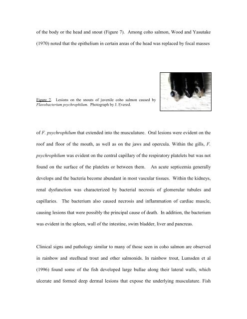
Figure 7. Lesions on the snouts <strong>of</strong> juvenile coho salmon <strong>cause</strong>d by<br />
<strong>Flavobacterium</strong> <strong>psychrophilum</strong>. Photograph by J. Evered.<br />
<strong>of</strong> F. <strong>psychrophilum</strong> that extended into the musculature. Oral lesions were evident on the<br />
ro<strong>of</strong> and floor <strong>of</strong> the mouth, as well as on the jaws and opercula. Within the gills, F.<br />
<strong>psychrophilum</strong> was evident on the central capillary <strong>of</strong> the respiratory platelets but was not<br />
found on the surface <strong>of</strong> the platelets or between them. An acute septicemia generally<br />
develops and the bacteria become abundant in most vascular tissues. Within the kidneys,<br />
renal dysfunction was characterized by bacterial necrosis <strong>of</strong> glomerular tubules and<br />
capillaries. The bacterium also <strong>cause</strong>d necrosis and inflammation <strong>of</strong> cardiac muscle,<br />
causing lesions that were possibly the principal <strong>cause</strong> <strong>of</strong> death. In addition, the bacterium<br />
was evident in the spleen, wall <strong>of</strong> the intestine, swim bladder, liver and pancreas.<br />
Clinical signs and pathology similar to many <strong>of</strong> those seen in coho salmon are observed<br />
in rainbow and steelhead trout and other salmonids. In rainbow trout, Lumsden et al<br />
(1996) found some <strong>of</strong> the fish developed large bullae along their lateral walls, which<br />
ulcerate and formed deep dermal lesions that expose the underlying musculature. Fish