

Embryology of the larynx - Dr. Nassem Talaat
Embryology of the larynx - Dr. Nassem Talaat
Embryology of the larynx - Dr. Nassem Talaat

You also want an ePaper? Increase the reach of your titles
YUMPU automatically turns print PDFs into web optimized ePapers that Google loves.
<strong>Embryology</strong> <strong>of</strong> <strong>the</strong> <strong>larynx</strong>:<br />
Larynx is derived from branchial arch system<br />
Anatomy <strong>of</strong> <strong>the</strong> <strong>larynx</strong>:<br />
It extends from tip <strong>of</strong> epiglottis C3, to lower edge <strong>of</strong> cricoid C6.<br />
It is if formed <strong>of</strong> cartilaginous framework, connected by ligaments and<br />
muscles, and lined by mucous membrane.<br />
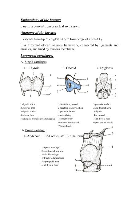
Laryngeal cartilages:<br />
A- Single cartilages<br />
1- Thyroid 2- Cricoid 3- Epiglottis<br />
1-thyroid notch 1-facet for arytenoid 1-posterior surface<br />
2-superior horn 2-facet for inf.thyroid horn 2-sup.thyroid horn<br />
3-thyroid lamina 3-posterior lamina 3-thyroid<br />
4-inferior horn 4-cricoid ring 4-arytenoid<br />
5-laryngeal prominence(adam apple) 5-upper border 5-inf.thyroid horn<br />
B- Paired cartilage<br />
6-narrow anterior arch 6-post.part <strong>of</strong> cricoid<br />
7-lower border<br />
1- Arytenoid 2-Corniculate 3-Cuneiform<br />
1-thyroid cartilage<br />
2-cricothyroid ligament<br />
3-cricoid cartilage<br />
4-thyrohyoid membrane<br />
5-sup.thyroid horn<br />
6-inf.thyroid horn
Laryngeal ligaments & membranes<br />
1- Crico thyroid membrane. 2-Thyrohyoid membrane.<br />
3-Crico tracheal ligament. 4-Thyro epiglottic<br />
ligament.<br />
5-Hyo epiglottic ligament<br />
6- Two intrinsic ligaments: form a broad sheet beneath lining<br />
mucosa:<br />
a- Superiorly: quadrangular ligament between lateral border <strong>of</strong><br />
epiglottis & arytenoid.<br />
b- Inferiorly: conus elasticus, from arch <strong>of</strong> cricoid to vocal ligament<br />
1-thyrohyoid membrane 1-epiglottos 1-laryngeal inlet<br />
2-cricothyroid ligament 2-aryepiglottic fold 2-aryepiglottic fold<br />
3-hyoid bone 3-cuneiform 3-epiglottis<br />
4-lateral part <strong>of</strong> thyrohyoid memb. 4-corniculate 4-quadrangular<br />
ligament<br />
5-thyroid cartilage 5- quadrangular ligament 5-vocal fold<br />
6-lat.part <strong>of</strong> cricothyroid lig. 6-vestibular ligament 6-glottis<br />
7-cricoid cartilage 7-vocal ligament 7-lat.part <strong>of</strong><br />
cricothyroid lig<br />
8-cricotracheal ligament 8-cricothyroid ligament<br />
9-first tracheal ring<br />
Laryngeal folds:<br />
1- Vocal folds: covering <strong>the</strong> vocal ligament that is attached on both<br />
sides from <strong>the</strong> vocal process <strong>of</strong> arytenoid to middle <strong>of</strong> inner surface<br />
<strong>of</strong> thyroid angle.<br />
2- Ventricular folds (false vocal cords): above vocal folds<br />
3- Ary epiglottic folds: between epiglottis and arytenoids.<br />
4- Glosso epiglottic folds two lat & one median.<br />
1-thyro-epiglottic ligament 2-epiglottis
3-yyo-epiglottic ligament 4-hyoid bone<br />
5-pre epiglottic space 6-thyrohyoid membrane<br />
7-thyriod cartilage<br />
Laryngeal inlet:<br />
Oblique, directed slightly posteriorly, bounded by: epiglottis, ary<br />
epiglottic folds, and arytenoids<br />
Cavity <strong>of</strong> <strong>the</strong> <strong>larynx</strong>: 3 areas<br />
1- Glottis space: between V.F extend to 1 cm below<br />
2- Supraglottis: above V.F(from floor <strong>of</strong> ventricle) extend to tip <strong>of</strong><br />
epiglottis<br />
3- Subglottis: from below glottis to lower border <strong>of</strong> cricoid<br />
Laryngeal muscles:<br />
A- Intrinsic muscles: responsible for vocal cord mobility & tension<br />
1- Abductor: Posterior crico arytenoid (posticus)<br />
2- Adductors: Lateral crico arytenoid - Inter arytenoid<br />
- Thyro arytenoid & vocalis (V.F bulk) - Crico thyroid<br />
muscle<br />
3- Tensors: Crico thyroid muscle.<br />
B- Extrinsic muscles<br />
Vocalis (internal part <strong>of</strong> thyro arytenoid).<br />
1- Depressors: sternohyoid, sterns thyroid, omohyoid.<br />
2- Elevators: mylohyoid, geniohyoid, digastric, hyoglossus.<br />
3- Inferior constrictor <strong>of</strong> <strong>the</strong> pharynx.
1-origin <strong>of</strong> PCA from back <strong>of</strong> cricoid lamina 1-origin <strong>of</strong> LCA from upper border <strong>of</strong> arytenoid<br />
2-insertion into muscular process <strong>of</strong> arytenoid 2-insertion into front <strong>of</strong> muscular process <strong>of</strong><br />
arytenoid<br />
Laryngeal spaces:<br />
- Pre epiglottic space - Paraglottic space<br />
Laryngeal mucosa<br />
Larynx is lined by pseudo stratified columner ciliated epi<strong>the</strong>lium, with<br />
goblet cells (respiratory epi<strong>the</strong>lium)<br />
Except: True vocal cords<br />
Nerve supply<br />
Tip <strong>of</strong> epiglottis<br />
Ary epiglottic fold<br />
1- Motor: all muscles by RLN, except cricothyroid by external branch<br />
<strong>of</strong> superior laryngeal nerve.<br />
2- Sensory: Above V.F: internal branch <strong>of</strong> superior laryngeal nerve<br />
Below V.F: RLN<br />
Blood supply: Arteries & veins are closely related to nerves<br />
a- Sup. Laryngeal A&V: branches <strong>of</strong> sup thyroid A&V, join internal<br />
branch <strong>of</strong> sup. laryngeal nerve to pierce thyrohyoid membrane.<br />
b- Inferior laryngeal A&V: branches from inferior thyroid A&V:<br />
accompany RLN<br />
Lymphatic drainage:<br />
- Supraglottis (rich): to UDCLN<br />
- Glottis : no lymphatics in Rienk’s space<br />
- Subglottis : pretracheal (delphian), LDCLN, supraclavicular, sup.<br />
mediastinal<br />
Physiology (Functions <strong>of</strong> <strong>the</strong> <strong>larynx</strong>):<br />
A: Air way protection: from aspiration by:
1- Closure <strong>of</strong> 3 sphincters: ary epiglottic fold, ventricular fold & true<br />
V.F.<br />
2- Laryngeal elevation during swallow.<br />
3- Reflex cough.<br />
4- Reflex inhibition <strong>of</strong> respiration during swallow.<br />
B: phonation<br />
C: Respiration: adjustment <strong>of</strong> glottic aperture helps to regulate acid base<br />
balance<br />
D: Fixation <strong>of</strong> chest during straining<br />
Laryngeal Symptoms & Examination<br />
Laryngeal symptoms:<br />
1- Change <strong>of</strong> voice (hoarseness)<br />
2- Dyspnea: difficulty in breathing.<br />
3- Stridor: noisy respiration.<br />
4- Pain: local or referred.<br />
5- Cough, expectoration, haemptysis<br />
6- Dysphagia<br />
7- 7- Chocking<br />
8- 8- Swelling<br />
Examination:<br />
A- External Examination<br />
inspection: position & movement, contour, swellings, and scars.<br />
palpation: tenderness, crepitus, crepitation, swellings, laryngeal<br />
click<br />
Side to side movement <strong>of</strong> <strong>larynx</strong> over <strong>the</strong> vertebral column, normally a<br />
click is felt, if lost (+ve moure’s sign) … mass between <strong>larynx</strong> & vertebral<br />
column e.g. post cricoid carcinoma.<br />
B- Indirect laryngoscopy
C- Flexible fiberoptic laryngoscopy<br />
- When I.L is difficult, it is introduced via <strong>the</strong> nose.<br />
- Photographic documentation.<br />
D- Direct laryngoscopy under GA:<br />
By rigid endoscope<br />
Operating microscope can be used: Microlaryngoscopy(MLS)<br />
N.B.: - Any lesion affect laryngeal patency: stridor<br />
- Any lesion affect vocal fold: hoarseness<br />
Laryngeal investigations:<br />
1- Direct laryngoscopy ± biopsy 2-Radiology (X<br />
ray.CT,MRI)<br />
Congenital diseases<br />
1- Congenital web<br />
Def and Aet: Incomplete canalization <strong>of</strong> <strong>the</strong> laryngeal lumen.<br />
Path: Commonest site is glottic , may extend to subglottis.<br />
Symptoms<br />
May be small, large, or imperforate fibrous ring.<br />
A: small: asymptomatic, hoarse cry, mild stridor.<br />
B: large: weak hoarse cry, evident stridor.<br />
C: imperforate: Cyanosis with no cry usually fatal.<br />
Signs: Flexible or direct: symmetrical grayish white membrane may be a<br />
semi translucent triangular band, or thick membrane.<br />
Treatment:<br />
A: mild if no symptoms ………… no ttt<br />
B: severe: - Tracheostomy if severe obstruction<br />
- Excision <strong>of</strong> web by:<br />
microlaryngosurgery using laser or cold instruments<br />
laryng<strong>of</strong>issure in very thick web
C: imperforate web: Urgent tracheostomy or rigid bronchoscope to<br />
rupture <strong>the</strong> web<br />
2- Laryngomalacia (congenital laryngeal strider)<br />
Def: stridor due to weak flaccid <strong>larynx</strong>.<br />
Aet: cartilaginous framework <strong>of</strong> <strong>larynx</strong> is abnormally s<strong>of</strong>t & gets<br />
collapsed during inspiration especially supraglottic structures.<br />
Incid: Most common congenital anomaly.<br />
Path: Elongated narrow folded epiglottic (omega), shortened<br />
approximated ary epiglottic fold, prominent elongated arytenoids & deep<br />
supraglottis.<br />
Symptoms:<br />
1- Inspiratory stridor: usually mild<br />
Increase on supine, decrease on prone.<br />
2-No hoarse cry<br />
Signs: (as pathology & aetiology)<br />
Treatment:<br />
1- Usually no ttt & condition improves by age <strong>of</strong> 12-18 months<br />
Give only vit D & Ca<br />
2- Rarely tracheostomy or intubation in severe distress<br />
3- Laser to remove excess mucosal folds, division <strong>of</strong> aryepiglottic<br />
fold<br />
3- Congenital subglottic stenosis<br />
4- Subglottic haemangioma<br />
Reddish subglottic s<strong>of</strong>t mass, situated posteriorly or laterally.<br />
Stridor at age <strong>of</strong> 3-6 months, no hoarseness or bleeding.<br />
Associated with cutaneous hemangioma in 50%, usually regress<br />
spontaneously but tracheostomy may be needed till <strong>the</strong>n<br />
5- Congenital V.C paralysis<br />
Usually due to birth trauma, may be due to congenital CNS<br />
anomalies (meningocele) or mediastinal mass.
6- Laryngo tracheo-oesophageal cleft<br />
Abnormal communication between <strong>larynx</strong> & trachea anteriorly and<br />
hypopharynx & oesophagus posteriorly…….. stridor,chocking &<br />
aspiration……….pneumonia<br />
7- Congenital cysts<br />
Laryngeal (saccular) cyst<br />
Mucous filled cysts,arise from ventricle, false folds or aryepiglottic<br />
fold.<br />
Types<br />
Asymptomatic if large: hoarseness& stridor.<br />
Ttt: endoscopic excision or marsupialisation.<br />
Laryngocele:<br />
Special type <strong>of</strong> congenital cyst develops from laryngeal saccule<br />
(anterior portion <strong>of</strong> <strong>the</strong> ventricle between false & true cords).<br />
May be internal, external, combined.<br />
Symptoms: hoarseness, stridor & neck swelling.<br />
Signs: bulge <strong>of</strong> <strong>the</strong> ventricle.<br />
S<strong>of</strong>t, expansile, compressible mass, on lateral neck<br />
Invest: X ray air filled sac CT :diagnostic<br />
ttt excision endoscopic: laser or MILS or external<br />
I- Mechanical<br />
Laryngeal trauma<br />
1- Sharp (penetrating): stab, gunshot, cut throat.<br />
2- Blunt: blow, strangulation, motor car accident.<br />
3- Inhaled F.B<br />
4- Surgical :rough endoscopy, high tracheostomy<br />
5- Intubation injury: rough intubation, prolonged intubation<br />
II- Chemical Corrosive ingestion (potash)<br />
Inhaled irritant gases
III- Physical: Irradiation injury<br />
Symptoms:<br />
History <strong>of</strong> trauma Dyspnea & stridor Hoarseness <strong>of</strong><br />
voice<br />
Severe local pain Dysphagia<br />
Haemptysis<br />
External swelling Hge, shock (hypovolaemic or neurogenic)<br />
Signs:<br />
1- External:<br />
Inspection: Swelling due to oedema, haematoma, surgical<br />
emphysema<br />
Deformity as depressed cartilage<br />
External wound<br />
Palpation: Localized tenderness & crepitus<br />
Surgical emphysema<br />
Deformity<br />
2- By flexible or direct laryngoscopy<br />
Mucosa: laceration Submucosa: oedema or haematoma<br />
V.F: paralysis or avulsion Cartilage: displaced<br />
Joints: dislocated Arytenoids: intubation granuloma<br />
investigations: - D.L - C.T<br />
treatment:<br />
Air way secure by tracheostomy or intubation<br />
Shock management<br />
<strong>Dr</strong>ugs antibiotics Steroids<br />
Open reduction: suture laceration & fractured cartilage + stent<br />
F.B<br />
In <strong>the</strong> <strong>larynx</strong><br />
Rare, mostly goes to trachea, sharp or large FB may be impacted<br />
Sudden severe respiratory obstruction
Ttt: Heimlich maneuver: sudden abdominal compression<br />
Turn child upside down & slap back<br />
Removal by DL<br />
Tracheostomy or cricothyrotomy<br />
Inhaled FB<br />
incid: children, mentally retarded<br />
Types: vegetable: seeds, beans non vegetable: pins, buttons.<br />
endogenous: vomitus, blood<br />
Sites: rare in <strong>larynx</strong> (passes to esophagus or bronchus) in bronchi<br />
usually right main bronchus (wider & in line with <strong>the</strong> trachea).<br />
Clinical picture:<br />
A- initial stage sudden severe cough, cyanosis, dyspnea and<br />
sometimes haemptysis, this stage may pass<br />
unnoticed<br />
B- Latent stage: mild cough & expectroation - localized<br />
wheeze<br />
C- Manifest stage:<br />
I- Mechanical obstruction<br />
Complete (collapse) Partial (emphysema)<br />
- Dyspnea, cough, expectoration - Dyspnea, cough, expectoration<br />
- mediastinum shift to same side - To opposite side<br />
- Dullness on percussion - Hyper resonance<br />
- No air entry - Localized wheeze<br />
F.B)<br />
II- Inflammatory changes<br />
Bronchitis, bronchopneumonia, lung abscess (more in organic<br />
D- Large F.B: Impacted in <strong>larynx</strong> may cause suffocation<br />
Investigations: 1- D.L & bronchoscopy 2- Xray neck &<br />
chest<br />
Ttt: 1-Urgent tracheostomy in large impacted F.B<br />
2- Removal by direct laryngoscopy or<br />
bronchoscopy<br />
Laryngeal stenosis
Def: Fibrotic narrowing <strong>of</strong> endo<strong>larynx</strong> leading to respiratory distress<br />
Aet: A-Congenital: Congenital laryngeal atresia, congenital laryngeal<br />
web<br />
B-Trauma (see before)<br />
congenital subglottic stenosis<br />
C-Chromic inflammations (granulomas): Scleroma, T.B, $, leprosy<br />
D-Laryngeal neoplasms: chondroma, fibroma, carcinoma<br />
Incid: most common is subglottic (& most difficult to treat)<br />
Path: 1- supra glottic 2- glottic (ant, post, complete). 3-<br />
Subglottic<br />
Clinical picture:<br />
- Asymptomatic Stridor on exertion<br />
- Biphasic stridor & dyspnea Exam shows <strong>the</strong> stenosis<br />
Investigations: 1-DL & bronchoscopy<br />
Treatment:<br />
2-C.T to show length & degree <strong>of</strong> stenosis<br />
3-Pulmonary functions for follow up<br />
A) Tracheostomy in severe cases<br />
B) Endoscopic procedure: Laser excision, repeated dilatation<br />
C) External: laryngoplasty: split cricoid, insert costal graf & put<br />
Montgomery tube<br />
Resection & end to end anastomosis.<br />
Laryngitis<br />
I- Acute<br />
(A) Non specific in adults, in children, epiglottistis & laryngo<br />
tracheobronclitis<br />
(B) Specific: Diph<strong>the</strong>ria<br />
(A) Non specific<br />
hypertrophic Diffuse<br />
II- Chronic<br />
Localized: nodule , polyp, and leukoplakia
Atrophic<br />
(B) Specific (granulomas): scleroma, TB, S, leprosy, & mycosis<br />
I- Acute Laryngitis<br />
(A) Acute non specific laryngitis<br />
(1) In adults<br />
Def.: acute catarrhal inflammation <strong>of</strong> laryngeal m.m.<br />
Aet.: follow upper respiratory tract infection<br />
Viral: influenza, rhinovirus Bacteria: Strept. pneumonia, strept.,<br />
straph.<br />
Predisposing factors:<br />
- Voice abuse. - Smoking.<br />
- Post nasal discharge as in sinusitis. - Dust & fumes.<br />
Incid.: see above Path: congestion and edema<br />
Symptoms:<br />
General: fever (occasionally), headache, malaise, and fatigue.<br />
Local: - hoarseness.<br />
- Discomfort & pain on phonation.<br />
- <strong>Dr</strong>y cough + yellow sputum.<br />
Signs: Diffuse symmetrical congestion & oedema, + mucoid secretions.<br />
Treatment:<br />
1- Voice rest. 2- Fluids.<br />
3- Humidificatoin :steam, tincture benzoini.<br />
4- Systemic antibiotics. 5- Mucolytics.<br />
(2) Acute non specific laryngitis in children<br />
Def.: as before.<br />
Aet.: as before.<br />
Path: Difference between adult & child <strong>larynx</strong>.<br />
1- Anatomical factors.<br />
- Lumen is relatively smaller.<br />
- Loose submucosal tissue.<br />
- Funnel shaped lumen.<br />
- More abundant lymphatic supply in mucosa.<br />
More oedema, minimal edema cause marked obstruction.
Symptoms:<br />
2- Immature immune system.<br />
3- Immature nervous regulation = spasm.<br />
General: as usual.<br />
Local: - Hoarseness. - <strong>Dr</strong>y cough. - Stridor,<br />
dyspnea.<br />
Signs: Diffuse congestion and oedema. + mucoid secretion.<br />
Subglottic oedema<br />
D.D.: causes <strong>of</strong> stridor in children<br />
Treatment: as adult +<br />
- Hospitalization, oxygen.<br />
- Steroids to decrease oedema.<br />
- Careful observation <strong>of</strong> respiratory obstruction if severe distress →<br />
intubation, or rarely tracheostomy (avoided in children unless no<br />
o<strong>the</strong>r way).<br />
(3) Acute Epiglottitis<br />
Def.: Acute non specific laryngitis affecting mainly epiglottis.<br />
Aet.: Haemophilus influenza usually.<br />
Incid.: more in infants & children.<br />
Symptoms:<br />
General: high fever, malaise, headache, and anorexia.<br />
Local: - Severe odynophagia, drippling <strong>of</strong> saliva.<br />
Signs:<br />
- Muffled voice (hot potato). -Rapidly progressive stridor.<br />
- Oedema <strong>of</strong> supraglottis. - Epiglottis appears as red<br />
swollen mass.<br />
- Pharyngeal oedema & congestion. - Enlarged UDCLN.<br />
Ttt: as before.<br />
(4) Acute laryngo tracheobronchitis<br />
Def.: Acute respiratory infection spreading to entire respiratory system.<br />
Clinical picture: In adult: as laryngitis + cough, expectoration.<br />
In children: as laryngitis + cough, expectoration.
Treatment: as laryngitis.<br />
(B) Laryngeal diph<strong>the</strong>ria.<br />
Symptoms:<br />
General: Toxaemia <br />
Local: - hoarseness, stridor, Dyspnea, cough.<br />
Signs: grayish, white, dirty membrane.<br />
Treatment: as usual. + Tracheostomy.<br />
II- Chronic Laryngitis.<br />
(a) Chronic non specific laryngitis.<br />
(1) Hypertrophic<br />
(a) Chronic diffuse hypertrophic laryngitis.<br />
Aet.: - Repeated acute laryngitis predisposing factors ----<br />
Symptoms:<br />
- Hoarseness <strong>of</strong> voice - Cough & expectoration.<br />
- Hemming (desire for frequent throat clearing).<br />
Signs: Congested thickened V.F<br />
in oedematous type (reinke oedema) :pale polypoidal VF.<br />
ttt: 1- Remove predisposing factor. 2- Like acute but no antibiotics.<br />
3- VF stripping: MLS or laser in resistant cases<br />
(b) Chronic localized hypertrophic laryngitis.<br />
(1) Vocal cord nodules (2) Vocal cord polyp.<br />
Aet.: - Voice abuse (singer). - Voice abuse.<br />
- reflux<br />
Incid.: More in females, children. - More in males.<br />
Path: Epi<strong>the</strong>lial hyperplasia <strong>of</strong> - Localized subepi<strong>the</strong>lial<br />
Free edge <strong>of</strong> V.F. oedema, or vascular eng-<br />
Fresh: s<strong>of</strong>t& red orgement, followed by<br />
fibrosis.<br />
mature:hard&white<br />
Symptoms: hoarseness, weakness - Hoarseness.
-Rarely stridor if large.<br />
- Chocking.<br />
Signs: Bilateral. - Unilateral<br />
Small tiny nodules. - Small or large<br />
Sessile -Usually pendunculated.<br />
White or pink. - Grey, white or red.<br />
At free margin <strong>of</strong> V.F. - From undersurface or V.F.<br />
Junction <strong>of</strong> ant 1/3 & post 2/3. - Site as nodule.<br />
Treatment: Voice rest, avoid misuse. - Removal by MLS<br />
(3) Leukoplakia.<br />
Speech <strong>the</strong>rapy. - Laser.<br />
+ removal by MLS - Speech <strong>the</strong>rapy.<br />
Laser. - Voice rest, avoid misuse.<br />
Def.: white patch on laryngeal mucosa.<br />
Aet.: Irritation.<br />
Pathology: Epi<strong>the</strong>lial hyper plasia & hyper keratinization.<br />
Symptoms: persistent hoarseness.<br />
Signs: white patch on V.F.<br />
May appear as diffuse, villous, or verrucous.<br />
ttt: Excision by MLS or Laser, follow up as it is pre cancerous.<br />
(2) Atrophic.<br />
Aet: dusty atmosphere - Industrial fumes - Post irradiation<br />
<strong>the</strong>rapy
Incid: more in women<br />
Symptoms: hoarseness, <strong>of</strong>fensive breath &dyspnea(crusts)<br />
Signs: dry pale atrophic mucosa, & crusts<br />
Ttt: Avoid predisposing factors, moisture, menthol inhalation,<br />
laryngoscopy to remove crusts<br />
(B) Chronic specific laryngitis (Granuloma).<br />
(1) Laryngoscleroma.<br />
Def.: chronic specific inflammation affecting upper respiratory tract.<br />
Aet.: gram – ve bacilli; klebsiella rhinoscleromatis.<br />
Incid.: endemic in Egypt. Usually 2ry to nasal scleroma may be 1ry.<br />
Usually subglottic region.<br />
Path: see nose.<br />
Symptoms: - stridor & dyspnea.<br />
- Hoarseness may be present.<br />
- Cough & expectoration.<br />
Signs: pale pinkish smooth swelling on both side <strong>of</strong> subglottis covered by<br />
greenish crusts fibrosis & subglottic stenosis.<br />
Investigations:<br />
ttt:<br />
- DL & biopsy. - CT for length & degree <strong>of</strong> stenosis.<br />
1- Medical as in rhinoscleroma. 2-Voice rest &<br />
humidification.<br />
3-Tracheostomy, if severe distress.<br />
4- Excision <strong>of</strong> <strong>the</strong> web by Laser.<br />
(2) T.B laryngitis.<br />
External: graft & stent<br />
Resection anastomosis<br />
Aet: 2ry to pulmonary T.B. Path.: affect post part <strong>of</strong> <strong>larynx</strong>.<br />
Symptoms:<br />
I- General: 1- T.B toxemia: night fever, night sweat, loss <strong>of</strong> weight, loss <strong>of</strong><br />
appetite.
II- Local:<br />
Signs:<br />
2- Pulmonary T. B cough, expectoration, haemptysis.<br />
1- Hoarseness: progressive, phonos<strong>the</strong>sia( weak voice)<br />
2- Stridor & dyspnea.<br />
3- Pain & referred otalgia.<br />
4- Odynophagia (marked).<br />
I- External: Tenderness due to perichondritis.<br />
II- by I.L or flexible:<br />
1- T.B granulations on arytenoids.<br />
2- T.B ulcer: thin undermined edge, yellow caseous floor.<br />
3- Impaired V.F mobility.<br />
Investigations:<br />
1- DL & biopsy. 2- Chest X ray for T.B. 3- Tuberculin test: good<br />
negative.<br />
Treatment:<br />
1- Tracheostomy in severe distress.<br />
2- Anti tuberculous drugs e.g. rifampicin, PASA, streptomycin &<br />
pyrazinamide.<br />
3- Local anes<strong>the</strong>tic spray before meals.<br />
(3) Syphilis <strong>of</strong> <strong>the</strong> <strong>larynx</strong>.<br />
Aet.: Treponema pallidum (spirochete). Incid.: Very rare.<br />
Path.: 1ry chancre. 2ry mucous patches. 3ry gumma<br />
(commonest).<br />
Symptoms: - Hoarseness.<br />
-Stridor.<br />
- Cough & discomfort (no pain).<br />
Signs: $ affects ant. part <strong>of</strong> <strong>larynx</strong><br />
Gumma <strong>of</strong> epiglottis appears swollen & ulcerated.
Diffuse symmetrical infiltration without ulcerated.<br />
Investigations:<br />
1- DL & biopsy. 2- Radiology. 3- Serology for $.<br />
Ttt: 1- Tracheostomy if needed.<br />
2- Penicillin.<br />
(4) Lupus<br />
Aet.: attenuated T.B. Incid.: mostly 2ry to nasal lupus.<br />
Path.: apple jelly nodule.<br />
Ulceration on one side.<br />
Fibrosis, notched epiglottis.<br />
Symptoms: vague discomfort.<br />
Signs: see pathology.<br />
Investigation: DL & biopsy. - X ray chest.<br />
ttt: like T.B + vit. D.<br />
(5) Leprosy<br />
- Lepromatous or tuberculoid types.<br />
- Nodules ulceration fibrosis.<br />
- Ttt: Dapson + rifampicin.<br />
(6) Fungal infection (mycosis).<br />
a) candidiasis (moniliasis)<br />
- Caused by candida albicans, whitish grey fibrinous membrane.<br />
- Ttt: Remove <strong>the</strong> underlying cause,topical nystatine or miconazole.<br />
b) Aspergillosis.<br />
Perichondritis<br />
Def: inflammation <strong>of</strong> <strong>the</strong> perichondrium <strong>of</strong> laryngeal cartilage<br />
Aet: trauma…………<br />
Infection: TB, S, typhoid<br />
Advanced carcinoma<br />
Path perichondritis……subperichondrial abscess… cartilage necrosis &<br />
stenosis<br />
Symptoms: fever, malaise, pain increase on swallowing, hoarseness, &<br />
stridor<br />
Signs: marked tenderness, enlarged laryngeal framework
Laryngeal mucosa is red, edematous, &covered with pus<br />
Ttt: hospitalization, high dose <strong>of</strong> antibiotics, steroids<br />
Incision <strong>of</strong> abscess, remove necrosed cartilage.<br />
Tracheostomy or intubation. - Laryngectomy in advanced cases.<br />
Laryngeal oedema<br />
Aet: trauma………..<br />
Infection: laryngitis, perichomdritis, spread <strong>of</strong> infection from<br />
quinsy, ludwig<br />
Angioneurotic oedema: allergic (foods, inhalants& drugs) or<br />
hereditary<br />
Non inflammatory oedema: heart renal or liver failure, myxodema .<br />
Symptoms: hoarseness, stridor & dyspnea<br />
Ttt: Rest, oxygen, ttt <strong>of</strong> <strong>the</strong> cause<br />
Tracheostomy or intubation in sever cases<br />
Antihistaminics, ,and hydrocortisone<br />
Tumors <strong>of</strong> <strong>the</strong> <strong>larynx</strong><br />
I- Benign<br />
A) Epi<strong>the</strong>lial Papilloma.Adenoma.<br />
B) Mesenchymal Haemangioma, Chondroma, Fibroma.<br />
Papilloma (commonest benign)<br />
(A) Single papilloma (B) Multiple papillomatosis<br />
Adult papolloma recurrent papillomatosis.<br />
Aet.: - True papilloma - Viral HPV<br />
Juvenile multiple papillomatosis<br />
Autoimmune<br />
Hormonal imbalance<br />
Incid.: - Adult 30-50 y - Children .<br />
Male: female 2/1. Equal sex.<br />
Path.: - Commonest site is free - V.F, any where in <strong>larynx</strong><br />
extend<br />
edge <strong>of</strong> V.F, may to trachea, bronchi,
Symptoms:<br />
affect o<strong>the</strong>r sites.<br />
- Vascular C.T core covered by - Similar.<br />
hyperplastic st. sq. epi<strong>the</strong>lium.<br />
- hoarseness - Stridor.<br />
+ stridor if large. Hoarseness.<br />
Signs: - single, white or pink. - Multiple white or pink.<br />
warty like growth sessile or warty, sessile affecting any<br />
pedunculated variable size. portion <strong>of</strong> <strong>the</strong> <strong>larynx</strong>.<br />
ttt: Surgical excision by: MLS using cold instruments or<br />
laser<br />
MLS using cold instruments or laser<br />
Malignancy: - 3% - no<br />
laryng<strong>of</strong>issure.<br />
Tracheostomy usually needed<br />
(try to avoid……spread).<br />
Interferon.<br />
Recurrence: - rare - very common<br />
Def.: malignant tumor <strong>of</strong> <strong>the</strong> <strong>larynx</strong>.<br />
II- Malignant (Cancer <strong>larynx</strong>)<br />
Aet Pre disposing factors tobacco: smoking <strong>the</strong> most important.
carcinoma.<br />
alcohol ingestion supra glottic<br />
irradiation.<br />
Precancerous lesions: Leukoplakia Adult papilloma.<br />
Incid: Old over 40 with peak in 60. Male to female 8:1(now<br />
less).<br />
Path.:<br />
Common neoplasm, 1% <strong>of</strong> all malignancies.<br />
(A) Site: glottic 70% Supraglottic 25% Subglottic<br />
5%<br />
(B) G.P: Malignant ulcer. Fungating mass.<br />
Infiltrating nodule.<br />
(C) M.P: commonest is squamous cell carcinoma.<br />
(D) Spread:<br />
1- Direct<br />
Glottic Supraglottic subglottic<br />
2- Lymphatic spread.<br />
a) Glottic: very rare, as <strong>the</strong>re is no lymphatics in Rinek’s space.<br />
b) Supraglottic: common & early due to rich lymphatics UDCLN.<br />
c) Subglottic: common may be bilateral pre laryngeal, pre<br />
tracheal, paratracheal middle & LDCLN.<br />
3- Blood spread rare & late : lungs, liver, bone, brain.<br />
(E) T.N.M classification<br />
(a) T for primary tumor
Tis Carcynoma in situ.<br />
T1 One region (in glottic T1a : one cord, T1b : two cords).<br />
T2 Two regions.<br />
T3 Fixed cord.<br />
T4 Extra laryngeal spread.<br />
(b) N: LN metastasis.<br />
No.: No palpable LN.<br />
N1: Single, 3 cm or less, ipsilateral.<br />
N2a: Single, 3-6, ipsilateral.<br />
N2b: Multiple, 3-6 ,ipsilateral.<br />
N2c: Bilateral < 6cm.<br />
N3: more than 6 cm.<br />
(c) M Distant metastasis.<br />
Symptoms:<br />
M0: No clinical or radiological evidence <strong>of</strong> metastasis.<br />
M1: Present clinical or radiological evidence <strong>of</strong> metastasis.<br />
1- Hoarseness <strong>of</strong> voice - progressive. -early in glottic<br />
cancer.<br />
2- Dyspnea & stridor due to air way obstruction.<br />
3- Discomfort in <strong>the</strong> throat.<br />
Late symptoms:<br />
4- Pain, referred otalgia along Arnold branch <strong>of</strong> X.<br />
5- Cough & irritation.<br />
6- Neck swelling: LN metastasis or direct tumor infiltration.<br />
7-Dysphagia, haemptysis. 8-Foetid breath, cachexia.<br />
Signs:<br />
I General: teeth for sepsis & oral hygiene. Chest : distant metastasis.<br />
II- Local: (Larynx)<br />
(1) External (Neck):<br />
- L.N, describe. - Swelling, broadening.<br />
- Tenderness. - Thyroid.
(2) by I.L flexible or D.L examine:<br />
Tumor: - hyper keratotic warty or papillary growth.<br />
- Malignant ulcer.<br />
- Raised nodule.<br />
V.F mobility: - Freely mobile.<br />
- Fixed: deep muscle invasion.<br />
- Limited: weight <strong>of</strong> tumor or moderate invasion.<br />
Extension: to hypopharynx, trachea or tongue.<br />
NB: some areas are difficult to be examined: ventricle, subglottis,<br />
posterior surface <strong>of</strong> epiglottis.<br />
Investigations:<br />
1- CT or MRI neck most important radiological diagnosis for accurate<br />
staging.<br />
2- DL & biopsy. Site, extent, biopsy.<br />
3- Metastatic work up.(chest X ray, abdominal sonar & bone scan)<br />
4- Routine preoperative investigations.<br />
Treatment:<br />
TIS: surgery: V.F stripping by MLS.or Laser , followed by regular<br />
follow up<br />
Radio<strong>the</strong>rapy (not preferred).<br />
T1, T2: surgery: Partial laryngectomy aiming at complete tumor excision<br />
with preservation <strong>of</strong> phonation, normal breathing °lutition<br />
Endoscopic Laser excision.<br />
Radio<strong>the</strong>rapy.<br />
T3: Total laryngectomy + post operative radio<strong>the</strong>rapy<br />
Also in subglottic tumors & recurrent cases after conservative surgery or<br />
radiation<br />
- Large T2 or small T3: subtotal laryngectomy<br />
(cricohyoidoepiglottopexy).<br />
T4: - Total laryngectomy + post operative radio<strong>the</strong>rapy.<br />
- Trail <strong>of</strong> chemo radio <strong>the</strong>rapy.
Management <strong>of</strong> cervical metastasis.<br />
If palpable LN RND.<br />
If no palpable LN Glottic (nothing)<br />
radio<strong>the</strong>rapy.<br />
radio<strong>the</strong>rapy<br />
Supra glottic prophylactic neck dissection, or<br />
Sub glottic prophylactic neck dissection, or<br />
Indications <strong>of</strong> total laryngectomy:<br />
1- T3, T4 glottic, supraglottic.<br />
2- All subglottic & transglottic.<br />
3- Recurrence or failure after conservative surgery.<br />
4- Recurrence or failure after radio<strong>the</strong>rapy.<br />
5- Contraindication for conservative or radio<strong>the</strong>rapy.<br />
6- Certain histological types.<br />
Contra indications <strong>of</strong> total laryngectomy:<br />
1- Poor general condition. 2-Patient refusal.<br />
3-Distant metastasis. 4-Involvement <strong>of</strong> unresectable<br />
structures.<br />
Disadvantages <strong>of</strong> total laryngectomy.<br />
1- Loss <strong>of</strong> voice. 2-Inability to increase intra thoracic<br />
pressure.<br />
3-Permanent tracheostomy. 4-Loss <strong>of</strong> nasal functions.<br />
2- Limitation <strong>of</strong> activities.<br />
Rehabilitation after surgery:<br />
1- After partial laryngectomy: voice <strong>the</strong>rapy.<br />
2- After total laryngectomy: Oesophageal speech.
Palliative ttt<br />
Tracheo oesophageal puncture.<br />
Electronic <strong>larynx</strong>.<br />
Indicated in: unresectable tumors, surgery refusal, distant metastasis &<br />
poor general condition<br />
1- Tracheostomy. 2- Ryle or gustrostomy for feeding.<br />
3- Palliative laser, radio<strong>the</strong>rapy, chemo<strong>the</strong>rapy.<br />
4- Pain killers. 5- Antibiotics.<br />
Prognosis: early cancer has good prognosis.<br />
Glottic cancer has best cure rate up to 90% due to early<br />
presentation (hoarseness) &absent lymphatic spread.<br />
Vocal Cord Paralysis<br />
a- Unilateral v.c paralysis<br />
Aet: Vagal or RLN injury.<br />
(A) Peripheral<br />
1- Congenital Hydrocephalus<br />
2- Surgical trauma 20%.<br />
- Thyroidectomy - Radical neck<br />
dissection.<br />
- Pharyngeal pouch - Esophageal surgery.<br />
- Cardio vascular surgery.<br />
3- Non surgical trauma 20%.<br />
- Neck trauma e.g. stangulation, open injuries.<br />
- Cricothyroid joint dislocation.<br />
- Fracture skull base.<br />
4- Inflammatory 5%.<br />
- Apical pulmonary T.B. - Meningitis,<br />
osteomyelitis.<br />
5- Peripheral neuritis
- Infections: herpes, influenza, and diph<strong>the</strong>ria. - DM<br />
- Chemicals: alcohol, lead. -Ascending polyneuritis<br />
6- Neoplastic 25%.<br />
- Thyroid malignancy. - Hypopharyngeal<br />
malignancy.<br />
carcinoma.<br />
SLE.<br />
- Bronchogenic carcinoma. - Oesophageal<br />
- Malignant L.N.<br />
7- Miscellaneous<br />
- Myas<strong>the</strong>nia gravis. - Rheumatoid arthritis,<br />
8- Idiopathic: unknown cause usually viral in aetiology.<br />
(B) Central causes.( Bulbar):<br />
- Head trauma.<br />
- Thrombosis, Hge, embolism.<br />
- Encephalitis, polio, diph<strong>the</strong>ria. -<br />
- Medullary tumors.<br />
Vocal cord positions<br />
1- Median: both cords in midline.<br />
2- Paramedian : glottis 3-5 mm.<br />
3- Cadaveric: glottis 7 mm.<br />
4- Slight abduction: glottis 14 mm.<br />
5- Full abduction: 18 mm.<br />
Explanation <strong>of</strong> vocal cord position:<br />
(A) Semon’s law<br />
In progressive RLN injury, abductor paralysis occurs 1 st v.c in<br />
median or paramedian position, <strong>the</strong>n adductor paralysis <br />
cadaveric position.<br />
(B) Adductors are more powerful than abductors so, when RLN is<br />
injured, adductors takes upper hand.
(C) Wegner – Grossman <strong>the</strong>rory.<br />
Symptoms:<br />
In RLN paramedian position by cricothyroid muscle.<br />
In vagal cadaveric position.<br />
1- May be asymptomatic when compansated.<br />
2- Hoarseness: may improve if compansation<br />
(o<strong>the</strong>r cord crosses midline to meet paralyzed one).<br />
3- No stridor.<br />
4- Aspiration: may improve.<br />
Signs: By indirect or flexible laryngoscopy:<br />
a) In RLN injury: median or paramedian position.<br />
b) In vagal injury: cadaveric position.<br />
The paralyzed cord appears bowed (flaccid), at a lower level,<br />
with <strong>the</strong> arytenoids leaning inwards<br />
Investigations:<br />
Full head, neck, chest exam for cause.<br />
(1) Radiology<br />
- X ray chest, nasopharynx, and neck. - CT from skull base to mid<br />
thorax.<br />
scan.<br />
- CT & MRI brain. - Barium swallow. - Thyroid<br />
(2) Endoscopy<br />
Panendoscopy: nasopharyngoscopy, laryngoscopy,<br />
hypopharyngoscopy, oesphagoscopy & bronchoscopy + biopsies.<br />
(3) Blood exam.<br />
Treatment:<br />
CBC, ESR, viral studies, blood sugar.<br />
1- Treat <strong>the</strong> causes if possible.<br />
2- Most cases requires no ttt: compansation or Spontaneous recovery<br />
may occur<br />
3- Surgical ttt, after 6-12 months.
Indicated in: persistent hoarseness or aspiration.<br />
Operation performed (medialization)<br />
a) Teflon or Collagen injection.<br />
b) Throplasty: external medialization.<br />
c) Nerve muscle pedicle reinnervation.<br />
b- Bilateral Vocal Cord Paralysis<br />
a- (Bilateral Abductor Paralysis)<br />
Aet.: injury to both RLN (peripheral).<br />
1- Surgical trauma. Thyroidectomy, esophageal surgery.<br />
2- Peripheral neuritis.<br />
3- Neoplastic: cancer thyroid.<br />
Symptoms:<br />
1- Good voice but tries easily.<br />
2- Stridor may be severe, increase by exertion & infection.<br />
Signs:<br />
1- By indirect or flexible laryngoscopy: Vocal cords are in median or<br />
paramedian position.<br />
2- Head, neck, chest exam for cause.<br />
Investigations: same<br />
Treatment:<br />
1- If sever stridor Tracheostomy.<br />
2- In established cases:laryngeal widening procedure ,3-6 months later.<br />
(a) Endoscopic arytenoidectomy with posterior cordectomy using MLS<br />
or laser.<br />
(b) Woodman’s operation. External operation to fix aryternoid<br />
laterally.<br />
(c) Reinnervation procedure.<br />
(d) Tracheostomy with speaking valve.<br />
b- Bilateral adductor paralysis<br />
Presents with aphonia & aspiration (fatal)<br />
Ttt: tracheostomy with speaking valve<br />
Laryngeal closure
Total laryngectomy<br />
Tetany neonatorum & laryngismus stridulus<br />
Def: active laryngeal spasm Aet: Ca deficiency<br />
Incid: young under nourished rickety children, commonly with adenoids<br />
Clinical picture: stridor suddenly at night, cyanosis, may be carpo-pedal<br />
spasm<br />
Ttt: A: during <strong>the</strong> attack: slapping child back, traction on <strong>the</strong> tongue<br />
B: in between attacks: adenoidectomy, Ca<br />
Laryngeal symptoms<br />
Stridor<br />
Def.: noisy respiration indicating partial airway obstruction in <strong>larynx</strong><br />
&/or trachea.<br />
Types: a. inspiratory laryngeal: at or above glottis.<br />
b. Biphasic: subglottic or tracheal.<br />
c. Expiratory: bronchial.<br />
Causes:<br />
I- Congenital:<br />
- Congenital web - laryngo malacia.<br />
- Subglottic stenosis - subglottic haemangioma.<br />
- Congenital cyst - congenital bil. cord palsy.<br />
II- Traumatic:<br />
Mechanical: sharp, blunt, surgical, intubation, F.B.<br />
Physical: radio<strong>the</strong>rapy, <strong>the</strong>rmal.<br />
Chemical: corrosives.<br />
III- Inflammatory:<br />
- Acute non-specific laryngitis, in children -Acute epiglottitis.<br />
- Acute laryngo tracheo brochitis in children. -Laryngeal<br />
diph<strong>the</strong>ria.<br />
- Chronic specific laryngitis (granulomas)<br />
IV- Neoplastic<br />
Benign: multiple papillomatosis. Subglottic chondroma.<br />
Malignant: carcinoma.<br />
V- Miscellaneous<br />
Neurological: bil abductor paralysis.
Laryngeal spasm e.g. tetany<br />
Tumor like conditions: Laryngeal cysts, laryngocele, amyloidosis.<br />
Laryngeal oedema.<br />
VI- Tracheal causes<br />
Retrosternal goiter, mediastinal LN, thymus hyperplasia,& deep neck<br />
infection<br />
Clinical picture:<br />
1- Stridor 2-Dyspnea &<br />
tachypnea.<br />
3-Irritability, restlessness, fatigue, sweating. 4-Working ale nasi.<br />
5-Suprasternal, supraclavicular & intercostal retraction.<br />
6-Working accessory muscles.<br />
7- Tachycardia. 8-Congested neck veins during<br />
expiration<br />
9-Late: cyanosis, bradycardia Finally: coma & cardiac<br />
arrest.<br />
+ Hoarseness <strong>of</strong> voice. NB: Stridor in children.<br />
Hoarseness <strong>of</strong> voice<br />
Def.: change in quality & timbre <strong>of</strong> voice, so it becomes rough, harsh &<br />
low pitch. caused by incomplete coaptation, tension or vibration.<br />
Causes <strong>of</strong> hoarseness.<br />
I- Congenital: web.<br />
II- Traumatic: Mechanical, physical, chemical + voice abuse.<br />
III- Inflammatory: all except subglottic scleroma.<br />
IV- Neoplastic: Benign & malignant.<br />
V- Miscellaneous: Unilateral cord paralysis<br />
Crico arytenoid arthritis.<br />
General weakness: myas<strong>the</strong>nia or convalescence<br />
Lack <strong>of</strong> mucus secretion: atropine<br />
Hysterical.
NB: Stridor without hoarseness.<br />
Hoarseness without stridor.<br />
Tracheostomy<br />
Laryngeal operations<br />
Creation <strong>of</strong> surgical opening, in anterior tracheal wall.<br />
Indications:<br />
I- Upper respiratory tract obstruction.<br />
a- Congenital b- Traumatic<br />
c- Inflammatory d- Neoplastic <br />
e- Miscellaneous <br />
f- Extra laryngeal causes (injury, infection, oedema, tumors)<br />
Injury: maxill<strong>of</strong>acial. Infection: retropharyngeal<br />
abscess.<br />
Edema: tongue & neck edema Tumors: oral cavity, tongue,<br />
pharynx.<br />
II- Lower respiratory tract obstruction<br />
1- Secretory obstruction.<br />
Value:<br />
1- Frequent accurate aspiration <strong>of</strong> secretion.<br />
2- Elimination <strong>of</strong> dead space.<br />
3- Prevent aspiration by cuffed tube.<br />
4- Avoids complications <strong>of</strong> prolonged intubation.<br />
Causes:<br />
A) Central<br />
1- Head injury: contusion, laceration. 2-<strong>Dr</strong>ug intoxication<br />
3- Uraemia, ketoacidosis. 4-Brain tumors &<br />
abscess.<br />
5-Stroke: Hge, embolism, thrombosis
B) Peripheral<br />
1)Respiratory muscle paralysis. 2) Chest injury e.g. ribs<br />
fracture.<br />
2- Respiratory failure:<br />
- COPD - Neurological disorders e.g. polio,<br />
myas<strong>the</strong>nia.<br />
III- Elective<br />
A) Before laryngeal surgery.<br />
B) Before major operations in head e.g. angi<strong>of</strong>ibroma,<br />
maxillectomy,<br />
Types:<br />
Site Advantages Disadvantages<br />
High 1 st , 2 nd rings Easy<br />
Rapid<br />
Emergency<br />
Low 5 th , 6 th Papillomatosis<br />
Subglottic Cancer<br />
Subglottic Stenosis<br />
Mid 3 rd , 4 th<br />
Technique:<br />
behind<br />
isthmus<br />
Avoid previous<br />
disadvantages.<br />
1- Anes<strong>the</strong>sia: Usually local infiltration.<br />
Cricoid injury<br />
Difficult<br />
Pleural injury.<br />
Innominate v.<br />
injury.<br />
Slips easily.<br />
General: in elective cases, better in children.<br />
No anes<strong>the</strong>sia in real emergency.(better do<br />
cricothyroidotomy).<br />
2- Position: Supine with extended neck.<br />
If patient distressed: sitting or semisitting.<br />
3- Incision: Vertical: rapid, lower border <strong>of</strong> thyroid….manubrium
Transverse: cosmetic,between lower border <strong>of</strong> cricoid and<br />
supra sternal notch<br />
4- Incise fat, fascia, separate pretracheal muscles.<br />
5- Dissect, incise & transfix isthmus.<br />
6- Open <strong>the</strong> trachea as a flap.<br />
7- Put a suitable tube.<br />
8- Adequate haemostasis.<br />
9- Close <strong>the</strong> wound not too tight, fix tube to skin.<br />
Types <strong>of</strong> Tracheostomy tubes:<br />
1- Metal <strong>of</strong> silastic. 2-Cuffed or not.<br />
3-Tubes with inner & outer layers. 4-Tubes with expiratory<br />
value.<br />
Complications:<br />
1- A nes<strong>the</strong>tic complications local or general.<br />
2- Apnea<br />
- When operation done under L.A<br />
- Due to rapid wash out <strong>of</strong> co2 which is stimulus for respiratory<br />
center.<br />
- ttt: close <strong>the</strong> opening for a short time, allow patient to breath 95%<br />
O2 in 5% co2 , or assisted ventilation.<br />
3- Bleeding<br />
a) Primary ant. jugular v. Thyroid gland. Innominate v.<br />
b) Reactionary slipped ligature, from previously collapsed v.(open<br />
<strong>the</strong> wound & ligate <strong>the</strong> vessel)<br />
c) Secondary due to infection.(antibiotics & fresh blood)<br />
4- Pneumothorax.<br />
Due to: pleural injury. Manifested by: dyspnea air entry, X ray.
ttt: intercostal tube connected to underwater seal.<br />
5- Pneumomediastinum.<br />
Due to: Excessive inferior dissection.<br />
If mild: resolve spontaneously. If severe: acute heart failure.<br />
6- Crustation.<br />
No filtration <strong>of</strong> inspired air. Decreased mucociliary<br />
clearance.<br />
7- Delayed complications:<br />
a) Subglottic stenosis: due to cricoid injury.<br />
b) Tracheal stenosis: due to erosion by tube or infection.<br />
c) Difficult extubation.<br />
d) Tracheo oesophageal fistula.<br />
e) Tracheo cutaneous fistula.<br />
8- Emphysema (surgical). Air accumulation under skin.<br />
Due to: - Improperly fitting tube. -Excessive lateral neck<br />
dissection.<br />
ttt: Remove a skin suture Insert a more fitting tube.<br />
9- Embolism (air embolism) :due to injury <strong>of</strong> large neck vein.<br />
ttt: Pour saline into wound. Compression <strong>of</strong> opened vein.<br />
Elevate foot <strong>of</strong> bed. Blood transfusion.<br />
10- Injury<br />
-Thyroid gland Hge. - Apex <strong>of</strong> pleura <br />
pneumothorax.<br />
- Cricoid cartilage subglottic stenosis.<br />
-Posterior tracheal wall Tracheo oesophageal fistula.<br />
ttt: Ryle feeding, surgical repair.<br />
-Big vessels Hge. - RLN V.F paralysis.<br />
11- Infection<br />
Wound infection. Chest infection.<br />
12- Tube complications:<br />
a) Slipped tube:<br />
Due to low tracheostomy, wide stoma, short neck, or short tube.<br />
ttt reposition.
) Blocked tube by dried secretions.<br />
ttt Frequent suction. Cleaning with NaHco3.<br />
Post Operative Care.<br />
1- Patient lies in semi sitting position.<br />
2- Observation for vital signs.<br />
3- Observation for bleeding.<br />
4- Observation for respiratory distress.<br />
Known by recurrence <strong>of</strong> stridor, absence <strong>of</strong> air current, absence <strong>of</strong><br />
mirror dimness, patient can speak without closing <strong>the</strong> tube.<br />
5- Humidification by steam inhalation.<br />
6- Antibiotics.<br />
7- Mucolytics.<br />
8- Care <strong>of</strong> tube:<br />
a) Frequent suction, NaHco3 to dissolve mucous.<br />
b) Regular removal <strong>of</strong> inner tube for cleaning.<br />
9- Extubation: tube is closed with cork for daytime, <strong>the</strong>n day & night<br />
and <strong>the</strong>n removed.<br />
Cricothyroidotomy(laryngotomy)<br />
A surgical opening made into <strong>the</strong> circothyroid membrance.<br />
Indications:<br />
It is done only in real emergency.<br />
If facilities or experience for tracheostomy are not available.<br />
Technique:<br />
A transverse stab is made with any available knife or even scissors.<br />
A special combined laryngostomy tube and introducer (if available) is<br />
used, however, if it is not present any tube is inserted e.g. a ball point pen<br />
barrel.<br />
Total laryngectomy (see before)<br />
Laryng<strong>of</strong>issure (Median thyrotomy):<br />
Opening <strong>the</strong> <strong>larynx</strong> in <strong>the</strong> mid-line through an external incision by<br />
splitting <strong>the</strong> thyroid cartilage.<br />
Indications:
1- Post-traumatic laryngeal stenosis 2- Impacted F.B.<br />
3-Post-inflammatory laryngeal stenosis e.g. scleroma. 4-Large<br />
benign tumors<br />
2- Malignant tumors: T1 glottic carcinoma.<br />
Partial laryngectomy<br />
A: partial vertical (for glottic carcinoma)<br />
Types: Vertical hemilaryngectomy: one cord<br />
Frontolateral hemilaryngectomy: extends to ant. Commissure, up<br />
to third <strong>of</strong> opposite cord<br />
Frontal partial laryngectomy: ant. Commissure, or third <strong>of</strong> one or<br />
both cords<br />
B: Partial horizontal: (for supraglottic tumors)<br />
Limited to supraglottis with free mobile cord<br />
C: Cricohyoidopexy or cricohyiodoepiglottopexy:a special type <strong>of</strong> partial<br />
laryngectomy, leaving only one or two arytenoids.<br />
Direct laryngoscopy<br />
This is direct visualization <strong>of</strong> <strong>the</strong> <strong>larynx</strong> under general anes<strong>the</strong>sia.<br />
Indications:<br />
Diagnostic<br />
1- Examination <strong>of</strong> <strong>the</strong> <strong>larynx</strong> in infants and children.<br />
2- Examination <strong>of</strong> <strong>the</strong> <strong>larynx</strong> in adults with difficult indirect<br />
laryngoscopy (mirror examination) is difficult.<br />
3- To asses extent & site <strong>of</strong> a lesion, as for staging <strong>of</strong> malignant tumors.<br />
4- To take a biopsy.<br />
Therapeutic<br />
1- Removal <strong>of</strong> a foreign body.<br />
2-Microlaryngosocpic sugery (MLS) e.g. to remove a polyp, nodules, or<br />
small localized tumors under magnification.<br />
Phoniatrics<br />
Phoniatrics: medical branch that deals with disorders <strong>of</strong> language, speech<br />
& voice
Language: <strong>the</strong> communication <strong>of</strong> meanings by means <strong>of</strong> symbols, it has 4<br />
modalities comprehension, speaking, reading, & writing.<br />
Speech: articulation <strong>of</strong> different speech sounds, controlled via: pyramidal<br />
tract, extra pyramidal, cranial nuclei (5, 7, 10&&12) &<br />
cerebellum.<br />
Voice: physical act <strong>of</strong> sound production by means <strong>of</strong> VF with <strong>the</strong> exhaled<br />
air stream<br />
A: language disorders<br />
1-Delayed language development (DLD)<br />
Causes: brain damage (mental retardation, cerebral palsy)<br />
Sensory deprivation (hearing or visual)<br />
Psychiatric disturbance<br />
Assessment: investigations for <strong>the</strong> cause, investigations for language level<br />
Management: correction <strong>of</strong> auditory, visual, nervous, or endocrinal<br />
problems<br />
2-Aphasia<br />
General language stimulation<br />
Def: acquired impairment <strong>of</strong> language processes.<br />
Causes: stroke, cranial trauma, cerebral tumors, degenerative diseases<br />
(Alzheimer)), metabolic, & infections<br />
Assessment: neurological examination, radiological investigations<br />
Psychological & language assessment<br />
Ttt:<strong>of</strong> <strong>the</strong> cause & language stimulation<br />
B: speech disorders<br />
1-stuttering<br />
Def: intraphoneme disruption resulting in part-sound, part-word<br />
repetition<br />
Aet: obscure, may be due to organic or a learnt behavior<br />
Clinical picture: may be associated with muscular activity, vocal<br />
abnormalities, breath disturbance<br />
Ttt: speech <strong>the</strong>rapy, tranquilizers or muscle relaxants<br />
2-velopharyngeal incompetence
Def: velum does not close oropharyngeal ithmus properly<br />
aet: (causes <strong>of</strong> nasal regurge)<br />
Clinically: hypernasality, nasal regurge, ear & psychological<br />
problems<br />
Diagnosis: history, endoscopy, radiology & speech analysis<br />
DD: hyponasality<br />
Ttt: correct cleft palate, pharyngeal flap & speech <strong>the</strong>rapy<br />
3-dyslalia<br />
Def: persistent defective speech sounds<br />
aet: tongue tie, dental problem, hearing loss or functional<br />
Ttt: <strong>of</strong> <strong>the</strong> cause & speech <strong>the</strong>rapy<br />
C: voice disorders<br />
1-Organic: congenital, traumatic, inflammatory, neoplastic or<br />
neurological.<br />
2-Minimal associated pathology: VF polyp, cyst, nodules, or edema.<br />
3-Functional: no organic cause, excessive muscular force or faulty<br />
respiration.
