Complications of ventilation tubes: - Dr. Nassem Talaat
Complications of ventilation tubes: - Dr. Nassem Talaat
Complications of ventilation tubes: - Dr. Nassem Talaat

Create successful ePaper yourself
Turn your PDF publications into a flip-book with our unique Google optimized e-Paper software.
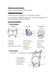
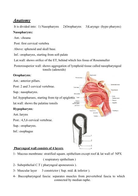
Anatomy<br />
It is divided into: 1) Nasopharynx 2)Oropharynx 3)Laryngo (hypo pharynx)<br />
Nasopharynx:<br />
Ant.: choana<br />
Post: first cervical vertebra<br />
Above: sphenoid and skull base.<br />
Inf.: oropharynx, starting from s<strong>of</strong>t palate<br />
Lat.wall: shows orifice <strong>of</strong> the ET, behind which lies fossa <strong>of</strong> Rosenmuller<br />
Posterosuperior wall: shows aggregation <strong>of</strong> lymphoid tissue called nasopharyngeal<br />
tonsils (adenoids)<br />
Oropharynx:<br />
Ant.: anterior pillars.<br />
Post: 2 and 3 cervical vertebrae.<br />
Sup.: nasopharynx.<br />
Inf.:hypopharunx, starting from tip <strong>of</strong> epiglottis<br />
lat.wall: shows the palatine tonsils<br />
Hypopharynx:<br />
Ant.:larynx<br />
Post.: 4,5,6 cervical vertebrae.<br />
Sup.: oropharynx.<br />
Inf.: esophagus<br />
Pharyngeal wall consists <strong>of</strong> 4 layers<br />
1- Mucous membrane: stratified squam. epithelium except ro<strong>of</strong> & lat wall <strong>of</strong> NPX<br />
( respiratory epithelium )<br />
2- Subepithelial C.T ( pharyngeal aponeurosis ).<br />
3- Muscular layer 3 constrictor ( Sup. mid. & inferior ).<br />
4- Buccopharyngeal fascia: separates muscles from prevertebral fascia to which<br />
connected by median raphe.
Blood Supply<br />
a- Arterial<br />
b- Venous: pharyngeal & pterygoid plexus ……… common facial ……IJV<br />
Nerve supply<br />
a- Motor: Cranial accessory (11), via vagus (10).<br />
b- Sensory glosso pharyngeal n. (9).<br />
Lymphatic drainage: retropharyngeal & lateral pharyngeal then UDCLN<br />
Waldayer’s ring :<br />
Def : ring <strong>of</strong> subepithelial lymphoid tissue that surrounds the pharynx<br />
Consists <strong>of</strong> :<br />
- Nasopharyngeal tonsil. -Tubal tonsils around orifice <strong>of</strong> E.T.<br />
- Palatine tonsils (the largest). -Lingual tonsils.<br />
- Discrete lymphoid nodules on lateral & posterior pharyngeal walls<br />
Characterized by :<br />
- lymphoid tissue lie in direct contact e mucosa<br />
- No afferent<br />
- <strong>Dr</strong>ain to retropharyngeal & UDCLN<br />
* Anatomy <strong>of</strong> the palatine tonsils<br />
- Two ovoid masses <strong>of</strong> lymphoid tissue lying in tonsillar fossa on each side <strong>of</strong><br />
oropharynx<br />
- Tonsillar fossa lies between ant. pillar , post. pillar & postero lat. third <strong>of</strong> the<br />
tongue<br />
- Tonsil has two surfaces :<br />
. Lat. surface covered by tonsillar capsule, which separates it from sup.<br />
constrictor muscle ( bed )<br />
. Med. Surface is free & covered by st. sq. epithelium that invaginates to form 12-<br />
15 crypts, largest is called crypta magna which is present near upper pole
Blood supply: as pharynx (mainly tonsillar artery)<br />
Venous drainage: para tonsillar veins …….. pharyngeal plexus<br />
Functions: Play a role in humoral & cell mediated immunity till age <strong>of</strong> 4-5 years<br />
Functions <strong>of</strong> the pharynx<br />
1- Respiratory channel 2-Voice resonance & speech articulation 3-Deglutition<br />
Adenoids<br />
Nasopharynx<br />
Def: Hypertrophy <strong>of</strong> naso pharyngeal tonsils sufficient to produce symptoms.<br />
Aet : Repeated URT infection.<br />
Incid : Childhood (2-12 y ), atrophy occurs at puberty.<br />
It is the commonest naso pharyngeal swelling<br />
Clinical picture:<br />
I. Effects <strong>of</strong> hypertrophy<br />
A-Nasal<br />
1- Bil nasal obstruction 2- Snoring, difficult suckling & may be O.S.A<br />
3- Bil mucoid nasal discharge 4- Nasal tone <strong>of</strong> voice<br />
5- Post nasal drip & foetor oris<br />
B- E.T obstruction<br />
1- Middle ear effusion ( S.O.M )…… deafness<br />
2- Recurrent acute otitis media….. pain & fever<br />
C- Adenoid facies<br />
- Narrow pinched ant. nares - Mucoid secretions over upper lip<br />
- Open dry mouth - Prominent incisors<br />
- High arched palate - Idiot look
II. Recurrent infections :<br />
Rhinitis – sinusitis – otitis media – pharyngitis – laryngo tracheo bronchitis .<br />
III. General manifestations :<br />
Examination<br />
- Chronic hypoxia , sleep apnea , night mares & nocturnal enuresis<br />
- Child is shy & friendless - Mental dullness & apathy<br />
A. Adenoid facies B.Ant. Rhinoscopy : ( see before )<br />
C. Oral cavity:<br />
- <strong>Dr</strong>y distorted decayed teeth - Enlarged tonsils<br />
- Egg white post nasal discharge - Fetor oris<br />
- Gums are inflamed - High arched palate<br />
- In marked adenoid hypertrophy its lower edge may be seen<br />
D. Ear: - retracted T.M - S.O.M<br />
- A.S.O.M - C.D by tuning fork<br />
E. Post. rhinoscopy dark pinkish swelling<br />
F. Digital palpation felt as punch <strong>of</strong> worms<br />
G. Endoscopy<br />
Investigations: Plain x ray nasopharynx lat. view: s<strong>of</strong>t tissue shadow<br />
Treatment : Adenoidectomy<br />
Indications : symptomatic adenoid ……see before<br />
Preoperative preparation & anesthesia : as tonsillectomy<br />
Position : patient on his back with head slightly flexed<br />
Technique :<br />
Post operative care : (as tonsillectomy)
<strong>Complications</strong> :<br />
I. Anesthetic complications<br />
II. Bleeding<br />
a) Primary: blood diseases, incomplete removal, and injury to muscles.<br />
ttt: - Complete removal - Suture injured muscles<br />
-Supply deficient factors - Blood transfusion if needed<br />
- Posterior nasal pack for 48 h.<br />
b) Reactionary (within 24 h): usually due to coagulation defect.<br />
ttt : Reanesthesia and as 1 y<br />
c) Secondary: after 5-7 days due to infection<br />
ttt antibiotics , blood transfusion, post nasal pack<br />
III. Incomplete removal due to<br />
Blunt curette, superficial anesthesia--- muscular contraction --- wrinkling <strong>of</strong> NPX<br />
This leads to: - post adenoidectomy bleeding - hypertrophy <strong>of</strong> residual tissue<br />
IV. Injury to surrounding structures<br />
V. Infection may cause<br />
Bleeding, local sepsis,<br />
Post nasal discharge & descending infection<br />
VI. Inhalation & aspiration<br />
Leads to lung collapse or abscess.<br />
Contra indications: as tonsillectomy + cleft palate & submucosal cleft to avoid<br />
velopharyngeal incompetence & rhinolalia aperta<br />
Tumors <strong>of</strong> the naso pharynx<br />
I. Benign Angi<strong>of</strong>ibroma<br />
Def : benign naso pharyngeal tumor<br />
Aet ! : a. true fibroma b. Hamartoma<br />
c. chemodectoma related to maxillary A. d.Endocrinal imbalance<br />
Incid : Most common benign tumor, only young males, peak 13-16 y<br />
Path : Site <strong>of</strong> origin is superior margin <strong>of</strong> spheno palatine foramen.
M/P sinusoidal spaces devoid <strong>of</strong> muscular coat, bundles <strong>of</strong> collagen<br />
Blood supply maxillary A<br />
Behavior: - Tumor extend due to pressure necrosis<br />
Clinical picture<br />
- Spontaneous regression may occur at age <strong>of</strong> sexual maturity<br />
A) General : anemia : easy fatigue , pallor<br />
B) Nasal manifestation<br />
Symptoms : - Intermittent , spontaneous , severe bleeding<br />
- Gradual progressive nasal obstruction<br />
- Purulent or bloody discharge<br />
- Nasal tone <strong>of</strong> voice - Hyposmia<br />
Signs: Ant. Rhinoscopy : MP discharge, D.S to opposite site.<br />
Unilateral nasal mass, bleeds on touch<br />
Post. rhinoscopy : pink , lobulated mass covered by intact mucosa with vessels<br />
on its surface, avoid digital palpation<br />
C) Aural manifestations<br />
Symptoms: deafness Signs: secretory otitis media<br />
D) oro pharyngeal manifestation: sagging <strong>of</strong> s<strong>of</strong>t palate<br />
E) External examination<br />
- proptosis - unilateral cheek swelling - frog face deformity<br />
Investigations:<br />
1- CT with contrast 2-MRI<br />
3-Carotid angiography (tumor blush) 4-Biopsy usually not needed<br />
D.D: Huge adenoid: no bleeding, not in the nose<br />
Treatment :<br />
Antro choanal polyp: no bleeding, C.T<br />
Malignant tumors: old age, cranial n. palsies, C.T<br />
A) surgical (mainly)<br />
1- Trans palatal. 2-Lateral rhinotomy.<br />
3-Trans nasal, trans antral via Weber-Furgusson or midfacial degloving.
4-Craniotomy if intra cranial extension<br />
5-Endoscopic nasal approach became widely accepted now in most cases.<br />
- To decrease bleeding: preoperative embolization is done 2-3 days before<br />
surgery, rapid technique, wide approach, hypotensive anesthesia, vessel ligation,<br />
remove all remnants & post operative pack<br />
- In extensive cases elective tracheostomy<br />
B) Hormonal!!<br />
C) Radio therapy: induce fibrosis but carcinogenic so used only in inoperable:<br />
intracranial extension or recurrence<br />
II- Malignant : carcinoma<br />
Def: Malignant tumor <strong>of</strong> the NPX.<br />
Aet: a) Environmental agents: Epstien barr virus, hydrocarbones , formaldehyde<br />
cigarette , smoke , fumes & Chinese herbal diet<br />
b) Genetic predisposition.<br />
Incid: - Most common malignant tumor (80%), highest among Chinese & Orientals<br />
-Peak (35-60) - Male: female 3/1<br />
path: site : commonest site is fossa <strong>of</strong> Rosenmuller<br />
M/P: commonest is squamous cell carcinoma then anaplastic carcinoma<br />
(lymphoepithelioma:carcinoma with lymphocytic infiltration)<br />
Spread: - Direct Ant, post, sup, inf. & lat.<br />
Clinical picture :<br />
- lymphatic : very early & common may be bilateral<br />
- Blood: rare& late.<br />
A) Aural mainfestation 1-Unilateral secretory otitis media 2- Referred otalgia<br />
B) Nodal manifestation : Enlarged UDCLN may be the presentation (occult primary ,<br />
silent area)
C) Nasal manifestation: 1- Nasal obstruction: usually unilateral.<br />
2-MP discharge.<br />
3-Mild epistaxis.<br />
4-Nasal tone <strong>of</strong> voice.<br />
On exam. Ant. rhinoscopy : may reveal a nasal mass<br />
Post rhinoscopy or endoscopy: fungating mass, ulcerative lesion, or<br />
submucosal swelling<br />
D) Neurological manifestation :<br />
1- Vidian nerve affection. 2-Cranial nerves: any can be affected.<br />
3- Sympathetic chain. 4-Increased ICT.<br />
NB the Diagnostic Trotters triad: Unilateral conductive deafness<br />
Investigations:<br />
1- CT<br />
Unilateral facial pain<br />
Immobile s<strong>of</strong>t palate<br />
2-Biopsy: endoscopic or direct if repeatedly negative: blind curettage<br />
3- Tympanomtry 4- Metastatic work up<br />
Treatment:<br />
- Radio therapy is treatment <strong>of</strong> choice (recently chemoradiotherapy).<br />
- Role <strong>of</strong> surgery: Biopsy.<br />
RND, if persistent after 1ry control.<br />
Salvage surgery <strong>of</strong> recurrent or residual disease is <strong>of</strong> limited<br />
role due to complex anatomy.<br />
NB: Other malignant tumors <strong>of</strong> NPX: Lymphoma, chordoma, & craniopharyngioma<br />
Congenital anomalies <strong>of</strong> the pharynx: Cleft palate<br />
Def: Failure <strong>of</strong> fusion <strong>of</strong> 2 halves forming the palate<br />
Types: Depends upon: depth & length<br />
1- Overt cleft: Bifid uvula, cleft s<strong>of</strong>t palate, complete cleft (s<strong>of</strong>t& hard), bipartite<br />
cleft (complete + unilateral gum cleft), tripartite cleft (complete + bilateral gum<br />
cleft).<br />
2-Submucos cleft: deficient palatal muscles with intact mucosa.<br />
Clinical picture: Nasal regurge, rhinolalia aperta
Ttt: at age <strong>of</strong> 1-2 year: surgery and speech therapy<br />
Traumatic conditions <strong>of</strong> the pharynx<br />
FB: As esophagus<br />
Lacerations: Suture if needed + antibiotics<br />
Penetrating wounds: Stab wounds or high velocity missiles, may be fatal due to<br />
damage to vital structures<br />
Caustics: as esophagus.<br />
I. Acute<br />
A) non specific<br />
Inflammation <strong>of</strong> the pharynx<br />
B) specific : 1- Diphtheria 2-Vincent’s angina 3- Moniliasis<br />
C) Blood Diseases : 1- Acute leakaemia 2- Agranulocytosis<br />
D) Systemic diseases : 1- IMN 2-Exanthemata 3-Aphthous ulceration<br />
II. Chronic :<br />
A) non specific<br />
B) Specific : 1- Scleroma 2-$ 3-T.B<br />
III. Inflammation <strong>of</strong> lymphoid tissue : Adenoiditis and tonsillitis<br />
Acute tonsillitis<br />
Def : Acute non specific inflammation <strong>of</strong> palatine tonsils.<br />
Aet : Predisposing factors : Recurrent URT infection<br />
Bad hygiene<br />
Low resistance<br />
Organism: group A beta haemolytic streptococci.
incid : More in children<br />
Path : 1- Acute catarrhal tonsillitis 2-Acute follicular 3-Acute parenchymatous<br />
Symptoms :<br />
General: high fever 39-40 , headache , malaise , anorexia & myalgia<br />
Local: - Sore throat & odynophagia -Referred otalgia<br />
Signs:<br />
- Fetor oris - Hot potato voice if huge tonsils<br />
General: Fever & proportionate tachycardia, patient looks ill.<br />
Local: 1- Swollen congested tonsils, oropharynx is red & edematous.<br />
2- Yellow white spots may be seen, yellow white membrane on the surface.<br />
3- Edema <strong>of</strong> s<strong>of</strong>t palate & foetor oris.<br />
4- Enlarged tender UDCLN ( jugulo digastric ).<br />
Investigations: - Swab for C&S - leucocytosis in CBC -High ESR<br />
D.D : other causes <strong>of</strong> membrane over tonsil……<br />
Treatment :<br />
Scarlet fever: erythematous rash, hypertrophy <strong>of</strong> posterior pharyngeal wall<br />
1- Rest, light diet, adequate fluids.<br />
2- Antibiotics : - Penicillin, you may start by I.M & continue on oral e.g -<br />
Amoxycillin –Amoxycillin clavulinate- Cephalosporins &<br />
macrolids<br />
3- Analgesics , antipyretics ,anti septic mouth wash<br />
<strong>Complications</strong>:<br />
A) local : 1- Quinsy, para & retropharyngeal abscess ,Ludwig's angina.<br />
2-Otitis media, laryngitis, bronchitis 3- chronic tonsillitis<br />
B) General : Rheumatic fever Acute glomerulonephritis
Chronic Tonsillitis<br />
Aet : repeated acute tonsillitis<br />
Symptoms :<br />
1- Recurrent attacks <strong>of</strong> acute tonsillitis<br />
2- Recurrent sore throat 3- Referred pain to the ear<br />
4- Recurrent enlarged cervical L.N 5- Fetor oris<br />
6- Snoring, sleep apnea, hot potato voice if huge tonsils<br />
7- Septic focus: low grade fever, fatigue, anorexia, headache, arthralgia .<br />
Signs:<br />
Investigations:<br />
1- High ESR<br />
2- High ASOT. +ve CRP<br />
Treatment: Tonsillectomy<br />
Acute pharyngitis<br />
A)Non specific :<br />
- Usually viral with common cold & exanthemata<br />
- Fever, headache, malaise - Sore throat & dysphagia<br />
- Generalized congestion <strong>of</strong> the pharynx - Treated like tonsillitis<br />
B)Specific :<br />
1) Diphtheria<br />
Def : Acute specific infection <strong>of</strong> the pharynx.<br />
Aet : Gram positive bacillus (corynebacterium diphtheriae) transmitted by droplets.<br />
Incid : Usually young 2-6 y (bellow 12 ), incubation period 2-6 days.<br />
Path : Site pharynx , larynx & nose (secondary), conjunctiva (rare)<br />
It is a pseudo membranous type <strong>of</strong> inflammation<br />
Symptoms : insidious onset<br />
Signs :<br />
General: low grade fever, headache, malaise, anorexia & may be vomiting.<br />
Local: severe sore throat & dysphasia.<br />
General: low grade fever, severe toxemia, tachycardia disproportionate to fever.
Local: - False membrane over the tonsil which is:<br />
- Unilateral -Yellow white or dirty gray.<br />
- Thick & firm , adherent & leaves a bleeding<br />
surface on removal & reforms rapidly<br />
- Often exceeds limits <strong>of</strong> the tonsils<br />
Enlarged tender cervical LN: Bull’s neck<br />
+ Clinical picture <strong>of</strong> laryngeal & nasal diphtheria<br />
Investigations : Throat swab: a: Direct smear b: Culture on loeffler’s serum<br />
<strong>Complications</strong> : due to effect <strong>of</strong> toxins<br />
A) Cardio vascular:<br />
1- Toxic myo carditis 2-vagal neuritis 3-acute heart failure<br />
B) Neurological (paralysis)<br />
1- S<strong>of</strong>t palate: earliest & most common.<br />
2- Occular paralysis: intrinsic more than extrinsic muscles.<br />
3- Laryngeal & pharyngeal muscles. 4- Diaphragm & inter costal muscles<br />
5- Peripheral neuritis.<br />
C) Respiratory :<br />
1- Laryngeal obstruction, lung collapse. 2-Peumonia , lung abscess<br />
2- Respiratory muscle paralysis & respiratory failure<br />
D) Renal: Toxic nephritis<br />
D.D membrane over tonsils<br />
1- Acute follicular tonsillitis 2- Diphtheria<br />
Onset Acute Gradual<br />
Fever high (39-40) low (38)<br />
Toxemia mild severe<br />
Face flushed pale<br />
Pulse full, rapid, proportionate weak rapid<br />
To fever Disproportionate<br />
Vomiting uncommon more common<br />
Exudation yellow spots dirty gray membrane<br />
confined to tonsils, exceeds the tonsils<br />
easily removed adherent<br />
No bleeding leaves bleeding surface<br />
Side bilateral unilateral<br />
Swab - ve for diphtheria + ve
3- Vincent’s angina<br />
4- I.M.N<br />
5- Acute leukaemia & agranulocytosis<br />
Treatment :<br />
1. Hospitalization, isolation , complete bed rest<br />
2. Diphtheria antitoxic serum<br />
To: neutralize circulating toxins<br />
Dose: 40000-100000 I.U I.M or I.V repeated after 24-48 h.<br />
When: diphtheria is suspected, within 48 h.<br />
Precautions: do skin sensitivity before, if positive start desensitization or shift<br />
to another serum, be ready with antihistaminics,and steroids<br />
3. Antibiotics: penicillin ½ million day IV/ IM for 10 days<br />
4. Glucose 25%, vitamins& antipyretics<br />
5. Observe airway if stridor………….tracheostomy<br />
6. Treatment <strong>of</strong> complications.<br />
prophylaxis:<br />
1- Active DPT vaccine<br />
2- Passive 5000-10000 IV antitoxic serum I.M for contacts<br />
2) Vincent’s angina<br />
Def : Acute specific inflammation <strong>of</strong> pharynx<br />
Aet : Symbiosis between a spirochaete : borrelia vincent & fusiform bacillus<br />
Symptoms: like diphtheria<br />
Signs: gingivitis & pharyngeal ulceration, deep punched out edges ulceration,<br />
covered by a dirty grayish membrane, extends beyond tonsils<br />
- enlarged tender submandibular L.N<br />
Investigations swab<br />
Treatment:<br />
1- Antibiotics e.g penicillin or erythrocin + metronidazole<br />
2- H2O2 mouth wash<br />
3- Oral hygiene, adequate nutrition
3) Moniliasis (oral thruth)<br />
Def: Acute specific inflammation <strong>of</strong> pharynx & oral cavity<br />
Aet : Organism: candida albicans Predisposing factors …..<br />
Symptons : Sore throat & dysphagia , no fever<br />
Signs: Milky white raised patches<br />
Treatment: - Stop antibiotics, adequate nutrition<br />
C) Blood diseases<br />
- Topical antifungal: mycostation , daktarin gel .<br />
-Systemic antifungal in severe cases e.q ketoconazole.<br />
1) Acute leukaemia neoplastic proliferation <strong>of</strong> the precursors <strong>of</strong> WBCS leading to<br />
- Anemia: fatigue pallor.<br />
- Thrombocytopenia : purpura , epistaxis, and bleeding tendency<br />
- Intercurrent infection : fever & sore throat, ulcers & membrane over the tonsils<br />
swollen purpulish gingiva & easy bleeding<br />
- Lymphadenopathy , splenomgaly - Sternal tenderness<br />
Investigations: 1- CBC 2- B.M aspiration<br />
Treatment: Cytotoxic drugs.<br />
2) Agrunulocytosis<br />
Def: marked reduction in formation <strong>of</strong> PNLs<br />
Aet : B.M depression, 1ry or 2ry to drugs (antibiotics: chloramphenicol,<br />
antimetabolites: methotrexate) or radiation.<br />
Clinical picture<br />
- Fever , malaise , rapid deterioration <strong>of</strong> general condition<br />
- Extensive ulceration with no or little surrounding inflammatory reaction<br />
Investigations: 1- CBC 2- MB aspiration<br />
Treatment :<br />
1- Stop <strong>of</strong>fending drug 2- Isolation<br />
3-Antibiotics & blood transfusion 4-B.M transplant
D) Systemic Diseases<br />
1) Infectious mononucleosis<br />
Def : Acute infective pharyngitis. Aet : Epstein barr virus ( EBV ) .<br />
Incid : young adults.<br />
Symptoms general:fever, headache, malaise (febrile type)<br />
Local: severe sore throat & dysphagia ( angionous type )<br />
Signs : - Congestion & edema <strong>of</strong> the pharynx<br />
- Shallow ulcers or grayish white membrane<br />
- Palatal petechiae in 30%<br />
- Tender enlarged cervical L.N ( glandular type )<br />
- Lymphadenopathy & splenomegaly<br />
Investigatios : lymphocytosis & monocytosis<br />
Serological test: Paul-Bunnel & monospot tests<br />
Treatment : Supportive, avoid penicillin as it forms rash + steroids in severe cases<br />
Chronic pharyngitis<br />
A)Non specific<br />
Aet : 1- Repeated acute tonsillitis 2- Tobacco, alcohol, spices<br />
3- Dusty atmosphere, mouth breathing 4- Reflux oesophagitis<br />
5-- Septic teeth, chronic tonsillitis, sinusitis<br />
Symptoms: - Persistent sore throat -A desire to clear the throat (hawking)<br />
Signs: Simple catarrhal pharyngitis<br />
Hypertrophic (granular) pharyngitis<br />
Atrophic pharyngitis<br />
Treatment : - Avoid predisposing factors<br />
- Local Treatment (gargles, cautery …..etc )<br />
- Antibiotics & H, antagonists
B)chronic specific pharyngitis<br />
1) Syphilis<br />
Primary: chancer, Rare but may affect tonsil<br />
Hard reddish painless nodule + cervical L.N<br />
secordary mucous patches<br />
Bluish Grey, slightly raised --- coalesce & ulcerate to form snail track ulcer<br />
Tertiary Gumma Hard purple swelling ---- ulcerate<br />
2) T.B<br />
Ulcers have deep punched out edge, indurated margins & necrotic floor<br />
Minute Grey tubercles that soon ulcerate: superficial, undermined edges, bluish<br />
margins & yellow caseous floor, severe odynophagia<br />
3) Scleroma : Uually 2ry to rhinoscleroma<br />
- Granulation & crustation in the pharynx<br />
- Painless induration without ulceration<br />
- Atrophic mucosa, lost uvula (Badrawy sign)<br />
- Fibrosis, leading to pharyngeal stenosis<br />
Suppurations related to the pharynx<br />
I. Peritonsillar abscess ( Quinsy )<br />
Def : Collection <strong>of</strong> pus between fibrous capsule <strong>of</strong> the tonsil, usually at its upper<br />
pole, & the superior constrictor muscle.<br />
Aet : - Usually as a complication <strong>of</strong> acute tonsillitis<br />
- Organisms : usually mixed aerobic & anaerobic infection<br />
incid : Usually young adult<br />
Path : Starts by infection in the depth <strong>of</strong> one <strong>of</strong> the crypts (usually crypta magna)<br />
Symptoms: (as tonsillitis but severe)<br />
Signs :<br />
General : Fever , headache , malaise ( if pus---hectic fever )<br />
Local : - Sore throat, severe & unilateral -Severe dysphagia & odynophagia<br />
-Unilateral neck pain & referred otalgia -Foetor oris<br />
General: Fever, tachycardia, toxic facies
Local: - Trismus , tonticollis<br />
Treatment :<br />
- Asymmetrical edema and congestion <strong>of</strong> s<strong>of</strong>t palate<br />
- Swelling above & lateral to tonsil<br />
- Tonsil is displaced downwards & medially<br />
- Uvula is edematous & pushed to other side<br />
- Large , firm , tender jugulodigastris LN<br />
A) during stage <strong>of</strong> peritonsillar cellulitis<br />
1- Parentral antibiotics<br />
2- Antipyretics , analgesics , bed rest , adequate fluids,& mouth wash<br />
B) During stage <strong>of</strong> peritonsillar abscess<br />
Indicated by: Hectic fever Throbbing pain<br />
Pitting edema on probing Aspiration brings pus<br />
Treated by: Incision & drainage Parentral antibiotics<br />
Site <strong>of</strong> incision: 1- Most bulging point<br />
2-Mid point <strong>of</strong> a line from base <strong>of</strong> uvula to last upper molar tooth<br />
3- 1/2 cm lat. To point <strong>of</strong> crossing <strong>of</strong> a vertical line along anterior<br />
pillar with a horizontal line along base <strong>of</strong> uvula<br />
Technique: use a guarded knife, Hilton method to open loculation usually under G.A<br />
C) Tonsillectomy should be done 4-6 weeks later.<br />
D.D :<br />
NB when to do quinsy tonsillectomy!!<br />
Anaplastic carcinoma<br />
Acute leukaemia<br />
Abscess related to upper molar tooth<br />
Para pharyngeal swelling<br />
<strong>Complications</strong> :<br />
Laryngeal edema & stridor. - Pyaemia & septicaemia -Para pharyngeal abscess<br />
II.Para pharyngeal abscess :<br />
Def. : Collection <strong>of</strong> pus in para pharyngeal space<br />
Aet. : - Peritonsillar abscess - tonsillitis - petrositis & mastoiditis
Symptoms like quinsy + unilateral neck swelling<br />
Signs : Fever, tachycardia, torticollis<br />
Becks triad: Swelling in lat. Pharyngeal wall pushing a normal tonsil medially<br />
Tender firm external swelling on lat. side <strong>of</strong> the neck<br />
Trismus<br />
Investigations : C T <strong>of</strong> pharynx & neck<br />
Treatment : As quinsy + incision & drainage by external incision along anterior<br />
border <strong>of</strong> sternomastoid<br />
D.D : All para pharyngeal swellings (salivary gland tumors neurogenic tumors,<br />
carotid aneurysm)<br />
<strong>Complications</strong> :<br />
- laryngeal oedema & stridor -Mediastinitis<br />
- Thrombosis <strong>of</strong> I.J.V -Erosion <strong>of</strong> carotid artery<br />
III. Retropharyngeal abscess:<br />
Between buccopharyngeal fascia <strong>of</strong> the post. pharyngeal wall & the prevertebral<br />
fascia<br />
A) Acute R.P.A :<br />
Aet: Suppuration <strong>of</strong> retroph. L.N (gland <strong>of</strong> Henle ) following URT infection<br />
Incid: Usually child, as gland atrophies later<br />
Path: Abscess occurs to one side <strong>of</strong> midline<br />
Symptoms:<br />
- Fever , headache , malaise<br />
- Severe dysphagia<br />
- Nasal obstruction if spreads up<br />
- Stridor due to laryngeal oedema
Signs :<br />
- Fever , tachycardia, torticollis with flexed neck<br />
- Swelling in the post.pharyngeal wall to one<br />
side <strong>of</strong> midline with hyperaemia & congestion<br />
- large tender cervical L.N.<br />
Investigations:<br />
- Xray :widening <strong>of</strong> prevertebral space - CT<br />
Treatment :<br />
1- Incision & drainage: trans oral route.<br />
2- Tracheostomy if stridor.<br />
3- Parentral antibiotics.<br />
B) Chronic R.P.A ( cold abscess , Pott’s disease )<br />
Aet: T.B <strong>of</strong> cervical spine<br />
Incid : In adult , uncommon<br />
Path : Cold abscess<br />
Symptoms: General: TB toxaemia night fever, night sweat loss <strong>of</strong> wt, loss <strong>of</strong><br />
appetite<br />
Local: sore throat & odynophagia<br />
Signs : General : neurological signs<br />
Investigation:<br />
Associated pulmonary T.B<br />
Local: bulge <strong>of</strong> post. Pharyngeal wall<br />
Tenderness over cervical spine<br />
1- Xray: destroyed vertebral bodies, chest x ray.<br />
2- Sputum analysis, tuberculin test, needle biopsy.<br />
Treatment :<br />
1- Anti tuberculous ttt.<br />
2- Incision & drainage along post. border <strong>of</strong> sternomastoid .<br />
3- Orthopaedic ttt .
IV. Ludwig’s angina<br />
Def: Suppuration in submandibular space.<br />
Aet: Dental causes in 90%, infection <strong>of</strong> lower tooth.<br />
extraction <strong>of</strong> septic tooth.<br />
Incid : More in diabetics.<br />
Symptoms :<br />
- General : Fever , headache , malaise<br />
- Local : Severe pain with dysphagia, muffled voice , difficult respiration<br />
Signs : -General : Fever , tachycardia<br />
-Local: - Massive indurated tender neck swelling<br />
- Swollen floor <strong>of</strong> mouth with the tongue pushed upwards<br />
Treatment : 1- Parentral antibiotics, antipyretics , analgesics<br />
I Traumatic<br />
2- Secure airway: tracheostomy<br />
3- <strong>Dr</strong>ainage: a free incision for decompression<br />
Stomatitis&OropharyngealUlceration<br />
A) Mechanical : Stiff bristles <strong>of</strong> tooth brush, fish bones , cheek biting , ill fitting<br />
denture, all have serrated edge with s<strong>of</strong>t base<br />
B) Chemical : Corrosives .<br />
C) Physical : Thermal due to hot foods , radio therapy (xero stomia)<br />
ttt : topical anti biotic with cortisone ( oro base )<br />
II. Infective:<br />
A)Bacterial : acute pharyngitis, tonsillitis, diphtheria, vincent’s<br />
B) Viral<br />
Chronic T.B. $<br />
1- Exanthemata:e.g. measles, Kopliks spots in the cheek opposite the molar teeth in<br />
the febrile stage, before rash appear.<br />
2- Herpes simplex : type I Prodroma <strong>of</strong> fever, headache, malaise followed by severe<br />
vesicular & ulcerative stomatitis, vesicles rupture to form multiple shallow ulcers
3- Herpes zoster<br />
- caused by varicella zoster<br />
- vesicular eruption occurs along 5,9,10 cranial nerves<br />
- strictly unilateral with severe dysphagia, vesicles & ulcers<br />
- usually accompanied by H.Z.oticus ttt: analgesics & acyclovir<br />
3- AIDS caused by HIV<br />
NB: E.N.T. manifestations <strong>of</strong> AIDS:<br />
Sever intercurrent infections (especially mucormycosis)<br />
Oropharyngeal ulceration Moniliasis<br />
Hairy leukoplakia cervical LN<br />
Kaposi sarcoma<br />
4- Herpangina (foot & mouth disease) caused by coxsakie virus, occurs in epidemics<br />
especially in children with vesicles & ulcers on the feet, hands, & oral cavity<br />
5-Infectious mononucleosis<br />
C) Fungal ( moniliasis )<br />
III .Neoplastic malignant ulcer: raised everted edge, necrotic floor& indurated<br />
base<br />
IV. Miscellaneous<br />
Allergic stomatitis<br />
- Chemical or contact allergy e.g lip stick , tooth paste<br />
- Vesicles rupture ……. ulcers<br />
Aphthous stomatitis<br />
- Aetiology is unknown may be abnormal immune reaction to oral bacteria viral<br />
infection, autoimmune, endocrinal disturbance.<br />
- Multiple superficial recurrent ulcers<br />
- ttt mouth wash, tetracycline, topical steroids, levamezole<br />
Blood diseases leukaemia agranulocytosis<br />
Behcet syndrome oral ulceration, genital ulceration, irido cyclitis<br />
Cancrum oris<br />
Ttt: steroids<br />
Rapidly spreading ulceration up to gangrene usually in children with low resistance
Dyspeptic ulcers:<br />
- Related to constipation , diarrhea, hyperacidity<br />
- Small, painful, superficial<br />
<strong>Dr</strong>ugs & metals: Epanutine, lead ….gingival hypertrophy & ulcerations<br />
Metabolic: D.M : xerostomia , red painful tongue uraemia : brown coated tongue<br />
Vitamin deficiency +B: glossitis & angular stomatitis<br />
+C: scurvy: swollen gums that bleeds easily<br />
Pre cancerous lesion leukoplakia raised white patches.<br />
Skin diseases<br />
- pemphigus ! autoimmune<br />
- Bullae ………rupture……..painful ulcers on the palate, buccal mucosa &<br />
tongue.<br />
- Rubbing <strong>of</strong> oral mucosa……bulla formation (diagnostic)<br />
- Ocular & neurological manifestations<br />
- Biopsy: acantholysis ttt by steroids.<br />
- Bullous pemphegoid less severe<br />
- Oral lesions similar to pemphigus with no other lesions<br />
- No acantholysis on biopsy- chronic course<br />
-Lichen planus: reticular: raised interlacing whitish lesion<br />
- Erosive: painful erythematous ulcer (premalignant)<br />
- Biopsy is diagnostic, ttt by steroids<br />
- Lupus erythematosis<br />
- Erythema multiformis<br />
! Delayed hypersensitivity …… bullae……..ulcers ttt by steroids<br />
oropharyngeal tumors<br />
Benign: papilloma: in tonsil or s<strong>of</strong>t palate Mixed salivary tumors<br />
Malignant:<br />
Squamous cell carcinoma: old males with risk factors, malignant ulcer<br />
Ttt: surgery &/or radiotherapy<br />
Sarcoma: mostly non Hodgkin lymphoma, Ttt: chemoradiotherapy
Indications<br />
1- Repeated attacks <strong>of</strong> acute tonsillitis<br />
Tonsillectomy<br />
2- Rheumatic fever, RHD, glomerulonephritis due to B haemolytic streptococci<br />
3- Septic focus with :<br />
Recurrent sore throat Recurrent URT infection<br />
Recurrent otitis media, pharyngitis, bronchitis<br />
Foetor oris Other manifestation <strong>of</strong> septic focus<br />
4- Tonsillar hypertrophy with<br />
Obstructive sleep apnea difficult swallow recurrent cough<br />
5- Trauma to the tonsils<br />
6- Tumors <strong>of</strong> the tonsil (unil. tonsillectomy )<br />
a. Benign: papilloma, fibroma<br />
b. Malignant : as biopsy<br />
c. At end stage in occult primary<br />
7- Tonsillolithiasis (impacted F.B)<br />
8-Abscess: quinsy to avoid recurrence<br />
9-Bleeding: persistent or recreant<br />
10-Cervical adenitis e.g T.B not resolving with medical ttt<br />
11-Diphtheria carrier<br />
Contraindications:<br />
1- Blood diseases e.g haemophilia, purpura<br />
2- Patient on aspirin or NSAID to avoid bleeding.<br />
4- Uncontrolled systemic disease e.g heart failure<br />
5- Active rheumatic fever 6- Acute infection e.g tonsillitis or URT infection<br />
7- Exanthemata e.g measles, chicken pox 8- Epidemics <strong>of</strong> polio<br />
9- Peritonsillar abscess<br />
Pre operative preparation<br />
A) History & exam To exclude contra indications (acute attack)
B) investigations<br />
1- CBC including HB %, blood group, ESR<br />
2- Coagulation pr<strong>of</strong>ile<br />
Bleeding time B.T ( N: 1-4 min ) Clotting time C.T ( N: 4-10 min )<br />
Prothrombin time P.T ( N: 12sec ) Prothrombin concentration P.C ( N: 100% )<br />
Partial thromboplastin time PTT (N: 25-45s)<br />
C) Fasting 6h before surgery:<br />
D) On the morning <strong>of</strong> surgery Check vital signs & tonsils<br />
Technique<br />
Anaesthesia usually general with cuffed tube<br />
Position supine with neck extended<br />
Procedure dissection method (usual one)<br />
Guillotine, cryosurgery: old, rarely<br />
Laser tonsillectomy: less pain & bleeding<br />
Coblation and radi<strong>of</strong>requency<br />
Post operative care:<br />
1-Patient is placed in tonsillectomy position<br />
2-Observe for:<br />
a. Vital signs weak rapid pulse & hypotension denote bleeding<br />
b. Bleeding : frequent swallow , spitting <strong>of</strong> blood or vomiting <strong>of</strong> dark blood<br />
c. Respiration : irregular respiration or cyanosis<br />
3-Medications:<br />
Antibiotics for 10 days Analgesics & antipyretics ( paracetamol )<br />
Vitamins Decongestant nasal drops (if adenoidectomy )<br />
4-Feeding: - Starts 4h. after surgery !<br />
- Semisolids & cold drinks in the first day<br />
- From 2 nd to 10 th day avoid hard, spicy & hot foods.
<strong>Complications</strong> <strong>of</strong> tonsillectomy :<br />
1- Anaesthetic complications<br />
Cardiac Arrest Aspiration <strong>of</strong> blood or vomitus<br />
Succinyl choline Apnea Anaphylaxis<br />
2- Post tonsillectomy bleeding<br />
a) primary during or immediately after<br />
Due to: - Bad technique -Bleeding tendency Bad preparation<br />
ttt: - ligation, suture or diathermy to bleeding point - Suture pillars together<br />
- ECA ligation may be needed - Correct shock (fluids & blood)<br />
b) Reactionary: During first 24 h<br />
Due to: - Slipped ligature, open <strong>of</strong> collapsed vessels, bleeding tendency<br />
ttt: conservative if mild : - Sedation, coagulants, H2O2 mouth wash<br />
- Remove blood clots from tonsillar bed<br />
- Firm pressure on the bed using tonsillar clamp<br />
Surgical: if bleeding is severe or persistent take patient back to theatre ttt as<br />
primary<br />
c) Secondary On 5th to 12 th day due to infection<br />
Ttt: Conservative Like reactionary + systemic parentral antibiotics for 4 days<br />
Surgical if bleeding is severe or persistent<br />
a. ligature or suture :difficult due to tissue friability<br />
b. suture pillars together over a pack<br />
c. ECA ligation may be needed<br />
d. Correct shock<br />
3- Respiratory complications<br />
A- Obstruction The most serious & may be fatal<br />
May be due to:<br />
- Laryngeal spasm : extubation spasm , or cord irritation by secretions or blood<br />
- Falling back <strong>of</strong> the tongue ( incomplete recovery )<br />
- Inhaled F.B or vomitus<br />
- laryngeal oedema from intubation<br />
b- Infection Pneumonia, lung abscess due to inhaled F.B
4- Injury:<br />
- Dental from intubation or mouth gag - TMJ dislocation<br />
- Injury to uvula, s<strong>of</strong>t palate, tongue - Atrophy <strong>of</strong> the uvula<br />
5- Infection: Otitis media, para pharyngeal abscess, cervical adenitis, bacteraemia<br />
6- Incomplete removal: Remnants regrow & become infected (revision surgery).<br />
Hypo pharynx<br />
Plummer vinson syndrome (paterson – brown kelly syndrome)<br />
Def: Chronic atrophic pharyngo oesophagitis<br />
Aet : Fe deficiency Incid : more in females<br />
Path : Atrophy <strong>of</strong> mucosa, submucosal fibrosis with stricture & web formation<br />
Clinical picture :<br />
1-Dysphagia: due to stricture or web<br />
2-Glossitis, fissured angle <strong>of</strong> the mouth<br />
3-Koilonychia: spooning <strong>of</strong> nails<br />
4-Achlorhydria due to atrophic gastritis<br />
5-splenomegaly<br />
6-Fe deficiency anemia<br />
Treatment : 1- Fe supplements & proper nutrition<br />
2-repeated dilatation 3-Regular follow up<br />
Complication: Condition is precancerous……..post cricoid carcinoma<br />
Hypo pharyngeal carcinoma<br />
Def: Malignant tumor <strong>of</strong> the hypo pharynx<br />
Aet: Predisposing factors: 1-tobacco smoking & alcohol consumption<br />
Incid : Old age > 50 more, in males<br />
Path :<br />
2- Irradiation 3- plummer vinson syndrome<br />
Post cricoid occurs in young ( 20-40 y ) female if on top <strong>of</strong> P.V.S<br />
- Site Piriform fossa 50%, post cricoid 40%, post pharyngeal wall 10%<br />
- G.P Malignant ulcer or fungating mass
- M.P Almost always squamous cell carcinoma<br />
- Spread: A) Direct: high tendency to sub mucosal extension<br />
Symptoms :<br />
B) Lymphatic : common , early & may be bilateral DCLN, paratracheal<br />
& mediastinal LN<br />
C) Blood: late lung, liver & bones<br />
1-Gradual progressive dysphagia first to solids later to fluids in addition.<br />
2-Pain in the throat & ipsilateral referred otalgia .<br />
3-Hoarseness <strong>of</strong> voice due to V.F fixation, infiltration <strong>of</strong> RLN<br />
4-Stridor: due to extension to larynx or bilateral V.F paralysis<br />
5-Painless neck mass gradual onset, progressive course: LN or extra laryngeal spread.<br />
6-Regurgitation, choking, cough<br />
7-Spitting <strong>of</strong> blood, WT loss<br />
8-Symptoms <strong>of</strong> distant spread.<br />
Signs :<br />
A) General: 1-Under weight, Fe deficiency anaemia<br />
B) Local:<br />
2-Respiratory distress 3-Signs <strong>of</strong> distant metastasis<br />
1-external neck examination<br />
- Enlarged cervical LN (Describe) - Larynx is pushed forwards<br />
- Absent laryngeal click (+ ve mour’s sign) - Fixation <strong>of</strong> larynx.<br />
2-Indirect larynqoscopy or flexible or rigid endoscope: may show:<br />
-Tumor itself: ulcer or fungating mass<br />
-Froth in piriform fossa or post cricoid<br />
-Invasion <strong>of</strong> the larynx<br />
Investigations :<br />
1-Radiology: Xray lat. View: widening <strong>of</strong> prevertebral space<br />
Barium swallow: filling defect<br />
CT <strong>of</strong> neck: shows tumor extensions<br />
2-Endoscopy & biopsy 3-Metastatic work up
Treatment :<br />
A) Surgery:<br />
-The standard is total laryngo pharyngectomy with esophagectomy, recently partial<br />
resection tailored to tumor extent<br />
-Neck dissection is mandatory<br />
-Reconstruction <strong>of</strong> the pharynx:<br />
a. Gastric pull up b. Colon inter position<br />
c. Myo cutaneous flaps d. Free vascularised flaps<br />
B) Radio therapy:<br />
As a palliative ttt in inoperable cases or as post operative adjuvant therapy<br />
C) Palliative ttt: In inoperable or recurrent cases<br />
- Adequate nutrition by ryle feeding or gastrostomy<br />
- Tracheostomy if stridor - Pain killer<br />
- Laser debulking - Radio & /or chems therapy<br />
Pharyngeal pouch (Zenker’s diverticulum)<br />
Def: herniation <strong>of</strong> the pharyngeal mucosa through a potentially weak area in the<br />
posterior pharyngeal wall ( killian dehisence )<br />
Aet : Spasm, failure <strong>of</strong> relaxation, pre mature closure <strong>of</strong> crico pharyngeal sphincter<br />
Incid : more in males > 40 y<br />
Symptoms:<br />
1-May be asymptomatic 2-Dysphagia<br />
3-Regurgitation <strong>of</strong> undigested food 4-Neck swelling usually on left side<br />
5-Loss <strong>of</strong> weight<br />
Signs: 1-unilateral neck swelling, usually LT sided, cystic, compressible, empties<br />
with gurgling sensation.<br />
Investigations:<br />
2-I.L: may show froth in the piriform fossa.<br />
1-Barium swallow: retort shaped smooth swelling<br />
2-Oesophagoscopy: may show the pouch orifice
Treatment:<br />
1-If asymptomatic: no ttt<br />
2-Symptomatic cases<br />
a. Small: repeated dilatation<br />
cricopharyngeal myotomy<br />
endoscopic crush & stapling<br />
b. Large: diverticulectomy with cricopharyngeal myotomy<br />
<strong>Complications</strong>:<br />
a. oesophageal obstruction b. chest infection c. malignant changes<br />
Septic focus<br />
Def: state <strong>of</strong> chronic bacteraemia or toxaemia<br />
Aet : chronic infection in a part <strong>of</strong> the body<br />
Chronic tonsillitis – chronic sinusitis<br />
Cholycystitis – colitis<br />
Prostatitis – salpingitis<br />
Path: Bacterial toxins produce systemic manifestation<br />
Clinical picture:<br />
1-Anemia, fatigue, anorexia 2-Headache, low grade fever<br />
3-Heart: Rheumatic fever, infective endocarditis 4-Lung: bronchiectasis<br />
5-Musculoskletal: myalgia, arthralgia & arthritis 6-Kidney: nephritis<br />
Occult primary<br />
Def: Enlarged cervical lymph node as the only presenting feature <strong>of</strong> a carcinoma<br />
(The primary is hidden)<br />
Aet: An occult primary may be one <strong>of</strong> the silent areas<br />
A) in the head & neck
B)Below the clavicle; Bronchogenic carcinoma, cancer breast, stomach &<br />
intestine (virchow’s gland)<br />
Management:<br />
I. History:<br />
1-Usually painless neck mass <strong>of</strong> insidious onset & rapid in size<br />
2-ask about symptoms <strong>of</strong>: larynx, pharynx, nose & naso pharynx, oral cavity,<br />
ear, chest & stomach.<br />
II. Examination:<br />
1-The lump site, size, shape, surface number, consistency, mobility…<br />
2-Other LN 3-Full H & N exam 4-Abdominal exam<br />
III. Investigations:<br />
A) Radiology: x ray to head & neck CT from skull base to chest<br />
B) Endoscopy: under GA<br />
Barium swallow, meal & enema Thyroid scan<br />
Pan endoscopy (naso pharyngoscopy, laryngoscopy hypo pharyngscopy<br />
Bronchoscopy & oesophagoscopy )<br />
If suspicious lesion …………..biopsy<br />
If no suspicious lesion ……….blind biopsy<br />
C) FNAC: (fine needle aspiration cytology) for the lump<br />
NB: never to excise the neck node before exhaustive search for the primary because:<br />
a. Biopsy does not give clue to site <strong>of</strong> the primary: as it is usually sq. cell carcinoma<br />
b. Spillage <strong>of</strong> tumor cells may occur<br />
c. Incision may interfere later with plane <strong>of</strong> neck dissection<br />
d. Patient may have false sense <strong>of</strong> security<br />
Ttt: If no primary was found: radical neck dissection with follow up<br />
Velopharyngeal incompetence<br />
Def: Failure <strong>of</strong> the s<strong>of</strong>t palate to close the NPX, during speech or swallowing<br />
Aet: congenital: cleft palate<br />
Traumatic: perforation, radiotherapy & post operative<br />
Inflammatory: scleroma(scarring), S (perforation)<br />
Neuromuscular: palatal paralysis<br />
Functional: faulty learning
Clinical picture: rhinolalia aperta & nasal regurge<br />
Ttt: Speech therapy Obturator: limited value<br />
Surgery: palatal repair Palatal push pack<br />
Pharyngeal augmentation to narrow the pharynx<br />
Pharyngopalatoplasty by various flaps<br />
Globus pharyngis( globus hystericus)<br />
Sensation <strong>of</strong> lump in the throat with no organic cause, mostly on swallowing saliva,<br />
more in females, normal barium swallow & endoscopy<br />
Ttt: Reassurance & psychotherapy<br />
Palatal & pharyngeal paralysis<br />
Aet: supranuclear, rare: requires bilateral cortical lesion<br />
Nuclear (nucleus ambiguous): bulbar palsy or cranial poliomyelitis<br />
Infranuclear: fracture base, parapharyngeal space tumor<br />
Symptoms: unilateral: palatal: no symptoms due to compensation<br />
Pharyngeal: patient sleeps on normal side to avoid aspiration<br />
<strong>of</strong> saliva<br />
Bilateral: palatal: nasal regurge & rhinolalia aperta<br />
Pharyngeal: aspiration on swallowing<br />
Signs: palate: unilateral: uvula shift to normal side on saying AHH<br />
Bilateral: immobile palate during phonation<br />
Pharyngeal: loss <strong>of</strong> pharyngeal reflex on the affected side & pooling <strong>of</strong><br />
secretion in hypopharynx<br />
Invest: modified barium swallow (video fluoroscopy)<br />
Ttt: <strong>of</strong> the cause<br />
1-unilateral: usually requires no ttt<br />
2-bilateral palatal: upper dental plate with s<strong>of</strong>t palate extension<br />
3-bilateral pharyngeal paralysis: nasogastric tube, suction, tracheostomy &<br />
gastrostomy.
Snoring & sleep apnea<br />
Def: * snoring sound produced during sleep due to partial air way obstruction.<br />
*Sleep apnea: cessation <strong>of</strong> respiration during sleep, at nostril & mouth > 10<br />
seconds it is 3 types<br />
- obstructive : no air flow in spite <strong>of</strong> respiratory effort ( commonest )<br />
- central: no air flow with no respiratory effort<br />
- Mixed : start as central then trial <strong>of</strong> respiratory effort<br />
* Sleep apnea syndrome (OSAS): 5 episodes <strong>of</strong> apnea / night h. sleep or<br />
30 episodes <strong>of</strong> apnea / night sleep<br />
* Apnea index periods <strong>of</strong> apnea / 1 h. night sleep<br />
Aet : any condition causing narrowing <strong>of</strong> the air way<br />
A)Naso pharyngeal<br />
1-Large naso pharyngeal tumor<br />
2-Huge adenoids<br />
3-Ant. & large post packs in children<br />
B) Oro pharyngeal<br />
1-Marked adenotonsillar hypertrophy<br />
2-Large lax uvula & s<strong>of</strong>t palate with excess mucosal folds<br />
3-Large oropharyngeal tumors<br />
C) Hypo pharyngeal:<br />
1-Macro glossia 2-Micro gnathia<br />
3-Hypo pharyngeal tumors 4-oedema due to radio therapy<br />
Exacerbating factors: 1-Nasal obstruction e.g. D.S, polypi , rhinitis or neoplasm<br />
Incid : more in male , at older age<br />
Pathogenesis :<br />
2-obesity 3-Alcohol & sedatives<br />
Partial narrowing <strong>of</strong> the upper air way ……...increased – ve intrathoracic pressure<br />
…….acceleration <strong>of</strong> air currents……….vibrations <strong>of</strong> s<strong>of</strong>t tissue………snoring.<br />
During sleep muscle tone decrease……..collapse<br />
Clinical picture: results from: oxygen desaturation & high negative intrathorathic<br />
pressure<br />
Symptoms: Snoring: disrupted sleep: frequent movement, nocturnal choking, day<br />
time sleep, memory loss<br />
Obstructive episodes
Less common:<br />
Morning headache, personality changes, nocturnal enuresis, impotence<br />
Systemic &pulmonary hypertension, arrhythmias, right heart failure, cardiovascular<br />
mortality<br />
Spouse (bed partner): sleep deprived, mood alteration, divorce<br />
-Examination may reveal:<br />
a. About 70% <strong>of</strong> patients are overweight.<br />
b. Short thick neck. c-Systemic hypertension.<br />
c. Oropharyngeal examination may show:<br />
- low hanging redundant palate and large uvula<br />
- Large (kissing) tonsils. - Excessive pharyngeal mucosal folds.<br />
- Narrow oropharyngeal isthmus. - Large tongue.<br />
d. Examin the larynx, nasopharyx and nose for any obstructive lesion<br />
N.B.: Although snoring indicates some degree <strong>of</strong> obstructed breathing, and although<br />
patients who have OSA are loud snorers, yet not all people who snore have OSA<br />
Investigations: to: assess general condition, differentiates between snoring & sleep<br />
apnea, site <strong>of</strong> obstruction<br />
1-Polysomnography, is the most sensitive and specific test in the evaluation <strong>of</strong> OSA.<br />
It measures eye movements (electro – occulography), brain activity (EEG), cardiac<br />
rhythm (ECG) pulse oximetery (to measure O2 and CO2 saturations), nasal and oral<br />
airflow, and respiratory movements (chest & abdominal movements). It allows<br />
correct diagnosis, estimation <strong>of</strong> the magnitude <strong>of</strong> the problem, and differentiates<br />
between obstructive and central sleep apnea.<br />
2-Flexible endoscopy <strong>of</strong> the naso, and hypopharynx. (Muller’s maneuver) is<br />
performed by asking the patient to snore with the mouth closed, which may show<br />
collapse in the area.<br />
3-Imaging: lateral cephalometry &CT.<br />
4-Pharyngeal manometry<br />
Treatment :<br />
Depends on: Is it simple snoring or apnea? , patient requests, severity &<br />
complications & level <strong>of</strong> obstruction<br />
A. Medical :<br />
1- Weight reduction.<br />
2- Avoid drugs that depress the CNS e.g. alcohol.
3- Progestin, which is respiratory stimulus (doubtful)<br />
4- Theophylline; increases the hypoxic drive.<br />
5- Protriptyline (non sedating tricyclic anti-depressant).<br />
6- Oxygen therapy. 7- Nasopharyngeal intubation.<br />
8- Tongue retaining devices. 9- Nasal continuous positive air pressure (CPAP).<br />
B. Surgical:<br />
Nasal surgery: for all cases for nasal obstruction<br />
Uvulopalatopharyngoplasty( U.P.P.P.)<br />
Laser assisted uvuloplasty(L.A.U.P.)<br />
Palate stiffening (somoplasty)<br />
Maxill<strong>of</strong>acial surgery: Mandibular advancement<br />
Tracheostomy: as a last resort<br />
Halitosis (foetor oris)<br />
Def: Bad mouth odour.<br />
Hyoid advancement<br />
Tongue advancement<br />
Aet: dental: bad hygiene, dental caries & pyorrhea.<br />
Oral: poor hygiene, stomatitis, ulcers (vincints), chronic tonsillitis with debris<br />
in the crypts, ulcerating tumors & mouth dryness (mouth breathing,<br />
irradiation, dehydration, smoking & atropine)<br />
Nasal: FB, atrophic rhinitis, sinusitis especially dental & ulcerating tumors<br />
Hypopharynx & esophagus: pharyngeal pouch & gastro-esophageal reflux<br />
Pulmonary: chronic bronchitis, bronchiectasis & lung abscess<br />
Metabolic: diabetic ketoacidosis(acetone like)<br />
Renal failure (urineferous)<br />
Hepatic failure<br />
Physiological: hunger<br />
Neurosis (non existing halitosis)
Trismus<br />
Def: Inability to open the mouth fully.<br />
Path: Lesion in either muscle <strong>of</strong> mastication or TMJ<br />
Aet: Local:<br />
1- Lesions causing reflex muscle spasm<br />
Dental infections, stomatitis, oral ulcers, quinsy, parapharyngeal abscess,<br />
ulcerating tumors, post operative (tonsillectomy)<br />
2-Lesion infiltrating the muscles<br />
Maxillary, nasopharyngeal, infratemporal & pterygopalatine tumors<br />
3-Fibrosis <strong>of</strong> the muscles<br />
4-TMJ disease<br />
Post irradiation & prolonged interdental fixation (ttt <strong>of</strong> mandibular fracture)<br />
Arthritis, ankylosis & fracture (neck, condyle or zygomatic arch)<br />
General:<br />
1- Tetanus<br />
2- Tetany (low Ca level)<br />
3- Hysteria<br />
4- Neurological: meningitis & bulbar palsy (increase muscle tone)<br />
Management: <strong>of</strong> the cause
Anatomy:<br />
The Oesophagus<br />
*it is a fibromuscular tube which extends from lower edge <strong>of</strong> hypo pharynx<br />
(C6) to stomach T11<br />
Consists <strong>of</strong> mucosa, submucosa & muscular layer (outer longitudinal & inner<br />
circular)<br />
Devoid <strong>of</strong> serosa exept for abdominal part making healing difficult<br />
*it consists <strong>of</strong> 3 parts:<br />
*it has 3 narrowings<br />
Physiology<br />
Cervical – thoracic –abdominal<br />
+ At upper end (15 cm from central incisors)<br />
+ At level <strong>of</strong> crossing <strong>of</strong> aortic arch & Lt Main bronchus (25 cm)<br />
+ At the diaphragm (40 cm)<br />
Peristaltic movement results in food propulsion towards the stomach<br />
The cricopharyngeus & cardia are normally closed, open on food passage, with<br />
positive pressure at rest (in the rest <strong>of</strong> esophagus: negative)<br />
Mechanism <strong>of</strong> swallowing (3 stages):<br />
1-Oral (voluntary): tongue pushed against palate,forcing food into pharynx, triggering<br />
reflex stages<br />
2-Pharyngeal (involuntary): food stimulates afferent in 5 & 9, efferent travel in 5, 9,<br />
10 & 12 to: elevate s<strong>of</strong>t palate, move palatopharyngeal wall medially, close glottis,<br />
elevates the larynx, relax cricopharyngeus & close superior constrictor as bolus<br />
passes into esophagus<br />
3- Esophageal (involuntary): solids falls by gravity, liquids pushed by peristalsis<br />
Clinically :<br />
I. History: dysphagia , pain & regurgitation .<br />
II. Neck examination.<br />
III. Investigations: Plain x ray, barium swallow, Oesophagoscopy.
Congenital: esophageal atresia<br />
Aet: Incomplete canalization <strong>of</strong> wall <strong>of</strong> foregut<br />
Incidence: In 85% associated with tracheo-esophageal fistula<br />
Results from incomplete separation <strong>of</strong> trachea from esophagus<br />
May present as esophageal atresia with proximal, distal, H shaped fistula<br />
Leads to recurrent pneumonia<br />
Other rare anomalies: duplicated esophagus, web, & stricture<br />
Types:<br />
F.B in the esophagus<br />
- In children: coins & buttons - In adults: fish or meet bone.<br />
- Old: lump <strong>of</strong> meat, dentures - Mental & prisoners: razors, pins & needles.<br />
Sites : - At the upper end (below cricopharyngeus)<br />
- Sites <strong>of</strong> anatomical constrictions<br />
- Sites <strong>of</strong> previous stricture<br />
Clinical picture: - Dysphagia & regurgitation <strong>of</strong> food<br />
- Retrosternal dull pain<br />
<strong>Complications</strong> : - Perforation: mediastinitis; fever & toxaemia<br />
- Ulceration & stenosis – TOF<br />
Investigation: -X ray neck & chest<br />
-Oesophagoscopy<br />
Treatment: a- Removal by oesophagoscope under GA<br />
b- Rarely external approach if perforation<br />
Corrosive oesophagitis & post corrosive stricture<br />
Aet : 1-Caustic potash (KOH)……liquefactive necrosis (more severe)<br />
Incid:<br />
2-lysol, phenol, H2SO4…….coagulative necrosis<br />
Accidental in children<br />
Suicidal in adult
A) Stage <strong>of</strong> corrosive oesophagitis<br />
Symptoms: -Severe pain in mouth, tongue, pharynx<br />
-Severe dysphagia & regurgitation<br />
-Stridor due to laryngeal oedema<br />
-Shock & dehydration<br />
Signs: -white sloughs, oedema -skin burns -shock<br />
<strong>Complications</strong>:<br />
1- Shock, dehydration, electrolyte imbalance 2- oesophageal perforation<br />
3- Esophageal stricture 4- T.O.F<br />
5- Stridor 6- Chest infection<br />
Treatment:<br />
1-milk & egg white<br />
2-management <strong>of</strong> shock & electrolyte imbalance<br />
3-Tracheostomy if severe obstruction<br />
4-parentral antibiotics<br />
5-cortisone to decrease edema & fibrosis<br />
6-Rubber naso gastric tube is inserted in 1 st few days to facilitate feeding & maintain<br />
the lumen<br />
7-Neutralization <strong>of</strong> the corrosive!!! (vingar)<br />
B) Stage <strong>of</strong> post corrosive stricture<br />
* Clinical picture: 1- Dysphagia reappears 2-3 weeks<br />
2- Dehydration & starvation<br />
3- Regurgitation & chest infection<br />
* Investigations: 1-plain x ray neck & chest<br />
2-barium swallow: irregular narrow segment<br />
3-oesophagoscopy<br />
* Treatment: A) Permeable stricture: Regular dilatation via rigid oesophagoscopy<br />
B) Impermeable stricture (non dilatable)<br />
1-Temporary gastrostomy<br />
2-Surgery: -Rsection <strong>of</strong> stricture & free jejunal loop reanastomosis<br />
or colon bypass<br />
-oesophagogastrostomy or jejunostomy -stents<br />
Indicated in: non dilatable stricture -complications(cachexia)
Achalasia Cancer<br />
Def marked dilatation <strong>of</strong> the malignant tumor<br />
lower 2/3 <strong>of</strong> the oes. due<br />
to closed cardia<br />
Aet neuromuscular incoordin- alcohol - smoking<br />
ation <strong>of</strong> lower oes. sphin- achalasia<br />
cter due to degenerated plumer - vinson<br />
auerbach’s plexus<br />
incid more in females 30-40 y more in males over 60<br />
Path failure <strong>of</strong> relaxation or commonest in middle<br />
spasm <strong>of</strong> cardia 1/3 ulcer,cauliflower,<br />
schirrous ,sq. cell carcinoma<br />
Clinical intermittent dysphagia progressive dysphagia<br />
Picture more to fluids more to solid<br />
regurgitation bloody regurgitation<br />
dull retrosternal pain severe pain<br />
normal built cachexia<br />
+hoarseness <strong>of</strong> voice<br />
Investigations:<br />
*Barium huge dilatation, fusiform with shouldering, rat tail<br />
smooth lower end, absent appearance<br />
air in stomach<br />
*oesophg- dilatation, stagnation biopsy<br />
oscopy CT : extension<br />
Treatment re assurance, sedatives -operable: radical resection<br />
amyl-nitrite before meals -palliative: gastrostomy or<br />
repeated dilatation, Heller’s stent, pain killer’s<br />
Cardiomyotomy cardioplasty<br />
(Vertical incision closed transversely)<br />
esophagogastrostomy in severe cases<br />
Stricture oesophagus<br />
I. congenital: rare<br />
II. Traumatic: a)accidental, corrosive, F.B b) Surgical after resection<br />
III. Inflammatory: reflux oesophagitis , peptic ulceration,TB, $<br />
IV. malignant stricture
Perforation <strong>of</strong> the oesophagus<br />
Aet : - During oesophagoscopy , dilatation , F.B extraction<br />
- Malignant growth or external injury<br />
Symptoms: -Severe retrosternal pain<br />
-Dyspnea (pneumothorax & empyema)<br />
-Dysphegia<br />
Signs: Fever, toxaemia, shock<br />
Tendernes & swelling in the neck Crepitation due to surgical emphysema<br />
ttt: -Control shock<br />
-Nothing by mouth (I.V alimentation or gastrostomy)<br />
-Heavy antibiotics<br />
-Intercostal tube connected to under water seal.<br />
*Indications:<br />
A) Diagnostic: examination - biopsy<br />
B) Theraputic: -FB extraction<br />
Oesophagoscopy<br />
-Dilatation <strong>of</strong> non malignant stricture<br />
-Excision <strong>of</strong> benign tumor<br />
-Stent in oesophageal carcinoma<br />
*Contraindications: -Acute necrotic ulceration from caustics<br />
-Marked kyphosis -Aortic aneurysm, vascular tumors.<br />
*<strong>Complications</strong>: -Perforation -Hge -Injury to teeth, tongue, pharynx<br />
Flexible esophagoscopy: Usually performed by gastroenterologists, to assess<br />
functional phenomenon suppressed by general anesthesia,<br />
or if rigid is contraindicated
Gastro esophageal reflux disease (G.E.R.D.)<br />
Def: Retrograde flow <strong>of</strong> gastric contents back into the esophagus<br />
Aet: Normally reflux is prevented by:<br />
Positive pressure <strong>of</strong> the cardia.<br />
Contraction <strong>of</strong> the crura <strong>of</strong> the diaphragm.<br />
The angle between the esophagus & the stomach.<br />
Decrease lower esophageal segment (LES) pressure is the major factor to<br />
GERD, it is affected by: smoking, alcohol, drugs, hormones, neuromuscular<br />
disease & delayed gastric emptying.<br />
ENT manifestations:<br />
- Heartburn, water brash (classic)<br />
- Choking, hoarseness, subglottic stenosis, globus hystericus<br />
- Asthma & chronic cough, otalgia, odynophagia<br />
Diagnosis: PH manometry (Ph less than 4 more than 6% <strong>of</strong> time is diagnostic)<br />
Management:<br />
Phase 1: Dietary & life style modification, with anti-acids<br />
Encourage protein meals (increase LES pressure)<br />
Discourage fatty meals (decrease LES pressure)<br />
Avoid chocolate, carbonated beverages & caffeine<br />
Avoid smoking, alcohol & over eating<br />
Last meal 3 hours before sleep<br />
Phase 2: if failed phase 1, give medication to decrease HCL production, increase LES<br />
pressure & promotes gastric emptying (H2 blockers e.g cimitidine,proton pump<br />
inhibitors e.g omeprazole)<br />
Phase 3: surgery in: failed medical ttt, complications or with hiatus<br />
hernia……correct hernia with fundoplication<br />
Dysphagia<br />
Def : Dysphasia is defined as difficulty on swallowing , when associated with pain is<br />
called odynophagia .<br />
Classification <strong>of</strong> the causes<br />
I. Oesophageal causes:<br />
A. Causes in the lumen: Foreign body
B. causes in the wall:<br />
1- Congenital diseases:<br />
a. congenital atresia or stenosis <strong>of</strong> the oesophagus.<br />
b.tracheo-oesophageal fistula (the commonest anomaly)<br />
2. Traumatic:<br />
a. foreign body<br />
b. oesophagoscopy and instrumentation<br />
c. Chemical: ingestion <strong>of</strong> corrosives which lead to stricture formation.<br />
d. External injury (rare).<br />
3. Inflammatory:<br />
a. acute ulcerations:<br />
-Corrosives.<br />
-<strong>Dr</strong>ugs, and specific fevers, e.g. typhoid and scarlet fever.<br />
-Persistent vomiting.<br />
-moniliasis<br />
b. chronic inflammatory changes in :<br />
4. Neoplastic :<br />
-Reflux oesophagitis<br />
-Peptic ulceration<br />
-T.B., syphilis, crhon’s disease. -scleroderma.<br />
-Pulmmer vinson syndrome.<br />
a. Benign tumours (rare) : e.g. leiomyoma, fibroma.<br />
b. Malignant tumours : carcinoma.<br />
5. Neurological: (Functional disordes <strong>of</strong> the swallowing mechanism)<br />
a. Paralytic : paralysis <strong>of</strong> the pharyngeal and oesophageal muscles due to<br />
b. Incoordinated motility :<br />
-Pharyngeal pouch<br />
-Achalasia <strong>of</strong> the cardia<br />
-Diffuse oesophageal spasm<br />
C. Pressure on the oesophagus from outside<br />
1. in the cervical region (upper 1/3)<br />
a. malignant thyroid tumor b.huge multinodular goiter<br />
c.enlarged cervical lymph nodes e.g. metastasis, and lymphoma
2. in the thorax (middle 1/3)<br />
a. Mediastinal tumours b.Pericardial effusion<br />
c.Enlarged left atrium d.Bronchogenic carcinoma<br />
e.Aneurysm <strong>of</strong> the aorta<br />
3. in the abdomen (lower 1/3)<br />
a. Enlarged left lobe <strong>of</strong> liver b.Paraoesophageal hiatus hernia<br />
II. Extraoesophageal causes:<br />
1. Nasal: nasal obstruction in infants (e.g. adenoids), and nasopharyngeal<br />
fibroma, they cause difficulty in feeding<br />
2. Oral:<br />
3. Pharyngeal:<br />
4. Laryngeal:<br />
a. Congenital : cleft palate<br />
b. Traumatic : injuries, corrosives, palatal tear<br />
c. Inflammatory:-stomatitis&ulcerations <strong>of</strong> mouth, gums and tongue<br />
-glossitis, dental sepsis<br />
-sialadenitis -ludwig’s angina<br />
d. Tumors <strong>of</strong> the oral cavity e.g. carcinoma <strong>of</strong> tongue, palate, tonsils.<br />
e. Miscellaneous: tongue paralysis<br />
a. Congenital : web, stricture<br />
b. Traumatic: lacerations, corrosives.<br />
c. Inflammatory :<br />
-Acute and chronic pharyngitis - Acute and chronic tonsillitis<br />
-Quinsy. -Retropharyngeal abscess<br />
-Parapharyngeal abscess. -Pulmmer vinson<br />
d. Tumors : Oropharyngeal and hypopharyngeal carcinoma<br />
e. Miscellaneous: pharyngeal pouch, globus hystericus<br />
a. Perichondritis b.T.B.<br />
b. Any lesion involving the inlet <strong>of</strong> the larynx (epiglottis, arytenoids,<br />
aryepiglottic folds) e.g.-supraglottic carcinoma, arytenoid edema<br />
c. Laryngopharyngeal malignancies<br />
d. Laryngeal incompetence: choking & cough
Def:<br />
Laser In ENT<br />
-Laser is the abbreviation <strong>of</strong> light Amplification by Stimulated Emission <strong>of</strong> Radiation<br />
-The history <strong>of</strong> laser begins in 1917 with Einstein who discovered stimulated<br />
emission, in 1960, Maiman made the first laser.<br />
- The radiant energy emitted by laser has 3 characters: monochromatic (one wave<br />
length), coherent (in one phase) & collimated (parallel)<br />
- Laser has 3 essential elements: lasing medium (gas, liquid or solids)<br />
Advantages <strong>of</strong> laser use:<br />
Excitation source (e.g. electrical)<br />
Two mirrors for optical feedback<br />
-Precise dissection - Non touch technique<br />
-Less operative bleeding -Less post operative pain<br />
-Minimal post operative edema & scarring<br />
-Possibility <strong>of</strong> local anesthesia<br />
-Less post operative hospital stay & cost<br />
Types <strong>of</strong> laser used in medicine: (according to lasing medium)<br />
1- CO2 laser 2- Argon laser 3- ND: YAG<br />
4- KTP laser 5- Diode 6- Dye laser<br />
Carbon dioxide laser:<br />
Most common type, wave length 10.6 um (invisible), site indicated by helium<br />
– neon aiming beam.<br />
Intracellular water absorbs light energy causing cell vaporization, used by hand<br />
piece, or connected to operating microscope<br />
Uses <strong>of</strong> CO2 laser in ENT<br />
A) Nasal surgery<br />
- Cong.: Choanal atresia<br />
- Inflammatory: laser turbinectomy<br />
-In seleroma : excision <strong>of</strong> localized mass ,widening <strong>of</strong> nostril after fibrosis<br />
-allergy: laser polypectomy
-septum: laser septoplasty<br />
-Neoplastic excision <strong>of</strong> benign tumors -Epistaxis photo coagulation <strong>of</strong> HHT<br />
-external rhinologic laser surgery: excision <strong>of</strong> rhinophyma, keloids, scars.<br />
B) Laryngeal surgery:<br />
Cong: laryngeal web, laryngo malacia<br />
Traumatic: subglottic stenosis<br />
Inflammatory: V.F nodules, polyps & cyst, Reink’s oedema<br />
Neoplastic: Benign papilloma especially in children<br />
Malignant: curative in T1, palliative in advanced cases<br />
Miscell: bil abductor paralysis: laser arytenoidectomy & post cordectomy<br />
C) Oral cavity & oro pharynx<br />
1-Tonsillectomy: dissection or cryptolysis<br />
2-LAUP (laser assisted uvulo palatoplasty) in snoring<br />
3-Partial tongue resection in OSA<br />
4-Lingual tonsillectomy<br />
5-Excision <strong>of</strong> haemangioma, laukoplakia<br />
D) Otological surgery<br />
1-Excision <strong>of</strong> auricular lesion<br />
2-Laser myringotomy: for <strong>ventilation</strong><br />
3-Laser tympanoplasty: removal <strong>of</strong> granulation tissue<br />
4-Laser stapedotomy (precise, haemostasis, visualization & less cochlear<br />
damage)<br />
5- Removal <strong>of</strong> cerebello pontine angle tumor<br />
Precautions for CO2 laser surgery<br />
1- Protect the eye: Patient eye by moist eye bad, operating room personnel by<br />
protective glasses, a sign placed outside laser room<br />
2- Protect adjacent tissues with wet cotton, gauze or drapes<br />
3- Avoid flammable anesthesia<br />
4- Use endotracheal <strong>tubes</strong> specially designed for laser
I- Lateral swellings<br />
1- Lymph nodes:<br />
a- Inflammatory:<br />
Acute : non specific lymphadenitis,<br />
Head & neck swellings<br />
specific infectious mononucleosis<br />
Chronic: non specific<br />
Specific: TB syphilis<br />
b- Primary malignancy (lymphomas)<br />
c- Blood diseases e.g. leukemia<br />
d- Metastatic<br />
e- Others: metabolic & autoimmune e.g. AIDS<br />
2- Branchial cyst:<br />
From remnants <strong>of</strong> second branchial cleft<br />
Cystic swelling under the anterior border <strong>of</strong> upper third <strong>of</strong> sternomastoid<br />
Aspirated fluid contains cholesterol crystals<br />
Track passes via bifurcation <strong>of</strong> CCA to lateral pharyngeal wall, behind the tonsil<br />
3- Pharyngeal pouch<br />
4-Parotid swellings<br />
Present below and infront <strong>of</strong> the auricle,<br />
Divided by facial nerve into superficial and deep lobes<br />
Parotid duct (stenson): open into inner aspect <strong>of</strong> cheek opposite second upper molar<br />
tooth<br />
Swelling may be 1-Inflammatory (sialadenitis)
2-Neoplastic a. benign: Pleomorphic adenoma (most common)<br />
Warthins tumor (adenolymphoma)<br />
Hemangioma and lymphangioma<br />
b. malignant:adenoid cystic carcinoma(commonest)<br />
mucoepidermoid<br />
adenocarcinoma<br />
Malignancy suspected if: rapid growth, facial palsy, pain, hardness, fixation, and LN<br />
5-Submandibular swellings<br />
Present below the mandible<br />
Divided by mylohyoid into superficial and deep lobes<br />
The duct (Wharton) opens into the floor <strong>of</strong> the mouth<br />
Related to lingual and hypoglossal nerves<br />
More commonly affected by calculi (viscid secretion and drainage against gravity)<br />
Swelling may be 1-Inflammatory (sialadenitis):swelling enlarges with meals<br />
2-Neoplastic (as parotid)<br />
6-Laryngocele: external or combined type<br />
II- Midline swellings<br />
7-Thyroid gland related swellings:<br />
a: Goiter: Enlarged thyroid gland, that present as a butterfly shaped swelling in the<br />
lower neck that moves up & down with deglutition<br />
-Simple physiological goiter<br />
-Simple nodular goiter: usually multinodular, may be solitary nodule.
- Colloid goiter: enlarged gland with irregular surface & s<strong>of</strong>t consistency<br />
- Toxic goiter (thyrotoxicosis)<br />
b- Thyroid neoplasms:<br />
Benign: follicular adenoma, presents as solitary nodule<br />
Malignant:<br />
Papillary carcinoma: spread to LN<br />
Follicular carcinoma: invade the capsule & spread by blood<br />
Anaplastic carcinoma: local, lymphatic & blood spread (lethal)<br />
medullary carcinoma: familial, secretes calcitonin( tumor marker),lymphatic &<br />
blood spread<br />
C- Thyroglossal cyst:<br />
Any where along the course <strong>of</strong> thyroglossal duct, commonly beneath the hyoid,<br />
moves up with swallowing as with tongue protrusion.<br />
e- Thyroglossal fistula:<br />
Follows infection or inadequate removal <strong>of</strong> thyroglossal cyst.<br />
8- Ranula: Retention cyst arise from sublingual salivary gland, contains a gelatinous<br />
material<br />
Form a cystic swelling on one side <strong>of</strong> floor <strong>of</strong> mouth, may present in<br />
Submandibular region as well (plunging or Thomson ranula)
Ear:<br />
Trauma: Auricle: cut wound, hematoma<br />
EAC: FB<br />
Emergencies in E.N.T<br />
TM: traumatic perforation, otitic barotrrauma<br />
Fracture skull base & CSF otorrhea<br />
Inflammation: <strong>Complications</strong> <strong>of</strong> otitis media<br />
Vestibular neuronitis<br />
Miscellaneous: Traumatic facial paralysis, accidental or post operative<br />
Nose<br />
Trauma: Fracture<br />
FB<br />
Sudden SNHL: give high dose <strong>of</strong> cortisone early<br />
Septal hematoma<br />
Septal abscess<br />
CSF rhinprrhea<br />
Inflammation: <strong>Complications</strong> <strong>of</strong> sinusitis<br />
Epistaxis<br />
Sudden blindness post FESS: immediate orbital & optic nerve decompression, high<br />
dose <strong>of</strong> steroids<br />
Throat & esophagus<br />
FB: larynx, trachea, pharynx & esophagus<br />
Corrosive ingestion<br />
Stridor