Laparoscopic retroperitoneal/mesenteric lymph ... - APAMED Central
Laparoscopic retroperitoneal/mesenteric lymph ... - APAMED Central
Laparoscopic retroperitoneal/mesenteric lymph ... - APAMED Central

You also want an ePaper? Increase the reach of your titles
YUMPU automatically turns print PDFs into web optimized ePapers that Google loves.
table as required. The ‘head up’ position was mostly<br />
used. The coeliac <strong>lymph</strong> nodes were approached by<br />
dissecting through the lesser sac. Para-aortic nodes<br />
were reached by dissecting the posterior peritoneum left<br />
of the duodenojejunal flexure. Using instruments with<br />
bipolar attachments and laparoscopic Babcock forceps<br />
(Mediflex, Islandia, NY, USA) to steady the mass of<br />
nodes with minimum manipulation, the nodes were<br />
dissected and sampled. On average, a 25 mm × 25 mm<br />
mass of tissue was aimed for. Haemostasis was achieved<br />
with bipolar rather than monopolar diathermy due to<br />
the close proximity of the bowel loops. The 10-mm port<br />
site was closed with a 1-0 PDS ® suture with a J needle<br />
(Ethicon, New Brunswick, NJ, USA) to the fascia. The<br />
skin and the remaining port sites were closed using skin<br />
glue. The tissue volume that was harvested was calculated<br />
Singapore Med J 2011; 52(10) : 759<br />
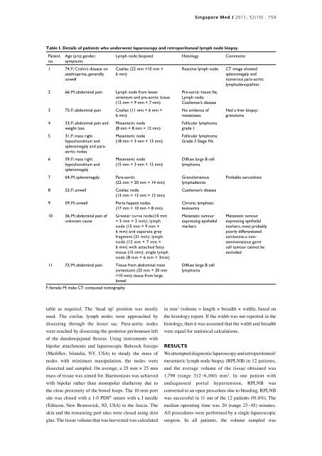
Table I. Details of patients who underwent laparoscopy and <strong>retroperitoneal</strong> <strong>lymph</strong> node biopsy.<br />
Patient<br />
no.<br />
Age (yrs); gender;<br />
symptoms<br />
1 74; F; Crohn’s disease on<br />
azathioprine, generally<br />
unwell<br />
Lymph node biopsied Histology Comments<br />
Coeliac (22 mm ×10 mm ×<br />
6 mm)<br />
2 66; M; abdominal pain Lymph node from lesser<br />
omentum and pre-aortic tissue<br />
(12 mm × 9 mm × 7 mm)<br />
3 75; F; abdominal pain Coeliac (11 mm × 6 mm ×<br />
6 mm)<br />
4 33; F; abdominal pain and<br />
weight loss<br />
5 31; F; mass right<br />
hypochondrium and<br />
splenomegaly and paraaortic<br />
nodes<br />
6 59; F; mass right<br />
hypochondrium and<br />
splenomegaly<br />
Mesenteric node<br />
(8 mm × 8 mm × 12 mm)<br />
Mesenteric node<br />
(18 mm × 3 mm × 13 mm)<br />
Mesenteric node<br />
(15 mm × 3 mm × 12 mm)<br />
7 64; M; splenomegaly Para-aortic<br />
(22 mm × 20 mm × 14 mm)<br />
8 52; F; unwell Coeliac node<br />
(13 mm × 12 mm × 12 mm)<br />
9 59; M; unwell Porta hepatis nodes<br />
(17 mm × 10 mm × 8 mm)<br />
10 36; M; abdominal pain of<br />
unknown cause<br />
Greater curve node(10 mm<br />
× 5 mm × 2 mm); <strong>lymph</strong><br />
node (15 mm × 9 mm ×<br />
6 mm) and separate grey<br />
fragment (21 mm); <strong>lymph</strong><br />
node (12 mm × 7 mm ×<br />
6 mm) with attached fatty<br />
tissue (15 mm); single <strong>lymph</strong><br />
node (8 mm × 6 mm × 3mm)<br />
11 72; M; abdominal pain Tissue from abdominal mass<br />
(omentum) (25 mm × 20 mm<br />
×10 mm); tissue from large<br />
bowel<br />
F: female; M: male; CT: computed tomography<br />
Reactive <strong>lymph</strong> node CT image showed<br />
splenomegaly and<br />
numerous para-aortic<br />
<strong>lymph</strong>adenopathies<br />
Pre-aortic tissue: fat,<br />
Lymph node:<br />
Castleman’s disease<br />
No evidence of<br />
metastases<br />
Follicular <strong>lymph</strong>oma<br />
grade 1<br />
Follicular <strong>lymph</strong>oma<br />
Grade 3 Stage IVa<br />
Diffuse large B cell<br />
<strong>lymph</strong>oma<br />
Granulomatous<br />
<strong>lymph</strong>adenitis<br />
Castleman’s disease<br />
Chronic <strong>lymph</strong>atic<br />
leukaemia<br />
Metastatic tumour<br />
expressing epithelial<br />
markers<br />
Diffuse large B cell<br />
<strong>lymph</strong>oma<br />
Had a liver biopsy:<br />
granuloma<br />
Probable sarcoidosis<br />
Metastatic tumour<br />
expressing epithelial<br />
markers, most probably<br />
poorly differentiated<br />
carcinoma; a nonseminomatous<br />
germ<br />
cell tumour cannot be<br />
excluded<br />
in mm3 (volume = length × breadth × width), based on<br />
the histology report. If the width was not reported in the<br />
histology, then it was assumed that the width and breadth<br />
were equal for statistical calculations.<br />
RESULTS<br />
We attempted diagnostic laparoscopy and <strong>retroperitoneal</strong>/<br />
<strong>mesenteric</strong> <strong>lymph</strong> node biopsy (RPLNB) in 12 patients,<br />
and the average volume of the tissue obtained was<br />
1,798 (range 312–6,160) mm3 . In one patient with<br />
undiagnosed portal hypertension, RPLNB was<br />
converted to an open procedure due to bleeding. RPLNB<br />
was successful in 11 out of the 12 patients (91.6%). The<br />
median operating time was 20 (range 23–45) minutes.<br />
All procedures were performed by a single laparoscopic<br />
surgeon. In all patients, the volume sampled was