
You also want an ePaper? Increase the reach of your titles
YUMPU automatically turns print PDFs into web optimized ePapers that Google loves.
Original Article<br />
Singapore Med J 2011; 52(9) : 673<br />
Anatomical study of the distal end<br />
of cadaveric human ulnae: a clinical<br />
consideration for the management of<br />
distal radioulnar joint injuries<br />
Sharma A, Kumar A, Singh P<br />
Department of<br />
Anatomy,<br />
Dayanand Medical<br />
College and Hospital,<br />
Tagore Nagar,<br />
Ludhiana 141001,<br />
Punjab,<br />
India<br />
Sharma A, MBBS, MS<br />
Associate Professor<br />
Kumar A, MBBS, MS<br />
Associate Professor<br />
Singh P, MBBS, MS<br />
Professor and Head<br />
Correspondence to:<br />
Dr Anu Sharma<br />
Tel: (91) 98722 02147<br />
Fax: (91) 161 4686609<br />
Email: anuashwani2003<br />
@yahoo.com<br />
ABSTRACT<br />
Introduction: Detailed anatomical knowledge<br />
of the distal end of the ulna plays a pivotal role in<br />
understanding post-injury instability and painful<br />
conditions at the distal radioulnar joint (DRUJ),<br />
which can be due to avulsion of the ulnar styloid<br />
process or ulnar styloid triquetral impaction<br />
syndrome. With this in mind, data on the<br />
morphological features of distal ulnae in the Indian<br />
population was collected.<br />
Methods: The distal end of 100 human ulnae (50<br />
right-sided and 50-left sided) of unknown gender<br />
from the anatomy department’s bone bank<br />
were studied with regard to the seat (articular<br />
circumference of the head of ulna), ulnar styloid<br />
process, fovea and pole (articular surface for<br />
articulation of triangular fibrocartilaginous<br />
complex of the wrist on the head of ulna).<br />
Results: The average length of the styloid process<br />
was 5.2 mm in the right-sided ulnae and 5 mm in<br />
the left. The mean maximum height of the seat<br />
was noted to be 5.9 mm and 6.9 mm on the right-<br />
and left-sided ulnae, respectively. The maximum<br />
width of the pole was calculated to be 5.4 mm<br />
(right-sided ulnae) and 6.1 mm (left-sided ulnae).<br />
The shapes of the pole and styloid process were<br />
also noted. Extensor carpi ulnaris groove was more<br />
commonly found on the left-sided ulnae.<br />
Conclusion: The study provides an anatomical<br />
database of the morphometry of parts of the distal<br />
end of the ulna in the Indian context, which will aid<br />
in the early management of DRUJ injuries.<br />
Keywords: distal end of ulna, distal radioulnar<br />
joint, head of ulna, styloid process of ulna<br />
Singapore Med J 2011; 52(9): 673–676<br />
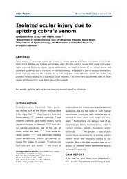
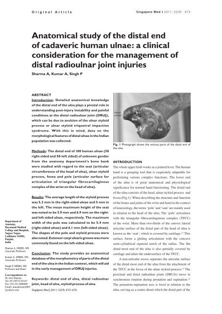
Styloid<br />
process<br />
Fovea<br />
INTRODUCTION<br />
Pole<br />
Seat<br />
Fig. 1 Photograph shows the various parts of the distal end of<br />
the ulna.<br />
The whole upper limb works as a jointed lever. The human<br />
hand is a grasping tool that is exquisitely adaptable for<br />
performing various complex functions. The lower end<br />
of the ulna is of great anatomical and physiological<br />
significance for normal hand functioning. The distal end<br />
of the ulna consists of the head, ulnar styloid process and<br />
fovea (Fig.1). When describing the structure and function<br />
of the bones and joints of the wrist and hand in the context<br />
of kinesiology, the terms ‘pole’ and ‘seat’ are mainly used<br />
in relation to the head of the ulna. The ‘pole’ articulates<br />
with the triangular fibrocartilaginous complex (TFCC)<br />
of the wrist. More than two-thirds of the convex lateral<br />
articular surface of the distal part of the head of ulna is<br />
known as the ‘seat’, which is covered by cartilage. (1) This<br />
surface forms a gliding articulation with the concave<br />
semi-cylindrical sigmoid notch of the radius. The flat<br />
distal-most end of the ulna is also partially covered by<br />
cartilage and abuts the undersurface of the TFCC.<br />
A non-articular recess separates the articular surface<br />
of the distal-most end of the ulna from the attachment of<br />
the TFCC at the fovea of the ulnar styloid process. (2) The<br />
proximal and distal radioulnar joints (DRUJs) move in<br />
synchronous rotation during pronation and supination. (3)<br />
The pronation-supination axis is fixed in relation to the<br />
ulna, serving as a centre about which the distal part of the
Singapore Med J 2011; 52(9) : 674<br />
Table I. Quantitative analysis of various parameters of the distal end of the ulnae.<br />
Component Measurement Mean ± SD (mm)<br />
Right-sided ulna<br />
Left-sided ulna<br />
Pole Maximum width 5.40 ± 0.99 6.10 ± 0.67<br />
Seat Maximum height 5.90 ± 0.69 6.90 ± 0.87<br />
Fovea Maximum width 4.50 ± 0.47 4.90 ± 1.10<br />
Styloid process Length 5.20 ± 0.82 5.00 ± 0.67<br />
SD: standard deviation<br />
radius rotates through a 150° arc; further lateral movement<br />
of the radius of around 30° in the direction of rotation<br />
enables this movement to go through approximately 180°<br />
for hand rotation. (4) At the extremes of the movement of<br />
pronation and supination, the ulnar head has little contact<br />
with the articulating sigmoid notch, making the joint<br />
vulnerable. Since the distal part of the ulna is a stable<br />
anatomical point of reference for rotation of the forearm,<br />
the distal part of the radius dislocates volarly or dorsally<br />
with respect to the ulna.<br />
There is general agreement that the mechanism<br />
of injury in an ulnar dorsal dislocation of the DRUJ is<br />
hyperpronation, while the mechanism of injury in ulnar<br />
volar dislocation is hypersupination. Galeazzi-fracture<br />
dislocations can also be associated with fracture of the<br />
ulnar shaft and styloid process in high-energy trauma.<br />
During sporting activities, forceful impact loading on the<br />
thenar side of the hand causes the wrist to be progressively<br />
levered into hyperextension, with ulnar deviation and<br />
intercarpal supination. (5)<br />
Many reports on the diagnosis and treatment of<br />
injuries of the DRUJ under-emphasised the role of<br />
the lower end of the ulna. Many of these irreducible<br />
dislocations commonly have displaced fracture of the<br />
ulnar styloid process as an associated feature. (6) Although<br />
instability of the DRUJ after radius end fracture did not<br />
receive much attention, the residual symptoms related to<br />
DRUJ instability are common. Our anatomical studies<br />
revealed the detailed structural anatomy of the lower<br />
end of the ulna. This study is one of the few to present the<br />
morphometric data of configurations of the lower end of the<br />
ulna. The detailed morphometric data on regular anatomy<br />
in this study would be helpful to the design of ulnar head<br />
prosthesis. Irreparable fracture dislocation of the ulnar<br />
head with concomitant fracture of the radius (Galeazzi<br />
lesion) had been treated by implantation of a Herbert Ulnar<br />
Head Prosthesis, which had yielded good results. (7)<br />
METHODS<br />
The present study, which was conducted over a period of<br />
one year, involved 50 right- and 50 left-sided dry adult<br />
human ulnae of unknown gender belonging to an Indian<br />
population. Approval for the study was obtained from<br />
the institutional ethics committee. A selected number<br />
of specimens were collected from the bone bank of the<br />
anatomy department at our institution, depending on the<br />
availability and time planned to conduct the study. Only<br />
bones that were intact and free from any pathological<br />
or congenital anomalies were selected. Anatomic<br />
measurements were taken using a vernier caliper<br />
(accurate to 0.1 mm). Appropriate statistical analysis<br />
was done wherever applicable. Each ulna was studied for<br />
different features, such as the shape of the pole (articular<br />
surface for articulation with TFCC), maximum width<br />
of the pole along the transverse axis (mm), slope of the<br />
seat (articular surface on the head of ulna for articulation<br />
with radius), maximum vertical height of the seat (mm),<br />
maximum width of the fovea (roughened depression at<br />
the base of the styloid process) along the transverse axis<br />
(mm) (Fig. 1). Also noted were the presence or absence of<br />
vascular foramina in the fovea, the presence or absence<br />
of styloid process, the shape and length (base to apex) of<br />
the styloid process, if any, and the presence or absence of<br />
grooves for extensor carpi ulnaris (ECU).<br />
RESULTS<br />
All quantitative measurements of the pole, seat, fovea<br />
and styloid process of the 50 right- and 50 left-sided<br />
ulnae were taken and tabulated. The mean and standard<br />
deviation of each parameter was calculated (Table I), and<br />
the various shapes of the poles and styloid processes were<br />
noted. The ulna seat (sloping or non-sloping surfaces),<br />
the presence or absence of vascular foramina of the fovea<br />
and that of ECU groove were also observed and their<br />
percentages tabulated (Table II).<br />
DISCUSSION<br />
DRUJ injury can occur in association with fracture of<br />
the forearm or as an isolated phenomenon. A dislocation<br />
of this joint may be simple or complex. The possibility<br />
of DRUJ injury should be kept in mind when treating<br />
wrist, forearm and elbow injuries. The distal end of the
Singapore Med J 2011; 52(9) : 675<br />
Table II. Measurements of various parts of the distal end<br />
of the ulna.<br />
Component No. (%)<br />
Pole<br />
Comma<br />
Semilunar<br />
Semicircular<br />
Kidney-shaped<br />
Seat<br />
Sloping<br />
Non-sloping<br />
Styloid process<br />
Present<br />
Absent<br />
Overall shape<br />
Curved<br />
Straight<br />
Shape of tip<br />
Blunt<br />
Pointed<br />
Fovea (vascular foramina)<br />
Present<br />
Absent<br />
ECU groove<br />
Present<br />
Absent<br />
ECU: extensor carpi ulnaris<br />
Right (n = 50) Left (n = 50)<br />
5 (10)<br />
30 (60)<br />
10 (20)<br />
5 (10)<br />
30 (60)<br />
20 (40)<br />
50 (100)<br />
-<br />
5 (10)<br />
45 (90)<br />
30 (60)<br />
20 (40)<br />
40 (80)<br />
10 (20)<br />
40 (80)<br />
10 (20)<br />
ulna comprises the head, fovea and styloid process. The<br />
ulnar head is the fixed point of the distal arm and wrist,<br />
around which the forearm, carpus and hand rotate. It is<br />
also an integral part of the ulnar-carpal wrist joint, which<br />
plays an important role in load transfer from hand to<br />
forearm. The kinematics and biomechanics of this region<br />
is unique and is found only in humans. The ulnar head<br />
consists of two parts, the dome (pole) and seat. The dome<br />
faces the carpus, while the seat faces the radius laterally.<br />
Radially, the slightly convex and triangular-shaped part is<br />
covered by the cartilage to articulate with the underside<br />
of the TFCC. <strong>Central</strong>ly, there is a roughened depression<br />
(fovea) for attachment of the apex of the TFCC. On the<br />
dorsal side, opposite to the fovea, a longitudinal sulcus<br />
is located for the ECU tendon and its sheath. Traumatic<br />
derangements around the ulnar head usually affect both<br />
the joint compartments, the distal radioulnar and ulnarcarpal<br />
joints. Therefore, the aim must be to restore normal<br />
anatomy in acute situations whenever possible. (8)<br />
15 (30)<br />
20 (40)<br />
10 (20)<br />
5 (10)<br />
30 (60)<br />
20 (40)<br />
50 (100)<br />
-<br />
-<br />
50 (100)<br />
35 (70)<br />
15 (30)<br />
40 (80)<br />
10 (20)<br />
50 (100)<br />
-<br />
Besides this, the anatomical relationships of the distal<br />
ulna with the distal radius and ulnar carpus are precise.<br />
These relationships are important from the functional<br />
point of view, e.g. minor modifications in these lead to<br />
significant load changes and resultant pain syndromes<br />
(ulnar-carpal abutment, ulnar styloid triquetral impaction<br />
syndromes and ulnar styloid impaction). These functional<br />
correlations as well as the treatment modalities of the<br />
region require detailed anatomical knowledge and<br />
morphometric data collection. The literature is replete<br />
with studies of the anatomy of bony configurations<br />
on dry bones, forming the wrist joint, which can aid in<br />
surgical interventions in the region of the wrist. Studies<br />
conducted by Berger et al (8) and Joshi et al (9) have targeted<br />
these relevant points. Keeping this relevance in mind, we<br />
compared our findings with their observations.<br />
The size and projection of the pole (articular surface<br />
for articulation of TFCC of the wrist on the head of<br />
ulna) may help in guiding the direction of transmission<br />
of forces through the ulna. We recorded the maximum<br />
width and various shapes of the pole. The average<br />
maximum widths of the pole from the radial to ulnar side<br />
on the right- and left-sided ulnae were documented as<br />
5.4 ± 0.99 mm and 6.1 ± 0.67 mm, respectively (Table I).<br />
An earlier study that reported a dimension of 8.2 (range<br />
5.1–13.2) mm, regardless of the side of the ulna, (8) is in<br />
line with the findings of our study. The widths recorded<br />
in the present study were varied, but they did not show<br />
such a wide range of variation. The confounding factor<br />
may be the overall medium stature of the people in the<br />
northern part of India. The variety of pole shapes may<br />
have affected the angulation between the seat and pole<br />
of the ulnar head. The most common shape observed on<br />
the right (60%) and left (40%) ulnae was semilunar. This<br />
observation concurs with Joshi et al’s study, which found<br />
a semilunar shape to be the most common on both sides. (9)<br />
The distribution of the shapes was found to be varied on<br />
both sides. The ‘comma’, the next most common shape,<br />
was found on the left side in 30% of ulnae and on the<br />
right, in just 10% of ulnae (Table II).<br />
More than two-thirds of the convex lateral articular<br />
surface of the distal part of the ulna, viz the seat, is the<br />
main determining factor for gliding articulation and<br />
complexity of movement at the DRUJ. In our study, the<br />
average maximum height of the seat was found to be<br />
5.9 ± 0.69 mm on the right side and 6.1 ± 0.67 mm<br />
on the left side (Table I). However, another study had<br />
documented the maximum height of the seat as 9.3 (range<br />
6.8–12.6) mm. (8) This could be attributed to the difference<br />
in the study population of the two studies. 60% sloping<br />
and 40% non-sloping surfaces on both the right and left<br />
sides were found in our study (Table II). The height and<br />
slope of the seat may be of great clinical significance<br />
in understanding any dysfunction of the site, as far as<br />
assessing the stability of the DRUJ is concerned.<br />
The fovea is a roughened depression at the base of the<br />
styloid process. The mean recorded maximum widths of
Singapore Med J 2011; 52(9) : 676<br />
the fovea in right-sided and left-sided ulnae were<br />
4.5 ± 0.47 mm and 4.9 ± 1.1 mm, respectively (Table I).<br />
20% of both the right- and left-sided ulnae showed an<br />
absence of any vascular formina in the fovea. A previous<br />
study has recorded an absence of any foramina in 24.63%<br />
of the cases involving right-sided ulnae. (9) A substantial<br />
amount of nutrition reaches the TFCC of the wrist from<br />
the synovial fluid of the DRUJ (on the superior surface)<br />
and the wrist joint (on the inferior surface), as well as<br />
from the vasulature in the ligament at the medial end of<br />
the disc by the vascular foramina.<br />
The mean length of the styloid process was<br />
5.2 ± 0.82 mm and 5.0 ± 0.67 mm on the right- and leftsided<br />
ulnae, respectively (Table I). If the length of the<br />
styloid process was > 6 mm, it was considered a criterion<br />
of long ulnar styloid process. (10) The length of the styloid<br />
process may act as a causative factor in ulnar styloid<br />
triquetral impaction, causing ulnar-sided wrist pain. (11) The<br />
dorsal surface on the head of the ulna showed a groove<br />
for ECU tendon in all left-sided ulnae and in 80% of<br />
right-sided ulnae (Table II). Its anatomical position is of<br />
great importance in the treatment of dislocation of the<br />
DRUJ.<br />
Over the last decade, there has been a greater<br />
appreciation of the anatomy and mechanics of the<br />
proximal radioulnar joint compared to the DRUJ.<br />
Morphometrical analysis of the DRUJ is still evolving.<br />
Traumatic derangements around the ulnar head<br />
usually affect the DRUJ. Most untreated injuries to<br />
the bony or stabilising ligamentous structures lead to<br />
arthrosis of the DRUJ. Therefore, the aim must be to<br />
restore normal anatomy in an acute situation, whenever<br />
possible, rather than delaying it until only excision<br />
or arthrodesis remains the treatment of choice. Also,<br />
in certain isolated intra-articular fractures of the<br />
head of ulna, open reduction and internal fixation<br />
with K-wires, screws or small plates is a common<br />
treatment. In all these cases, if the congruency of the<br />
joint is not restored, permanent functional impairment<br />
will ensue. (8) The dislocations of the ulnar head with<br />
concomitant fracture of the radius (Galeazzi lesion)<br />
had been treated by implantation of prosthesis such<br />
as the Herbert Ulnar Head instead of arthrodesis, as<br />
commonly found in the literature. (7) These metrical<br />
values of structural anatomy of the lower end of the<br />
ulna are valuable for reconstruction of the DRUJ with<br />
prosthesis. While the literature is replete with detailed<br />
anatomical descriptions of the distal end of the radius,<br />
the lower end of the ulna is a neglected area of research<br />
in the Indian population. Therefore, our study aimed at<br />
providing the morphometric database for configurations<br />
and kinematics of the distal end of the ulna, which is a<br />
less frequently researched area in the Indian context.<br />
REFERENCES<br />
1. Oatis CA. Kinesiology in structure and function of the bones<br />
and joints of the wrist and hand. 2nd Ed. The Mechanics and<br />
Pathomechanics of Human Movement. Baltimore: Williams and<br />
Wilkins, Lippincott, 2003:243-54.<br />
2. af Ekenstam F. Anatomy of the distal radioulnar joint. Clin Orthop<br />
Relat Res 1992; 275:14-8.<br />
3. Almquist EE. Evolution of the distal radioulnar joint. Clin Orthop<br />
Relat Res 1992; 275:5-13.<br />
4. King GJ, McMurtry RY, Rubenstein JD, Gertzbein SD.<br />
Kinematics of the distal radioulnar joint. J Hand Surg Am 1986;<br />
11:798-804.<br />
5. Tulley E. Sports physiotherapy applied science and practice in<br />
the wrist and hand. 1st Ed. Sports Physiotherapy Melbourne:<br />
Churchill Livingstone, 1995: 439-42.<br />
6. Spinner M, Kaplan EB. Extensor carpi ulnaris. Its relationship to<br />
the stability of the distal radio-ulnar joint. Clin Orthop Relat Res;<br />
1970; 68:124-9.<br />
7. Grechenig W, Peicha G, Fellinger M. Primary ulnar head<br />
prosthesis for the treatment of an irreparable ulnar head fracture<br />
dislocation. J Hand Surg Br 2001; 26:269-71.<br />
8. Berger RA, Weiss APC. Hand Surgery vol 1. 1st Ed. Philadelphia<br />
USA: Williams and Wilkins: Lippincott 2004: 297.<br />
9. Joshi SD, Joshi SS, Athavale SA, Kishve PS, Jadhav SD. Metrical<br />
and non-metrical study of lower end of ulna. J Anat Soc India<br />
2009; 58:156-60.<br />
10. van Der Heijden B, Groot S, Schuurman AH. Evaluation of ulnar<br />
styloid length. J Hand Surg Am 2005; 30:954-9.<br />
11. Giachino AA, Mclntyre AI, Guy KJ, Conway AF. Ulnar styloid<br />
triquetral impaction. Hand Surg 2007; 12:123-34.