
You also want an ePaper? Increase the reach of your titles
YUMPU automatically turns print PDFs into web optimized ePapers that Google loves.
Singapore Med J 2011; 52(9) : 674<br />
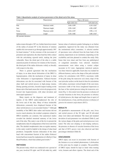
Table I. Quantitative analysis of various parameters of the distal end of the ulnae.<br />
Component Measurement Mean ± SD (mm)<br />
Right-sided ulna<br />
Left-sided ulna<br />
Pole Maximum width 5.40 ± 0.99 6.10 ± 0.67<br />
Seat Maximum height 5.90 ± 0.69 6.90 ± 0.87<br />
Fovea Maximum width 4.50 ± 0.47 4.90 ± 1.10<br />
Styloid process Length 5.20 ± 0.82 5.00 ± 0.67<br />
SD: standard deviation<br />
radius rotates through a 150° arc; further lateral movement<br />
of the radius of around 30° in the direction of rotation<br />
enables this movement to go through approximately 180°<br />
for hand rotation. (4) At the extremes of the movement of<br />
pronation and supination, the ulnar head has little contact<br />
with the articulating sigmoid notch, making the joint<br />
vulnerable. Since the distal part of the ulna is a stable<br />
anatomical point of reference for rotation of the forearm,<br />
the distal part of the radius dislocates volarly or dorsally<br />
with respect to the ulna.<br />
There is general agreement that the mechanism<br />
of injury in an ulnar dorsal dislocation of the DRUJ is<br />
hyperpronation, while the mechanism of injury in ulnar<br />
volar dislocation is hypersupination. Galeazzi-fracture<br />
dislocations can also be associated with fracture of the<br />
ulnar shaft and styloid process in high-energy trauma.<br />
During sporting activities, forceful impact loading on the<br />
thenar side of the hand causes the wrist to be progressively<br />
levered into hyperextension, with ulnar deviation and<br />
intercarpal supination. (5)<br />
Many reports on the diagnosis and treatment of<br />
injuries of the DRUJ under-emphasised the role of<br />
the lower end of the ulna. Many of these irreducible<br />
dislocations commonly have displaced fracture of the<br />
ulnar styloid process as an associated feature. (6) Although<br />
instability of the DRUJ after radius end fracture did not<br />
receive much attention, the residual symptoms related to<br />
DRUJ instability are common. Our anatomical studies<br />
revealed the detailed structural anatomy of the lower<br />
end of the ulna. This study is one of the few to present the<br />
morphometric data of configurations of the lower end of the<br />
ulna. The detailed morphometric data on regular anatomy<br />
in this study would be helpful to the design of ulnar head<br />
prosthesis. Irreparable fracture dislocation of the ulnar<br />
head with concomitant fracture of the radius (Galeazzi<br />
lesion) had been treated by implantation of a Herbert Ulnar<br />
Head Prosthesis, which had yielded good results. (7)<br />
METHODS<br />
The present study, which was conducted over a period of<br />
one year, involved 50 right- and 50 left-sided dry adult<br />
human ulnae of unknown gender belonging to an Indian<br />
population. Approval for the study was obtained from<br />
the institutional ethics committee. A selected number<br />
of specimens were collected from the bone bank of the<br />
anatomy department at our institution, depending on the<br />
availability and time planned to conduct the study. Only<br />
bones that were intact and free from any pathological<br />
or congenital anomalies were selected. Anatomic<br />
measurements were taken using a vernier caliper<br />
(accurate to 0.1 mm). Appropriate statistical analysis<br />
was done wherever applicable. Each ulna was studied for<br />
different features, such as the shape of the pole (articular<br />
surface for articulation with TFCC), maximum width<br />
of the pole along the transverse axis (mm), slope of the<br />
seat (articular surface on the head of ulna for articulation<br />
with radius), maximum vertical height of the seat (mm),<br />
maximum width of the fovea (roughened depression at<br />
the base of the styloid process) along the transverse axis<br />
(mm) (Fig. 1). Also noted were the presence or absence of<br />
vascular foramina in the fovea, the presence or absence<br />
of styloid process, the shape and length (base to apex) of<br />
the styloid process, if any, and the presence or absence of<br />
grooves for extensor carpi ulnaris (ECU).<br />
RESULTS<br />
All quantitative measurements of the pole, seat, fovea<br />
and styloid process of the 50 right- and 50 left-sided<br />
ulnae were taken and tabulated. The mean and standard<br />
deviation of each parameter was calculated (Table I), and<br />
the various shapes of the poles and styloid processes were<br />
noted. The ulna seat (sloping or non-sloping surfaces),<br />
the presence or absence of vascular foramina of the fovea<br />
and that of ECU groove were also observed and their<br />
percentages tabulated (Table II).<br />
DISCUSSION<br />
DRUJ injury can occur in association with fracture of<br />
the forearm or as an isolated phenomenon. A dislocation<br />
of this joint may be simple or complex. The possibility<br />
of DRUJ injury should be kept in mind when treating<br />
wrist, forearm and elbow injuries. The distal end of the