Nonobstructive Acute Pyelonephritis Presenting as Acute Renal ...
Nonobstructive Acute Pyelonephritis Presenting as Acute Renal ...
Nonobstructive Acute Pyelonephritis Presenting as Acute Renal ...
You also want an ePaper? Increase the reach of your titles
YUMPU automatically turns print PDFs into web optimized ePapers that Google loves.
J Med Sci 2003;23(1):61J64<br />
http://jms.ndmctsgh.edu.tw/2301061.pdf<br />
Copyright © 2003 JMS<br />
<strong>Nonobstructive</strong> <strong>Acute</strong> <strong>Pyelonephritis</strong> <strong>Presenting</strong><br />
<strong>as</strong> <strong>Acute</strong> <strong>Renal</strong> Failure in the Elderly<br />
Yuen-Hua Ni 1 , Ning-Chi Wang 1 , Ann Chen 2 , Yuh-Feng Lin 3 , and Feng-Yee Chang 1*<br />
1 Division of Infectious Dise<strong>as</strong>es and Tropical Medicine, Department of Medicine,<br />
2 Department of Pathology, 3 Division of Nephrology, Department of Medicine,<br />
Tri-Service General Hospital, National Defense Medical Center, Taipei, Taiwan, Republic of China<br />
Received: May 16, 2002; Revised: July 17, 2002; Accepted:<br />
July 23, 2002.<br />
* Corresponding author: Feng-Yee Chang, Division of Infectious<br />
Dise<strong>as</strong>es and Tropical Medicine, Department of<br />
Medicine, Tri-Service General Hospital, 325, Chung-Kung<br />
Road Section 2, Taipei 114, Taiwan, Republic of China. Tel:<br />
+886-2-87927257; Fax: +886-2-87927258; e-mail :<br />
fychang@ndmctsgh.edu.tw<br />
Yuen-Hua Ni, et al.<br />
<strong>Acute</strong> renal failure is a rare, but less recognized complication of acute bacterial pyelonephritis in patients who do not<br />
have urinary obstruction. We describe an aged woman who developed acute renal failure suffered from severe bilateral<br />
flank pain for one week and progressive anorexia, general weakness, decre<strong>as</strong>ed urine output and short of breath. She<br />
did not have hypotension, fever, leukocytosis and thrombocytopenia. Urinalysis showed numerous leukocytes in the<br />
urine. Biochemistry revealed marked renal function impairment with BUN 98 mg/dL and creatinine 7.2 mg/dL.<br />
Abdominal sonography demonstrated bilateral enlarged kidney without the evidence of obstruction. Blood and urine<br />
cultures grew Escherichia coli. The renal biopsy revealed predominant leukocyte filtration in the interstitium and<br />
tubules, compatible with acute pyelonephritis. Antimicrobial therapy and hemodialysis led to rapid improvement in<br />
clinical symptoms and renal function. The clinician should be aware that acute bacterial pyelonephritis can cause acute<br />
renal failure in the elderly even with no urinary obstruction and hypotension.<br />
Key words: acute renal failure, bacteremia, pyelonephritis<br />
INTRODUCTION<br />
<strong>Acute</strong> renal failure is commonly seen in hospitalized<br />
patients. B<strong>as</strong>ed on the anatomical site of involvement, it<br />
can be cl<strong>as</strong>sified into postrenal, prerenal and renal. <strong>Renal</strong><br />
sites of involvement can be subdivided into v<strong>as</strong>cular,<br />
glomerular, interstitial, and tubular lesions. Infection-related<br />
acute renal failure is usually related to septic shock,<br />
urinary tract obstruction, immune-mediated glomerular or<br />
tubulointerstitial nephritis, and direct bacterial inv<strong>as</strong>ion 1,2 .<br />
Although urinary tract infections are common in the elderly,<br />
acute pyelonephritis without an <strong>as</strong>sociation of obstruction<br />
and hypotension is rarely considered in the differential<br />
diagnosis of acute renal failure 3,4 . Herein, we report on the<br />
c<strong>as</strong>e of an elderly woman with no urinary tract obstruction<br />
who developed acute bacterial pyelonephritis with acute<br />
renal failure requiring hemodialysis support.<br />
CASE REPORT<br />
An 82-year-old female w<strong>as</strong> admitted to our hospital<br />
with a 1-week history of bilateral flank pain, back pain and<br />
progressive l<strong>as</strong>situde, decre<strong>as</strong>ing urine output and shortness<br />
of breath. There w<strong>as</strong> no history of fever, chills, or<br />
dysuria. She also denied any history of previous urinary<br />
tract infection. She w<strong>as</strong> noted to have a serum creatinine of<br />
1.0 mg/dL and BUN 11 mg/dL six months ago. However,<br />
she had been using a nonsteroidal anti-inflammatory drug<br />
(ibuprofen) to control back pain for one week prior to<br />
admission.<br />
On admission, her blood pressure w<strong>as</strong> 120/70 mmHg,<br />
pulse rate 88/minute, respiration 20/minute and temperature<br />
36.5 o C. On physical examination she w<strong>as</strong> lethargic but<br />
arousable. Her mucous membranes appeared dry. Lungs<br />
were clear to auscultation and examination of the heart<br />
revealed a normal sinus rhythm, with grade II systolic<br />
ejection murmur. There w<strong>as</strong> no gallop or pericardial rub.<br />
Abdomen w<strong>as</strong> soft with marked knocking tenderness over<br />
bilateral costo-vertebral angle. There w<strong>as</strong> no pitting edema.<br />
Complete blood count showed white blood cell count<br />
(WBC) 11,200/uL, with 87% segmented neutrophils<br />
predominant, hemoglobin 8.4 g/dL, hematocrit 25.3%,<br />
platelet count 176,000/uL. Urinalysis revealed a pH of 5.5,<br />
protein (2+), no eosinophiluria, positive reaction for nitrates,<br />
and numerous WBC/HPF. Urine Bence-Jones protein w<strong>as</strong><br />
61
<strong>Acute</strong> pyelonephritis and acute renal failure<br />
negative. Her blood urea nitrogen (BUN) w<strong>as</strong> 98 mg/dL,<br />
creatinine 7.2 mg/dL, serum sodium 135 mmol/L, pot<strong>as</strong>sium<br />
4.8 mmol/L, chloride 96 mmol/L, total serum calcium<br />
7.4 mg/dL, phosphate 5.2 mg/dL, cholesterol 164<br />
mg/dL, total protein 6.1 g/dL, and albumin 3.4 g/dL. Liver<br />
function w<strong>as</strong> normal. Serum c-reactive protein (CRP) w<strong>as</strong><br />
elevated at 9.5 mg/dL. Arterial blood g<strong>as</strong>es revealed pH<br />
7.335, PCO 2 26.6 mmHg, PO 2 84.8 mmHg, HCO 3 13.9<br />
mmol/L. Serum iron and TIBC levels were 46 g/dL and<br />
212 mg/dL, respectively. Serum ferritin concentration w<strong>as</strong><br />
540 ng/L. Immunological studies showed normal C3, C4,<br />
IgM, IgA, ASOT, RF and negative ANA except mildly<br />
incre<strong>as</strong>ed serum IgG levels (1,670 mg/dL). Tumor markers<br />
were normal . Chest X-ray revealed cardiomegaly and<br />
torturosity of the aorta. KUB demonstrated bilateral enlarged<br />
kidney shadow, severe degenerative joint dise<strong>as</strong>e<br />
and osteoporosis. A renal ultr<strong>as</strong>ound indicated incre<strong>as</strong>ed<br />
size of kidneys bilaterally (right kidney: 12 cm, left kidney<br />
11.8 cm) and incre<strong>as</strong>ed echogenicity, without evidence of<br />
obstruction.<br />
This initial impression w<strong>as</strong> urinary tract infection with<br />
sepsis and acute renal failure although she had no fever.<br />
Intravenous flumarine 1.0 gm every 8 hours w<strong>as</strong> initiated<br />
along with volume repletion with 1 liter normal saline.<br />
Nevertheless, renal function remained severely impaired<br />
and urine output w<strong>as</strong> still oligouric despite hydration and<br />
antibiotic therapy. Since she progressed to shortness of<br />
breath, vomiting, anorexia, and severe metabolic acidosis,<br />
hemodialysis w<strong>as</strong> started on hospital day 3. A diagnostic<br />
percutaneous renal biopsy w<strong>as</strong> performed on hospital day<br />
5. Light microscopic examination of the renal biopsy<br />
showed a predominant neutrophil infiltration in the interstitium<br />
and renal tubules. Occ<strong>as</strong>ional intratubular neutrophils<br />
were also seen. The glomeruli and vessels appeared<br />
normal. Immunofluorescent staining for IgG, IgA, IgM,<br />
C3, C1q, and fibrinogen w<strong>as</strong> negative. Cultures of urine<br />
and blood raised Escherichia coli. By the end of the first<br />
week, her serum creatinine began to decline, and with<br />
improving urine routine and negative urine and blood<br />
cultures, dialysis w<strong>as</strong> discontinued on day 9. She required<br />
a total of four hemodialysis treatments. During her hospital<br />
stay, the patient remained afebrile. At the time of discharge<br />
on hospital day 12, her BUN w<strong>as</strong> 24 mg/dL and serum<br />
creatinine 1.5 mg/dL.<br />
DISCUSSION<br />
The patient presented above showed many of the signs<br />
and symptoms of acute pyelonephritis with acute renal<br />
failure. She had typical presentations such <strong>as</strong> bilateral<br />
62<br />
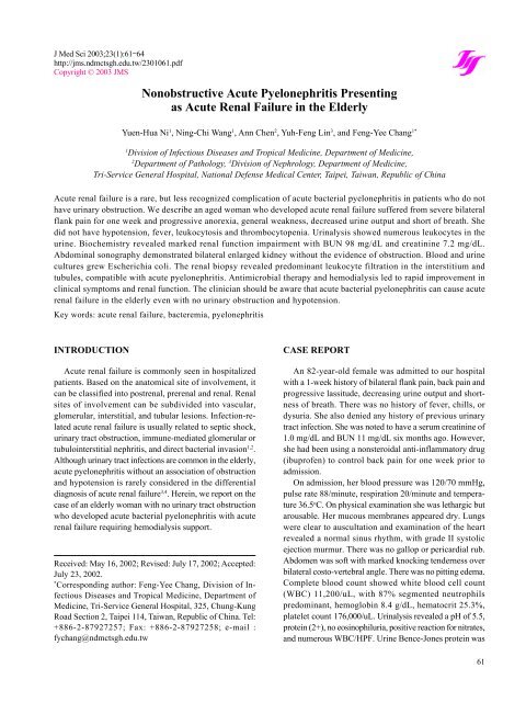
Fig. 1 <strong>Renal</strong> biopsy showing a diffuse prominent polymorphonuclear<br />
leukocyte infiltration in the interstitium<br />
and renal tubules in high power field. WBC c<strong>as</strong>ts were<br />
present in the lumen of renal tubules.<br />
flank pain, enlarged kidneys, along with a positive urine<br />
and blood culture. These distinct features have been noted<br />
frequently in earlier c<strong>as</strong>e reports 5,6 . However, she lacked<br />
hypotension, any fever, leukocytosis, and thrombocypenia<br />
despite the presence of severe bacteremia. In addition, no<br />
predisposing factors such <strong>as</strong> a structural abnormality of the<br />
urinary tract, indwelling catheter, solitary kidney or renal<br />
transplant were identified in this patient 7-9 . Finally, compared<br />
with the more common pattern of ischemic or toxininduced<br />
acute renal failure, the recovery of acute renal<br />
failure due to acute pyelonephritis appears to be almost<br />
complete and much quicker 10 . Consistent with previous<br />
reports, most of all c<strong>as</strong>es occurred in individuals with no<br />
history of urinary tract infections were caused by Escherichia<br />
coli. Nevertheless, acute oligouric renal failure <strong>as</strong>sociated<br />
with bacterial pyelonephritis is still an ignored and<br />
unrecognized clinical entity.<br />
The pathogenesis of acute renal failure <strong>as</strong>sociated with<br />
acute pyelonephritis in this patient remains uncertain. True<br />
extracellular fluid volume depletion w<strong>as</strong> not a major contributing<br />
factor because volume repletion did not reverse<br />
her oligouria and renal function. <strong>Acute</strong> tubular necrosis<br />
may occur in systemic bacteremia without the accompaning<br />
hypotension through the actions of v<strong>as</strong>oactive hormones,<br />
endotoxins, and cytokines that cause intrarenal<br />
v<strong>as</strong>oconstriction 11,12 . Although intrarenal v<strong>as</strong>oconstriction<br />
due to bacteremia could not be completely ruled out, the<br />
predominant interstitial edema, microabscess formation,<br />
leukocyte interstitial infiltration and tubular obstruction by<br />
cellular debris demonstrated by the renal biopsy favored<br />
that bacterial pyelonephritis directly caused renal function<br />
in both kidneys. Furthermore, acute bacterial pyelonephritis<br />
per se may result in the reduction of the glomerular<br />
filtration rate secondary to high hydrostatic pressures within<br />
the renal interstitium 13 .
This patient had no prior history of renal dise<strong>as</strong>e and<br />
w<strong>as</strong> taking an NSAID at the time of presentation. NSAID<br />
use is a well-known cause of both acute and chronic renal<br />
failure 14 . The chronic c<strong>as</strong>e usually involves the development<br />
of papillary necrosis resulting from the habitual<br />
consumption of NSAIDs 15 . The acute scenario is either<br />
NSAID-induced hemodynamic deterioration of renal function<br />
or NSAID- <strong>as</strong>sociated tubulointerstitial nephritis 16 .<br />
The latter is commonly accompanied by nephrotic syndrome<br />
where<strong>as</strong> the former often needs risk factors plus a<br />
higher dose of NSAIDs and usually develops with typical<br />
acute tubular necrosis. Although the possibility of NSAIDinduced<br />
acute renal failure can not be entirely excluded, the<br />
typical findings of acute pyelonephritis from renal biopsy<br />
mitigate against both NSAID-induced hemodynamic and<br />
immune-mediated acute renal failure.<br />
<strong>Acute</strong> renal failure <strong>as</strong>sociated with enlarged kidney in<br />
the absence of obstruction is usually <strong>as</strong>sociated with infection<br />
(leptospirotis, HIV infection, malacoplakia, et al.),<br />
tumor-infiltration (multiple myeloma, leukemia and<br />
lymphoma), diabetic mellitus, nephrotic syndrome (focal<br />
segment glomerulosclerosis), renal vein thrombosis, or<br />
drugs (heroins). In this c<strong>as</strong>e, enlargement of the kidneys is<br />
typically <strong>as</strong>sociated with acute bacterial pyelonephritis.<br />
Enlarged kidneys <strong>as</strong>sociated with anemia, back pain and<br />
acute renal failure seen in this patient are also the common<br />
clinical presentations in multiple myeloma. However, the<br />
immune study and no evidence of M protein in the pl<strong>as</strong>ma<br />
did not favor the possibility of multiple myeloma.<br />
In conclusion, the present c<strong>as</strong>e highlights the fact that<br />
acute bacterial pyelonephritis alone can result in an uncommon<br />
but serious complication of acute renal failure,<br />
which requires hemodialytic intervention in the elderly<br />
with no urinary tract obstruction. It should be stressed that<br />
acute bacterial pyelonephritis even without hypotension<br />
should be kept in the differential diagnosis of acute renal<br />
failure. Rapid diagnosis and treatment can lead to a more<br />
complete recovery of renal function.<br />
REFERENCES<br />
1. Baker LR, Cattell WR, Fry IK, Mallinson WJ. <strong>Acute</strong><br />
renal failure due to bacterial pyelonephritis. QJM 1979;<br />
192:603J612.<br />
2. Greenhill AH, Norman ME, Cornfield D, Chatten J,<br />
Buck B, Witzleben CC. <strong>Acute</strong> renal failure secondary<br />
to pyelonephritis. Clin Nephrol 1977;8:400J403.<br />
Yuen-Hua Ni, et al.<br />
3. Olsson PJ, Black JR, Gaffney E, Alexander RW, Mars<br />
DR, Fuller TJ. Reversible acute renal failure secondary<br />
to acute pyelonephritis. South Med J 1980;73:374J<br />
376.<br />
4. Aller SN. <strong>Nonobstructive</strong> pyelonephritis initially seen<br />
<strong>as</strong> acute renal failure. Arch Int Med 1978;138:816J<br />
817.<br />
5. Thompson C, Verani R, Evanoff G, Weinman E.<br />
Supporative bacterial pyelonephritis <strong>as</strong> a cause of acute<br />
renal failure. Am J Kidney Dis 1986;8:271J273.<br />
6. Jones SR. <strong>Acute</strong> renal failure in adults with uncomplicated<br />
acute pyelonephritis: c<strong>as</strong>e reports and review.<br />
Clin Infect Dis 1992;14:243J246.<br />
7. Yang CW, Kim YS, Yang KH, Chang YS, Yoon YS,<br />
Bang BK. <strong>Acute</strong> focal bacterial nephritis presented <strong>as</strong><br />
acute renal failure and hepatic dysfunction in a renal<br />
transplant recipient. Am J Nephrol 1994;14:72J75.<br />
8. Kooman JP, Barendregt JN, van der Sande FM, van<br />
suylen R. <strong>Acute</strong> pyelonephritis: a cause of acute renal<br />
failure? Neth J Med 2000;57:185J189.<br />
9. Richet G, Mayaud C. The course of acute renal failure<br />
in pyelonephritis. Nephron 1978;22:124J127.<br />
10. Lorentz WB, Iskandar S, Browning MC, Reynolds GD.<br />
<strong>Acute</strong> renal failure due to pyelonephritis. Nephron<br />
1990;54:256J258.<br />
11. Bones BF, Nanar RS, Shite KH. <strong>Acute</strong> renal failure<br />
secondary to acute pyelonephritis. Am J Nephrol 1991;<br />
11:257J259.<br />
12. Nunez JE, Perez E, Gun<strong>as</strong>ekaran S, Narvarte J,<br />
Ramirez G. <strong>Acute</strong> renal failure secondary to acute bacterial<br />
pyelonephritis. Nephron 1992;62:240J241.<br />
13. Atkinson LK, Goodship TH, Ward MK. <strong>Acute</strong> renal<br />
failure <strong>as</strong>sociated with acute pyelonephritis and consumption<br />
of non-steroidal anti-inflammatory drugs. Br<br />
Med J 1986;292:97J98.<br />
14. Sturmer T, Elseviers MM, de Broe ME. Nonsteroidal<br />
anti-inflammatory drugs and the kidney. Curr Opin<br />
Nephrol Hypert 2001;10:161J163.<br />
15. Shankel SW, Johnson DC, Clark PS, Shankel TL,<br />
O’Neil WM Jr. <strong>Acute</strong> renal failure and glomerulopathy<br />
caused by nonsteroid anti-inflammatory drugs. Arch<br />
Intern Med 1992;152:986J990.<br />
16. Wen SF. Nephrotoxicity of nonsteroid anti-inflammatory<br />
drugs. J Formosan Med Ass 1997;96:157J171.<br />
63
<strong>Acute</strong> pyelonephritis and acute renal failure<br />
64


