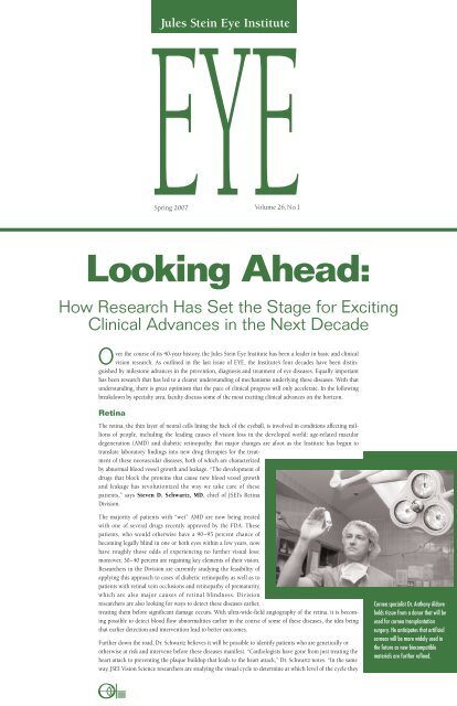
Looking Ahead: - Jules Stein Eye Institute
Looking Ahead: - Jules Stein Eye Institute
Looking Ahead: - Jules Stein Eye Institute

Create successful ePaper yourself
Turn your PDF publications into a flip-book with our unique Google optimized e-Paper software.
EYE<br />
Spring 2007 Volume 26,No.1<br />
Over the course of its 40-year history, the <strong>Jules</strong> <strong>Stein</strong> <strong>Eye</strong> <strong>Institute</strong> has been a leader in basic and clinical<br />
vision research. As outlined in the last issue of EYE, the <strong>Institute</strong>’s four decades have been distinguished<br />
by milestone advances in the prevention, diagnosis and treatment of eye diseases. Equally important<br />
has been research that has led to a clearer understanding of mechanisms underlying these diseases. With that<br />
understanding, there is great optimism that the pace of clinical progress will only accelerate. In the following<br />
breakdown by specialty area, faculty discuss some of the most exciting clinical advances on the horizon.<br />
Retina<br />
<strong>Jules</strong> <strong>Stein</strong> <strong>Eye</strong> <strong>Institute</strong><br />
<strong>Looking</strong> <strong>Ahead</strong>:<br />
How Research Has Set the Stage for Exciting<br />
Clinical Advances in the Next Decade<br />
The retina, the thin layer of neural cells lining the back of the eyeball, is involved in conditions affecting millions<br />
of people, including the leading causes of vision loss in the developed world: age-related macular<br />
degeneration (AMD) and diabetic retinopathy. But major changes are afoot as the <strong>Institute</strong> has begun to<br />
translate laboratory findings into new drug therapies for the treatment<br />
of these neovascular diseases, both of which are characterized<br />
by abnormal blood vessel growth and leakage. “The development of<br />
drugs that block the proteins that cause new blood vessel growth<br />
and leakage has revolutionized the way we take care of these<br />
patients,” says Steven D. Schwartz, MD, chief of JSEI’s Retina<br />
Division.<br />
The majority of patients with “wet” AMD are now being treated<br />
with one of several drugs recently approved by the FDA. These<br />
patients, who would otherwise have a 90–95 percent chance of<br />
becoming legally blind in one or both eyes within a few years, now<br />
have roughly those odds of experiencing no further visual loss;<br />
moreover, 30–40 percent are regaining key elements of their vision.<br />
Researchers in the Division are currently studying the feasibility of<br />
applying this approach to cases of diabetic retinopathy as well as to<br />
patients with retinal vein occlusions and retinopathy of prematurity,<br />
which are also major causes of retinal blindness. Division<br />
researchers are also looking for ways to detect these diseases earlier,<br />
treating them before significant damage occurs. With ultra-wide-field angiography of the retina, it is becoming<br />
possible to detect blood flow abnormalities earlier in the course of some of these diseases, the idea being<br />
that earlier detection and intervention lead to better outcomes.<br />
Further down the road, Dr. Schwartz believes it will be possible to identify patients who are genetically or<br />
otherwise at risk and intervene before these diseases manifest. “Cardiologists have gone from just treating the<br />
heart attack to preventing the plaque buildup that leads to the heart attack,” Dr. Schwartz notes. “In the same<br />
way, JSEI Vision Science researchers are studying the visual cycle to determine at which level of the cycle they<br />
Cornea specialist Dr. Anthony Aldave<br />
holds tissue from a donor that will be<br />
used for cornea transplantation<br />
surgery. He anticipates that artificial<br />
corneas will be more widely used in<br />
the future as new biocompatible<br />
materials are further refined.
“<strong>Looking</strong> <strong>Ahead</strong>...” continued<br />
EYE<br />
NEWSLETTER<br />
IS APUBLICATION<br />
OF THE<br />
JULES STEIN EYE INSTITUTE<br />
D IRECTOR<br />
Bartly J. Mondino, MD<br />
E DITORS<br />
Debora B. Farber, PhD, DPhhc<br />
Gary N. Holland, MD<br />
M ANAGING E DITOR<br />
Gloria P. Jurisic<br />
C ONTRIBUTING E DITORS<br />
Teresa Closson<br />
Nancy Graydon<br />
Debbie Sato<br />
Lori Twitchell<br />
Melania Vartanian<br />
G UEST E DITOR<br />
Dan Gordon<br />
P HOTOGRAPHY<br />
J. Charles Martin<br />
D ESIGN<br />
Robin Weisz /Graphic Design<br />
P RODUCTION C OORDINATION<br />
Coniglio & Associates<br />
©2007, by The Regents of the<br />
University of California.<br />
All rights reserved.<br />
<strong>Jules</strong> <strong>Stein</strong> <strong>Eye</strong> <strong>Institute</strong><br />
100 <strong>Stein</strong> Plaza, UCLA<br />
Box 957000<br />
Los Angeles, CA 90095–7000<br />
(310) 206-6035<br />
www.jsei.org<br />
can intervene. In addition, the Retina Division is beginning to<br />
investigate targeted drug therapies that will block the buildup of<br />
materials at the back of the eye that lead to age-related macular<br />
degeneration.” The Retina Division has recruited Michael Gorin,<br />
MD, PhD, a clinician scientist credited with identifying genetic<br />
regions that contribute to AMD. “Developing a molecular means<br />
of identifying and treating people at risk for vision loss caused by<br />
AMD, a long-term goal of the JSEI Retina Division, is now much<br />
closer to fruition,” says Dr. Schwartz.<br />
Cornea and Uveitis<br />
JSEI researchers are also making great progress in treating problems<br />
occurring in the cornea, the eye’s outer covering. Already, this is<br />
starting to enable greater precision in surgical and medical treatment<br />
through the use of new materials, new instruments, and<br />
new drugs.<br />
Improved materials are beginning to make a difference in a<br />
number of areas. For patients who need new corneas but are at<br />
high risk for rejection of a donor organ, artificial corneas have<br />
become a more viable option, according to Gary N. Holland, MD,<br />
chief of JSEI’s Cornea and Uveitis Division. “We anticipate that<br />
with further refinements in materials, these procedures will be<br />
even more successful and will be more widely adopted,” agrees<br />
Anthony J. Aldave, MD, director of the Division’s cornea service.<br />
New materials that are biocompatible will also allow future clinicians<br />
to close wounds after corneal transplants with glue-like<br />
substances rather than sutures. “It is going to be a better-sealed<br />
wound, closed faster, without the problem of sutures coming<br />
loose and being exposed, and without the need to have the<br />
sutures removed,” Dr. Holland explains.<br />
Dr. Holland also foresees increased use of lasers for creating<br />
incisions during surgery—an approach that has also begun to<br />
be used at the <strong>Institute</strong>. “This is leading to an improvement in<br />
precision over making corneal incisions with a blade,” he says.<br />
“It will also help us to customize the shape of the wound for an<br />
individual patient, which will allow better visual results and<br />
fewer complications following corneal transplantation.<br />
With continuing progress in understanding disease mechanisms<br />
and the genetic basis for ocular problems, the future also looks<br />
bright for non-surgical treatments that target the specific problem<br />
causing a disease. For example, if a specific enzyme is known to<br />
be deficient or abnormal in a patient with certain kinds of corneal<br />
opacities, it can be addressed through targeted gene or drug<br />
therapies. With regard to uveitis (inflammation inside the eye),<br />
most current therapies suppress inflammation non-specifically<br />
with corticosteroids; in the future, a more direct approach will<br />
take aim at the specific part of the immune system causing the<br />
problem. This approach is already beginning to occur at JSEI,<br />
with new drugs for the treatment of disorders such as uveitis in<br />
children with juvenile rheumatoid arthritis, Dr. Holland notes.<br />
Orbital and Ophthalmic<br />
Plastic Surgery<br />
Many problems of the eye are both functional and aesthetic.<br />
Research in JSEI’s Orbital and Ophthalmic Plastic Surgery Division<br />
has tackled both concerns. Particularly noteworthy are the strides<br />
being made in treating—and, potentially, preventing—Graves’<br />
disease, a disorder of the immune system that causes the majority<br />
of its victims to experience problems with sight and/or appearance<br />
(typically, bulging eyes).<br />
In the last 10 years, treatment of Graves’ disease symptoms has<br />
evolved. The <strong>Institute</strong> has pioneered an approach known as deep<br />
lateral wall orbital decompression surgery, leading to significantly<br />
fewer complications, less double vision and more rapid recovery.<br />
Increasingly, this surgery is now performed on an outpatient basis.<br />
Researchers in the Division are continuing to refine surgical techniques,<br />
using endoscopic, laser, and robotic technologies. Robert<br />
A. Goldberg, MD, chief of the Division, notes that future<br />
advances in molecular biology are likely to bring major improvements<br />
in the area of wound healing after surgery. The other assist<br />
will come from advances in imaging and surgical technology. “We<br />
A patient with Graves’ disease benefits from deep lateral wall<br />
orbital decompression surgery, which was pioneered at the<br />
<strong>Jules</strong> <strong>Stein</strong> <strong>Eye</strong> <strong>Institute</strong>. Current research at the <strong>Institute</strong> could<br />
soon enable Graves’ disease to be treated non-surgically before<br />
it begins to cause problems.<br />
are going to have better ways to diagnose problems and more<br />
sophisticated surgical instrumentation,” says Dr. Goldberg, who<br />
sees a movement toward more minimally invasive surgeries not<br />
only for Graves’ disease patients, but also for patients with orbital<br />
tumors or diseases of the tear system, and those who seek aesthetic<br />
surgery.<br />
Even more exciting is research that could soon enable Graves’<br />
disease to be treated before it begins to cause problems. A JSEI<br />
research group headed by Terry J. Smith, MD, has discovered an<br />
antibody associated with the disease and, in the laboratory, has<br />
successfully tested a therapy to block the process triggered by the<br />
antibody. Dr. Smith is now teaming with Raymond Douglas, MD,<br />
PhD, in an effort to translate these laboratory findings into a new<br />
non-surgical treatment that could stave off the disease before it<br />
takes hold. “I am optimistic that within a couple of years, we will<br />
be ready to test patients in clinical trials, and that soon, Graves’<br />
will be seen as a treatable disease,” says Dr. Goldberg.<br />
Glaucoma<br />
Approximately 2.2 million people in the United States have<br />
glaucoma, the nation’s second-leading cause of adult blindness,<br />
and half are unaware that they have the condition because early<br />
symptoms are rare. Current research is paving the way toward<br />
earlier detection of glaucoma, which would enable patients to be<br />
treated before the disease robs them of much of their vision. With<br />
more sophisticated image-analysis techniques and laser scanning<br />
devices, future specialists will be better able to recognize early<br />
damage by evaluating the appearance of the optic nerve.<br />
In addition, efforts are aimed at finding better ways of predicting<br />
how the disease will progress. “With glaucoma, it is not only a<br />
matter of recognizing it early,” says Joseph Caprioli, MD, chief<br />
of JSEI’s Glaucoma Division. “It is also important to know the rate<br />
of the individual’s disease.” The rate at which glaucoma progresses<br />
varies widely, he notes. While younger patients in whom the disease<br />
is rapidly advancing need aggressive treatment, older patients<br />
with slowly progressing glaucoma could die with their vision<br />
intact, and can be spared the side effects and potential complications<br />
of aggressive treatment. The <strong>Institute</strong> is developing sensitive<br />
Improved laser scanning of a patient’s eye shows no evidence of glaucoma<br />
(left) and borderline glaucoma (right) years later. The <strong>Institute</strong> is<br />
developing sensitive measures of the disease’s progression to enable future<br />
specialists to treat glaucoma patients before vision has been compromised.
measures to detect small amounts of progression so that glaucoma<br />
patients can be monitored to determine when treatment needs to<br />
be intensified.<br />
Ultimately, Dr. Caprioli believes the <strong>Institute</strong> will move toward<br />
glaucoma treatments that make the optic nerve more resistant to<br />
damage from the disease, a strategy known as neuroprotection.<br />
“This is the most exciting area for us and it will represent a new<br />
dimension of treatment,” Dr. Caprioli explains. “Up to now, the<br />
only treatment we have is to lower the eye pressure, but in as<br />
many as half of the patients, the disease continues to progress<br />
despite our best efforts.” In the future, Dr. Caprioli sees a twopronged<br />
treatment that would combine pressure reduction with<br />
medications protecting the optic nerve.<br />
Pediatric Ophthalmology<br />
and Strabismus<br />
The future may also include gene therapy approaches to conditions<br />
that are all too common in children, including a number of<br />
congenital eye diseases affecting the retina; optic nerve abnormalities;<br />
and cases in which the eye muscles are not formed properly<br />
or not properly innervated, including strabismus, according to<br />
Arthur L. Rosenbaum, MD, chief of the Pediatric Ophthalmology<br />
and Strabismus Division. In the years to come, innovative methods<br />
may also further reduce blindness in children.<br />
Division members Sherwin J. Isenberg, MD, and Leonard Apt, MD,<br />
(emeritus) are initiating studies of an inexpensive medication to<br />
treat corneal infections caused by fungi and trachoma, an infection<br />
of the conjunctiva that blinds millions in developing countries.<br />
Dr. Isenberg and associates are also developing and testing<br />
an instrument that measures blood gases (oxygen and carbon<br />
dioxide) as well as bicarbonate and pH from the surface of the eye,<br />
decreasing the need to draw blood. The device has the potential to<br />
reduce childhood blindness by decreasing the incidence of<br />
Retinopathy of Prematurity and postnatal cerebral palsy.<br />
JSEI pediatric ophthalmology specialists Drs. Leonard Apt (left) and Sherwin<br />
Isenberg (right) completed a study of treating bacterial corneal infections with an<br />
effective and inexpensive iodine based medication in India and the Philippines.<br />
They will next study the treatment of fungus caused corneal infections, which blind<br />
millions in developing countries.<br />
Dr. Rosenbaum and <strong>Institute</strong> colleague Federico Velez, MD, are<br />
conducting laboratory research to determine how to re-innervate<br />
paralyzed eye muscles by utilizing computer chips and innervational<br />
signals from adjacent muscles. “These studies could have<br />
important implications for treating many adult and childhood<br />
strabismic disorders in which the nerve to the eye muscle is<br />
injured or a brain tumor or other neurologic cause prevents the<br />
nerve from functioning correctly,” Dr. Rosenbaum explains.<br />
Researchers are also seeking more accurate ways to assess visual<br />
function in infants so that ophthalmologists can better determine<br />
whether certain eye disorders, such as congenital cataracts and<br />
drooping eyelid, are interfering with infants’ vision and know<br />
whether to be more aggressive in treatment.<br />
An investigative team led by Joseph L. Demer, MD, PhD, is<br />
continuing to develop special magnetic resonance imaging (MRI)<br />
techniques that provide the most detailed view of the eye muscles.<br />
This will further improve surgical outcomes by enhancing<br />
the precision of diagnosis for complicated strabismus cases. Dr.<br />
Demer is also refining the hardware and software used for imaging<br />
the eye and eye socket. “We expect to have microscopic resolution<br />
by MRI of the ocular structures within five to 10 years,” he<br />
says. “That would have enormous clinical implications by noninvasively<br />
providing pictures of small tumors, inflammations, and<br />
abnormalities of muscles behind the eye that could not be detected<br />
in other ways, except by doing surgery and biopsy.”<br />
Vision Science<br />
Fundamental to these and other future advances are studies in<br />
<strong>Institute</strong> laboratories that enhance the understanding of ophthalmic<br />
disorders. Treatment for age-related macular degeneration<br />
has moved from highly<br />
invasive measures to the<br />
drug injections described in<br />
the Retina section on the<br />
first page, which have dramatically<br />
improved care for<br />
the 10 percent of patients<br />
with the more dangerous<br />
“wet” form of the disease.<br />
Now, researchers in JSEI’s<br />
Division of Vision Science<br />
are poised to parlay their<br />
AMD understanding of the Autoflourescent images show a normal retina (left) and the retina of a patient who has<br />
genetics of into treatments, recessive Stargardt macular dystrophy (right). The large bright spots represent the presence<br />
if not cures, for the full of a toxic molecule called A2E that accumulates as a result of a gene mutation. Scientists at<br />
spectrum of the disease, the <strong>Institute</strong> are working on methods to reduce or prevent the accumulation of A2E.<br />
including the “dry” form.<br />
“There has been a great leap<br />
forward in the last year on<br />
the genetics of the disease, which makes it possible to start studying<br />
the cell biology to determine how to intervene in a less invasive<br />
way,” says Division member Dean Bok, PhD.<br />
Research in the laboratory of Debora B. Farber, PhD, DPhhc,<br />
has described several genes that, when mutated, cause different<br />
forms of retinitis pigmentosa and other retinal degenerations in<br />
people. In her laboratory as well as several others at the <strong>Institute</strong>,<br />
including those of Gabriel H. Travis, MD, Xianjie Yang, PhD,<br />
and Dr. Bok, fundamental work also has been done on the genes<br />
causing Usher syndrome and Stargardt macular dystophy. This<br />
understanding has paved the way for breakthrough treatments.<br />
Scientists at JSEI have begun studying gene therapy in the laboratory<br />
for inherited eye diseases in animals, and at the national<br />
level, two clinical trials are already scheduled for a potentially<br />
curative approach to Leber congenital amaurosis, another retinal<br />
degenerative disease. The research of Joseph Horwitz, PhD, is<br />
also having a fundamental impact on vision science. Dr. Horwitz<br />
has discovered that one of the major proteins in the eye lens,<br />
alpha-crystallin, acts as a molecular chaperone protecting other<br />
proteins from aggregating— a finding that could lead to new<br />
approaches to cataracts.<br />
The Stage is Set<br />
It is an exciting time at the <strong>Jules</strong> <strong>Stein</strong> <strong>Eye</strong> <strong>Institute</strong>. Many of the<br />
fundamental advances that have taken place in the <strong>Institute</strong>’s laboratories<br />
in the last 40 years are now being translated into better<br />
approaches to diagnosing, treating and preventing eye diseases.<br />
There is a sense that the clinical progress thus far represents only<br />
the tip of the iceberg, and that the next decade will see even<br />
more dramatic improvements that will lead to better vision and<br />
enhanced quality of life for people all over the world.
JSEI MEMBERS A SSUME<br />
AUPO LEADERSHIP P OSITIONS<br />
INSTITUTE NEWS<br />
The <strong>Jules</strong> <strong>Stein</strong> <strong>Eye</strong> <strong>Institute</strong> at UCLA is proud to announce the appointment of three of its own<br />
members to key posts within the Association of University Professors of Ophthalmology (AUPO).<br />
AUPO represents departments of ophthalmology nationwide. The organization provides support and<br />
information to departmental chairs and other faculty members, promotes excellence in ophthalmic<br />
education and vision research, and promotes excellence in eye care in order to ensure the best possible<br />
vision for the public.<br />
Chairman of the UCLA Department of Ophthalmology and Director of the <strong>Jules</strong> <strong>Stein</strong> <strong>Eye</strong> <strong>Institute</strong>,<br />
Bartly J. Mondino, MD, is the current AUPO Executive Vice President. Professor of Clinical<br />
Ophthalmology, Anthony C. Arnold, MD, serves as the President of the AUPO Program Directors<br />
Council, which advances ophthalmology residency education on a national level. Finally, JSEI Chief<br />
Operating Officer Jonathan Smith serves as the President of the AUPO University Administrators of<br />
Ophthalmology, which promotes effective and professional administrative support of medical education,<br />
research and patient care, particularly as it concerns departments of ophthalmology.<br />
(Left to right) Mr. Jonathan Smith, Dr. Bartly Mondino and Dr. Anthony Arnold were appointed to key posts within AUPO.<br />
Dolly Green Professor of<br />
Ophthalmology and Professor<br />
of Neurobiology at UCLA<br />
Dean Bok, PhD, presented the<br />
keynote address, “The Retinal<br />
Pigment Epithelium: Its Role<br />
in Inherited Retinal Diseases,”<br />
at the 1st International Congress<br />
of the International<br />
Society for Ocular Cell Biology<br />
at Homerton College, in<br />
Cambridge, England, on<br />
September 6, 2006. Dr. Bok<br />
also received the Paul Kayser<br />
International Award in Retina<br />
Research at the XVII International<br />
Congress for <strong>Eye</strong><br />
Research held in Buenos Aires,<br />
Argentina, on October 29–<br />
November 3, 2006. The award<br />
included a prize of $50,000<br />
and a Plenary Lecture entitled,<br />
“The Retinoid (visual) Cycle<br />
in Health and Disease.”<br />
Joseph Caprioli, MD, David<br />
May II Professor of<br />
Ophthalmology, received an<br />
Editors’ Choice Award from<br />
the Editors-in-Chief of the<br />
three major clinical ophthalmology<br />
journals: American<br />
Faculty Honors and Awards<br />
Journal of Ophthalmology, the<br />
Archives of Ophthalmology<br />
and Ophthalmology, for his<br />
paper “Trabeculectomy with<br />
Mitomycin C in Pseudophakic<br />
Patients with Open-Angle<br />
Glaucoma: Outcomes and Risk<br />
Factors for Failure.” The paper<br />
was presented at the American<br />
Academy of Ophthalmology<br />
(AAO) in Las Vegas, Nevada,<br />
on November 11–14, 2006.<br />
Anne L. Coleman, MD, PhD,<br />
Frances and Ray Stark Professor<br />
of Ophthalmology at the<br />
<strong>Jules</strong> <strong>Stein</strong> <strong>Eye</strong> <strong>Institute</strong>, presented<br />
the 14th Arthur Light,<br />
MD Memorial Lecture in<br />
Ophthalmology at Loyola<br />
University Medical Center,<br />
Stritch School of Medicine, in<br />
Chicago, Illinois, on<br />
September 6, 2006. The<br />
subject of the lecture was,<br />
“Predicting Glaucomatous<br />
Progression with Imaging.”<br />
Laraine and David Gerber<br />
Professor of Pediatric Ophthalmology<br />
Sherwin J. Isenberg,<br />
MD, presented the Second<br />
Eugene R. Folk Memorial<br />
Lecture at the Pediatric<br />
Ophthalmology Symposium<br />
at the University of Illinois,<br />
in Chicago, Illinois, on<br />
September 27, 2006. The title<br />
of the lecture was,”Long Term<br />
Results of Strabismus Surgery.”<br />
Dr. Isenberg, a former Heed<br />
Fellow, also received the<br />
prestigious Heed Award in<br />
recognition of his contributions<br />
to ophthalmology. The<br />
Award was presented at the<br />
annual AAO meeting<br />
in Las Vegas, Nevada, on<br />
November 11–14, 2006.<br />
Kevin M. Miller, MD,<br />
Kolokotrones Professor of<br />
Ophthalmology at the <strong>Jules</strong><br />
<strong>Stein</strong> <strong>Eye</strong> <strong>Institute</strong>, received<br />
the American Academy of<br />
Ophthalmolgy 2006 Senior<br />
Achievement Award at the<br />
AAO annual meeting in Las<br />
Vegas, Nevada, on November<br />
11–14, 2006. The Award was<br />
presented in recognition of<br />
his significant contributions<br />
to the Academy, its scientific<br />
and educational programs<br />
and to ophthalmology.<br />
JSEI WELCOMES N EW FACULTY<br />
M ICHAEL B. GORIN, MD, PHD<br />
The UCLA Department of Ophthalmology and <strong>Jules</strong> <strong>Stein</strong><br />
<strong>Eye</strong> <strong>Institute</strong> proudly announce the appointment of<br />
Michael B. Gorin, MD, PhD, as Professor of Ophthalmology<br />
in the Retina and Vision Science<br />
Divisions, and Harold and<br />
Pauline Price Chair in Ophthalmology,<br />
effective September 5,<br />
2006. Dr. Gorin comes to the<br />
<strong>Jules</strong> <strong>Stein</strong> <strong>Eye</strong> <strong>Institute</strong> with<br />
outstanding qualifications and<br />
accomplishments. He obtained<br />
his medical and doctor of<br />
philosophy degrees from the<br />
University of Pennsylvania at<br />
Philadelphia and completed his<br />
internship at the Center for<br />
Health Sciences at the<br />
University of California at Los Dr. Michael Gorin<br />
Angeles (UCLA). He stayed on<br />
at UCLA for postdoctoral fellowship and ophthalmology residency<br />
training at the <strong>Jules</strong> <strong>Stein</strong> <strong>Eye</strong> <strong>Institute</strong>, and then finished<br />
a fellowship in Medical Retina and Genetics at Moorfields <strong>Eye</strong><br />
Hospital in London, England.<br />
In 1990, Dr. Gorin began his academic career as Assistant Professor<br />
of Ophthalmology and Human Genetics at the University of<br />
Pittsburgh School of Medicine. He has the unique distinction of<br />
having been an interim chair for both a basic science department<br />
(Department of Human Genetics at the Graduate School of Public<br />
Health) and a clinical department (Department of Ophthalmology<br />
in the School of Medicine) at the University of Pittsburgh. More<br />
recently, he served as the Assistant Vice Chancellor for Strategic<br />
Initiatives for the University of Pittsburgh Schools of the Health<br />
Sciences and Professor of Ophthalmology, Human Genetics and<br />
Bioengineering, from which positions he was recruited to the<br />
<strong>Jules</strong> <strong>Stein</strong> <strong>Eye</strong> <strong>Institute</strong>.<br />
Dr. Gorin’s primary research focus is in the field of molecular<br />
genetics of hereditable eye disorders, specifically in the complex<br />
genetics of age-related maculopathy. His research group was the<br />
first to identify genetic regions that contribute to macular degeneration,<br />
which then led to the identification of several macular<br />
degeneration genes by multiple investigators. He also investigates<br />
monogenic disorders such as hereditary retinal degenerations,<br />
glaucoma, cataracts and ocular syndromes. Other basic research<br />
areas include genetic modifiers of disease, neurobiology of pain,<br />
and ocular toxicities of systemic medications. Applied research<br />
areas include bioinformatics in clinical ophthalmic practice, new<br />
diagnostic technologies for retinal disorders, and the use of insurance<br />
and health care data to assess clinical practice patterns and<br />
public health issues pertaining to ocular disease.<br />
He is the recipient of many honors including election to the<br />
Omega Delta Honor Society for Public Health, the Lew R.<br />
Wasserman Merit Award and the Senior Scientist Investigator<br />
Award from Research to Prevent Blindness. He has published<br />
over 100 scientific papers in refereed journals, and is author of<br />
five book chapters.<br />
As a full-time faculty member, Dr. Gorin divides his time between<br />
patient care for diseases of the retina, research into the genetics of<br />
inherited eye disorders and training young ophthalmologists.<br />
It is a great pleasure to welcome Dr. Gorin back to UCLA and to<br />
the full-time faculty at the <strong>Jules</strong> <strong>Stein</strong> <strong>Eye</strong> <strong>Institute</strong>.
JSEI VISITING F ELLOW R ECEIVES R OYAL S EAL OF A PPROVAL<br />
Former visiting Winston Churchill Trust Fellow Dr. Tuyen Ong received a silver<br />
medallion at Buckingham Palace from Her Majesty Queen Elizabeth II on June<br />
6, 2006. In 2004, Dr Ong used his Traveling Fellowship to visit the <strong>Jules</strong> <strong>Stein</strong><br />
<strong>Eye</strong> <strong>Institute</strong> under the supervision of Professor of Ophthalmology Dr. Marc<br />
Yoshizumi, observing ophthalmic surgical procedures performed by glaucoma<br />
specialist Dr. Joseph Caprioli and retina specialist Dr. Steven Schwartz. He<br />
received the silver medallion in recognition of his achievements.<br />
EYE LINES<br />
UCLA Department of Ophthalmology Association<br />
The UCLA Department of Ophthalmology Association hosted its<br />
annual reception at the American Academy of Ophthalmology meeting<br />
in Las Vegas, Nevada, on Sunday, November 12, 2006, at the<br />
Westin Cuasarina Hotel. Over 150 JSEI faculty members, staff, and<br />
resident and fellow alumni from around the world gathered to renew<br />
acquaintances and reconnect with old friends.<br />
UCLA Department of Ophthalmology Association Treasurer<br />
Dr. Robert Goldberg (left) greets fellow JSEI faculty member<br />
Dr. Marc Yoshizumi.<br />
(Left to right) JSEI Director Dr. Bartly Mondino welcomes JSEI<br />
alumnus Dr. David Aizuss and current resident Dr. F. Jacob<br />
Khoubian.<br />
JSEI FOUNDING M EMBER R ETIRES<br />
Michael O. Hall, PhD, Professor of Ophthalmology in<br />
the Vision Sciences Division and Founding Member<br />
of the <strong>Jules</strong> <strong>Stein</strong> <strong>Eye</strong> <strong>Institute</strong> has retired after more than<br />
40 years of service.<br />
Dr. Hall began his career at the University of California at<br />
Los Angeles (UCLA) after earning his degree in<br />
Biochemistry at the University of Natal in South Africa. He<br />
earned his doctor of philosophy degree in Physiological<br />
Chemistry at UCLA in 1961. He continued at the university<br />
first as an Assistant Research Biological Chemist, subsequently<br />
as an Assistant Professor of Ophthalmology and<br />
Biochemistry, and later as Professor of Ophthalmology and<br />
Biochemistry.<br />
Dr. Michael Hall<br />
Dr. Hall was a founding member of the <strong>Jules</strong> <strong>Stein</strong> <strong>Eye</strong><br />
<strong>Institute</strong> (1965), and served as Associate Director of the <strong>Institute</strong> for several years (1971–1972;<br />
1974–1975; 1978–1985). He also chaired or co-chaired numerous research, fellowship training<br />
and educational program committees during his many years at the <strong>Institute</strong>.<br />
Dr. Hall’s research has focused on retinal biochemistry, retinal degeneration, cellular interaction<br />
and metabolism of retinal pigment epithelium, authoring many papers on these topics. His<br />
illustrious career has taken him to various regions of the world; he has been a visiting fellow,<br />
professor and lecturer in England, South Africa and Australia.<br />
<strong>Jules</strong> <strong>Stein</strong> <strong>Eye</strong> <strong>Institute</strong> and UCLA paid tribute to Dr. Hall at a reception in The Adam Room<br />
on October 5, 2006. The faculty and staff thank him for his years of service and lasting contributions<br />
to vision science. We wish him the best on his well-deserved retirement. (See retirement<br />
reception photos on last page.)<br />
(Left to right) Drs. Henry Baylis, Rona Silkiss with husband Neil Jacobstein (second from left), Robert Goldberg<br />
and Kenneth <strong>Stein</strong>sapir enjoy the evening’s festivities.<br />
(Left to right) Drs. Ronald Mancini, Tanju Nakra and Robert Goldberg test<br />
fate with Lady Luck!<br />
Thank you JSEI Alumni and UCLA<br />
Department of Ophthalmology<br />
Association Members!<br />
The UCLA Department of Ophthalmology<br />
Association extends a special thank you to its<br />
2006–07 dues paying members. Annual<br />
dues provide the Association with the<br />
resources to support the annual Research<br />
Grant Awards, JSEI Clinical and Research<br />
Seminar, Video Library Project, Alumni<br />
Directory, reception at the Annual AAO<br />
Meeting and other important alumni events.<br />
If you have not yet submitted your dues,<br />
registration forms can be requested by<br />
contacting us at alumni@jsei.ucla.edu.<br />
Thank you!
P HILANTHROPY<br />
Focus on<br />
If you would like to make a<br />
contribution to the <strong>Institute</strong>, you<br />
may do so by means of the<br />
remittance envelope included in<br />
this issue of EYE. For additional<br />
information, please call or write<br />
to the following:<br />
Development Office<br />
<strong>Jules</strong> <strong>Stein</strong> <strong>Eye</strong> <strong>Institute</strong><br />
100 <strong>Stein</strong> Plaza, UCLA<br />
Box 957000<br />
Los Angeles, California<br />
90095–7000<br />
(310) 206-6035<br />
giving@jsei.ucla.edu<br />
Paying tribute to<br />
Friends &Family<br />
Richard B. Shapiro Vision Fund<br />
For more than 15 years, Richard B. Shapiro had been<br />
struggling with uveitis, an intraocular inflammatory disease.<br />
He stated, “To have your sight threatened is very debilitating. I<br />
wanted to find the best place in the world to be treated, and I<br />
found that was at the <strong>Jules</strong> <strong>Stein</strong> <strong>Eye</strong> <strong>Institute</strong>.” Meeting with<br />
Gary N. Holland, MD, Chief of the Cornea and Uveitis Division,<br />
and Anne L. Coleman, MD, PhD, Professor of Ophthalmology and<br />
Epidemiology, Richard was inspired to support their research in this<br />
field of medicine. He was particularly impressed by Dr. Holland’s<br />
thorough explanation of and promising ideas regarding uveitis.<br />
In addition to his own gift, Richard then asked friends, family<br />
and colleagues to contribute to the establishment of the “Richard<br />
B. Shapiro Vision Fund.” He felt that “a grassroots approach<br />
would be the most effective plan” to raise the necessary funds to<br />
support groundbreaking investigations that would broaden the<br />
understanding of uveitis and its complications, such as glaucoma,<br />
and ultimately find new treatment options. “This was an easy sell.<br />
I just told people about what an incredible place <strong>Jules</strong> <strong>Stein</strong> is, and<br />
the important work being done to preserve people’s sight. The<br />
benefits of the scientists’ work will not likely change my situation,<br />
but it will help others in the future.” To date, more than $170,000<br />
has been raised from more than 90 donors, many of whom have<br />
given multiple gifts.<br />
Dr. Holland stated, “Through Mr. Shapiro’s commitment and<br />
dedicated fund-raising efforts, the Shapiro Fund has been established<br />
as a permanent endowment that will serve as a continuing<br />
source of support for our investigations dealing with uveitis,<br />
which can be a particularly devastating eye disease. Income from<br />
the Fund has already allowed us to conduct a study that has provided<br />
a better understanding of mechanisms that may contribute<br />
to the development of glaucoma in uveitis patients. We are grateful<br />
to both Richard, his wife Colleen and his friends and family<br />
who have made generous donations to this important initiative.”<br />
In addition to his involvement with the <strong>Jules</strong> <strong>Stein</strong> <strong>Eye</strong> <strong>Institute</strong>,<br />
Richard serves as the Vice Chairman of the Parkinson’s <strong>Institute</strong> in<br />
Sunnyvale, California. He also is the Chairman of the California<br />
Horse Racing Board to which Governor Schwarzenegger appointed<br />
him in 2004. For more than 20 years, he has been active in all<br />
aspects of commercial real estate including development, leasing,<br />
management and repositioning. Currently, he is the owner<br />
of Winco Real Estate Services, Inc., and Chairman of Bridge<br />
Capital Finance.<br />
David Schumacher<br />
The friends and family of David Schumacher, PhD, who<br />
passed away at 86 on July 26, 2006, honored his legacy<br />
with memorial contributions to benefit glaucoma research at the<br />
<strong>Jules</strong> <strong>Stein</strong> <strong>Eye</strong> <strong>Institute</strong>. To date, more than $10,000 has been contributed<br />
from 75 donors. Margaret, Dr. Schumacher’s wife of 29<br />
years, was not surprised at this generosity.<br />
“David was a kind and gentle man<br />
loved by so many. The tributes to my<br />
dear David have been monumental. He<br />
would never have believed it.”<br />
Born in Los Angeles in 1919, David<br />
Theodore Schumacher was a real estate<br />
giant, author, entrepreneur, scholar and<br />
philanthropist. At Pacific Western<br />
University, he majored in real estate and<br />
earned his PhD. He authored two books,<br />
David Schumacher, PhD<br />
Buy and Hold: Real Estate Strategy and<br />
more recently, Buy and Hold: 7 Steps to<br />
Real Estate Fortune, offering practical advice on how to invest and<br />
attain financial security. In 1960, Dr. Schumacher began investing<br />
Paul J. Vicari Endowed<br />
Cataract Research Fund<br />
Stewart Resnick was looking for an opportunity to honor<br />
Paul J. Vicari, his good friend and former business partner.<br />
Paul suggested a gift to support cataract research under the<br />
direction of Kevin M. Miller, MD, Professor of Clinical Ophthalmology<br />
at UCLA’s <strong>Jules</strong> <strong>Stein</strong> <strong>Eye</strong> <strong>Institute</strong>. In May 2006, Stewart<br />
and his wife Lynda made a generous one-million dollar donation<br />
through the Resnick Family Foundation to establish the Paul J.<br />
Vicari Endowed Cataract Research Fund. Paul said he was “proud<br />
to be honored by Stewart, especially in an ongoing endeavor to<br />
help others.” Stewart mentioned that “Paul could have designated<br />
this gift anywhere, but he chose the <strong>Jules</strong> <strong>Stein</strong> <strong>Eye</strong> <strong>Institute</strong>; he is<br />
very committed to Dr. Miller’s work.”<br />
This essential resource will underwrite investigations in cataract<br />
surgery and promote scientific breakthroughs in this important<br />
field of ophthalmology. Dr. Miller stated, “I am incredibly grateful<br />
to both Paul Vicari and the Resnick Family Foundation for their<br />
combined efforts to provide a permanent source of funding to<br />
support my research in alternative treatments for cataract.”<br />
A gift to create an endowment demonstrates a long-term commitment<br />
to the <strong>Jules</strong> <strong>Stein</strong> <strong>Eye</strong> <strong>Institute</strong>, as the fund is maintained in<br />
perpetuity. Over the past 40 years, more than 70 endowment funds<br />
have been established to support education, research, patient care,<br />
and public service programs. These resources are vital to uphold<br />
JSEI’s mission to preserve sight and prevent blindness.<br />
(Left to right) Dr. Bartly Mondino, Paul Vicari, Stewart Resnick and Dr. Kevin<br />
Miller recently attended a lunch at JSEI to thank Mr. Vicari and Mr. Resnick for<br />
their involvement in establishing this important endowment.<br />
in real estate in Hermosa Beach and became one of its largest and<br />
most beloved residential landlords.<br />
Born with progressive myopia, Dr. Schumacher had poor eyesight<br />
throughout his life. After suffering a detached retina in 1997, he<br />
lost sight in his left eye. Then, 10 years ago, after undergoing<br />
trabeculectomy surgery to control pressure in his right eye and<br />
minimize the effects of glaucoma, the vision in his right eye failed.<br />
Despite this major setback, Dr. Schumacher never lost faith and<br />
continued to relish each and every day.<br />
Margaret and David Schumacher took an active part in the<br />
“Impaired Vision” group at their retirement facility in Aliso Viejo.<br />
The members thought of it as a way to support each other, discuss<br />
their needs, and keep abreast of the latest vision research. The<br />
Schumachers led a rich life, traveling around the world and<br />
enjoying time with their family and friends. Dr. Schumacher will<br />
be greatly missed by all those who knew him.<br />
If you are interested in honoring a loved one with a tribute or<br />
memorial donation, please call the JSEI Development Office at<br />
(310) 206-6035.
Community Outreach<br />
JSEI AFFILIATES H OSTS H OLIDAY<br />
V OLUNTEER R ECOGNITION L UNCHEON<br />
Marti Oppenheimer and Cherie Hubbell, Co-Chairs of the<br />
JSEI Affiliates, hosted a festive holiday luncheon on<br />
Monday, December 11, 2006, at the Hotel Bel-Air honoring the JSEI<br />
Affiliates Advisory Board members, volunteers and special guests.<br />
“The strength of the JSEI Affiliates programs depends on our<br />
dedicated volunteers whom we recognize at this special annual<br />
recognition event,” Ms. Hubbell remarked. “The Affiliates<br />
accomplished new levels this year in each of our community<br />
outreach programs, results that would not have been possible<br />
without the commitment of our advisory board and dedication<br />
of our volunteers.”<br />
Educating children about one of their most precious assets—<br />
their eyes—the Affiliates offer the Vision In-School program and<br />
Preschool Vision Screenings free of charge to elementary schools<br />
and preschools in the community. The Affiliates also support<br />
several patient programs including the Make Surgery Bearable<br />
and Shared Vision programs.<br />
If you would like more information about joining or volunteering<br />
with the <strong>Jules</strong> <strong>Stein</strong> <strong>Eye</strong> <strong>Institute</strong> Affiliates, please contact us at<br />
(310) 825-4148.<br />
Affiliates President Cherie Hubbell welcomes volunteers Dr. Jule Lamm and<br />
Dr. Alex Yuan<br />
Vision IN-School Volunteers (left to right) Roxanna Radu,<br />
Sheryll Mangahas and Dr. Ned Van Eps<br />
JSEI Affiliates member Gloria Jurisic<br />
(left) and Affiliates Advisory Board<br />
member Tracey Tatkin<br />
Affiliates Advisory Board members and volunteers<br />
Linda Valliant (left) and Maude Feil<br />
Academic News & Views<br />
UCLA/AUPO INTRODUCTORY C OURSE ON C LINICAL R ESEARCH<br />
As a function of the JSEI Clinical Research Center, Gary N. Holland, MD, Chief of the Cornea and Uveitis Division,<br />
and Bartly J. Mondino, MD, Director of the <strong>Jules</strong> <strong>Stein</strong> <strong>Eye</strong> <strong>Institute</strong> and Chairman of the UCLA Department of<br />
Ophthalmology, organized an “Introduction to Clinical Research” course at UCLA from September 15–17, 2006. The<br />
course was co-sponsored by the Association of University Professors of Ophthalmology (AUPO) and endorsed by the<br />
Association for Research in Vision and Ophthalmology (ARVO). Designed to assist new investigators who are beginning<br />
their academic careers and to help physicians read and interpret scientific literature more critically, the course offered a<br />
comprehensive overview of research methods, interpretation of statistical tests, regulatory issues and manuscript preparation.<br />
It was attended by ophthalmology residents, clinical fellows, and junior faculty from across the nation and involved<br />
instructors from various UCLA departments, as well as guest speakers from other institutions.<br />
“Introduction to Clinical Research” course participants gather on the front steps of the <strong>Jules</strong> <strong>Stein</strong> <strong>Eye</strong> <strong>Institute</strong>.<br />
G RAVES’ DISEASE<br />
I NTERNATIONAL S YMPOSIUM<br />
The Graves’ Disease International<br />
Symposium was held at the <strong>Jules</strong> <strong>Stein</strong><br />
<strong>Eye</strong> <strong>Institute</strong> on November 17–18, 2006. The<br />
symposium attracted participants with diverse<br />
areas of expertise including Graves’ disease,<br />
endocrinology, orbital disease and autoimmune<br />
research, and encouraged active audience<br />
participation in current disease concepts,<br />
research and disease management. The focus<br />
of the meeting was to formulate a Consensus<br />
Statement regarding future clinical and translation<br />
research of Graves’ disease. Through<br />
small group discussions, participants outlined<br />
disease diagnostic and longitudinal parameters,<br />
potential immunomodulatory therapies,<br />
improvements to existing therapies, and novel<br />
medical and surgical treatments. The<br />
Consensus Statement will provide a guide for<br />
multi-center clinical trial investigations. The<br />
meeting also facilitated participant enrollment<br />
in multi-center trials though a private listserv,<br />
website and IRB assistance.<br />
Course Directors:<br />
Raymond S. Douglas, MD, PhD<br />
Robert Alan Goldberg, MD<br />
Terry J. Smith, MD
Retirement Reception<br />
(Left to right) Dr. Bradley Straatsma, Founding Director of the <strong>Jules</strong> <strong>Stein</strong> <strong>Eye</strong><br />
<strong>Institute</strong>, Gerald Oppenheimer, JSEI Trustee, and Norman Abrams, Acting<br />
Chancellor of UCLA, were among those present to pay tribute to Dr. Hall.<br />
EYE<br />
<strong>Jules</strong> <strong>Stein</strong> <strong>Eye</strong> <strong>Institute</strong><br />
100 <strong>Stein</strong> Plaza, UCLA<br />
Box 957000<br />
Los Angeles, California, 90095-7000<br />
U.S.A.<br />
Forwarding Service Requested<br />
&<br />
Special Events<br />
Activities<br />
On Thursday, October 5, 2006, the <strong>Jules</strong> <strong>Stein</strong> <strong>Eye</strong> <strong>Institute</strong><br />
paid tribute to retiring professor of ophthalmology, Michael<br />
O. Hall, PhD. Friends and faculty members gathered in The Adam<br />
Room to recognize Dr. Hall’s contributions to vision science and to<br />
reminisce about his personal and professional milestones. JSEI<br />
faculty and staff extend their best wishes to Dr. Hall and his wife,<br />
Jill, for a happy retirement. (See article on fifth page.)<br />
IMPORTANT JSEI PHONE NUMBERS<br />
P ATIENT C ARE<br />
JSEI Ophthalmology Referral Service (310) 825-5000<br />
JSEI Ophthalmology Emergency Service (310) 825-3090<br />
after hours (310) 825-2111<br />
JSEI SPECIALTY AREAS:<br />
Aesthetic <strong>Eye</strong> & Facial Surgery (310) 794-9341<br />
Contact Lens Service (310) 206-6351<br />
Cornea and Uveitis (310) 206-7202<br />
Glaucoma (310) 794-9442<br />
Neuro-Ophthalmology (310) 825-4344<br />
Pediatric Ophthalmology & Strabismus (310) 825-5000<br />
Refractive Surgery (Custom LASIK) (310) 825-2737<br />
Retina<br />
F UND R AISING<br />
(310) 825-5000<br />
JSEI Development Office (310) 206-6035<br />
JSEI Affiliates (310) 825-4148<br />
Dr. Michael Hall and his wife Jill enjoy the festivities at the retirement reception.<br />
Dr. Bartly Mondino presents Ms. Jill Hall with a token<br />
of appreciation in recognition of her husband’s many<br />
years of contribution to vision science and the <strong>Jules</strong><br />
<strong>Stein</strong> <strong>Eye</strong> <strong>Institute</strong>.<br />
SPECIAL THANKS TO<br />
DR. TEDDY HOLIDAY DONORS!<br />
The JSEI Affiliates thank the many<br />
donors who contributed to its annual<br />
Make Surgery Bearable Holiday<br />
Sponsorship Campaign. The winter<br />
campaign raised funds to sponsor<br />
over 150 Dr. Teddy MD teddy bears<br />
for pediatric surgery patients at the<br />
<strong>Jules</strong> <strong>Stein</strong> <strong>Eye</strong> <strong>Institute</strong>. Dr. Teddy<br />
sponsorships can be submitted year<br />
round in honor or memory of a loved<br />
one, or to celebrate anniversaries or<br />
birthdays. Contact the JSEI Affiilates<br />
at (310) 825-4148 or<br />
www.jseiaffiliates.com for further<br />
information.