Pulvinar Sign in Wernicke's Encephalopathy - MBL Communications
Pulvinar Sign in Wernicke's Encephalopathy - MBL Communications
Pulvinar Sign in Wernicke's Encephalopathy - MBL Communications

You also want an ePaper? Increase the reach of your titles
YUMPU automatically turns print PDFs into web optimized ePapers that Google loves.
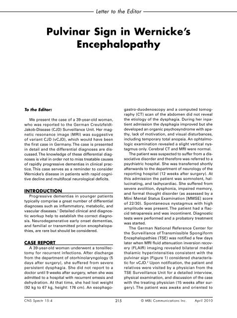
To the Editor:<br />
<strong>Pulv<strong>in</strong>ar</strong> <strong>Sign</strong> <strong>in</strong> Wernicke’s<br />
<strong>Encephalopathy</strong><br />
We present the case of a 39-year-old woman,<br />
who was reported to the German Creutzfeldt-<br />
Jakob-Disease (CJD) Surveillance Unit. Her magnetic<br />
resonance image (MRI) was suggestive<br />
of variant CJD (vCJD), which would have been<br />
the first case <strong>in</strong> Germany. The case is presented<br />
<strong>in</strong> detail and the differential diagnoses are discussed.<br />
The knowledge of these differential diagnoses<br />
is vital <strong>in</strong> order not to miss treatable causes<br />
of rapidly progressive dementias <strong>in</strong> cl<strong>in</strong>ical practice.<br />
This case serves as a rem<strong>in</strong>der to consider<br />
Wernicke’s disease <strong>in</strong> patients with rapid cognitive<br />
decl<strong>in</strong>e and multifocal neurological deficits.<br />
INTRODUCTION<br />
Progressive dementias <strong>in</strong> younger patients<br />
typically comprise a great number of differential<br />
diagnoses such as <strong>in</strong>flammatory, metabolic, and<br />
vascular diseases. 1 Detailed cl<strong>in</strong>ical and diagnostic<br />
workup help to establish the correct diagnosis.<br />
Neurodegenerative early onset dementias,<br />
and familial or transmitted prion encephalopathies,<br />
are rare but should be considered.<br />
CASE REPORT<br />
A 39-year-old woman underwent a tonsillectomy<br />
for recurrent <strong>in</strong>fections. After discharge<br />
from the department of otorh<strong>in</strong>olaryngology (5<br />
days after surgery), she suffered from severe<br />
persistent dysphagia. She did not report to a<br />
doctor until 9 weeks after surgery, when she was<br />
admitted to a hospital with recurrent emesis and<br />
dehydration. At that time, she had lost weight<br />
(92 kg to 67 kg, height: 176 cm). An esophago-<br />
Letter to the Editor<br />
gastro-duodenoscopy and a computed tomography<br />
(CT) scan of the abdomen did not reveal<br />
the etiology of the dysphagia. Dur<strong>in</strong>g her <strong>in</strong>patient<br />
admission the dysphagia improved but she<br />
developed an organic psychosyndrome with apathy,<br />
lack of motivation, and visual disturbances,<br />
<strong>in</strong>clud<strong>in</strong>g temporary total anopsia. An ophtalmologic<br />
exam<strong>in</strong>ation revealed a slight vertical nystagmus<br />
only. Cerebral CT and MRI were normal.<br />
The patient was suspected to suffer from a dissociative<br />
disorder and therefore was referred to a<br />
psychiatric hospital. She was transferred shortly<br />
afterwards to the department of neurology of the<br />
report<strong>in</strong>g hospital (12 weeks after surgery). At<br />
this admission the patient was somnolent, halluc<strong>in</strong>at<strong>in</strong>g,<br />
and tachycardiac. She suffered from<br />
severe avolition, dysphonia, impaired memory,<br />
and formal thought disorder (as assessed by a<br />
M<strong>in</strong>i Mental Status Exam<strong>in</strong>ation [MMSE] score<br />
of 22/30). Spontaneous nystagmus with high<br />
amplitude was present. The patient had a flaccid<br />
tetraparesis and was <strong>in</strong>cont<strong>in</strong>ent. Diagnostic<br />
tests were performed and a probatory treatment<br />
was started.<br />
The German National Reference Center for<br />
the Surveillance of Transmissible Spongiform<br />
Encephalopathies (TSE) was notified a few days<br />
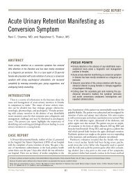
later when MRI fluid attenuation <strong>in</strong>version recovery<br />
(FLAIR) imag<strong>in</strong>g revealed bilateral medial<br />
thalamic hyper<strong>in</strong>tensities consistent with the<br />
pulv<strong>in</strong>ar sign (Figure 1) considered characteristic<br />
for vCJD. 2 Upon notification, the patient and<br />
relatives were visited by a physician from the<br />
TSE Surveillance Unit for a detailed <strong>in</strong>terview,<br />
physical exam<strong>in</strong>ation, and discussion of the case<br />
with the treat<strong>in</strong>g physician (15 weeks after surgery).<br />
The patient was awake and oriented to<br />
CNS Spectr 15:4 215<br />
© <strong>MBL</strong> <strong>Communications</strong> Inc. April 2010
situational qualities. Cognitive deficits ma<strong>in</strong>ly<br />
affected short-term memory (MMSE score:<br />
25/30). Spontaneous vertical upbeat nystagmus<br />
was present. The cranial nerve exam<strong>in</strong>ation was<br />
otherwise normal. Dysmetria was apparent <strong>in</strong> all<br />
limbs as well as <strong>in</strong>tention tremor <strong>in</strong> both arms.<br />
The patient was bedbound due to paraparesis<br />
of the lower extremities. Motor reflexes were all<br />
decreased and were not <strong>in</strong>ducible at the lower<br />
extremities. No Bab<strong>in</strong>ski sign was provokable.<br />
Sensitivity was not impaired.<br />
Medical, Social, and Familiy History<br />
The patient had a history of allergic asthma<br />
and hyperlipidemia, nasal s<strong>in</strong>us surgeries 4 and<br />
6 years before, a coccygeal fracture 14 years and<br />
pulmonary embolism 15 years before. She neither<br />
misused alcohol, nicot<strong>in</strong>e, or illicit drugs nor<br />
did she take any medication prior to the tonsillectomy.<br />
She had not received blood transfusions,<br />
growth hormones, or heterologous grafts. The<br />
patient had never been <strong>in</strong> regular contact with<br />
cattle or chemicals such as solvents. She had<br />
never been to the United K<strong>in</strong>gdom nor had she<br />
lived abroad for any significant period of time.<br />
The family’s medical history comprises allergic<br />
asthma, psoriasis, cerebrovascular disease, and<br />
colon cancer, but no early onset dementia or<br />
other severe neurologic or psychiatric disorder.<br />
Imag<strong>in</strong>g, Laboratory Tests,<br />
Electrophysiological Tests<br />
An MRI FLAIR study revealed the pulv<strong>in</strong>ar<br />
sign (Figure 1) and hyper<strong>in</strong>tensity <strong>in</strong> the periaqueductal<br />
grey matter. A sp<strong>in</strong>al MRI was normal.<br />
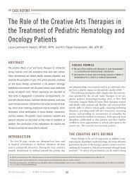
Generalized theta-activity but no triphasic waves<br />
or epileptic potentials were recorded on electroencephalography<br />
(EEG) (Figure 2). Celiac disease<br />
associated antibodies, paraneoplastic ant<strong>in</strong>euronal<br />
antibodies, and thyreoperoxidase antibodies<br />
were with<strong>in</strong> the normal range. Vitam<strong>in</strong> B1 level<br />
was 54 ng/ml (range: 20–100 ng/ml), erythrocyte<br />
transketolase activity was decreased with 38 U/L<br />
(range: 60–85 U/L). Cerebrosp<strong>in</strong>al fluid (CSF) was<br />
clear, and no signs of acute or chronic <strong>in</strong>flammation<br />
were present. Test<strong>in</strong>g for Prote<strong>in</strong>s 14-3-3<br />
was negative. CSF Tau was not elevated (110 pg/<br />
ml). Neurography detected signs of recent axonal<br />
damage. Extra- and transcranial sonography<br />
excluded arterial stenosis or dissection.<br />
Management of the Case<br />
Important differential diagnoses of the MRI<br />
Letter to the Editor<br />
lesions were vCJD and Wernicke’s <strong>Encephalopathy</strong><br />
(WE). Familial prion diseases such as Gerstmann-<br />
Straeussler-Sche<strong>in</strong>ker-Synrome or Fatal Familial<br />
Insomnia were also considered because of the<br />
patient’s young age. She was supplemented with<br />
<strong>in</strong>travenous vitam<strong>in</strong> B1, B12, benfotiam<strong>in</strong>e, and<br />
folic acid. Tachycardia was treated with β-blockers.<br />
A few days later, the cognitive status improved,<br />
the psychotic symptoms regressed, and the<br />
patient started to eat.<br />
Results of prion prote<strong>in</strong> (PRNP) codon 129<br />
test<strong>in</strong>g arrived show<strong>in</strong>g methion<strong>in</strong>e/val<strong>in</strong>e (M/V)<br />
heterozygosity. This <strong>in</strong>formation made the diagnosis<br />
of vCJD appear unlikely because it has only<br />
been seen <strong>in</strong> methion<strong>in</strong>e homozygous patients 3<br />
with one exception. Pathological changes related<br />
to vCJD <strong>in</strong> lymphatic tissue have been reported<br />
for one M/V heterozygous patient who died early<br />
from a non-neurological disorder 5 years after<br />
hav<strong>in</strong>g received blood from a vCJD patient. 4<br />
In the follow<strong>in</strong>g weeks, the EEG activity<br />
accelerated, the MRI lesions improved, muscle<br />
strength returned, nystagmus subsided, and at<br />
17 weeks after surgery, the patient was transferred<br />
<strong>in</strong>to a rehabilitation unit. In week 18 after<br />
surgery the patient still had some cognitive<br />
impairment but was aga<strong>in</strong> able to sit, and to control<br />
bladder and bowel function. Further genetic<br />
FIGURE 1.<br />
Cerebral MRI FLAIR axial sequence<br />
The pulv<strong>in</strong>ar sign is marked by arrows.<br />
MRI=magnetic resonance image; FLAIR=fluid attenuation <strong>in</strong>version recovery.<br />
Schmidt C, Plickert S, Summers D, Zerr I. CNS Spectr. Vol 15, No 4. 2010.<br />
CNS Spectr 15:4 216<br />
© <strong>MBL</strong> <strong>Communications</strong> Inc. April 2010
test<strong>in</strong>g for familial forms of prion disorders<br />
(PRNP sequenc<strong>in</strong>g) was not considered useful<br />
due to the negative family history and successful<br />
response to treatment of thiam<strong>in</strong>e deficiency.<br />
DISCUSSION<br />
vCJD is a transmissible prion disorder associated<br />
with consumption of contam<strong>in</strong>ated meat<br />
from cattle suffer<strong>in</strong>g from the Bov<strong>in</strong>e Spongiform<br />
FIGURE 2.<br />
EEG of generalized theta-activity<br />
EEG=electroencephalography.<br />
Schmidt C, Plickert S, Summers D, Zerr I. CNS Spectr. Vol 15, No 4. 2010.<br />
Letter to the Editor<br />
<strong>Encephalopathy</strong>. 5 The patients are often young<br />
(around 30 years of age) and present with progressive<br />
psychiatric symptoms, visual disturbance,<br />
ataxia, and dementia. 6 The cumulative<br />
case number is
<strong>in</strong>itial symptom of WE. This possibly genetically<br />
predisposed patient then entered a vicious cycle<br />
of dysphagia and malnutrition.<br />
CONCLUSION<br />
It is easy to be mislead by isolated diagnostic<br />
f<strong>in</strong>d<strong>in</strong>gs, such as the apparent pulv<strong>in</strong>ar sign <strong>in</strong><br />
this case, to suspect vCJD. Misdiagnoses of fatal<br />
diseases result <strong>in</strong> far reach<strong>in</strong>g consequences<br />
for the patient and relatives. The laboratory and<br />
imag<strong>in</strong>g f<strong>in</strong>d<strong>in</strong>gs <strong>in</strong> vCJD should always be<br />
<strong>in</strong>terpreted <strong>in</strong> the context of all the available cl<strong>in</strong>ical<br />
<strong>in</strong>formation. This requires thorough identification<br />
and exclusion of potentially reversible<br />
causes of encephalopathies. WE should always<br />
be considered a treatable differential diagnosis<br />
of vCJD. CNS<br />
S<strong>in</strong>cerely,<br />
Christian Schmidt, MD<br />
Steffen Plickert, MD<br />
David Summers, MD<br />
and Inga Zerr, MD, PhD<br />
REFERENCES<br />
1. Harvey RJ, Skelton-Rob<strong>in</strong>son M, Rossor MN. The prevalence and causes of dementia <strong>in</strong><br />
people under the age of 65 years. J Neurol Neurosurg Psychiatry. 2003;74:1206-1209.<br />
Letter to the Editor<br />
2. Brown P. Transmissible spongiform encephalopathy <strong>in</strong> the 21st century: neuroscience for<br />
the cl<strong>in</strong>ical neurologist. Neurology. 2008;70:713-722.<br />
3. Wadsworth JDF, Coll<strong>in</strong>ge J. Update on human prion disease. Biochim Biophys Acta.<br />
2007;1772:598-609.<br />
4. Peden AH, Head MW, Ritchie DL, Bell JE, Ironside JW. Precl<strong>in</strong>ical vCJD after blood<br />
transfusion <strong>in</strong> a PRNP codon 129 heterozygous patient. Lancet. 2004;364:527-529.<br />
5. Will RG, Ironside JW, Zeidler M, et al. A new variant of Creutzfeldt-Jakob disease <strong>in</strong> the<br />
UK. Lancet. 1996;347:921-925.<br />
6. Zeidler M, Stewart GE, Barraclough CR, et al. New variant Creutzfeldt-Jakob disease:<br />
neurological features and diagnostic tests. Lancet. 1997;350:903-907.<br />
7. The European and Allied Countries Collaborative Study Group of CJD (EUROCJD). http://<br />
www.eurocjd.ed.ac.uk/vcjdworldeuro.htm. Accessed: March 5, 2010.<br />
8. Zeidler M, Sellar RJ, Collie DA, et al. The pulv<strong>in</strong>ar sign on magnetic resonance imag<strong>in</strong>g<br />
<strong>in</strong> variant Creutzfeldt-Jakob disease. Lancet. 2000;355:1412-1418.<br />
9. Summers DM, Collie DA, Zeidler M, Will RG. The pulv<strong>in</strong>ar sign <strong>in</strong> variant Creutzfeldt-<br />
Jakob disease. Arch Neurol. 2004;61:446-447.<br />
10. Tschampa HJ, Zerr I, Urbach H. Radiological assessment of Creutzfeldt-Jakob disease.<br />
Eur Radiol. 2007;17:1200-1211.<br />
11. Hak S, Brandel JP, Oppenheim C, et al. Sporadic CJD cl<strong>in</strong>ically mimick<strong>in</strong>g variant CJD<br />
with bilateral <strong>in</strong>creased signal <strong>in</strong> the pulv<strong>in</strong>ar. Neurology. 2002;58:148-149.<br />
12. Krasnianski A, Schulz-Schaeffer WJ, Kallenberg K, et al. Cl<strong>in</strong>ical f<strong>in</strong>d<strong>in</strong>gs and diagnostic<br />
tests <strong>in</strong> the MV2 subtype of sporadic CJD. Bra<strong>in</strong>. 2006;129(Pt 9):2288-2296.<br />
13. Nolli M, Barbieri A, P<strong>in</strong>na C, Pasetto A, Nicosia F. Wernicke’s encephalopathy <strong>in</strong> a<br />
malnourished surgical patient: cl<strong>in</strong>ical features and magnetic resonance imag<strong>in</strong>g. Acta<br />
Anaesthesiol Scand. 2005;49:1566-1570.<br />
14. Doss A, Mahad D, Romanowski CAJ:.Wernicke encephalopathy: unusual f<strong>in</strong>d<strong>in</strong>gs <strong>in</strong><br />
non-alcoholic patients. J Comput Assist Tomogr. 2003;27:235-240.<br />
15. Weidauer S, Nichtweiss M, Lanfermann H, Zanella FE. Wernicke encephalopathy: MR<br />
f<strong>in</strong>d<strong>in</strong>gs and cl<strong>in</strong>ical presentation. Eur Radiol. 2003;13:1001-1009.<br />
16. Donn<strong>in</strong>o MW, Vega J, Miller J, Walsh M. Myths and misconceptions of Wernicke’s<br />
encephalopathy: what every emergency physician should know. Ann Emerg Med.<br />
2007;50:715-721.<br />
17. Sechi G, Serra A. Wernicke’s encephalopathy: new cl<strong>in</strong>ical sett<strong>in</strong>gs and recent advances<br />
<strong>in</strong> diagnosis and management. Lancet Neurol. 2007;6:442-455.<br />
18. Pearce JMS. Wernicke-Korsakoff encephalopathy. Eur Neurol. 2008;59:101-104.<br />
19. Karaiskos I, Katsarolis I, Stefanis L. Severe dysphagia as the present<strong>in</strong>g symptom of<br />
Wernicke-Korsakoff syndrome <strong>in</strong> a non-alcoholic man. Neurol Sci. 2008;29:45-46.<br />
Dr. Schmidt is a resident <strong>in</strong> neurology at Georg August University of Goett<strong>in</strong>gen Medical School, German National Reference Center for the Surveilance<br />
of Prion Diseases <strong>in</strong> Goett<strong>in</strong>gen, Germany. Dr. Plickert is attend<strong>in</strong>g/senior physician <strong>in</strong> the Department of Neurology at St. Kathar<strong>in</strong>en Hospital <strong>in</strong> Frechen,<br />
Germany. Dr. Summers is an attend<strong>in</strong>g neuroradiologist at the University of Ed<strong>in</strong>burgh Medical School <strong>in</strong> the United K<strong>in</strong>gdom. Dr. Zerr is an attend<strong>in</strong>g neurologist<br />
and head of the German National Reference Center for the Surveilance of Prion Diseases <strong>in</strong> Goett<strong>in</strong>gen, Germany.<br />
Faculty Disclosures: The authors do not have an affiliation with or f<strong>in</strong>ancial <strong>in</strong>terest <strong>in</strong> any organization that might pose a conflict of <strong>in</strong>terest.<br />
Fund<strong>in</strong>g/Support: This study was supported by grants from the Robert Koch-Institute through funds of the German Federal M<strong>in</strong>istry of Health (grant no<br />
1369-341).<br />
Submitted for publication: March 18, 2009; Accepted for publication: July 31, 2009.<br />
Please direct all correspondence to: Christian Schmidt, MD, Georg August University Hospital, Dept. of Neurology, Prionresearch, Robert-Koch Str. 40,<br />
D-37075 Goett<strong>in</strong>gen, Germany; Tel: 49-551-398955, Fax: 49-551-397020; E-mail: Christian.Schmidt@mediz<strong>in</strong>.uni-goett<strong>in</strong>gen.de.<br />
CNS Spectr 15:4 218<br />
© <strong>MBL</strong> <strong>Communications</strong> Inc. April 2010