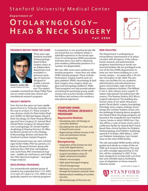
HEAD & NECK SURGERY - Stanford University School of Medicine
HEAD & NECK SURGERY - Stanford University School of Medicine
HEAD & NECK SURGERY - Stanford University School of Medicine

Create successful ePaper yourself
Turn your PDF publications into a flip-book with our unique Google optimized e-Paper software.
<strong>Stanford</strong> <strong>University</strong> Medical Center<br />
D epartment <strong>of</strong><br />
O TOLARYNGOLOGY–<br />
H EAD &<strong>NECK</strong> S URGERY<br />
PROGRESS REPORT FROM THE CHAIR<br />
Robert K. Jackler, MD,<br />
Sewall Pr<strong>of</strong>essor & Chair<br />
Three years ago,<br />
<strong>Stanford</strong> awarded<br />
Otolaryngology –<br />
Head & Neck<br />
Surgery status as<br />
an independent<br />
department and<br />
provided it a<br />
generous package<br />
<strong>of</strong> resources<br />
designed to<br />
enable a period<br />
<strong>of</strong> rapid expansion.<br />
The explicit<br />
mandate received from Dean Philip Pizzo<br />
was to create world class clinical and<br />
translational research programs.<br />
FACULTY GROWTH<br />
Over the last few years we have rapidly<br />
grown from 6 to 16 faculty members (on<br />
the way to at least 21). These include 6<br />
new division chiefs: Dr. Peter Koltai (Pediatric<br />
OHNS), Dr Michael Kaplan (Head &<br />
Neck Oncology), Dr. Peter Hwang (Rhinology<br />
and Sinus Surgery), Dr. Samuel Most<br />
(Facial Plastic Surgery), Dr. Edward Damrose<br />
(Laryngology), Dr. Gerald Popelka<br />
(Audiology & Hearing Devices). Dr. Nikolas<br />
Blevins joined me in the Otology<br />
& Neurotology division and leads the<br />
<strong>Stanford</strong> Cochlear Implant Program.<br />
World renowned inner ear stem cell biologist<br />
Stefan Heller, PhD has joined us to<br />
lead our Research Division along with<br />
Anthony Ricci, PhD (hair cell biophysicist),<br />
Sunil Puria, PhD (mechanical engineer<br />
- middle ear mechanics), and Yuling<br />
Yan, PhD (electrical engineer – laryngeal<br />
image analysis).<br />
EDUCATIONAL PROGRAMS<br />
Our extraordinarily talented group <strong>of</strong><br />
residents has expanded from 15 in 2003<br />
(3 in each <strong>of</strong> 5 years) to 19 in 2006 as we<br />
gradually expand our training programs<br />
in proportion to our growing faculty. We<br />
are proud that our residents obtain a<br />
splendid experience in the broad spectrum<br />
<strong>of</strong> contemporary OHNS procedures<br />
and have done very well in obtaining<br />
post-residency fellowship positions in a<br />
number <strong>of</strong> subspecialties.<br />
We now <strong>of</strong>fer seven post-residency fellowship<br />
programs – more than any other<br />
OHNS training program. These include:<br />
facial plastic surgery, head & neck surgery,<br />
pediatric OHNS, neurotology & skull<br />
base surgery, sinus surgery, sleep surgery,<br />
and laryngology (added in 2006).<br />
These programs not only provide advanced<br />
training for promising young academicians,<br />
but as junior faculty members<br />
the fellows also enhance the residency<br />
educational experience.<br />
STANFORD OHNS:<br />
TRANSLATIONAL RESEARCH<br />
PROGRAMS<br />
Regenerative <strong>Medicine</strong><br />
• Developing stem cell therapy to<br />
overcome deafness<br />
• Elucidating the role <strong>of</strong> <strong>of</strong> stem cells<br />
in head & neck cancer<br />
• Regenerating ciliated mucosa in the<br />
nose and paranasal sinuses<br />
• Tissue engineering<br />
Bioengineering<br />
• Integration <strong>of</strong> the human ear and<br />
voice with digital devices<br />
• Biophysical properties <strong>of</strong> hair cells<br />
• Surgical simulation using 3D -<br />
haptic enhanced simulators<br />
• Robotic microsurgery<br />
• High speed laryngeal imaging<br />
• Virtual laryngoscopy<br />
• Mechanics <strong>of</strong> sound transmission<br />
through the tympano-ossicular chain<br />
• Mathematical modeling <strong>of</strong> cochlear<br />
function<br />
• Microendoscopy <strong>of</strong> the inner ear<br />
Fall 2006<br />
NEW FACILITIES<br />
The Department is undergoing an<br />
extraordinary improvement in its physical<br />
plant with all aspects <strong>of</strong> the educational,<br />
research, and administrative<br />
programs transitioning to newly constructed<br />
facilities. We are privileged to be<br />
one <strong>of</strong> the few OHNS departments to<br />
have our own home building on a university<br />
campus – occupied after a $4 million<br />
renovation in late 2004. This provides<br />
core facilities for our academic,<br />
administrative, and educational programs<br />
and includes state-<strong>of</strong>-the-art<br />
library-conference facilities (The Willard<br />
E. Fee Jr., M.D. Library) and a superb 10<br />
station educational microdissection laboratory<br />
(The Rodney Perkins, M.D. Microsurgical<br />
Laboratory). The building also<br />
houses some <strong>of</strong> our adult clinical programs<br />
(facial plastic surgery, laryngology,<br />
rhinology-sinus surgery, otology-neurotology,<br />
the cochlear implant center, and<br />
the audiology & hearing device program).<br />
Our Head & Neck Oncology programs are<br />
housed in the magnificent new <strong>Stanford</strong><br />
Cancer Center, in which OHNS has 3 faculty<br />
<strong>of</strong>fices, a suite <strong>of</strong> exam rooms, and a<br />
conference facility for Head & Neck Tumor<br />
Board. New clinical facilities for Pediatric<br />
Otolaryngology and Pediatric Audiology<br />
opened in October, 2004 (Mary L. Johnson<br />
Pediatric Ambulatory Care Center)<br />
close to the main OHNS facility.<br />
The old outmoded OHNS facility will be<br />
gutted and rebuilt as a state-<strong>of</strong>-the-art<br />
7000 sq ft research laboratory. This project,<br />
with a budget approaching $5 million,<br />
will be completed in 2007. It has<br />
been designed to include both core facilities<br />
and a flexible layout, facilitating<br />
ease <strong>of</strong> adaptation as future research<br />
interests evolve.<br />
(continued on page 2)
S TANFORD U NIVERSITY M EDICAL C ENTER D EPARTMENT OF O TOLARYNGOLOGY – <strong>HEAD</strong> & N ECK S URGERY<br />
RESEARCH EMPHASIS<br />
The research division <strong>of</strong> <strong>Stanford</strong> OHNS<br />
is in the midst <strong>of</strong> rapid expansion. <strong>Stanford</strong><br />
has made a substantial investment<br />
in new laboratory space, endowment,<br />
and additional basic science faculty positions.<br />
The intention is to create a highly<br />
productive, innovative, and collaborative<br />
center which takes full advantage <strong>of</strong> the<br />
surrounding <strong>Stanford</strong> bioscience and<br />
engineering communities. The priority <strong>of</strong><br />
our laboratory programs is to produce<br />
high quality, innovative research in areas<br />
<strong>of</strong> inquiry relevant to human disease.<br />
Growth in our research programs will<br />
emphasize two central themes: Regenerative<br />
<strong>Medicine</strong> and Bioengineering.<br />
<strong>Stanford</strong> OHNS has come a long way in a<br />
short 3 years since emerging as an independent<br />
department: more than doubling<br />
the size <strong>of</strong> the faculty with recruitment<br />
<strong>of</strong> a number <strong>of</strong> highly talented<br />
individuals; abandoning antiquated facilities<br />
for new ones triple their size; sizable<br />
expansion <strong>of</strong> both residency and fellowship<br />
programs; and development <strong>of</strong><br />
dynamic, cutting edge research programs.<br />
It is a credit to a large team <strong>of</strong><br />
hard working individuals that we have<br />
made much progress in such a relatively<br />
short period. We look forward to sharing<br />
our progress with you in the coming<br />
years. We plan to keep things hopping<br />
on “The Farm.”<br />
BUILDING WORLD CLASS PROGRAMS<br />
2
I NTRODUCING<br />
O UR FACULTY (Fall 2006)<br />
NIKOLAS H. BLEVINS, MD<br />
Assistant Pr<strong>of</strong>essor<br />
Otology & Neurotology<br />
College: <strong>Stanford</strong> <strong>University</strong><br />
Medical <strong>School</strong>: Harvard<br />
<strong>University</strong>.<br />
Residency: <strong>University</strong> <strong>of</strong><br />
California at San Francisco<br />
Fellowship: Neurotology/Skull<br />
Base Surgery – <strong>University</strong> <strong>of</strong><br />
California at San Francisco<br />
Former Faculty Position: Tufts<br />
<strong>University</strong> (1995-2003)<br />
Clinical Interests: Otology,<br />
neurotology, skull base surgery,<br />
cochlear implants<br />
Research Interests: Surgical<br />
simulation and robotics,<br />
microendoscopy <strong>of</strong> the inner ear<br />
KAY W. CHANG, MD<br />
Assistant Pr<strong>of</strong>essor<br />
Pediatric OHNS<br />
College: Brown <strong>University</strong><br />
Medical <strong>School</strong>: Brown <strong>University</strong><br />
Residency: <strong>University</strong> <strong>of</strong><br />
Washington<br />
Fellowship: Pediatric<br />
Otolaryngology at the Children’s<br />
Hospital <strong>of</strong> Pittsburgh<br />
Clinical Interests: Pediatric<br />
otology, Ear reconstruction<br />
Research Interests: Pediatric<br />
hearing loss, cis-platinum<br />
ototoxicity<br />
EDWARD J. DAMROSE, MD<br />
Assistant Pr<strong>of</strong>essor<br />
Chief <strong>of</strong> Laryngology Division<br />
College: Yale <strong>University</strong><br />
Medical <strong>School</strong>: UCLA <strong>School</strong> <strong>of</strong><br />
<strong>Medicine</strong><br />
Residency: <strong>University</strong> <strong>of</strong><br />
California at Los Angeles<br />
Fellowship: Laryngology/<br />
Bronchoesophagology –<br />
<strong>University</strong> <strong>of</strong> California at Los<br />
Angeles<br />
Clinical Interests: Voice and<br />
swallowing disorders<br />
Research Interests: High speed<br />
laryngeal imaging, spasmodic<br />
dysphonia, rehabilitation <strong>of</strong> vocal<br />
cord palsy<br />
WILLARD E. FEE, JR., MD<br />
Edward C. and Amy H. Sewall<br />
Pr<strong>of</strong>essor<br />
Head & Neck Surgery<br />
College: <strong>University</strong> <strong>of</strong> San<br />
Francisco<br />
Medical <strong>School</strong>: <strong>University</strong> <strong>of</strong><br />
Colorado<br />
Residency: <strong>University</strong> <strong>of</strong><br />
California, Los Angeles<br />
Clinical Interests: Tumors <strong>of</strong> the<br />
head and neck<br />
Research Interests: Clinical<br />
outcomes in head & neck cancer<br />
RICHARD L. GOODE, MD<br />
Pr<strong>of</strong>essor<br />
Sleep Surgery & Facial Plastic<br />
Surgery<br />
Chief VA Service<br />
College: <strong>University</strong> <strong>of</strong> California at<br />
Santa Barbara, California<br />
Medical <strong>School</strong>: <strong>University</strong> <strong>of</strong><br />
Southern California<br />
Residency: <strong>Stanford</strong> <strong>University</strong><br />
Fellowship: NIH Fellow –<br />
Vestibular Physiology<br />
Clinical Interests: Facial plastic<br />
surgery, sleep surgery<br />
Research Interests: Mechanics <strong>of</strong><br />
middle ear function, innovations<br />
in sleep surgery<br />
STEFAN HELLER, PHD<br />
Associate Pr<strong>of</strong>essor<br />
Head <strong>of</strong> Research<br />
M.S. Biology: Johannes<br />
Gutenberg <strong>University</strong>, Mainz,<br />
Germany<br />
PhD Genetics: Johannes<br />
Gutenberg <strong>University</strong>, Mainz,<br />
Germany<br />
and Max-Planck-Institute for<br />
Brain Research, Frankfurt/M.,<br />
Germany<br />
Post-Doctoral Training: The<br />
Rockefeller <strong>University</strong>, New York<br />
Former Faculty Position: Harvard<br />
<strong>University</strong> (2000-2005)<br />
Research Interests: Hair cell<br />
regeneration to overcome<br />
deafness, structure and function<br />
<strong>of</strong> mechanosensitive ion channel<br />
proteins.<br />
Fall 2006<br />
PETER H. HWANG , MD<br />
Associate Pr<strong>of</strong>essor<br />
Chief <strong>of</strong> Rhinology<br />
College: <strong>Stanford</strong> <strong>University</strong><br />
Medical <strong>School</strong>: <strong>University</strong> <strong>of</strong><br />
California at San Francisco<br />
Residency: <strong>University</strong> <strong>of</strong><br />
California at San Francisco<br />
Fellowship: Rhinology and Sinus<br />
Disorders, Hospital <strong>of</strong> the<br />
<strong>University</strong> <strong>of</strong> Pennsylvania<br />
Former Faculty Position: Oregon<br />
Health & Science <strong>University</strong><br />
(1997-2005)<br />
Clinical Interests: Endoscopic<br />
sinus surgery, endoscopic tumor<br />
and skull base surgery<br />
Research Interests: Mucosal wound<br />
healing, novel drug delivery<br />
technologies, clinical outcomes<br />
ROBERT K. JACKLER, MD<br />
Sewall Pr<strong>of</strong>essor and Chair<br />
Otology & Neurotology<br />
College: Brandeis <strong>University</strong><br />
Medical <strong>School</strong>: Boston <strong>University</strong><br />
Residency: <strong>University</strong> <strong>of</strong><br />
California at San Francisco<br />
Fellowship: Neurotology, House<br />
Ear Clinic, Los Angeles, CA<br />
Former Faculty Position: UCSF<br />
(1986 - 2003)<br />
Clinical Interests: Neurotology<br />
and skull base surgery<br />
Research Interests: Innovation in<br />
skull base surgery, cholesteatoma<br />
pathogenesis, history <strong>of</strong> otology<br />
3
S T ANFORD U NIVERSITY D EPARTMENT OF O TOLARYNGOLOGY– <strong>HEAD</strong> & N ECK S URGERY<br />
MICHAEL J. KAPLAN, MD<br />
Pr<strong>of</strong>essor<br />
Chief <strong>of</strong> Head & Neck Surgery<br />
College: Harvard College<br />
Medical <strong>School</strong>: Harvard Medical<br />
<strong>School</strong><br />
Residency: Massachusetts Eye<br />
and Ear Infirmary<br />
Fellowship: <strong>University</strong> <strong>of</strong> Virginia<br />
(Head & Neck Surgery)<br />
Former Faculty Position: UCSF<br />
(1984 - 2003)<br />
Clinical Interests: Tumors <strong>of</strong> the<br />
head and neck<br />
Research Interests: Clinical<br />
outcomes for head and neck<br />
malignancy, advanced imaging,<br />
head & neck cancer stem cells<br />
PETER J. KOLTAI, MD<br />
Pr<strong>of</strong>essor<br />
Chief <strong>of</strong> Pediatric OHNS<br />
College: Queens College<br />
Medical <strong>School</strong>: The Albany<br />
Medical College<br />
Residency: <strong>University</strong> <strong>of</strong> Texas<br />
Medical Branch<br />
Fellowship: Pediatric<br />
Otolaryngology/Hospital for Sick<br />
Children at Great Ormond Street<br />
Former Faculty Position: Albany<br />
Med. (1982-1998), Cleveland<br />
Clinic (1998-2004)<br />
Clinical Interests: Pediatric airway<br />
obstruction, sleep apnea<br />
Research Interests: Developing<br />
new techniques in managing<br />
sleep apnea<br />
4<br />
ANNA H. MESSNER, MD<br />
Associate Pr<strong>of</strong>essor<br />
Pediatric OHNS<br />
Vice Chair<br />
College: Duke <strong>University</strong><br />
Medical <strong>School</strong>: Wake Forest<br />
<strong>University</strong><br />
Residency: Wake Forest<br />
<strong>University</strong><br />
Fellowship: Pediatric OHNS,<br />
Hospital for Sick Children,<br />
Toronto, Canada<br />
Clinical Interests: Pediatric OHNS<br />
Research Interests: Neonatal<br />
hearing screening, ankyloglossia<br />
SAM MOST, MD<br />
Associate Pr<strong>of</strong>essor<br />
Chief <strong>of</strong> Facial Plastic Surgery<br />
College: <strong>University</strong> <strong>of</strong> Michigan<br />
Medical <strong>School</strong>: <strong>Stanford</strong><br />
Residency: <strong>University</strong> <strong>of</strong><br />
Washington<br />
Fellowship: Facial Plastic Surgery<br />
(U Washington)<br />
Former Faculty Position:<br />
<strong>University</strong> <strong>of</strong> Washington (2002 -<br />
2006),<br />
Clinical Interests: Aesthetic<br />
surgery <strong>of</strong> the face<br />
Research Interests: Minimally<br />
invasive improvement <strong>of</strong> the<br />
aging face, facial nerve biology<br />
SUNIL PURIA, PHD<br />
Consulting Associate Pr<strong>of</strong>essor<br />
Research<br />
College: The City College <strong>of</strong> NY<br />
MS: Columbia <strong>University</strong><br />
PhD: City <strong>University</strong> <strong>of</strong> NY<br />
Postdoctoral Fellowships: MIT,<br />
Harvard<br />
Former Faculty Position: Harvard<br />
(1995-1997)<br />
Research Interests: Biomechanics,<br />
physiology, and imaging <strong>of</strong> the<br />
middle ear and the cochlea.<br />
GERALD R. POPELKA, PHD<br />
Consulting Pr<strong>of</strong>essor<br />
Research<br />
Chief <strong>of</strong> Audiology<br />
College: Kent State <strong>University</strong><br />
MA: Audiology/Kent State<br />
<strong>University</strong><br />
PhD: Communication Sciences/<br />
<strong>University</strong> <strong>of</strong> Wisconsin<br />
Postdoctoral Fellowship:<br />
<strong>University</strong> <strong>of</strong> California at Los<br />
Angeles<br />
Former Faculty Position:<br />
Washington <strong>University</strong> (1980 -<br />
2004)<br />
Clinical Interests: Advanced<br />
hearing devices, advanced<br />
measures <strong>of</strong> auditory function<br />
Research Interests: The<br />
developing auditory system,<br />
hyperbilirubinemia<br />
ANTHONY RICCI, PHD<br />
Associate Pr<strong>of</strong>essor<br />
Research<br />
College: Case Western Reserve<br />
<strong>University</strong><br />
PhD: Neuroscience/Tulane<br />
<strong>University</strong><br />
Post-doctoral Fellowships: UTMB,<br />
<strong>University</strong> <strong>of</strong> Wisconsin<br />
Former Faculty Position:<br />
Louisiana State <strong>University</strong> (1999 -<br />
2006)<br />
Research Interests: Hair cell<br />
biophysics<br />
YULING YAN, PHD<br />
Consulting Assistant Pr<strong>of</strong>essor<br />
Research<br />
PhD: Mechanical Engineering/<br />
Keio <strong>University</strong>, Yokohama, Japan<br />
Post-Doctoral Fellowship: McGill<br />
<strong>University</strong><br />
Former Faculty Position:<br />
<strong>University</strong> <strong>of</strong> Hawaii, U Wisconsin<br />
Research Interests: High speed<br />
laryngeal imaging
STANFORD FACULTY AT SANTA CLARA<br />
VALLEY MEDICAL CENTER (Fall 2006)<br />
JOHN B. SHINN, MD<br />
Clinical Pr<strong>of</strong>essor<br />
Santa Clara Valley Medical Center<br />
College: <strong>University</strong> <strong>of</strong> North<br />
Carolina<br />
Medical <strong>School</strong>: <strong>University</strong> <strong>of</strong><br />
North Carolina, Chapel Hill<br />
Residency: <strong>Stanford</strong><br />
Clinical Interests: Otology &<br />
Neurotology<br />
M. LAUREN LALAKEA, MD<br />
Clinical Associate Pr<strong>of</strong>essor<br />
Santa Clara Valley Medical Center<br />
College: Harvard<br />
Medical <strong>School</strong>: Boston <strong>University</strong><br />
Residency: <strong>Stanford</strong><br />
Clinical Interests: Pediatric OHNS,<br />
laryngology<br />
Research Interests: Ankyloglossia<br />
KIMBERLY G. SHEPARD, MD<br />
Clinical Assistant Pr<strong>of</strong>essor<br />
Santa Clara Valley Medical Center<br />
College: UC San Diego<br />
Medical <strong>School</strong>: Dartmouth<br />
Residency: <strong>Stanford</strong><br />
Clinical Interests: Head & Neck<br />
Oncology; Sleep apnea<br />
Research Interests: Non-surgical<br />
treatment <strong>of</strong> tonsillar hypertrophy,<br />
Medical management <strong>of</strong> postoperative<br />
pain<br />
CARRIE ROLLER, MD<br />
Clinical Assistant Pr<strong>of</strong>essor<br />
Santa Clara Valley Medical Center<br />
College: UC Berkeley<br />
Medical <strong>School</strong>: Georgetown<br />
<strong>University</strong><br />
Residency: Baylor<br />
Clinical Interests: Head & neck<br />
cancer, trauma<br />
Research Interests: Functional<br />
outcomes after Head & Neck<br />
Surgery<br />
Fall 2006<br />
5
S TANFORD U NIVERSITY D EPARTMENT OF O TOLARYNGOLOGY– <strong>HEAD</strong> & N ECK S URGERY<br />
R ESEARCH P ROGRAMS<br />
CURING HEARING LOSS<br />
AND UNLOCKING THE SECRETS<br />
OF HOW THE EAR WORKS<br />
Stefan Heller, PhD<br />
All hearing sensation is derived from the<br />
electrical output <strong>of</strong> a remarkably small<br />
number <strong>of</strong> sensory cells: fewer that<br />
15,000 per inner ear at birth. These hair<br />
cells are the mechanoelectrical transducers<br />
<strong>of</strong> the inner ear: deflections <strong>of</strong> the<br />
sterociliary bundles on their apical surfaces<br />
lead to transmitter release from<br />
their basolateral poles, leading, in turn, to<br />
signal generation in the peripheral axons<br />
<strong>of</strong> the auditory nerve fibers.<br />
Most types <strong>of</strong> congenital and acquired<br />
hearing loss arise from damage to, or<br />
loss <strong>of</strong>, these sensory cells or their associated<br />
neurons. The incidence <strong>of</strong> heritable<br />
deafness is high: one child in a thousand<br />
is born deaf; another one in a thousand<br />
becomes deaf before adulthood. The<br />
prevalence <strong>of</strong> acquired hearing loss is rising,<br />
as the population ages, and as noise<br />
pollution steadily increases. It is estimated<br />
that one in three adults over the age<br />
<strong>of</strong> 65 has a handicapping hearing loss,<br />
and this impairment is largely due to the<br />
irreversible loss <strong>of</strong> sensory cells.<br />
Underlying the irreversibility <strong>of</strong> hearing<br />
loss in mammals is the incapacity to<br />
replace lost hair cells by cell division or<br />
by regeneration from endogenous cells<br />
6<br />
The image shows integration and differentiation <strong>of</strong> inner ear progenitor cells derived<br />
from mouse embryonic stem cells after injection into the chicken’s developing<br />
inner ear (otic vesicle). In (A), some <strong>of</strong> the injected cells, which are expressing<br />
the ß-Gal marker gene are found to integrate into the epithelium <strong>of</strong> the otic<br />
vesicle (arrow). Shown in (B) and (C) are embryonic stem cell-derived cells found<br />
3 days after injection (in green). These cells start expressing markers <strong>of</strong> hair cells,<br />
such as myosin VIIA (red) and appear to have integrated well into the chicken<br />
auditory epithelium where they are surrounded by endogenous chicken hair cells<br />
(non green cells, labeled red). (D) and (E) show that the mouse cells, here visualized<br />
with a blue ß-Gal staining with developed hair bundles that are immunopositive<br />
for the hair bundle marker protein espin.<br />
in the inner ear epithelia. Hair cell<br />
replacement, either by stimulation <strong>of</strong><br />
regeneration (as occurs naturally in nonmammalian<br />
vertebrates) or by transplantation<br />
<strong>of</strong> progenitor cells capable <strong>of</strong> differentiating<br />
into hair cells, remains<br />
therefore the ultimate goal in the development<br />
<strong>of</strong> treatment applications to<br />
reconstruct the damaged inner ear.<br />
Our recent work has focused on creating<br />
inner ear cell types, in particular hair cells<br />
and auditory neurons, from a renewable<br />
source.<br />
We have shown that embryonic stem<br />
(ES) cells and adult inner ear stem cells<br />
can serve as such a<br />
source, and we are<br />
currently exploring<br />
signaling pathways<br />
that control hair cell<br />
and neuronal (re-)<br />
generation in vitro<br />
and in vivo.<br />
Our research does<br />
not only focus on<br />
generating inner<br />
ear replacement<br />
parts. We are also<br />
exploring novel<br />
methods to deliver<br />
such replacement<br />
parts into the damaged<br />
cochlea or<br />
whether it is possible<br />
to use drugs to coax<br />
cells <strong>of</strong> the adult<br />
damaged cochlea<br />
into a prenatal status,<br />
which could result<br />
in self-repair <strong>of</strong> the damaged mammalian<br />
cochlea.<br />
In an independent line <strong>of</strong> research, we<br />
are pursuing the structural basis <strong>of</strong> hair<br />
cell mechanoreception. Although the<br />
individual molecular components <strong>of</strong> the<br />
mechanoelectrical transduction apparatus<br />
are not known, we and other laboratories<br />
have identified a number <strong>of</strong><br />
potential candidates. Our goal is to solve<br />
the atomic structure <strong>of</strong> the different<br />
components <strong>of</strong> the transduction machinery<br />
to study the molecular hinges and<br />
mechanisms that<br />
mechanically gate the<br />
elusive ion channel<br />
that is central to our<br />
senses <strong>of</strong> hearing and<br />
balance.<br />
THE THERAPEUTIC RELEVANCE<br />
OF HAIR CELL BIOPHYSICS<br />
Anthony Ricci, PhD<br />
Hair cells are the sensory cells <strong>of</strong> the<br />
inner ear. They are responsible for converting<br />
mechanical signals into one recognized<br />
by the brain. Hair cells get their<br />
name from having a tuft <strong>of</strong> hair-like cilia<br />
at their apical surface. Deflection <strong>of</strong> these<br />
stereocilia opens mechanically-gated ion<br />
channels that convert the mechanical<br />
signal into an electrical signal. This<br />
process, termed mechanotransduction is<br />
critical to both hearing and balance. Perturbations<br />
<strong>of</strong> this system are associated<br />
with both temporary and permanent<br />
hearing loss, with age-related and noiseinduced<br />
hearing loss and may even be<br />
associated with tinnitus and vertigo.<br />
Understanding the mechanisms involved<br />
in regulating mechanotransduction may<br />
elucidate new and novel sites for intervention.<br />
Characterizing these pathways<br />
might also lead to new technological<br />
breakthroughs in hearing aid and<br />
cochlear implant development as well as<br />
in the design <strong>of</strong> novel noise prevention<br />
devices. Over the past several years my<br />
laboratory has identified several important<br />
attributes <strong>of</strong> mechanotransduction.<br />
First, we have discovered a new process,<br />
called fast adaptation that is critical for<br />
establishing frequency discrimination, or<br />
the ability <strong>of</strong> the ear to separate sound<br />
into its individual frequency components.<br />
Second, we demonstrated for the<br />
first time, biochemical regulation <strong>of</strong><br />
mechanotransduction. This pathway may<br />
provide a new site for pharmacological<br />
intervention in preventing noise-induced<br />
hearing loss. Third, we have recently<br />
demonstrated the importance <strong>of</strong><br />
mechanotransduction in controlling the<br />
electrical properties <strong>of</strong> hair cells. As<br />
mechanotransduction is very sensitive to<br />
its ionic environment, particularly the<br />
concentration <strong>of</strong> calcium, the importance<br />
<strong>of</strong> maintaining low levels <strong>of</strong> calcium<br />
bathing the hair bundle has become<br />
more apparent. Characterizing the regulation<br />
<strong>of</strong> extracellular calcium may provide<br />
a new pathway for intervention and<br />
may shed light on pathologies related to<br />
loss <strong>of</strong> ionic homeostasis within the ear.<br />
And finally we are involved in identifying<br />
a mechanism associated with mechanotransduction<br />
and the hair bundle that
may act to amplify low levels <strong>of</strong> sound<br />
and be a major contributor to determining<br />
how the ear can be sensitive to such<br />
low energy sound waves.<br />
Aside from converting a mechanical signal<br />
into an electrical signal, the hair cell<br />
must communicate with the brain<br />
through synaptic transmission. The hair<br />
cell-afferent nerve synapse is very sensitive<br />
to overstimulation where the nerve<br />
ending can swell and retract leading to<br />
hair cell loss. My laboratory has been<br />
investigating the functional properties <strong>of</strong><br />
this synapse to identify the properties<br />
that make it unique in its ability to operate<br />
with such high fidelity and at such<br />
high rates. Here too, the goal is to identify<br />
pathways and mechanisms that might<br />
<strong>of</strong>fer sites for intervention and modification.<br />
And finally, my laboratory is trying to<br />
determine whether hair cells require<br />
extrinsic factors, like innervation,<br />
mechanical stimulation or growth factors<br />
to mature into their final form. This is a<br />
critical question to answer in order to<br />
judge feasibility <strong>of</strong> therapies where hair<br />
cell regeneration from supporting cells<br />
or hair cell development from stem cells<br />
is being investigated as a new therapy to<br />
replace hair cells in damaged cochlea<br />
Isolated outer hair cell with sensory hair bundle (left).<br />
Upper right is patch electrode on hair cell body to<br />
measure electrical responses elicited by mechanically<br />
stimulating the sensory hair bundle (lower right).<br />
INNER EAR FLUORESCENCE<br />
MICROENDOSCOPY<br />
Nikolas Blevins, MD, Mark Schnitzer, PhD,<br />
Eunice Cheung, PhD<br />
We are developing a method to visualize<br />
functional hair cells and other cellular<br />
elements <strong>of</strong> the inner ear within the<br />
intact mammalian cochlea using fluorescence<br />
microendoscopy. Our laboratory<br />
has begun work on this minimally invasive<br />
in vivo imaging technique to provide<br />
high-resolution images <strong>of</strong> deep tissues<br />
previously inaccessible in live<br />
subjects. Using microendoscopes as<br />
small as 0.3 mm in diameter, we have<br />
successfully imaged individual red blood<br />
cells flowing within capillaries inside the<br />
mammalian cochlea. We are extending<br />
this work by labeling functional neural<br />
elements with fluorescent dyes to concurrently<br />
reveal mechanotransduction as<br />
well as microanatomy.<br />
Current work includes microendoscopy<br />
using a styryl dye (FM-143) in the guinea<br />
pig. It is anticipated that this will allow us<br />
View <strong>of</strong> live cochlear hair cells from a microendoscope<br />
to accurately map hair cell injury following<br />
ototoxin exposure, and to observe<br />
the recovery <strong>of</strong> hair cells from temporary<br />
threshold shift noise damage. The use<br />
<strong>of</strong> microendoscopes should provide an<br />
opportunity to observe specific hair cell<br />
populations over time – information<br />
previously unavailable through conventional<br />
techniques.<br />
An imaging technology to observe functional<br />
hair cells and dendrites within live<br />
MIcroendoscope<br />
Fall 2006<br />
mammalian subjects will provide considerable<br />
benefit, and enable progress in a<br />
broad range <strong>of</strong> previously intractable<br />
hearing science questions. The success <strong>of</strong><br />
inner ear microendoscopy will provide a<br />
basis on which inner ear surgery can be<br />
established. The inner ear is one <strong>of</strong> the<br />
last areas <strong>of</strong> the human body to remain<br />
largely inaccessible to direct examina-<br />
tion and surgical intervention. This is<br />
because <strong>of</strong> the combination <strong>of</strong> its small<br />
size and its extraordinary fragility to<br />
mechanical manipulation. The development<br />
<strong>of</strong> non-destructive imaging techniques<br />
will enable diagnostic and therapeutic<br />
manipulations, including the<br />
optimal placement <strong>of</strong> cochlear implant<br />
arrays, or the specific delivery <strong>of</strong> stem<br />
cells or growth factors to enable hearing<br />
restoration.<br />
7
S T ANFORD U NIVERSITY D EPARTMENT OF O TOLARYNGOLOGY– <strong>HEAD</strong> & N ECK S URGERY<br />
R ESEARCH P ROGRAMS<br />
THE POSSIBLE ROLE OF STEM<br />
CELLS IN <strong>HEAD</strong> & <strong>NECK</strong> CANCER<br />
Michael Kaplan, MD, Michael Clarke, MD,<br />
Laurie Ailles, PHD Willard Fee, MD, Ranjiv<br />
Sivanidan, MD, Mark Prince, MD<br />
Head and neck cancer affects 50,000<br />
Americans annually and remains a devastating<br />
world-wide killer. In India, for<br />
example, it is the leading cause <strong>of</strong> cancer<br />
deaths. Cisplatin-based concomitant<br />
chemotherapy and selected monoclonal<br />
antibodies have improved locoregional<br />
controls compared to irradiation alone,<br />
but there has been little to no impact on<br />
survival because <strong>of</strong> distant metastasis<br />
and the therapy-resistant recurrences.<br />
Why has there been so little progress? Is<br />
it because we have not adequately<br />
enough appreciated the underlying<br />
tumor biology so as to develop more<br />
effective therapeutic approaches?<br />
H&E and immunoperodixase staining <strong>of</strong> CD44 in<br />
passaged cells growing as explant<br />
It has been understood for quite some<br />
time that leukemias and lymphomas<br />
arise within hematopoietic stem cells, yet<br />
it has been only recently that a cancer<br />
stem cell (CSC) hypothesis has been<br />
extended to solid epithelial cells, including<br />
head and neck squamous cell carcinomas<br />
(HNSCC).<br />
Normal stem cells maintain an organ’s<br />
stem cell pool by self-renewing, while<br />
generating large numbers <strong>of</strong> mature<br />
daughter differentiated cells that in their<br />
life cells in time undergo apoptosis (programmed<br />
cell death). Squamous epithelium<br />
is comprised <strong>of</strong> a basal layer <strong>of</strong> cells<br />
that contain some stem cells and overlying<br />
layers that contain daughter cells<br />
that die as they approach the surface.<br />
When stem cells divide they asymmetrically<br />
give rise to both a committed<br />
daughter cell as well as another stem<br />
cell. This stem cell must avoid programmed<br />
cell death (apoptosis)<br />
throughout the organism’s lifetime.<br />
Under normal circumstances it also must<br />
recognize its neighbors in order to know<br />
8<br />
when to divide and when not to; in other<br />
words cell-cell signaling pathways are<br />
likely important. Stem cells (or their<br />
immediate daughters) also must be able<br />
to migrate, both upward in the epithelium<br />
and in response to trauma. These<br />
three properties – avoiding apoptosis,<br />
critical cell-cell signaling, and migration –<br />
are also key attributes <strong>of</strong> developmental<br />
(embryonic, fetal) stem cells. An important<br />
insight is that avoiding apoptosis<br />
and migration are also key characteristics<br />
<strong>of</strong> cancer cells; and aberrant cell-cell<br />
signaling pathways are beginning to be<br />
shown as well.<br />
A cancer stem cell hypothesis suggests<br />
that cancer is a result <strong>of</strong> inadequately<br />
controlled proliferation <strong>of</strong> the stem cells<br />
themselves, and not the heterogeneous<br />
mix <strong>of</strong> daughter cells. Such aberrant<br />
growth would lead to subpopulations<br />
that contain a small percentage <strong>of</strong><br />
daughter stem cells but many more<br />
committed differentiated cells that will<br />
undergo apoptosis in time. In other<br />
words, one should expect to see functional<br />
heterogeneity, with only a small<br />
fraction <strong>of</strong> cells harboring tumorigenic<br />
potential. This had been known in hematological<br />
malignancies for 40 years, but<br />
was shown in solid tumors (medulloblastoma,<br />
breast cancer) only in the past<br />
three years.<br />
Only CD44+ cells serially maintain clonogenicity.<br />
Working along similar lines using methods<br />
employed to identify cancer stem<br />
cells in breast cancer, collaborating laboratories<br />
at the <strong>University</strong> <strong>of</strong> Michigan<br />
and at <strong>Stanford</strong> showed in 2006 (in<br />
press) that this is also true for HNSCC.<br />
HNSCC contains a distinct population <strong>of</strong><br />
cancer stem cells with the exclusive ability<br />
to produce tumors in mice and recreate<br />
the original tumor heterogeneity.<br />
They are clonogenic in vitro (Fig 1) and<br />
initiate tumors in vivo (Fig 2), while the<br />
remaining cells in the tumor do not<br />
share these properties. This population is<br />
distinguished by a cell surface marker<br />
(CD44) that distinguishes these cells<br />
from the other epithelial cells within the<br />
tumor that lack clonogenicity.<br />
The identification <strong>of</strong> cancer stem cells in<br />
HNSCC has important ramifications. Most<br />
basically, it lends further support to the<br />
general concept that a CSC model is true<br />
for all cancers. Second, it suggests one<br />
possible reason why chemotherapy has<br />
been disappointing is that the assay<br />
used clinically (and in some clonogenic<br />
assays) is overall tumor response, whereas<br />
it is not overall but rather stem cell<br />
response that is key. Third, it suggests<br />
that characterizing selected key molecular<br />
pathways that are involved in selfrenewal,<br />
such as cell-cell signaling pathways,<br />
should provide insights into<br />
specifically what is abnormally regulated.<br />
Insights such as these hold promise for<br />
the development <strong>of</strong> new treatment<br />
strategies targeted not against the<br />
majority <strong>of</strong> tumor cells (which have limited<br />
tumorigenicity) but against the critical<br />
population <strong>of</strong> cancer stem cells that is<br />
the key culprit.<br />
A fourth striking implication <strong>of</strong> validating<br />
a CSC model is that one cannot help but<br />
see the striking similarities between nor-<br />
mal embryonic development and normal<br />
stem cells and their dysregulation that is<br />
cancer. This suggests that understanding<br />
normal development should lead to<br />
insight into cancer, and vice versa. The<br />
cellular genetic control mechanisms<br />
seen in normal development, stem cell<br />
regulation, and cancer are likely to be<br />
the same.<br />
In the past decade an entirely new layer<br />
<strong>of</strong> intranuclear genetic control has been<br />
identified-small RNAs regulate gene<br />
expression by suppressing homologous<br />
or near-homologous DNA sequences.
These microRNAs (miRNA) are normal,<br />
and derive from pre-miRNA genes within<br />
us, and the RNA-protein machinery all<br />
organisms use for this is being increasingly<br />
identified. MiRNA gene chips are<br />
available, and abnormal quantities <strong>of</strong><br />
selected miRNAs have begun to be identified<br />
in a few malignancies. That miRNAs<br />
are stable in paraffin will allow investigation<br />
<strong>of</strong> archived material as well as easier<br />
investigation <strong>of</strong> fresh tumor-banked<br />
material.<br />
The recognition <strong>of</strong> cancer stem cells in<br />
solid tumors, including head and neck<br />
carcinomas, and the advances in DNA<br />
control by miRNAs suggest reasons for<br />
optimism that fundamental insights will<br />
lead to genetic therapies targeting cancer<br />
stem cells in the future. Our lab, as<br />
well as others, will investigate whether<br />
CD44 (a complex molecule, with multiple<br />
splice variants, that is involved in cell<br />
adhesion and mobility) is simply a marker<br />
for CSCs, or whether it plays an essential<br />
function. More specific markers are<br />
likely to be found, which in aggregate<br />
will better identify the stem cell pool. As<br />
identification <strong>of</strong> stem cells becomes<br />
more precise, investigation <strong>of</strong> abnormal<br />
miRNAs will be a goal. As abnormalities<br />
in cell-cell signaling pathways become<br />
better understood, it will be <strong>of</strong> interest to<br />
look at differences between tumors and<br />
pre-malignant conditions such as dysplasia<br />
and inverted papilloma, as well as the<br />
differences in pathways associated with<br />
motility between primary tumors and<br />
both nodal and distant metastases.<br />
In summary, the initial validation <strong>of</strong> cancer<br />
stem cells in head and neck carcinomas<br />
<strong>of</strong>fers myriad opportunities both to<br />
understand the fundamental nature <strong>of</strong><br />
cancer and to develop stem cell targets<br />
for genetic in the future.<br />
HIGH SPEED LARYNGEAL<br />
IMAGING & THE VIRTUAL<br />
LARYNGOSCOPE<br />
Yuling Yan, PhD & Edward Damrose MD<br />
The primary objective <strong>of</strong> our research<br />
program is to understand the mechanism<br />
<strong>of</strong> phonation for normal and for<br />
pathological voice conditions. We<br />
employ an interdisciplinary approach to<br />
these studies that borrows and integrates<br />
concepts and methodologies<br />
from bioengineering, biophysics, mathematical<br />
modeling and physiology.<br />
Functional Analysis and Modeling <strong>of</strong><br />
Phonation in Normal and Diseased States<br />
Vibration <strong>of</strong> the vocal folds is an essential<br />
yet poorly understood event in human<br />
voice production. An important aspect <strong>of</strong><br />
our research program is to characterize<br />
the dynamic behavior <strong>of</strong> the vocal folds<br />
during phonation – the ultimate goal for<br />
these studies is to understand the mech-<br />
Figure 1 – (Top) A montage <strong>of</strong> 10 image frames from an HSKI recording <strong>of</strong> a normal<br />
subject while producing a sustained vowel phonation; (Bottom) Spatially resolved<br />
vocal fold vibration representing diplophonic voice, and Nyquist pattern showing the<br />
bifurcation (transition from a normophonic [red] to a diplophonic phase [black]).<br />
anism <strong>of</strong> phonation in terms <strong>of</strong> the generation<br />
and interaction <strong>of</strong> sound waves<br />
in the vocal system; these studies will<br />
lead to the development <strong>of</strong> quantitative<br />
biomechanical models <strong>of</strong> vocal fold<br />
dynamics and acoustic interactions in<br />
the vocal tract for the detection, diagnosis<br />
and assessment <strong>of</strong> treatments for specific<br />
voice disorders.<br />
Fall 2006<br />
Quantitative analysis <strong>of</strong> vocal fold<br />
dynamics using High Speed Digital<br />
Imaging (HSDI)<br />
HSDI with simultaneously acquired acoustic<br />
recordings are being used to characterize<br />
vocal fold dynamics. We have<br />
developed new methods and s<strong>of</strong>tware<br />
platforms to generate comprehensive,<br />
functional analysis <strong>of</strong> vocal fold vibrations<br />
from HSDI and acoustic recordings.<br />
For example, our analytical platform that<br />
integrates automatic image segmentation<br />
<strong>of</strong> the vocal folds and detection <strong>of</strong><br />
vocal fold edge (Figure 1) with the generation<br />
<strong>of</strong> glottal waveforms that include<br />
the glottal area waveform, glottal width<br />
function and displacements <strong>of</strong> the leftright<br />
vocal fold edges at specific anterior-medial-posterior<br />
locations. The<br />
approach also integrates our ‘Nyquist’<br />
plot based waveform analysis (Yan et al.,<br />
2005. J. Voice), which provides not only<br />
an at-a-glance assessment <strong>of</strong> the vibratory<br />
properties <strong>of</strong><br />
the vocal fold (Figure<br />
1, bottom right)<br />
but a comprehensive<br />
and quantitative,high-resolution<br />
description <strong>of</strong><br />
the vibratory<br />
properties <strong>of</strong> the<br />
vocal fold for<br />
diagnosing specific<br />
voice disorders<br />
and assessment <strong>of</strong><br />
therapies. A related<br />
analysis has<br />
been described<br />
for acoustic signals<br />
(Yan et al,<br />
2006. J. Voice).<br />
These studies are<br />
advancing towards<br />
a better<br />
understanding <strong>of</strong><br />
voicing and are<br />
currently under<br />
clinical evaluation for the differential<br />
diagnosis <strong>of</strong> voice disorders associated<br />
with neurological disease and the aging<br />
process. A near-term research goal is to<br />
develop a large, comprehensive and<br />
comparative database <strong>of</strong> dynamic characteristics<br />
<strong>of</strong> vocal folds derived from our<br />
image and acoustic-based analyses that<br />
will be used to correlate changes in the<br />
9
S T ANFORD U NIVERSITY D EPARTMENT OF O TOLARYNGOLOGY– <strong>HEAD</strong> & N ECK S URGERY<br />
R ESEARCH P ROGRAMS<br />
vibratory properties <strong>of</strong> the vocal fold<br />
with specific voice condition and pathologies.<br />
The database can be used for online<br />
clinical diagnoses and for training<br />
voice researchers, clinicians and medical<br />
students.<br />
Virtual Laryngoscopy<br />
Endoscopy is a routine, minimally invasive<br />
imaging technique for evaluating<br />
the three-dimensional (3D) features and<br />
properties <strong>of</strong> the inner surface <strong>of</strong> the<br />
larynx. We are extending principles and<br />
methods from Virtual Endoscopy (VE), to<br />
develop a virtual laryngoscope (VL) for<br />
non-invasive exploration <strong>of</strong> the laryngeal<br />
system for specific applications in medicine,<br />
medical education and surgery. The<br />
VL uses s<strong>of</strong>tware to assemble data from<br />
diverse imaging techniques (e.g., CT and<br />
MRI) to reconstruct the internal anatomy<br />
in 3D. Computer rendering provides a<br />
continuous luminal view, within which<br />
one can navigate along inner surfaces,<br />
just as in conventional laryngoscopy<br />
(Figure 2). In addition, the VL can display<br />
a perspective global view in 3D and a<br />
view <strong>of</strong> the related CT and MRI slices for<br />
informative and interactive examination<br />
for diagnosis and treatment <strong>of</strong> disease.<br />
The VL <strong>of</strong>fers several benefits over optical<br />
endoscopes that include both internal<br />
unconventional views and external<br />
anatomical views <strong>of</strong> the airway and the<br />
sub-glottal cavity in patients with infection,<br />
inflammation and neoplasia <strong>of</strong> the<br />
lumen. VL maybe especially useful for<br />
patients with stenosis, congenital defects<br />
or those unfit for general anesthesia. We<br />
are exploiting the advantages <strong>of</strong> the VL<br />
for applications in surgical examinations<br />
<strong>of</strong> sub-glottal cavity and diagnoses <strong>of</strong><br />
laryngeal and airway diseases.<br />
Figure 2 – Conventional endoscopic view (left) and<br />
the virtual endoscopic view (right) <strong>of</strong> the vocal folds in<br />
a patient with laryngeal tumor.<br />
10<br />
RHINOLOGY RESEARCH AT THE<br />
STANFORD SINUS CENTER<br />
Peter H. Hwang, MD<br />
We are evaluating the role <strong>of</strong> retinoic<br />
acid in mucosal wound healing and ciliogenesis.<br />
The process <strong>of</strong> mucosal wound<br />
healing in the nose and paranasal sinuses<br />
is complex yet poorly understood.<br />
Retinoids have been shown to be important<br />
co-factors in regulating the differentiation<br />
and proliferation <strong>of</strong> ciliated<br />
epithelial cells <strong>of</strong> the respiratory tract.<br />
Using a rabbit model <strong>of</strong> sinus surgery, we<br />
are studying reciliation patterns <strong>of</strong><br />
regenerated sinus mucosa after surgical<br />
demucosalization <strong>of</strong> the maxillary sinus.<br />
When evaluated by scanning electron<br />
microscopy, rabbits receiving topical<br />
retinoic acid showed greater density and<br />
uniformity <strong>of</strong> regenerated cilia compared<br />
to controls. We are also evaluating functional<br />
aspects <strong>of</strong> regenerated mucosa<br />
through studies <strong>of</strong> ciliary beat frequency<br />
and mucociliary transport times. In addition,<br />
we are pursuing quantitative analysis<br />
<strong>of</strong> marker proteins associated with ciliogenesis<br />
in this model <strong>of</strong> mucosal<br />
wound healing.<br />
5000x scanning electron<br />
micrographs show<br />
a) normal rabbit maxillary<br />
sinus mucosa<br />
b) regenerated mucosa<br />
without topical retinoic<br />
acid<br />
c) regenerated mucosa<br />
with topical retinoic<br />
acid. Retinoic acidtreated<br />
sinuses showed<br />
improved ciliary morphology,<br />
density, and<br />
orientation compared<br />
to controls.<br />
We are also actively engaged in a variety<br />
<strong>of</strong> clinical research topics. Among these<br />
include longitudinal outcomes <strong>of</strong> endoscopic<br />
sinus surgery for chronic rhinosinusitis;<br />
novel drug delivery technologies;<br />
efficacy <strong>of</strong> sublingual immunotherapy<br />
for seasonal allergic rhinitis; and histologic<br />
correlates <strong>of</strong> symptomatic improvement<br />
after endoscopic sinus surgery.<br />
AUDITORY FUNCTION IN THE<br />
DEVELOPING HUMAN NEONATE<br />
Gerald Popelka, PhD, David Stevenson, MD<br />
The overall research effort centers on<br />
increasing our understanding <strong>of</strong> auditory<br />
function in the developing human<br />
neonate. This effort is driven by the critical<br />
role audition plays in the normal<br />
development <strong>of</strong> language and speech<br />
and the need to optimize all interventions<br />
for pre-lingual hearing loss including<br />
hearing aids and cochlear implants.<br />
Human auditory development differs<br />
significantly from that <strong>of</strong> most other<br />
organisms necessitating innovative<br />
experiments be carried out directly on<br />
newborns in well baby, special care and<br />
intensive care nurseries. Measurement<br />
systems must be non-invasive, integrated,<br />
very small and insensitive to the<br />
many forms <strong>of</strong> ambient acoustic and<br />
electrical noise found in these environments,<br />
yet remain precise and repeatable.<br />
Under a series <strong>of</strong> carefully controlled<br />
experiments we recently showed<br />
that the auditory system undergoes systematic<br />
and repeatable neural maturation<br />
during the first two days after birth,<br />
both across subjects and in individual<br />
neonates.<br />
This effect clearly is associated with<br />
neural development at the level <strong>of</strong> the<br />
brainstem because the experimental<br />
approach allowed control <strong>of</strong> maturational<br />
effects associated with other auditory<br />
structures such as the external ear,<br />
the middle ear and the cochlea, nonauditory<br />
developmental factors such as<br />
birth weight, gestational age, and general<br />
health <strong>of</strong> the neonate, and a variety <strong>of</strong><br />
exogenous variables such as exposure to<br />
maternal anesthetic at delivery. This early<br />
auditory neural maturation may be associated<br />
with apoptosis (programmed cell<br />
death) or dendritic pruning. Current<br />
research involves understanding the<br />
relationship between auditory function<br />
and exposure to bilirubin, a molecule<br />
that results from the normal catabolism
<strong>of</strong> maternal senescent red blood cells<br />
and a potential detriment to normal<br />
auditory development. Significant bilirubin<br />
exposure is experienced by 60% <strong>of</strong><br />
well babies and much higher percentages<br />
in the remaining neonates. This<br />
molecule is known to permanently affect<br />
auditory function at extremely high<br />
exposures. However, its chronic or acute<br />
effects at lower exposures are largely<br />
unknown.<br />
Our experimental approach is to measure<br />
auditory function simultaneously<br />
with precise measures <strong>of</strong> bilirubin exposure<br />
at several points in time during the<br />
first few days after birth. A correlation <strong>of</strong><br />
these two measures, after compensating<br />
for normal neural development, will<br />
establish the relation between auditory<br />
neural function and bilirubin exposure.<br />
Several related projects support these<br />
experiments. We are developing a lifesized<br />
neonatal hearing simulator that<br />
contains computers, electronics and<br />
transducers that can be programmed to<br />
simulate normal and impaired neonatal<br />
cochlear and auditory neural responses.<br />
This device will help us to understand<br />
the measurement process by determining<br />
the effects <strong>of</strong> known sources <strong>of</strong><br />
acoustic and electrical noise and by<br />
investigating interactions among the<br />
various measures. We also are developing<br />
and incorporating measures <strong>of</strong> bilirubin<br />
production derived from measures <strong>of</strong><br />
carbon monoxide concentration in the<br />
breath and measures <strong>of</strong> bilirubin accumulation<br />
derived from transcutaneous<br />
optical techniques.<br />
Future efforts will involve the development<br />
<strong>of</strong> improved non-invasive measures<br />
<strong>of</strong> bilirubin production and accumulation<br />
and improved auditory neural<br />
measures. Potentially useful clinical procedures<br />
resulting from this research<br />
include improvements in neonatal hearing<br />
screening achieved from simulatorbased<br />
training <strong>of</strong> nursery personnel,<br />
expansion <strong>of</strong> neonatal breath analysis to<br />
include other hemolytic conditions,<br />
improvements in non-invasive measures<br />
<strong>of</strong> bilirubin concentration, and the use <strong>of</strong><br />
non-invasive auditory neural measures<br />
for early detection <strong>of</strong> impending toxic<br />
bilirubin exposure to improve intervention<br />
for hyperbilirubinemia<br />
OTOBIOMECHANICS GROUP<br />
Sunil Puria PhD, Charles Steele PhD,<br />
Richard L. Goode, MD<br />
The OtoBiomechanics Group at <strong>Stanford</strong><br />
is developing three-dimensional and<br />
multiscale bio-computational models <strong>of</strong><br />
the middle ear and the inner ear and<br />
their applications to understanding disease<br />
processes and interventions.<br />
Middle Ear Mechanics – Our goal is to<br />
understand the relationship between<br />
anatomical structures and physiological<br />
responses <strong>of</strong> the human middle ear. We<br />
combine dynamical measurements <strong>of</strong><br />
the middle ear with advances in medical<br />
imaging <strong>of</strong> anatomical structures, and<br />
three-dimensional bio-computational<br />
modeling tailored to the anatomy and<br />
physiology <strong>of</strong> individual ears. This<br />
approach allows us to asses quantitatively<br />
the effect <strong>of</strong> the middle ear anatomy<br />
on sound transmission in the forward<br />
and reverse directions, from high-resolution<br />
microCT imaging based morphometry,<br />
tailored to the individual anatomy.<br />
Such an approach allows quantification<br />
<strong>of</strong> precise causes <strong>of</strong> conductive hearing<br />
loss due to damage, based on imaging<br />
data and computational biomechanics. It<br />
also allows the possibility to predict the<br />
outcome <strong>of</strong> a particular surgical plan to<br />
repair the damage, or reconstruct it with<br />
a passive or active prosthetic.<br />
Inner-ear Mechanics – Our plan is to<br />
build a three-dimensional and multiscale<br />
computational model <strong>of</strong> the human<br />
organ <strong>of</strong> Corti with associated vestibular<br />
canals and ducts on a mm scale, the<br />
hair cell soma on a um scale and hair cell<br />
tip links on a nm scale. This will be the<br />
first biomechanical model valid for both<br />
air and bone conducted sound, a vital<br />
distinction, because <strong>of</strong> its application to<br />
a broader scope <strong>of</strong> hearing health issues<br />
Fall 2006<br />
than with previous models. The computational<br />
framework will allow modification<br />
<strong>of</strong> structural parameters and provide the<br />
power to analyze resulting functions in a<br />
fast and efficient manner on a desktop<br />
computer. The bio-computational framework<br />
will be used to systematically understand<br />
a variety <strong>of</strong> inner ear pathologies.<br />
We also plan to integrate the cochlear<br />
model with the human middle ear<br />
model. Such a unified model can be used<br />
to better understand the generation and<br />
detection <strong>of</strong> otoacoustic emissions and<br />
how pathology <strong>of</strong> the organ <strong>of</strong> Corti can<br />
affect their clinical measurements in the<br />
ear canal. With our model, the mechanical<br />
etiology <strong>of</strong> inner ear disease and<br />
potential strategies for its repair can be<br />
explored systematically. An important<br />
future technology is the regeneration <strong>of</strong><br />
cochlear sub structures through the<br />
introduction and differentiation <strong>of</strong> stem<br />
cells. The yet unknown mechanical consequences<br />
<strong>of</strong> these regeneration efforts<br />
on hearing also can be explored in the<br />
proposed biomechanical framework.<br />
The research being performed by the<br />
OtoBiomechanics Group at <strong>Stanford</strong> and<br />
funded in part by the NIDCD <strong>of</strong> NIH, are<br />
therefore the core foundation for multiple<br />
projects that are expected to fundamentally<br />
alter our understanding <strong>of</strong> middle<br />
and inner ear function, pathology<br />
and intervention.<br />
11
S T ANFORD U NIVERSITY D EPARTMENT OF O TOLARYNGOLOGY– <strong>HEAD</strong> & N ECK S URGERY<br />
R ESEARCH P ROGRAMS<br />
THE EVALUATION, MANAGE-<br />
MENT, AND PREVENTION<br />
OF CISPLATIN OTOTOXICITY IN<br />
PEDIATRIC PATIENTS.<br />
Kay Chang, MD<br />
Cisplatin is a commonly administered<br />
chemotherapeutic agent in multiple<br />
pediatric neoplasms. The ototoxicity <strong>of</strong><br />
this agent is well-documented, though<br />
poorly characterized. Reports <strong>of</strong> ototoxicity<br />
rates in children vary from 1% to 82%.<br />
This disparity is due to extreme variability<br />
between institutions in the audiologic<br />
assessment <strong>of</strong> sick pediatric patients, as<br />
well as the lack <strong>of</strong> a well established and<br />
clinically validated classification for<br />
degrees <strong>of</strong> ototoxicity. The Common Terminology<br />
Criteria for Adverse Events<br />
(CTCAE v3.0) widely used by oncologists,<br />
fails to classify ototoxicity in a clinically<br />
consistent, or relevant manner.<br />
Due to a lack <strong>of</strong> a robust grading system<br />
for ototoxicity, it is difficult to design ototoxicity<br />
studies in patients that can be<br />
easily compared to other studies. I have<br />
developed a more clinically useful grading<br />
system for pediatric ototoxicity and<br />
have validated it to a large 5-year cohort<br />
<strong>of</strong> children treated by the Lucile Packard<br />
Children’s Hospital (LPCH) at <strong>Stanford</strong><br />
Pediatric Oncology department. By examining<br />
details such as dose delivery schedule<br />
and co-administered drugs, a number<br />
<strong>of</strong> interesting revelations regarding optimal<br />
methods <strong>of</strong> reducing ototoxicity in<br />
children have been discovered, and will<br />
be presented at the next ASCO meeting.<br />
As an active member <strong>of</strong> the Children’s<br />
Oncology Group (COG), a national organization<br />
involved in improving the oncologic<br />
care <strong>of</strong> children, I have been intimately<br />
involved in the ototoxicity<br />
assessment <strong>of</strong> multiple large multi-institutional<br />
studies administered by COG<br />
12<br />
20<br />
15<br />
10<br />
5<br />
0<br />
dB<br />
-5<br />
-10<br />
-15<br />
-20<br />
-25<br />
Post-Treatment DPOAEs<br />
2000 2378 2828 3364 4000 4757 5657 6727 8000 9514 11314 13454 16000<br />
Hz<br />
(including the Intergroup Hepatoblastoma<br />
Study P9645 and the ARAR0331<br />
Nasopharyngeal Carcinoma Study). This<br />
grading system has been a valuable tool<br />
and has helped to improve methodologies<br />
for accurately assessing and characterizing<br />
ototoxic effects. This is particularly<br />
important since children seem to be<br />
much more susceptible to ototoxicity<br />
than adults. Furthermore, while the effects<br />
<strong>of</strong> ototoxicity may be quite limited in<br />
adults who have mastered speech and<br />
language, in pre-lingual young children,<br />
ototoxicity may result in severe speech<br />
delay and the inability to ever assume a<br />
normal role in society. So while the children<br />
may be cured <strong>of</strong> their cancer, they<br />
really never fully recover from their treatment<br />
to live normal unhindered lives.<br />
While accurately monitoring cisplatin<br />
ototoxicity may provide some insights<br />
into improved dosing strategies for<br />
reducing adverse effects in children, a<br />
more exciting approach is actual prevention<br />
<strong>of</strong> ototoxicity by administering various<br />
“otoprotective” agents. In the course<br />
<strong>of</strong> investigating the protective effect <strong>of</strong><br />
the anti-oxidant N-acetylcysteine in the<br />
guinea pig cochlea, my laboratory discovered<br />
a novel otoprotective effect<br />
induced by the transtympanic administration<br />
<strong>of</strong> lactate to the middle ear. In<br />
this experiment, guinea pigs treated with<br />
cisplatin that were administered either<br />
lactated Ringer’s solution or N-acetylcysteine<br />
had significantly improved cochlear<br />
function, as measured by DPOAE, compared<br />
to the normal saline and negative<br />
control groups (see figure). Currently,<br />
with the collaboration the LPCH Pediatric<br />
Oncology department, standardized protocols<br />
utilizing my grading scale are<br />
being developed to investigate these as<br />
well as other otoprotective agents, including<br />
EPO and several gene therapy<br />
agents. Our goal is with these<br />
efforts is to eliminate this most<br />
devastating late-effect <strong>of</strong> chemotherapy<br />
in young children.<br />
Legend<br />
IR<br />
N-AC<br />
NS<br />
Control<br />
Mean post-treatment DPOAE data. Stimulus<br />
parameters were L2 = 55 dB and F2 ranging<br />
from 2 to 16 kHz. Error bars represent one<br />
SEM, and are plotted for the Control and LR<br />
groups; however they were comparable<br />
across all 4 groups (average SEM across frequencies<br />
measured 2.88, 3.27, 2.93, and 2.78<br />
for the 4 groups). The light dotted line at the<br />
bottom <strong>of</strong> the graph represents the average<br />
noise floor during emission recording.<br />
CLINICAL RESEARCH IN SLEEP<br />
<strong>SURGERY</strong><br />
Richard L. Goode, MD, Jose E. Barrera, MD,<br />
Nelson Powell, MD, Robert Riley, MD<br />
The Division <strong>of</strong> sleep surgery aims to<br />
develop improved diagnostic methods<br />
in evaluating site <strong>of</strong> obstruction in sleep<br />
apnea patients. We are taking two<br />
approaches to improve our understanding<br />
<strong>of</strong> the anatomic reasons for collapse<br />
<strong>of</strong> the upper airway in obstructive sleep<br />
apnea. A protocol which is being coordinated<br />
with Dr. Gerald Popelka will utilize<br />
cine real-time MRI scanning <strong>of</strong> the upper<br />
airway in patients with sleep disordered<br />
breathing and normal volunteers, and<br />
correlate these findings with the endoscopic<br />
evaluation <strong>of</strong> patients before and<br />
after surgery. Electroencephalogram,<br />
actigraphy, and pulse oximetry data in<br />
combination with MRI images will be<br />
collected from patients with upper airway<br />
resistance syndrome, obstructive<br />
sleep apnea, and normal controls. We expect<br />
to be able to significantly improve<br />
our understanding <strong>of</strong> the anatomic reasons<br />
for a given patient’s obstructive<br />
symptoms, and thus improve clinical<br />
staging and surgical decision-making.<br />
Real time MRI scan<br />
Our second project aims to evaluate<br />
functional obstruction during sleep as a<br />
measure <strong>of</strong> pressure manometry. The use<br />
<strong>of</strong> a multi-site pressure probe tube that<br />
is worn during sleep will be utilized to<br />
determine the site <strong>of</strong> obstruction based<br />
on pressure changes across five transducers<br />
within the probe tube precisely<br />
located in the upper airway. We hope to<br />
characterize what causes multi-site<br />
obstruction and to what degree patients<br />
with with obstructive sleep apnea are<br />
affected.
EVIDENCE-BASED MEDICINE IN<br />
FACIAL PLASTIC <strong>SURGERY</strong><br />
Sam P. Most, MD<br />
The primary goal <strong>of</strong> this research program<br />
is to develop a higher standard <strong>of</strong><br />
care for facial plastic surgery patients.<br />
The approach to this goal is two-fold. The<br />
first involves development <strong>of</strong> prospective<br />
studies that examine the efficacy <strong>of</strong><br />
new or existing surgical techniques in<br />
facial plastic surgery. One clinical problem<br />
we have already begun to examine<br />
is nasal obstruction. Functional rhinoplasty<br />
techniques have been a mainstay<br />
<strong>of</strong> otolaryngology, and facial plastic surgery<br />
in particular, for decades. While<br />
many have attempted, with mixed success,<br />
to examine nasal function using<br />
quantitative measures, few prospective<br />
studies <strong>of</strong> quality <strong>of</strong> life have been performed.<br />
To this end, we have begun to<br />
examine prospectively various functional<br />
rhinoplasty techniques.<br />
The second approach to development <strong>of</strong><br />
a higher standard <strong>of</strong> care for our patients<br />
is the testing <strong>of</strong> various over-the-counter<br />
‘cosmeceutical’ products. Generally, products<br />
that are touted as effective by industry<br />
have little or no clinical evidence<br />
to back up said claims. Two <strong>of</strong> these<br />
studies have been completed and have<br />
resulted in remarkable response from<br />
industry as well as the media. More<br />
importantly, these types <strong>of</strong> studies provide<br />
valuable information about product<br />
efficacy to physicians and patients alike.<br />
Facial Nerve Recovery after Injury –<br />
Facial nerve injury after trauma or extirpative<br />
surgery can be devastating to<br />
patients. The Division seeks to develop a<br />
clinical and basic research program<br />
studying facial nerve recovery after such<br />
injuries. The basic research program<br />
within the Division will use a previously<br />
developed animal (mouse) model for<br />
facial nerve injury to examine the agedependence<br />
<strong>of</strong> motor neuron survival in<br />
the facial nucleus and its correlation to<br />
facial nerve recovery. Furthermore, the<br />
role <strong>of</strong> apoptotic cell death in the facial<br />
nerve nucleus will be studied, with the<br />
hope that anti-apoptotic processes may<br />
aid in facial nerve recovery. The clinical<br />
research program will study quality <strong>of</strong><br />
life issues in facial nerve injury patients.<br />
Anterior septal reconstruction, a modified extracorporeal<br />
septoplasty technique.<br />
A) Murine facial nerve nucleus (outlined with arrowheads);<br />
B) Facial motor neurons stained with anti-bcl2<br />
antibody (arrows).<br />
Fall 2006<br />
CLINICAL RESEARCH IN<br />
LARYNGOLOGY<br />
Edward Damrose, MD and Yuling Yan, PHD<br />
The Division <strong>of</strong> Laryngology is currently<br />
performing research in several fields.<br />
Since the arrival <strong>of</strong> Dr. Yuling Yan, PhD,<br />
we have begun investigations into vocal<br />
fold vibration using high-speed digital<br />
imaging. High-speed digital imaging can<br />
capture motion at a rate <strong>of</strong> more than<br />
2000 frames per second, allowing the<br />
resolution <strong>of</strong> a single vibration <strong>of</strong> the<br />
vocal folds. Because pathological voicing<br />
represents the generation <strong>of</strong> an aperiodic<br />
signal, traditional laryngostroboscopy<br />
has been somewhat limited in allowing<br />
extensive analysis <strong>of</strong> the factors that<br />
cause abnormal voicing. Coupled with<br />
algorithms developed by Dr. Yan, highspeed<br />
imaging techniques can resolve<br />
the vibratory properties <strong>of</strong> an individual<br />
vocal cord through individual cycles,<br />
affording us insight into the mechanism<br />
<strong>of</strong> voice production that has never been<br />
possible before (Figure 1).<br />
Figure 1 – A single cycle <strong>of</strong> vocal fold vibration as seen<br />
by high-speed digital imaging.<br />
When multiple vibratory cycles are analyzed<br />
in regards to symmetry and regularity,<br />
a visual graphic <strong>of</strong> periodicity<br />
can be generated, called a Nyquist plot<br />
(Figure 2).<br />
With the incorporation <strong>of</strong> these new analytical<br />
tools into the armamentarium <strong>of</strong><br />
the <strong>Stanford</strong> Voice Center, it is hoped<br />
that new insight may be provided into<br />
the causes and treatments <strong>of</strong> a variety <strong>of</strong><br />
voice disorders.<br />
Our division is also partnering with the<br />
Departments <strong>of</strong> Electrical Engineering<br />
and Radiology to apply an experimental<br />
13
S T ANFORD U NIVERSITY D EPARTMENT OF O TOLARYNGOLOGY – <strong>HEAD</strong> & N ECK S URGERY<br />
R ESEARCH P ROGRAMS<br />
Figure 2 – Generation <strong>of</strong> the Nyquist plot, an objective<br />
representation <strong>of</strong> normal vocal fold vibration.<br />
form <strong>of</strong> high-resolution MRI imaging for<br />
the detection <strong>of</strong> early invasive cancer <strong>of</strong><br />
the vocal cords (Figure 3). High-resolution<br />
MRI scanning may allow images <strong>of</strong><br />
the larynx at 8 to 10 times the resolution<br />
<strong>of</strong> standard MRI, and a clinical trial is currently<br />
underway to determine its role in<br />
predicting invasion <strong>of</strong> the cartilage by<br />
cancer.<br />
Earlier detection <strong>of</strong> advanced disease<br />
may afford higher rates <strong>of</strong> laryngeal conservation<br />
in the future.<br />
Figure 3 – High-resolution MRI <strong>of</strong> the human larynx.<br />
14<br />
CLINICAL RESEARCH IN<br />
OTOLOGY-NEUROTOLOGY<br />
Nikolas H. Blevins and Robert K. Jackler<br />
Planned Subtotal Resection and<br />
Stereotactic Radiotherapy for Acoustic<br />
Neuroma<br />
The advent <strong>of</strong> stereotactic radiosurgery<br />
has provided the clinician with a nonsurgical<br />
option to control the growth <strong>of</strong><br />
acoustic tumors. With the development<br />
<strong>of</strong> the Cyberknife linear accelerator system,<br />
<strong>Stanford</strong> has long been a pioneer in<br />
the non-surgical management <strong>of</strong> skull<br />
base disease. Despite considerable experience<br />
with small acoustic tumors, the<br />
role <strong>of</strong> radiosurgery into the treatment<br />
<strong>of</strong> large tumors remains to be fully<br />
defined. The potentially synergistic effect<br />
<strong>of</strong> combined microsurgical resection and<br />
stereotactic radiotherapy could <strong>of</strong>fer<br />
effective new options to individuals who<br />
remain at most risk given conventional<br />
treatment.<br />
The <strong>Stanford</strong> Department <strong>of</strong> Otolaryngology<br />
in collaboration with the departments<br />
<strong>of</strong> Neurosurgery and Radiation<br />
Oncology, is leading a prospective multicenter<br />
trial to assess the efficacy <strong>of</strong> managing<br />
large acoustic neuromas (over 3<br />
cm) with a combination <strong>of</strong> planned<br />
subtotal resection followed by stereotactic<br />
radiosurgery. Patients enrolled in the<br />
protocol will undergo planned subtotal<br />
resection avoiding potentially injurious<br />
dissection <strong>of</strong> the facial nerve from the<br />
tumor capsule. Patients will be followed<br />
with serial MRI scans, and will receive<br />
stereotactic radiation to the tumor remnant<br />
if growth is detected.<br />
The prospective nature <strong>of</strong> this study will<br />
provide valuable data towards establishing<br />
optimal treatment <strong>of</strong> advanced disease,<br />
while minimizing the risk <strong>of</strong> postoperative<br />
facial nerve dysfunction.<br />
Planned subtotal resection <strong>of</strong> an acoustic neuroma<br />
The <strong>Stanford</strong> Cyberknife.<br />
The Use <strong>of</strong> Stacked ABR for the<br />
Assessment <strong>of</strong> Hearing Preservation in<br />
Acoustic Neuroma Resection<br />
Patients with small acoustic neuromas<br />
and good hearing are faced with a<br />
choice <strong>of</strong> treatment options. Whether<br />
they choose to undergo microsurgical<br />
resection or stereotactic radiotherapy<br />
(such as with Cyberknife) can be largely<br />
influenced by the likelihood <strong>of</strong> hearing<br />
preservation. The short-term rates <strong>of</strong><br />
hearing preservation with stereotactic<br />
radiation are excellent, but given the persistence<br />
<strong>of</strong> tumor and possible long-term<br />
neurovascular changes, hearing levels<br />
may deteriorate with time. Microsurgery<br />
in contrast, <strong>of</strong>ten places additional risks<br />
to hearing in the short term. However,<br />
the expectation for maintaining hearing<br />
that is present post-operatively is quite<br />
favorable. Unfortunately, there is a current<br />
lack <strong>of</strong> preoperative predictors <strong>of</strong><br />
which patients are more likely to retain<br />
hearing through a surgical procedure.<br />
The <strong>Stanford</strong> Departments <strong>of</strong> Otolaryngology<br />
and Audiology are engaged in a<br />
prospective clinical trial to assess innovative<br />
<strong>of</strong> electrophysiologic testing to predict<br />
the success <strong>of</strong> hearing-preservation<br />
attempts. The study applies highly sensitive<br />
auditory brainstem response techniques<br />
(“stacked ABR”) that may be an<br />
accurate predictor <strong>of</strong> the potential for an<br />
involved cochlear nerve to withstand surgical<br />
manipulation and tumor extraction.<br />
The development <strong>of</strong> such non-invasive<br />
preoperative predictors will substantively<br />
assist with patient counseling and<br />
treatment planning in patients with<br />
acoustic neuromas. Given this additional<br />
information, patients and their clinicians<br />
may make better-informed decisions<br />
about the pursuit <strong>of</strong> treatment options.
Non-invasive Diagnosis <strong>of</strong><br />
Cholesteatoma using High-Resolution<br />
Diffusion-Weighted MRI Sequences<br />
The Department <strong>of</strong> Otolaryngology, in<br />
conjunction with the Department <strong>of</strong> Radiology,<br />
is engaged in a prospective study<br />
to establish the efficacy <strong>of</strong> innovative MRI<br />
techniques in the diagnosis <strong>of</strong> cholesteatoma.<br />
Standard diffusion-weighted MRI is<br />
capable <strong>of</strong> detecting intratemporal squamous<br />
epithelium. However, it is suboptimal<br />
in its anatomic resolution and is<br />
subject to significant artifacts, both <strong>of</strong><br />
which limit its clinical utility.<br />
Our protocol involves the use <strong>of</strong> new signal<br />
processing techniques (SENSE-DWI).<br />
The resulting improved images have the<br />
potential to make MRI clinically useful in<br />
treatment planning for patients with<br />
possible occult cholesteatoma. Patients<br />
in whom a second-look procedure is<br />
contemplated may benefit greatly from<br />
this non-invasive imaging modality.<br />
Innovations in Cochlear Implant<br />
Technology<br />
In 1964, the first human multichannel<br />
cochlear implant was placed at <strong>Stanford</strong>.<br />
The Department <strong>of</strong> Otolaryngology continues<br />
this long history <strong>of</strong> innovation in<br />
the development<br />
and<br />
application<br />
<strong>of</strong> inner ear<br />
prostheses.<br />
Related<br />
basic science<br />
projects<br />
include the<br />
application<br />
<strong>of</strong> stem-cells for inner ear regeneration,<br />
computational modeling <strong>of</strong> inner ear<br />
function, and inner ear microendoscopy<br />
for therapeutics and inner ear microrobotics.<br />
The LPCH/<strong>Stanford</strong> Cochlear Implant<br />
Center is actively involved in clinical trials<br />
for cochlear implants. We are a center<br />
for the clinical trial <strong>of</strong> the Nucleus Electrical-Acoustic<br />
Hybrid implant.These devices<br />
<strong>of</strong>fer the potential benefits <strong>of</strong> cochlear<br />
implantation to the vast number <strong>of</strong> individuals<br />
who suffer from high frequency<br />
hearing loss, since residual acoustic hearing<br />
in the lower frequencies can be<br />
maintained.<br />
CLINICAL RESEARCH IN<br />
AUDIOLOGY<br />
Gerald Popelka, PhD<br />
Clinical research in Audiology complements<br />
the services that are provided and<br />
consequently covers both diagnostics<br />
and rehabilitation. All clinical audiology<br />
research is performed under strict IRB<br />
protocols with patients providing signed<br />
consents and receiving compensation in<br />
most cases and complete protection <strong>of</strong><br />
patient privacy<br />
Our current auditory diagnostic research<br />
projects center around the development<br />
and improvement <strong>of</strong> clinical information<br />
derived from auditory evoked potentials.<br />
A new auditory evoked potential measure<br />
reported last year has the possibility<br />
<strong>of</strong> indicating whether cochlear fluid<br />
pressure is abnormally high. The measure<br />
is based on the theory that a prominent<br />
peak in the waveform <strong>of</strong> the auditory<br />
brainstem response will shift in<br />
latency (about 1 msec) with selective<br />
stimulation <strong>of</strong> different portions <strong>of</strong> the<br />
basilar membrane. This latency shift is<br />
due primarily to the normal stiffness gradient<br />
along the length <strong>of</strong> the basilar<br />
membrane. High fluid pressure such as<br />
that associated with endolymphatic<br />
hydrops is expected to eliminate this<br />
gradient stiffness and therefore eliminate<br />
the latency shift. Currently, several<br />
parametric variables are being investigated<br />
including electrode location, automated<br />
response quantification, methods<br />
for reducing electromyogenic artifacts,<br />
and other practical considerations before<br />
the method can be deemed reliable<br />
Fall 2006<br />
enough for routine clinical use. It is<br />
hoped that the improved procedure will<br />
provide a repeatable and useful indication<br />
<strong>of</strong> cochlear fluid pressure.<br />
Our current rehabilitative research projects<br />
center on the development <strong>of</strong><br />
advanced digital signal processing that<br />
can be implemented with contemporary<br />
digital hearing aids. These advanced<br />
hearing aids now constitute over 90% <strong>of</strong><br />
all hearing aids sold nationally and 100%<br />
<strong>of</strong> those dispensed in our clinic and continue<br />
to add new functionality such as<br />
incorporating cell phone capability.<br />
Current research projects involve both<br />
the development <strong>of</strong> new digital processing<br />
for improving speech understanding<br />
in noise and measuring and understanding<br />
the sources <strong>of</strong> alterations in other<br />
auditory tasks caused by the specific digital<br />
processing such as errors in locating<br />
sounds in the environment. We wish to<br />
make sure that an enhancement in one<br />
type <strong>of</strong> auditory performance, improved<br />
understanding in noise, eg, is not<br />
obtained at the expense <strong>of</strong> a detriment<br />
in another type <strong>of</strong> auditory performance,<br />
poor speech understanding through the<br />
internal cell phone function, eg. We also<br />
wish to determine if the particular digital<br />
processing interacts with the individual<br />
hearing status such that a particular processing<br />
may be beneficial for certain<br />
types <strong>of</strong> hearing impairment but detrimental<br />
for other<br />
types <strong>of</strong> hearing<br />
impairment.<br />
Patients with well<br />
documented hearing<br />
loss are<br />
recruited to listen<br />
to speech and<br />
other sounds<br />
processed digitally<br />
under highly-controlled<br />
conditions<br />
either in real time<br />
or with prerecordedrecordings.<br />
It is hoped<br />
that this systematic<br />
approach will result in an understanding<br />
<strong>of</strong> the optimal digital processing for<br />
individual patients allowing us to fulfill<br />
our goal <strong>of</strong> providing customized and<br />
optimized solutions for our hearing<br />
impaired patients.<br />
15
S TANFORD U NIVERSITY D EPARTMENT OF O TOLARYNGOLOGY – <strong>HEAD</strong> & N ECK S URGERY F ALL 2006<br />
C ONTINUING<br />
M EDICAL E DUCATION<br />
The Departments <strong>of</strong> Otolaryngology –<br />
Head & Neck Surgery and Neurosurgery,<br />
<strong>Stanford</strong> <strong>University</strong> <strong>School</strong> <strong>of</strong> <strong>Medicine</strong><br />
and <strong>Stanford</strong> Hospital and Clinics<br />
present<br />
STANFORD COURSE IN<br />
CRANIAL BASE <strong>SURGERY</strong>:<br />
Minimally Invasive Approaches<br />
to Inaccessible Intracranial Lesions<br />
Friday and Saturday<br />
March 4th and 5th, 2005<br />
Clark Center Auditorium<br />
<strong>Stanford</strong> Campus<br />
2005<br />
To help support our mission to educate our colleagues, <strong>Stanford</strong> OHNS has developed a series <strong>of</strong> high quality CME courses. To<br />
enrich the curriculum, aside from <strong>Stanford</strong> faculty we invite nationally and internationally renowned guest instructors to teach<br />
in our programs.<br />
STANFORD OTOLARYNGOLOGY – <strong>HEAD</strong> & <strong>NECK</strong> <strong>SURGERY</strong><br />
801 Welch Road, Palo Alto, CA 94304<br />
(650) 723-5281 (clinic) • (650) 725-6500 (academic)<br />
http://med.stanford.edu/ohns<br />
2006<br />
The Department <strong>of</strong> Otolaryngology –<br />
Head & Neck Surgery<br />
<strong>Stanford</strong> <strong>University</strong> <strong>School</strong> <strong>of</strong> <strong>Medicine</strong><br />
presents<br />
STANFORD<br />
OTOLOGY<br />
& NEUROTOLOGY<br />
UPDATE 2006<br />
<strong>Stanford</strong> Court Hotel, San Francisco<br />
November 2-4, 2006<br />
S TANFORD O TOLARYNGOLOGY D EPARTMENT 2006