HEAD & NECK SURGERY - Stanford University School of Medicine
HEAD & NECK SURGERY - Stanford University School of Medicine
HEAD & NECK SURGERY - Stanford University School of Medicine

You also want an ePaper? Increase the reach of your titles
YUMPU automatically turns print PDFs into web optimized ePapers that Google loves.
S TANFORD U NIVERSITY D EPARTMENT OF O TOLARYNGOLOGY– <strong>HEAD</strong> & N ECK S URGERY<br />
R ESEARCH P ROGRAMS<br />
CURING HEARING LOSS<br />
AND UNLOCKING THE SECRETS<br />
OF HOW THE EAR WORKS<br />
Stefan Heller, PhD<br />
All hearing sensation is derived from the<br />
electrical output <strong>of</strong> a remarkably small<br />
number <strong>of</strong> sensory cells: fewer that<br />
15,000 per inner ear at birth. These hair<br />
cells are the mechanoelectrical transducers<br />
<strong>of</strong> the inner ear: deflections <strong>of</strong> the<br />
sterociliary bundles on their apical surfaces<br />
lead to transmitter release from<br />
their basolateral poles, leading, in turn, to<br />
signal generation in the peripheral axons<br />
<strong>of</strong> the auditory nerve fibers.<br />
Most types <strong>of</strong> congenital and acquired<br />
hearing loss arise from damage to, or<br />
loss <strong>of</strong>, these sensory cells or their associated<br />
neurons. The incidence <strong>of</strong> heritable<br />
deafness is high: one child in a thousand<br />
is born deaf; another one in a thousand<br />
becomes deaf before adulthood. The<br />
prevalence <strong>of</strong> acquired hearing loss is rising,<br />
as the population ages, and as noise<br />
pollution steadily increases. It is estimated<br />
that one in three adults over the age<br />
<strong>of</strong> 65 has a handicapping hearing loss,<br />
and this impairment is largely due to the<br />
irreversible loss <strong>of</strong> sensory cells.<br />
Underlying the irreversibility <strong>of</strong> hearing<br />
loss in mammals is the incapacity to<br />
replace lost hair cells by cell division or<br />
by regeneration from endogenous cells<br />
6<br />
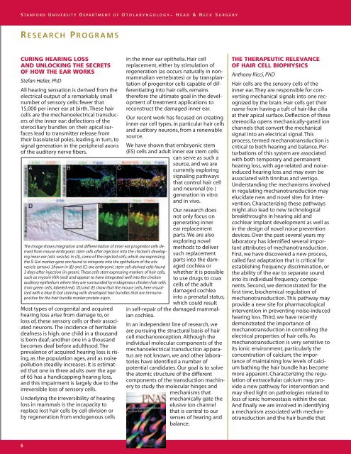
The image shows integration and differentiation <strong>of</strong> inner ear progenitor cells derived<br />
from mouse embryonic stem cells after injection into the chicken’s developing<br />
inner ear (otic vesicle). In (A), some <strong>of</strong> the injected cells, which are expressing<br />
the ß-Gal marker gene are found to integrate into the epithelium <strong>of</strong> the otic<br />
vesicle (arrow). Shown in (B) and (C) are embryonic stem cell-derived cells found<br />
3 days after injection (in green). These cells start expressing markers <strong>of</strong> hair cells,<br />
such as myosin VIIA (red) and appear to have integrated well into the chicken<br />
auditory epithelium where they are surrounded by endogenous chicken hair cells<br />
(non green cells, labeled red). (D) and (E) show that the mouse cells, here visualized<br />
with a blue ß-Gal staining with developed hair bundles that are immunopositive<br />
for the hair bundle marker protein espin.<br />
in the inner ear epithelia. Hair cell<br />
replacement, either by stimulation <strong>of</strong><br />
regeneration (as occurs naturally in nonmammalian<br />
vertebrates) or by transplantation<br />
<strong>of</strong> progenitor cells capable <strong>of</strong> differentiating<br />
into hair cells, remains<br />
therefore the ultimate goal in the development<br />
<strong>of</strong> treatment applications to<br />
reconstruct the damaged inner ear.<br />
Our recent work has focused on creating<br />
inner ear cell types, in particular hair cells<br />
and auditory neurons, from a renewable<br />
source.<br />
We have shown that embryonic stem<br />
(ES) cells and adult inner ear stem cells<br />
can serve as such a<br />
source, and we are<br />
currently exploring<br />
signaling pathways<br />
that control hair cell<br />
and neuronal (re-)<br />
generation in vitro<br />
and in vivo.<br />
Our research does<br />
not only focus on<br />
generating inner<br />
ear replacement<br />
parts. We are also<br />
exploring novel<br />
methods to deliver<br />
such replacement<br />
parts into the damaged<br />
cochlea or<br />
whether it is possible<br />
to use drugs to coax<br />
cells <strong>of</strong> the adult<br />
damaged cochlea<br />
into a prenatal status,<br />
which could result<br />
in self-repair <strong>of</strong> the damaged mammalian<br />
cochlea.<br />
In an independent line <strong>of</strong> research, we<br />
are pursuing the structural basis <strong>of</strong> hair<br />
cell mechanoreception. Although the<br />
individual molecular components <strong>of</strong> the<br />
mechanoelectrical transduction apparatus<br />
are not known, we and other laboratories<br />
have identified a number <strong>of</strong><br />
potential candidates. Our goal is to solve<br />
the atomic structure <strong>of</strong> the different<br />
components <strong>of</strong> the transduction machinery<br />
to study the molecular hinges and<br />
mechanisms that<br />
mechanically gate the<br />
elusive ion channel<br />
that is central to our<br />
senses <strong>of</strong> hearing and<br />
balance.<br />
THE THERAPEUTIC RELEVANCE<br />
OF HAIR CELL BIOPHYSICS<br />
Anthony Ricci, PhD<br />
Hair cells are the sensory cells <strong>of</strong> the<br />
inner ear. They are responsible for converting<br />
mechanical signals into one recognized<br />
by the brain. Hair cells get their<br />
name from having a tuft <strong>of</strong> hair-like cilia<br />
at their apical surface. Deflection <strong>of</strong> these<br />
stereocilia opens mechanically-gated ion<br />
channels that convert the mechanical<br />
signal into an electrical signal. This<br />
process, termed mechanotransduction is<br />
critical to both hearing and balance. Perturbations<br />
<strong>of</strong> this system are associated<br />
with both temporary and permanent<br />
hearing loss, with age-related and noiseinduced<br />
hearing loss and may even be<br />
associated with tinnitus and vertigo.<br />
Understanding the mechanisms involved<br />
in regulating mechanotransduction may<br />
elucidate new and novel sites for intervention.<br />
Characterizing these pathways<br />
might also lead to new technological<br />
breakthroughs in hearing aid and<br />
cochlear implant development as well as<br />
in the design <strong>of</strong> novel noise prevention<br />
devices. Over the past several years my<br />
laboratory has identified several important<br />
attributes <strong>of</strong> mechanotransduction.<br />
First, we have discovered a new process,<br />
called fast adaptation that is critical for<br />
establishing frequency discrimination, or<br />
the ability <strong>of</strong> the ear to separate sound<br />
into its individual frequency components.<br />
Second, we demonstrated for the<br />
first time, biochemical regulation <strong>of</strong><br />
mechanotransduction. This pathway may<br />
provide a new site for pharmacological<br />
intervention in preventing noise-induced<br />
hearing loss. Third, we have recently<br />
demonstrated the importance <strong>of</strong><br />
mechanotransduction in controlling the<br />
electrical properties <strong>of</strong> hair cells. As<br />
mechanotransduction is very sensitive to<br />
its ionic environment, particularly the<br />
concentration <strong>of</strong> calcium, the importance<br />
<strong>of</strong> maintaining low levels <strong>of</strong> calcium<br />
bathing the hair bundle has become<br />
more apparent. Characterizing the regulation<br />
<strong>of</strong> extracellular calcium may provide<br />
a new pathway for intervention and<br />
may shed light on pathologies related to<br />
loss <strong>of</strong> ionic homeostasis within the ear.<br />
And finally we are involved in identifying<br />
a mechanism associated with mechanotransduction<br />
and the hair bundle that