HEAD & NECK SURGERY - Stanford University School of Medicine
HEAD & NECK SURGERY - Stanford University School of Medicine
HEAD & NECK SURGERY - Stanford University School of Medicine

Create successful ePaper yourself
Turn your PDF publications into a flip-book with our unique Google optimized e-Paper software.
may act to amplify low levels <strong>of</strong> sound<br />
and be a major contributor to determining<br />
how the ear can be sensitive to such<br />
low energy sound waves.<br />
Aside from converting a mechanical signal<br />
into an electrical signal, the hair cell<br />
must communicate with the brain<br />
through synaptic transmission. The hair<br />
cell-afferent nerve synapse is very sensitive<br />
to overstimulation where the nerve<br />
ending can swell and retract leading to<br />
hair cell loss. My laboratory has been<br />
investigating the functional properties <strong>of</strong><br />
this synapse to identify the properties<br />
that make it unique in its ability to operate<br />
with such high fidelity and at such<br />
high rates. Here too, the goal is to identify<br />
pathways and mechanisms that might<br />
<strong>of</strong>fer sites for intervention and modification.<br />
And finally, my laboratory is trying to<br />
determine whether hair cells require<br />
extrinsic factors, like innervation,<br />
mechanical stimulation or growth factors<br />
to mature into their final form. This is a<br />
critical question to answer in order to<br />
judge feasibility <strong>of</strong> therapies where hair<br />
cell regeneration from supporting cells<br />
or hair cell development from stem cells<br />
is being investigated as a new therapy to<br />
replace hair cells in damaged cochlea<br />
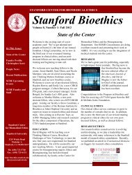
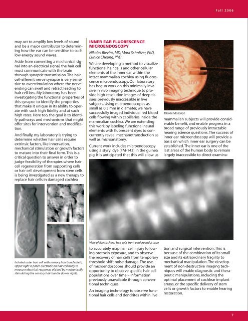
Isolated outer hair cell with sensory hair bundle (left).<br />
Upper right is patch electrode on hair cell body to<br />
measure electrical responses elicited by mechanically<br />
stimulating the sensory hair bundle (lower right).<br />
INNER EAR FLUORESCENCE<br />
MICROENDOSCOPY<br />
Nikolas Blevins, MD, Mark Schnitzer, PhD,<br />
Eunice Cheung, PhD<br />
We are developing a method to visualize<br />
functional hair cells and other cellular<br />
elements <strong>of</strong> the inner ear within the<br />
intact mammalian cochlea using fluorescence<br />
microendoscopy. Our laboratory<br />
has begun work on this minimally invasive<br />
in vivo imaging technique to provide<br />
high-resolution images <strong>of</strong> deep tissues<br />
previously inaccessible in live<br />
subjects. Using microendoscopes as<br />
small as 0.3 mm in diameter, we have<br />
successfully imaged individual red blood<br />
cells flowing within capillaries inside the<br />
mammalian cochlea. We are extending<br />
this work by labeling functional neural<br />
elements with fluorescent dyes to concurrently<br />
reveal mechanotransduction as<br />
well as microanatomy.<br />
Current work includes microendoscopy<br />
using a styryl dye (FM-143) in the guinea<br />
pig. It is anticipated that this will allow us<br />
View <strong>of</strong> live cochlear hair cells from a microendoscope<br />
to accurately map hair cell injury following<br />
ototoxin exposure, and to observe<br />
the recovery <strong>of</strong> hair cells from temporary<br />
threshold shift noise damage. The use<br />
<strong>of</strong> microendoscopes should provide an<br />
opportunity to observe specific hair cell<br />
populations over time – information<br />
previously unavailable through conventional<br />
techniques.<br />
An imaging technology to observe functional<br />
hair cells and dendrites within live<br />
MIcroendoscope<br />
Fall 2006<br />
mammalian subjects will provide considerable<br />
benefit, and enable progress in a<br />
broad range <strong>of</strong> previously intractable<br />
hearing science questions. The success <strong>of</strong><br />
inner ear microendoscopy will provide a<br />
basis on which inner ear surgery can be<br />
established. The inner ear is one <strong>of</strong> the<br />
last areas <strong>of</strong> the human body to remain<br />
largely inaccessible to direct examina-<br />
tion and surgical intervention. This is<br />
because <strong>of</strong> the combination <strong>of</strong> its small<br />
size and its extraordinary fragility to<br />
mechanical manipulation. The development<br />
<strong>of</strong> non-destructive imaging techniques<br />
will enable diagnostic and therapeutic<br />
manipulations, including the<br />
optimal placement <strong>of</strong> cochlear implant<br />
arrays, or the specific delivery <strong>of</strong> stem<br />
cells or growth factors to enable hearing<br />
restoration.<br />
7