Fournier's gangrene: Be alert for this medical emergency
Fournier's gangrene: Be alert for this medical emergency
Fournier's gangrene: Be alert for this medical emergency

Create successful ePaper yourself
Turn your PDF publications into a flip-book with our unique Google optimized e-Paper software.
CME<br />
44 JAAPA • NOVEMBER 2007 • 20(11) • www.jaapa.com<br />
EARN CATEGORY I CME CREDIT by reading <strong>this</strong> article and the article beginning on page 21 and successfully<br />
completing the posttest on page 49. Successful completion is defined as a cumulative score of at least 70%<br />
correct. This material has been reviewed and is approved <strong>for</strong> 1 hour of clinical Category I (Preapproved) CME credit<br />
by the AAPA. The term of approval is <strong>for</strong> 1 year from the publication date of November 2007.<br />
LEARNING OBJECTIVES<br />
● Describe Fournier’s <strong>gangrene</strong>, including its comorbidities, multiple causes, and multi-organism nature<br />
● Understand the clinical presentation of Fournier’s <strong>gangrene</strong> and how it can be diagnosed<br />
● Outline the treatment modalities, including aggressive resuscitation, utilization of broad-spectrum<br />
antibiotics, and emergent surgical therapy<br />
Fournier’s <strong>gangrene</strong>: <strong>Be</strong> <strong>alert</strong><br />
<strong>for</strong> <strong>this</strong> <strong>medical</strong> <strong>emergency</strong><br />
Intense pain and tenderness in the genitalia are hallmarks of <strong>this</strong> infection. Early diagnosis,<br />
prompt antibiotic administration, and surgical debridement are essential.<br />
Draion M. Burch, DO; Timothy J. Barreiro, DO, FCCP,<br />
FACOI; Vincent W. Vanek, MD, FACS, CNSP<br />
Fournier’s <strong>gangrene</strong> is an aggressive and rapidly<br />
spreading infection of soft tissue, or necrotizing<br />
fasciitis, that involves the deep and superficial fascia<br />
of the perineum. 1 The rate of fascial necrosis<br />
in Fournier’s <strong>gangrene</strong> is reported to be 2 to 3<br />
cm/h. 1 Thrombosis of subcutaneous and cutaneous blood<br />
vessels produces <strong>gangrene</strong>, but the fascial necrosis is usually<br />
more extensive than the visible <strong>gangrene</strong> suggests. 2 Classic<br />
findings are necrosis of the superficial and deep fascial<br />
planes, fibrinoid coagulation of the nutrient arterioles, polymorphonuclear<br />
cell infiltration, and positive microorganism<br />
culture of involved tissues.<br />
An estimated 750 cases have been reported in the literature<br />
since Fournier first described the disease in 1883. 3 Fournier’s<br />
<strong>gangrene</strong>, an uncommon disease with no seasonal variation,<br />
is not indigenous to any region of the world; however, the<br />
largest number of cases originates from the African continent.<br />
The typical patient is a male in his sixth or seventh<br />
decade with comorbidities (see Table 1, page 46). Fournier’s<br />
<strong>gangrene</strong> is 10 times more common in men than women. 4<br />
Women tend to develop the disease following childbirth. 5<br />
Regardless of gender predominance, multiple risk factors are<br />
associated with the disease 6 (see Table 2, page 46).<br />
EPIDEMIOLOGY AND ETIOLOGY<br />
Fournier’s <strong>gangrene</strong> was originally thought to be an idiopathic<br />
<strong>gangrene</strong> of the genitalia; however, a specific etiology is<br />
found in approximately 95% of cases. 7 Anorectal abscess, genitourinary<br />
infection, and traumatic injury are the most common<br />
causes. 8 Comorbid diseases that compromise the immune<br />
system are a predisposing factor. The most frequent<br />
systemic illness associated with Fournier’s <strong>gangrene</strong> is diabetes<br />
mellitus, which is seen in 10% to 60% of cases. 9 Diabetes<br />
causes defective phagocytosis, decreased cellular immunity,<br />
and microvascular disease with resultant ischemia. 10,11<br />
An iatrogenic or noniatrogenic injury to the perineum can<br />
initiate the development of Fournier’s <strong>gangrene</strong>. Cases have<br />
been reported following hydrocele aspiration, blunt thoracic<br />
trauma, vasectomy, sparganosis (parasitic infection) of the<br />
scrotum, transrectal prostate biopsy, and penile self-injection<br />
with cocaine. Other case reports include the following etiologies:<br />
complications of varicella in a child, steroid enema use<br />
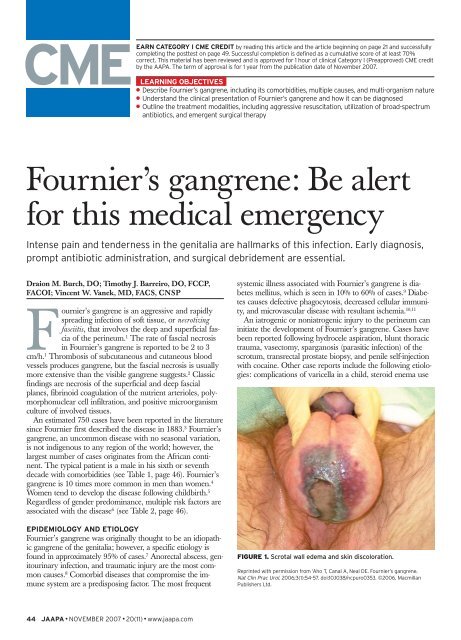
FIGURE 1. Scrotal wall edema and skin discoloration.<br />
Reprinted with permission from ‘Aho T, Canal A, Neal DE. Fournier’s <strong>gangrene</strong>.<br />
Nat Clin Prac Urol. 2006;3(1):54-57. doi:10.1038/ncpuro0353. ©2006. Macmillan<br />
Publishers Ltd.
with radiation treatment <strong>for</strong> proctitis, spinal cord injury, and<br />
femoral heroin injection. 12<br />
The necrotizing process commonly originates with an<br />
infection in the anorectum, the urogenital tract, or the skin<br />
around the perineum. 13 Anorectal causes include an infection<br />
in the perianal glands, colonic diverticulitis, decubitus ulcers,<br />
or a colorectal injury or malignancy. Urogenital etiologies<br />
include an infection in the bulbourethral glands, a urethral<br />
injury, a lower urinary tract infection, or an iatrogenic injury<br />
secondary to stricture manipulation. Dermatologic causes<br />
include hidradenitis suppurativa, scrotal pressure ulceration,<br />
trauma, a surgical complication, or intentional trauma such<br />
as skin-popping—a <strong>for</strong>m of injection drug abuse. Other diseases<br />
in addition to diabetes that increase the risk of developing<br />
Fournier’s <strong>gangrene</strong> are systemic lupus erythematosus,<br />
Crohn’s disease, and HIV infection. A less commonly<br />
reported cause is bone marrow malignancy. 14,15<br />
Fournier’s <strong>gangrene</strong>, like most cases of necrotizing infection,<br />
has a multiorganism nature; 16 the disease is due to polymicrobial<br />
infection with a mixture of aerobic and anaerobic organisms.<br />
17 The majority of cases are caused by normal flora of<br />
the lower GI tract, 18 most commonly Escherichia coli. 19 Other<br />
causative micro-organisms include Staphylococcus, Streptococcus,<br />
and Enterobacteriaceae species, anaerobic organisms, and fungi.<br />
The infection is rarely caused by one organism; as many<br />
as five species may be cultured.<br />
The hallmark of Fournier’s <strong>gangrene</strong> is intense pain and<br />
tenderness in the genitalia. The clinical course progresses<br />
through several phases. First, fever and lethargy develop.<br />
Next, patients experience intense genital pain and tenderness<br />
associated with edema of the overlying skin, which appears<br />
dusky, indicating subcutaneous crepitance. Soft tissue gas, a<br />
byproduct of anaerobic metabolism, is produced. 20 As genital<br />
pain and tenderness increase, obvious <strong>gangrene</strong> in a portion<br />
of the genitalia and purulent drainage materialize (see Figure<br />
1). Systemic effects range from local tenderness to septic<br />
shock, depending on necrotic progression.<br />
MAKING THE DIAGNOSIS<br />
Fournier’s <strong>gangrene</strong> is diagnosed primarily on clinical<br />
grounds, as diagnostic studies risk postponing treatment. In<br />
cases of rapid accessibility, uncertain diagnosis, or suspicion<br />
KEY POINTS<br />
■ Fournier’s <strong>gangrene</strong> was originally thought to be an idiopathic <strong>gangrene</strong> of the genitalia;<br />
however, a specific etiology is found in approximately 95% of cases. Anorectal abscesses,<br />
genitourinary infections, and traumatic injuries are the most common causes.<br />
■ Typically, fluctuance, soft-tissue crepitance, localized tenderness, or occult wounds in the<br />
genitalia, perineum, and anorectal area should <strong>alert</strong> the examiner to the possibility of<br />
Fournier’s <strong>gangrene</strong>. A CBC, comprehensive metabolic panel, coagulation profile, and blood<br />
cultures should be obtained.<br />
■ Once the diagnosis is established, emergent surgical excision of all necrotic tissue must be<br />
per<strong>for</strong>med. Given the potential fulminant nature of <strong>this</strong> necrotizing process, repeat debridement<br />
procedures are usually needed to completely eradicate the infection.<br />
FIGURE 2. CT shows small pockets of gas in the rectum (arrows).<br />
FIGURE 3. Fluid collections along the deep fascial planes (arrows)<br />
are demonstrated on CT.<br />
of retroperitoneal or intra-abdominal sources of infection,<br />
imaging studies should be considered. 21 Careful palpation of<br />
the genitalia and perineum and a digital rectal examination<br />
are important parts of the physical examination. Table 3<br />
(page 46) lists the signs and symptoms of Fournier’s <strong>gangrene</strong>.<br />
Typically, fluctuance, soft-tissue crepitance, localized<br />
tenderness, or occult wounds should <strong>alert</strong> the examiner to<br />
the possibility of Fournier’s <strong>gangrene</strong>. A CBC, comprehensive<br />
metabolic panel, coagulation profile, and blood cultures<br />
should be obtained.<br />
Continued on page 46<br />
COMPETENCIES<br />
●●●●● Medical knowledge<br />
● Interpersonal & communication skills<br />
●●● Patient care<br />
● Professionalism<br />
● Practice-based learning and improvement<br />
● Systems-based practice<br />
NOVEMBER 2007 • 20(11) • www.jaapa.com • JAAPA 45
CME Fournier’s <strong>gangrene</strong><br />
Imaging studies are more likely to detect gas within the<br />
soft tissues than is physical examination. An initial imaging<br />
study includes a plain radiograph, which may show moderate-to-large<br />
amounts of soft-tissue gas or <strong>for</strong>eign bodies.<br />
Ultrasonography also detects fluid or gas within the soft tissues<br />
22 and is the preferred method. 23 Small pockets of softtissue<br />
gas are more readily detected on CT (see Figure 2,<br />
page 45). CT also can demonstrate fluid collections tracking<br />
along deep fascial planes (see Figure 3, page 45). MRI gives<br />
greater soft tissue detail than CT; however, it creates greater<br />
logistical challenges, especially in critically ill patients.<br />
The definitive diagnosis of Fournier’s <strong>gangrene</strong> is established<br />
by surgical examination under anesthesia, with an<br />
incision into the area of greatest clinical concern. If gangre-<br />
TABLE 1. Common predisposing comorbidities<br />
Cirrhosis<br />
Diabetes mellitus<br />
High-risk behaviors (alcohol or IV drug abuse)<br />
Immunosuppression<br />
Malignancies<br />
Malnutrition<br />
Morbid obesity<br />
Vascular disease of the pelvis<br />
TABLE 2. Risk factors <strong>for</strong> Fournier’s <strong>gangrene</strong><br />
Circumcision<br />
Episiotomy<br />
Extravasations of urine (periurethrally or through cutaneous fistula)<br />
Hernioplasty<br />
Hysterectomy<br />
Local trauma or instrumentation to the perineum<br />
Paraphimosis<br />
Septic abortion<br />
Urethral stricture caused by sexually transmitted diseases<br />
TABLE 3. Signs and symptoms of Fournier’s <strong>gangrene</strong><br />
Crepitant skin (“spongy” to the touch)<br />
Dead and discolored (gray-black) tissue; pus weeping from injury<br />
Fever and lethargy<br />
Increasing genital pain and erythema or severe genital pain accompanied<br />
by tenderness and swelling of the penis and scrotum<br />
46 JAAPA • NOVEMBER 2007 • 20(11) • www.jaapa.com<br />
nous tissue is present or purulence is drained, the diagnosis<br />
is established. Tissue samples should be sent <strong>for</strong> anaerobic<br />
and aerobic cultures, as well as histopathologic assessment.<br />
Ultimately, early identification of Fournier’s <strong>gangrene</strong> is essential<br />
<strong>for</strong> a good prognosis. 24<br />
TREATMENT<br />
In patients who present with systemic toxicity manifesting as<br />
hypoperfusion and/or organ failure, aggressive resuscitation<br />
to return normal organ perfusion and function must take<br />
precedence. 25 Antibiotics with broad-spectrum coverage<br />
against staphylococci, streptococci, Enterobacteriaceae species,<br />
and anaerobes should be administered. If initial tissue stains<br />
show fungi, an antifungal should be included in the regimen.<br />
Empiric antibiotic regimens should be adjusted when the<br />
infective organisms are identified.<br />
Once the diagnosis is established, emergent surgical excision<br />
of all necrotic tissue is required. The skin should be<br />
opened wide to expose the full extent of underlying fascial<br />
and subcutaneous tissue necrosis. Given the potential fulminant<br />
nature of <strong>this</strong> necrotizing process, repeat debridement<br />
procedures are usually needed to eradicate the infection. If<br />
perineal involvement is extensive, fecal diversion should be<br />
per<strong>for</strong>med to eliminate potential contamination of the<br />
wounds; urinary diversion is accomplished via a urethral<br />
catheter. Hyperbaric oxygen (HBO) therapy has a theoretical<br />
role in treating Fournier’s <strong>gangrene</strong>, but results of <strong>this</strong> therapy<br />
are mixed. 26 HBO therapy increases tissue-oxygen tension,<br />
leukocyte activation, oxygen free-radical production,<br />
capillary angiogenesis, fibroblast proliferation, and vasoconstriction<br />
and decreases anaerobe multiplication. 27 Prompt<br />
antibiotic administration and surgical debridement (with or<br />
without HBO) are the cornerstones of therapy. 28<br />
CONCLUSION<br />
In the pre-antibiotic era, Fournier’s <strong>gangrene</strong> was commonly<br />
fatal; even today, it poses a significant risk of morbidity and<br />
mortality. 29 Despite aggressive therapy, the mortality rate <strong>for</strong><br />
patients with Fournier’s <strong>gangrene</strong> is nearly 50% because of<br />
the aggressive nature of the infection and the presence of<br />
underlying comorbidities. 30 Delays in diagnosis or treatment<br />
increase the mortality rate. For example, a 24-hour delay in<br />
radical debridement increases the mortality rate by 11.5%;<br />
a 6-day delay is associated with a mortality rate of 76%. 31<br />
Additional factors associated with high mortality include<br />
anorectal origin, advanced age, extensive disease, shock or<br />
sepsis at presentation, renal failure, and hepatic dysfunction.<br />
32 Multiorgan system failure secondary to gram-negative<br />
sepsis is the most common cause of death. 33 Early clinical<br />
identification and prompt, aggressive treatment are essential<br />
<strong>for</strong> reducing mortality and morbidity in patients presenting<br />
with <strong>this</strong> disease. JAAPA<br />
Draion Burch is an intern in the Department of Obstetrics at St. John Health<br />
System, Michigan State University College of Osteopathic Medicine, Warren.<br />
Timothy Barreiro is assistant professor of internal medicine and Vincent
Vanek is professor of surgery at Northeastern Ohio Universities Colleges of<br />
Medicine and Pharmacy, Youngstown. The authors have indicated no relationships<br />
to disclose relating to the content of <strong>this</strong> article.<br />
REFERENCES<br />
1. Yaghan RJ, Al-Jaberi TM, Bani-Hani I. Fournier’s <strong>gangrene</strong>: changing face of the disease. Dis<br />
Colon Rectum. 2000;43(9):1300-1308.<br />
2. Harden SP, Creasy TS. Case of the month. All that glistens isn’t gold (so do be sure the surgeon’s<br />
told!). Br J Radiol. 2003;76(911):841-842.<br />
3. Morrison D, Blaivas M, Lyon M. Emergency diagnosis of Fournier’s <strong>gangrene</strong> with bedside ultrasound.<br />
Am J Emerg Med. 2005;23(4):544-547.<br />
4. Ekelius L, Björkman H, Kalin M, Fohlman J. Fournier’s <strong>gangrene</strong> after genital piercing. Scand J<br />
Infect Dis. 2004;36(8):610-612.<br />
5. Quatan N, Kirby RS. Improving outcomes in Fournier’s <strong>gangrene</strong>. BJU Int. 2004;93(6):691-692.<br />
6. David JE, Yale SH, Goldman IL. Urology: scrotal pain. Clin Med Res. 2003;1(2):159-160.<br />
7. Clayton MD, Fowler JE Jr, Sharifi R, Pearl RK. Causes, presentation and survival of<br />
fifty-seven patients with necrotizing fasciitis of the male genitalia. Surg Gynecol Obstet.<br />
1990;170(1):49-55.<br />
8. Bronder CS, Cowey A, Hill J. Delayed stoma <strong>for</strong>mation in Fournier’s <strong>gangrene</strong>. Colorectal Dis.<br />
2004;6(6):518-520.<br />
9. Bayar S, Unal AE, Demirkan A, et al. Fournier’s <strong>gangrene</strong> complicating blunt thoracic trauma.<br />
Surgery. 2004;135(6):693-694.<br />
10. Eke N. Fournier’s <strong>gangrene</strong>: a review of 1726 cases. Br J Surg. 2000;87(6):718-728.<br />
11. Jean-Charles N, Sadler MA. Necrotizing perineal fasciitis in two paraplegic nursing-home residents:<br />
CT imaging findings. Abdom Imaging. 2001;26(4):443-446.<br />
12. Nambiar PK, Lander S, Midha M, Ha C. Fournier <strong>gangrene</strong> in spinal cord injury: a case report.<br />
J Spinal Cord Med. 2005;28(2):121-124.<br />
13. Bakshi C, Banavali S, Lokeshwar N, et al. Clustering of Fournier (male genital) <strong>gangrene</strong> cases in<br />
a pediatric cancer ward. Med Pediatr Oncol. 2003;41(5):472-474.<br />
14. Faber JH, Girbes AR, Daenen S. Fournier’s <strong>gangrene</strong> as first presentation of promyelocytic<br />
leukemia. Leuk Res. 1998;22(5):473-476.<br />
15. Martinelli G, Alessandrino EP, <strong>Be</strong>rnasconi P, et al. Fournier’s <strong>gangrene</strong>: a clinical presentation of<br />
necrotizing fasciitis after bone marrow transplantation. Bone Marrow Transplant. 1998;22(10):<br />
1023-1026.<br />
16. Hejase MJ, Simonin JE, Bihrle R, Coogan CL. Genital Fournier’s <strong>gangrene</strong>: experience with 38<br />
patients. Urology. 1996;47(5):734-739.<br />
17. Maguiña P, Palmieri TL, Greenhalgh DG. Split thickness skin grafting <strong>for</strong> recreation of the scrotum<br />
following Fournier’s <strong>gangrene</strong>. Burns. 2003;29(8):857-862.<br />
18. Tleyjeh IM, Routh J, Qutub MO, et al. Lactobacillus gasseri causing Fournier’s <strong>gangrene</strong>. Scand J<br />
Infect Dis. 2004;36(6-7):501-503.<br />
19. Kiliç A, Aksoy Y, Kiliç L. Fournier’s <strong>gangrene</strong>: etiology, treatment and complications. Ann Plast<br />
Surg. 2001;47(5):523-527.<br />
20. Uppot RN, Levy HM, Patel PH. Case 54: Fournier <strong>gangrene</strong>. Radiology. 2003;226(1):115-117.<br />
21. Maréchal R, Taccone F. Diagnosis and treatment of an unusual cause of sepsis in a diabetic<br />
patient: a Fournier’s <strong>gangrene</strong>. Acta Clin Bleg. 2005;60(1):17-21.<br />
22. Kane CJ, Nash P, McAninch JW. Ultrasonographic appearance of necrotizing <strong>gangrene</strong>: aid in<br />
early diagnosis. Urology. 1996;48(1):142-144.<br />
23. Fan CM, Whitman GJ, Chew FS. Radiologic-Pathologic Conferences of the Massachusetts General<br />
Hospital. Necrotizing fasciitis of the scrotum (Fournier’s <strong>gangrene</strong>). AJR Am J Roentgenol. 1996;<br />
166(5):1164.<br />
24. Tayib AM, Mosli HA, Abdulwahab MH, Atwa MA. Fournier’s <strong>gangrene</strong> in diabetic and renal failure<br />
patients. Saudi Med J. 2003;24(10):1105-1108.<br />
25. Salvino C, Har<strong>for</strong>d FJ, Dobrin PB. Necrotizing infections of the perineum. South Med J. 1993;<br />
86(8):908-911.<br />
26. Hollabaugh RS Jr, Dmochowski RR, Hickerson WL, Cox CE. Fournier’s <strong>gangrene</strong>: therapeutic<br />
impact of hyperbaric oxygen. Plast Reconstr Surg. 1998;101(1):94-100.<br />
27. Ali MZ. Fournier’s <strong>gangrene</strong>—a rare complication of hydrocele aspiration. J Coll Physicians Surg<br />
Pak. 2004;14(5):304-305.<br />
28. Marinella MA. Group C streptococcal sepsis complicating Fournier <strong>gangrene</strong>. Southern Medical<br />
Journal. 2005;98(9):921-923.<br />
29. Erikoglu M, Tavli S, Turk S. Fournier’s <strong>gangrene</strong> after renal transplantation. Nephrol Dial<br />
Transplant. 2005;20(2):449-450.<br />
30. Riedler I, Primus G, Trummer H, et al. Fournier’s <strong>gangrene</strong> after tension-free vaginal tape (TVT)<br />
procedure. Int Urogynecol J Pelvic Floor Dysfunct. 2004;15(2):145-146.<br />
31. Paty R, Smith AD. Gangrene and Fournier’s <strong>gangrene</strong>. Urol Clin North Am. 1992;19(1):149-162.<br />
32. Lehnhardt M, Steinstraesser L, Druecke D, et al. Fournier’s <strong>gangrene</strong> after Milligan-Morgan hemorrhoidectomy<br />
requiring subsequent abdominoperineal resection of the rectum: report of a<br />
case. Dis Colon Rectum. 2004;47(10):1729-1733.<br />
33. Murphy BL, Pezzullo JA. Images in medicine. Fournier’s <strong>gangrene</strong>. Med Health. 2003;86(4):121.<br />
NOVEMBER 2007 • 20(11) • www.jaapa.com • JAAPA 47