Chapter 25 :: Parapsoriasis and Pityriasis Lichenoides :: Gary S ...
Chapter 25 :: Parapsoriasis and Pityriasis Lichenoides :: Gary S ...
Chapter 25 :: Parapsoriasis and Pityriasis Lichenoides :: Gary S ...

Create successful ePaper yourself
Turn your PDF publications into a flip-book with our unique Google optimized e-Paper software.
4<br />
Section 4 :: Inflammatory Disorders Based on T-Cell Reactivity <strong>and</strong> Dysregulation<br />
8<br />
whether these agents are actively involved in disease<br />
pathogenesis or merely coincidental byst<strong>and</strong>ers; however,<br />
several cases associated with toxoplasmosis have<br />
cleared fairly quickly in response to specific therapy. 69<br />
A tenfold higher level of maternal keratinocytes have<br />
been reported in the epidermis of children with PL<br />
compared to controls. 79<br />
Immunohistologic studies have shown a reduction in<br />
CD1a + antigen-presenting dendritic (Langerhans) cells<br />
within the central epidermis of pityriasis lichenoides<br />
lesions. 80 Keratinocytes <strong>and</strong> endothelial cells are HLA-<br />
DR + , which suggests activation by T-cell cytokines. 80<br />
CD8 + T cells predominate in PLEVA, whereas either<br />
CD8 + or CD4 + T cells predominate in PLC. 80–82 Many<br />
of these T cells express memory proteins (CD45RO)<br />
<strong>and</strong> cytolytic proteins (TIA-1 <strong>and</strong> granzyme B). 72,73<br />
Dominant T-cell clonality has been demonstrated<br />
in about half of PLEVA cases <strong>and</strong> a minority of PLC<br />
cases. 32,83,84 In aggregate, these findings raise the possibility<br />
that pityriasis lichenoides is a variably clonal<br />
cytolytic memory T-cell lymphoproliferative response<br />
to one or more foreign antigens. Deposition of immunoglobulin<br />
M, C3, <strong>and</strong> fibrin in <strong>and</strong> around blood vessels<br />
<strong>and</strong> along the dermal–epidermal junction in early<br />
acute lesions suggests a possible concomitant humoral<br />
immune response, although this could be a secondary<br />
phenomenon.<br />
The relationship of pityriasis lichenoides to lymphomatoid<br />
papulosis remains controversial 10,51,80 (see<br />
also <strong>Chapter</strong>s 144 <strong>and</strong> 145). Common features include<br />
dominant T-cell clonality <strong>and</strong> spontaneous resolution<br />
of papular, predominantly lymphoid lesions. Furthermore,<br />
individual lesions with the clinicopathologic<br />
characteristics of either pityriasis lichenoides or lymphomatoid<br />
papulosis can coexist in the same patient,<br />
either concurrently or serially. It remains to be determined<br />
whether this can be explained as an artifact of<br />
sampling lymphomatoid papulosis lesions at various<br />
stages of their evolution. The presence of large CD30 +<br />
atypical lymphoid cells is the hallmark of lymphomatoid<br />
papulosis (at least types A <strong>and</strong> C). 84 Furthermore,<br />
these cells are typically CD4 + <strong>and</strong> often lack one or<br />
more mature T-cell antigens such as CD2, CD3, <strong>and</strong><br />
CD5. These features serve to distinguish lymphomatoid<br />
papulosis from pityriasis lichenoides. Although<br />
occasional CD30 + cells can be seen in a wide variety<br />
of dermatoses, the presence of any appreciable number<br />
should favor lymphomatoid papulosis over pityriasis<br />
lichenoides as a matter of definition. It may be<br />
that the “PLC-PLEVA” <strong>and</strong> “lymphomatoid papulosis–CD30<br />
+ anaplastic large cell lymphoma” disease<br />
spectra are intersecting rather than overlapping entities,<br />
i.e., although pityriasis lichenoides is a distinct<br />
cutaneous T-cell disorder, it is possible that it may<br />
sometimes serve as fertile soil for the development of<br />
the CD30 + T-cell clone characteristic of lymphomatoid<br />
papulosis.<br />
CLINICAL FINDINGS<br />
CUTANEOUS LESIONS. PLC <strong>and</strong> PLEVA exist<br />
on a clinicopathologic continuum. 2,51 Therefore,<br />
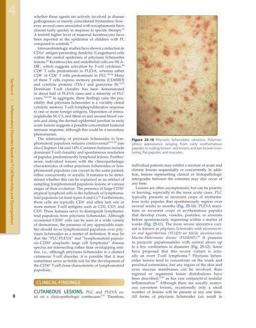
Figure <strong>25</strong>-10 <strong>Pityriasis</strong> lichenoides chronica. Polymorphous<br />
appearance ranging from early erythematous<br />
papules to scaling brown–red lesions <strong>and</strong> tan-brown involuting,<br />
flat papules, <strong>and</strong> macules.<br />
individual patients may exhibit a mixture of acute <strong>and</strong><br />
chronic lesions sequentially or concurrently. In addition,<br />
lesions representing clinical or histopathologic<br />
intergrades between the extremes may also occur at<br />
any time.<br />
Lesions are often asymptomatic but can be pruritic<br />
or burning, especially in the more acute cases. PLC<br />
typically presents as recurrent crops of erythematous<br />
scaly papules that spontaneously regress over<br />
several weeks to months (Fig. <strong>25</strong>-10). PLEVA manifests<br />
as recurrent crops of erythematous papules<br />
that develop crusts, vesicles, pustules, or erosions<br />
before spontaneously regressing within a matter of<br />
weeks (Fig. <strong>25</strong>-11). The more severe ulcerative variant<br />
is known as pityriasis lichenoides with ulceronecrosis<br />
<strong>and</strong> hyperthermia (PLUH) or febrile ulceronecrotic<br />
Mucha–Habermann disease (FUMHD). 85<br />
It presents<br />
as purpuric papulonodules with central ulcers up<br />
to a few centimeters in diameter (Fig. <strong>25</strong>-12). Some<br />
have proposed that this severe variant is actually<br />
an overt T-cell lymphoma. 31 <strong>Pityriasis</strong> lichenoides<br />
lesions tend to concentrate on the trunk <strong>and</strong><br />
proximal extremities, but any region of the skin <strong>and</strong><br />
even mucous membranes can be involved. Rare<br />
regional or segmental lesion distributions have<br />
been described, 10,86 as has rare conjunctival nodular<br />
inflammation. 87 Although there are usually numerous<br />
coexistent lesions, occasionally only a small<br />
number of lesions will be present at any one time.<br />
All forms of pityriasis lichenoides can result in