Paediatric Protocols for Malaysian Hospitals 3rd Edition 2012.pdf
Paediatric Protocols for Malaysian Hospitals 3rd Edition 2012.pdf
Paediatric Protocols for Malaysian Hospitals 3rd Edition 2012.pdf

You also want an ePaper? Increase the reach of your titles
YUMPU automatically turns print PDFs into web optimized ePapers that Google loves.
PAEDIATRIC<br />
PROTOCOLS<br />
For <strong>Malaysian</strong> <strong>Hospitals</strong><br />
Hussain Imam Hj Muhammad Ismail<br />
Ng Hoong Phak<br />
Terrence Thomas<br />
<strong>3rd</strong> <strong>Edition</strong><br />
Kementerian Kesihatan Malaysia
PAEDIATRIC<br />
PROTOCOLS<br />
For <strong>Malaysian</strong> <strong>Hospitals</strong><br />
Hussain Imam Hj Muhammad Ismail<br />
Ng Hoong Phak<br />
Terrence Thomas<br />
i<br />
<strong>3rd</strong> <strong>Edition</strong><br />
Scan this QR code to<br />
download the electronic<br />
<strong>Paediatric</strong> Protocol <strong>3rd</strong> <strong>Edition</strong><br />
Kementerian Kesihatan Malaysia
FOREWORD BY THE DIRECTOR GENERAL OF HEALTH<br />
Malaysia like the rest of the world has 3 more years to achieve the Millennium<br />
Developmental Goals (MDG). MDG 4 is concerned with under 5 mortality. Although<br />
we have done very well since lndependence to reduce our infant and toddler<br />
mortality rates, we are now faced with some last lap issues in achieving this goal.<br />
Despite urbanization there are still many children in the rural areas. This constitutes<br />
a vulnerable group in many ways. Among the factors contributing to this vulnerability<br />
is the distance from specialist care.<br />
There is a need to ensure that doctors in the frontline are well equipped to handle<br />
common paediatric emergencies so that proper care can be instituted from the<br />
very beginning.<br />
Although all doctors are now required to do 4 months of pre-registration training in<br />
<strong>Paediatric</strong>s, this is insufficient to prepare them <strong>for</strong> all the conditions they are likely<br />
to meet as Medical Officers in district hospitals and health clinics. Hence the ef<strong>for</strong>t<br />
made by the paediatricians to prepare a protocol book covering all the common<br />
paediatric problems is laudable. I would also like to congratulate them <strong>for</strong><br />
bringing out a third edition within 4 years of the previous edition.<br />
l am confident that this third edition will contribute to improving the care of<br />
children attending the Ministry’s facilities throughout the country.<br />
Dato’Sri Dr Hasan Bin Abdul Rahman<br />
Director General of Health, Malavsia<br />
iii
FOREWORD TO THE THIRD EDITION<br />
It has been 7 years since we produced the first edition of a national protocol<br />
book <strong>for</strong> <strong>Paediatric</strong>s. This ef<strong>for</strong>t was of course inspired by the Sarawak <strong>Paediatric</strong><br />
<strong>Protocols</strong> initiated by Dr Tan Poh Tin. The 2nd edition in 2008 has proven to be<br />
very popular and we have had to recruit the services of the <strong>Malaysian</strong> <strong>Paediatric</strong><br />
Association (MPA) to produce extra copies <strong>for</strong> sale. It is now the standard<br />
reference <strong>for</strong> House officers in <strong>Paediatric</strong>s.<br />
In producing a third edition we have retained the size and style of the current version,<br />
essentially only updating the contents. Again it is targeted at young doctors<br />
in the service many of whom seem to have had a suboptimal exposure to<br />
paediatrics in their undergraduate years. It is hoped that the protocol book will<br />
help them fill in the gaps as they prepare to serve in district hospitals and health<br />
clinics.<br />
The Ministry of Health has once again agreed to sponsor the printing of 1000<br />
books and 500 CDs <strong>for</strong> distribution to MOH facilities. We shall be soliciting the<br />
help of the MPA in producing extra books to be sold to those who wish to have a<br />
personal copy. As a result of the full PDF version being available on the MPA<br />
website, we have had requests from as far away as Kenya and Egypt to download<br />
and print the material <strong>for</strong> local distribution. We have gladly allowed this in the<br />
hope that it will contribute to better care of ill children in those and other neighbouring<br />
countries.<br />
As previously this new edition is only possible because of the willingness of busy<br />
clinicians to chip in and update the content <strong>for</strong> purely altruistic reasons and we<br />
hope this spirit will persist in our fraternity. Prof Frank Shann has gracefully agreed<br />
<strong>for</strong> the latest edition of his drug dosages handbook to be incorporated into the<br />
new edition. The Director General of Health has also kindly provided a <strong>for</strong>eword<br />
to this edition.<br />
We wish to thank all who have made this new edition possible and hope this<br />
combined ef<strong>for</strong>t will help in improving the wellbeing of the children entrusted to<br />
our care.<br />
Hussain Imam B. Hj Muhammad Ismail<br />
Ng Hoong Phak<br />
Terrence Thomas<br />
iv
Dr Airena Mohamad Nor,<br />
<strong>Paediatric</strong>ian<br />
Hospital Tuanku Jaafar, Seremban.<br />
Dr. Alex Khoo Peng Chuan<br />
<strong>Paediatric</strong> Neurologist<br />
Hospital Raja Permaisuri Bainun, Ipoh.<br />
Dr. Amar-Singh HSS<br />
Consultant Community <strong>Paediatric</strong>ian &<br />
Head, Dept. of <strong>Paediatric</strong>s<br />
Hospital Raja Permaisuri Bainun, Ipoh<br />
Dr. Angeline Wan<br />
Consultant Neonatologist<br />
Head, Dept. of <strong>Paediatric</strong>s<br />
Hospital Pakar Sultanah Fatimah, Muar<br />
Ms. Anne John<br />
Consultant <strong>Paediatric</strong> Surgeon<br />
Hospital Umum Sarawak, Kuching<br />
Dr. Bina Menon<br />
Consultant <strong>Paediatric</strong> Haemato-Oncologist<br />
Hospital Kuala Lumpur (Sessional)<br />
Dr. Chan Lee Gaik<br />
Consultant Neonatologist<br />
& Head, Dept. of <strong>Paediatric</strong>s<br />
Hospital Umum Sarawak, Kuching<br />
Dr. Chee Seok Chiong<br />
Consultant Neonatologist<br />
Hospital Selayang<br />
Dr. Chin Choy Nyok<br />
Consultant Neonatologist<br />
& Head, Dept. of <strong>Paediatric</strong>s<br />
Hospital Tengku Ampuan Afzan, Kuantan<br />
Dr. Chong Sze Yee<br />
<strong>Paediatric</strong> Gastroenterology & Hepatology<br />
Fellow<br />
Hospital Selayang<br />
Dr. Eni Juraida<br />
Consultant <strong>Paediatric</strong> Haemato-Oncologist<br />
Hospital Kuala Lumpur<br />
Dr. Farah Inaz Syed Abdullah<br />
Consultant Neonatologist<br />
Hospital Kuala Lumpur<br />
LIST OF CONTRIBUTORS<br />
v<br />
Dr. Fazila Mohamed Kutty<br />
Neonatologist<br />
Hospital Serdang<br />
Dr. Fong Siew Moy<br />
<strong>Paediatric</strong> Infectious Disease Consultant<br />
Sabah Women &Children’s Hospital, Kota<br />
Kinabalu<br />
Dr. Fuziah Md. Zain<br />
Consultant <strong>Paediatric</strong> Endocrinologist<br />
& Head, Dept. of <strong>Paediatric</strong>s<br />
Hospital Putrajaya<br />
Dr. Hasmawati Hassan<br />
Consultant Neonatologist,<br />
Hospital Raja Perempuan Zainab II,<br />
Kota Bharu<br />
Dr. Hishamshah b. Mohd Ibrahim<br />
Consultant <strong>Paediatric</strong> Haemato-Oncologist<br />
Hospital Kuala Lumpur<br />
Dr. Hung Liang Choo<br />
Consultant <strong>Paediatric</strong> Cardiologist<br />
Hospital Kuala Lumpur<br />
Dato’ Dr. Hussain Imam B. Hj Muhammad<br />
Ismail<br />
Consultant <strong>Paediatric</strong> Neurologist &<br />
Head, Dept. of <strong>Paediatric</strong>s<br />
Hospital Kuala Lumpur<br />
Dr. Heng Hock Sin<br />
<strong>Paediatric</strong> Neurologist<br />
Hospital Kuala Lumpur<br />
Dr. Irene Cheah Guat Sim<br />
Consultant Neonatologist<br />
Hospital Kuala Lumpur<br />
Dr. Janet Hong Yeow Hua<br />
Consultant <strong>Paediatric</strong> Endocrinologist<br />
Hospital Putra Jaya<br />
Dr. Jeyaseelan Nachiappan<br />
Pediatric Infectious Disease Consultant<br />
Hospital Raja Perempuan Bainun, Ipoh.<br />
Dato’ Dr. Jimmy Lee Kok Foo<br />
Consultant <strong>Paediatric</strong>ian &<br />
Head, Dept. of <strong>Paediatric</strong>s<br />
Hospital Sultanah Nur Zahirah,<br />
Kuala Terengganu
Dr. Kamarul Razali<br />
<strong>Paediatric</strong> Infectious Disease Consultant<br />
Hospital Kuala Lumpur<br />
Dr. Kew Seih Teck<br />
<strong>Paediatric</strong> Gastroenterology & Hepatology<br />
Fellow<br />
Hospital Selayang<br />
Dr. Khoo Teik Beng<br />
Consultant <strong>Paediatric</strong> Neurologist<br />
Hospital Kuala Lumpur<br />
Datuk Dr. Kuan Geok Lan<br />
Consultant General <strong>Paediatric</strong>ian<br />
Hospital Melaka.<br />
Dr. Lee Ming Lee<br />
Consultant <strong>Paediatric</strong> Nephrologist<br />
Hospital Tuanku Ja’far, Seremban<br />
Dr. Leow Poy Lee<br />
Consultant Neonatologist<br />
Hospital Melaka.<br />
Dr. Lim Chooi Bee<br />
Consultant <strong>Paediatric</strong> Gastroenterologist<br />
Hospital Selayang<br />
Dr. Lim Yam Ngo<br />
Consultant <strong>Paediatric</strong> Nephrologist<br />
Hospital Kuala Lumpur<br />
Dr. Lynster Liaw<br />
Consultant <strong>Paediatric</strong> Nephrologist<br />
Hospital Pulau Pinang<br />
Dr Mahfuzah Mohamed<br />
Consultant <strong>Paediatric</strong> Haemato-Oncologist<br />
Hospital Kuala Lumpur<br />
Dr. Maznisah Bt Mahmood<br />
Pediatric Intensivist<br />
Hospital Kuala Lumpur<br />
Dr. Martin Wong<br />
Consultant <strong>Paediatric</strong> Cardiologist<br />
Hospital Umum Sarawak, Kuching<br />
Dr. Mohd Nizam Mat Bah<br />
Consultant <strong>Paediatric</strong> Cardiologist<br />
Head, Dept. of <strong>Paediatric</strong>s<br />
Hospital Sultanah Aminah, Johor Bharu<br />
Dr. Nazrul Neezam Nordin<br />
<strong>Paediatric</strong> Gastroenterologist & Hepatologist<br />
Hospital Kuala Lumpur<br />
Dr. Neoh Siew Hong<br />
Consultant Neonatologist<br />
Hospital Kuala Lumpur<br />
Dr. Ng Hoong Phak<br />
Consultant in General <strong>Paediatric</strong>s and Child<br />
Health,<br />
Hospital Umum Sarawak, Kuching<br />
Dr. Ngu Lock Hock<br />
Consultant in <strong>Paediatric</strong> Metabolic Diseases<br />
Hospital Kuala Lumpur<br />
Dr. Nik Khairulddin<br />
<strong>Paediatric</strong> Infectious Disease Consultant &<br />
Head, Dept. of <strong>Paediatric</strong>s<br />
Hospital Raja Perempuan Zainab II, Kota Bharu<br />
Dr Noor Khatijah Nurani<br />
Consultant in General <strong>Paediatric</strong>s and Child<br />
Health,<br />
Hospital Raja Permaisuri Bainun, Ipoh<br />
Dr. Nor Azni bin Yahya<br />
Consultant <strong>Paediatric</strong> Neurologist,<br />
Hospital Raja Perempuan Zainab II, Kota Bharu.<br />
Dr. Norzila Bt. Mohd Zainudin<br />
Consultant, <strong>Paediatric</strong> Respiratory Disease<br />
Hospital Kuala Lumpur<br />
Dr. Ong Gek Bee<br />
Consultant <strong>Paediatric</strong> Haemato-Oncologist<br />
Hospital Umum Sarawak, Kuching<br />
Dr. Pauline Choo<br />
Neonatologist<br />
Hospital Tuanku Jaafar, Seremban<br />
Dr Raja Aimee Raja Abdullah<br />
<strong>Paediatric</strong> Endocrinologist<br />
Hospital Putrajaya<br />
Dr. Revathy Nallusamy<br />
<strong>Paediatric</strong> Infectious Disease Consultant &<br />
Head, Dept. of <strong>Paediatric</strong>s<br />
Hospital Pulau Pinang<br />
Dr. Rozitah Razman<br />
<strong>Paediatric</strong>ian<br />
Hospital Kuala Lumpur<br />
vi
Dr. Sabeera Begum Bt Kader Ibrahim<br />
Consultant <strong>Paediatric</strong> Dermatologist<br />
Hospital Kuala Lumpur<br />
Dr. See Kwee Ching<br />
Neonatologist<br />
Hospital Sungai Buloh<br />
Dr. Sharifah Ainon Bt Ismail Mokhtar<br />
Consultant <strong>Paediatric</strong> Cardiologist,<br />
Hospital Pulau Pinang<br />
Dr. Sheila Gopal Krishnan<br />
Specialist in General <strong>Paediatric</strong>s and<br />
Child Health<br />
Head, Dept. of <strong>Paediatric</strong>s<br />
Hospital Kulim<br />
Dr. Siti Aishah Bt Saidin<br />
Adolescent Medicine Specialist<br />
Hospital Raja Permaisuri Bainun, Ipoh<br />
Dr. Soo Thian Lian<br />
Consultant Neonatologist & Head,<br />
Dept. of Pediatrics<br />
Sabah Women &Children’s Hospital,<br />
Kota Kinabalu<br />
Dr. Susan Pee<br />
Consultant <strong>Paediatric</strong> Nephrologist &<br />
Head, Dept. of <strong>Paediatric</strong>s,<br />
Hosp Sultan Ismail, Pandan<br />
Dr. Tan Kah Kee<br />
<strong>Paediatric</strong> Infectious Disease<br />
Consultant & Head, Dept. of <strong>Paediatric</strong>s<br />
Hospital Tuanku Ja’far, Seremban<br />
Assoc. Prof. Dr. Tang Swee Fong<br />
Consultant <strong>Paediatric</strong> Intensivist<br />
Hospital University Kebangsaan Malaysia<br />
Dr. Tang Swee Ping<br />
Consultant <strong>Paediatric</strong> Rheumatologist<br />
Hospital Selayang<br />
Dr. Teh Chee Ming<br />
<strong>Paediatric</strong> Neurologist<br />
Hospital Pulau Pinang<br />
Dato’ Dr. Teh Keng Hwang<br />
Consultant <strong>Paediatric</strong> Intensivist<br />
Hospital Sultanah Bahiyah, Alor Star<br />
Dr Thahira Jamal Mohamed<br />
<strong>Paediatric</strong> Infectious Disease Consultant<br />
Hospital Kuala Lumpur<br />
Dr. N. Thiyagar<br />
Consultant, Adolescent Medicine &<br />
Head, Dept. of <strong>Paediatric</strong>s<br />
Hospital Sultanah Bahiyah, Alor Setar<br />
Dr. Terrence Thomas<br />
Consultant <strong>Paediatric</strong> Neurologist<br />
KK Women’s & Children’s Hospital, Singapore<br />
Dr. Vidya Natthondan<br />
Neonatologist<br />
Hospital Putrajaya<br />
Dr. Vigneswari Ganesan<br />
Consultant <strong>Paediatric</strong> Neurologist<br />
Hospital Pulau Pinang<br />
Dr. Wan Jazilah Wan Ismail<br />
Consultant <strong>Paediatric</strong> Nephrologist &<br />
Head, Dept. of <strong>Paediatric</strong>s<br />
Hospital Selayang<br />
Dr. Wong Ann Cheng<br />
<strong>Paediatric</strong>ian<br />
Hospital Kuala Lumpur<br />
Dr Wong Ke Juin<br />
Pediatrician<br />
Sabah Women & Children’s Hospital,<br />
Kota Kinabalu<br />
Dr. Yap Yok Chin<br />
Consultant <strong>Paediatric</strong> Nephrologist<br />
Hospital Kuala Lumpur<br />
Dr. Yogeswery Sithamparanathan<br />
Consultant Neonatologist<br />
Hospital Tuanku Ampuan Rahimah, Klang<br />
Dr Zainah Sheikh Hendra,<br />
Consultant in General <strong>Paediatric</strong>s and Child<br />
Health, Hospital Batu Pahat<br />
Dr. Zuraidah Bt Abd Latif<br />
Consultant Neonatologist & Head,<br />
Dept. of <strong>Paediatric</strong>s, Hospital Ampang<br />
Dr. Zurina Zainudin<br />
Consultant <strong>Paediatric</strong>ian<br />
Universiti Putra Malaysia<br />
Hospital Kuala Lumpur<br />
vii
TABLE OF CONTENTS<br />
Section 1 General <strong>Paediatric</strong>s<br />
Chapter 1: Normal Values in Children 1<br />
Chapter 2: Immunisations 5<br />
Chapter 3: <strong>Paediatric</strong> Fluid and Electrolyte Guidelines 19<br />
Chapter 4: Developmental Milestones in Normal Children 27<br />
Chapter 5: Developmental Assessment 31<br />
Chapter 6: Developmental Dyslexia 37<br />
Chapter 7: The H.E.A.D.S.S. Assessment 45<br />
Chapter 8: End of Life Care in Children 49<br />
Section 2 Neonatalogy<br />
Chapter 9: Principles of Transport of the Sick Newborn 55<br />
Chapter 10: The Premature Infant 63<br />
Chapter 11: Enteral Feeding in Neonates 67<br />
Chapter 12: Total Parenteral Nutrition <strong>for</strong> Neonates 71<br />
Chapter 13: NICU - General Pointers <strong>for</strong> Care and Review of Newborn Infants 77<br />
Chapter 14: Vascular Spasm and Thrombosis 85<br />
Chapter 15: Guidelines <strong>for</strong> the Use of Surfactant 91<br />
Chapter 16: The Newborn and Acid Base Balance 93<br />
Chapter 17: Neonatal Encephalopathy 97<br />
Chapter 18: Neonatal Seizures 101<br />
Chapter 19: Neonatal Hypoglycemia 107<br />
Chapter 20: Neonatal Jaundice 111<br />
Chapter 21: Exchange Transfusion 117<br />
Chapter 22: Prolonged Jaundice in Newborn Infants 121<br />
Chapter 23: Apnoea in the Newborn 125<br />
Chapter 24: Neonatal Sepsis 127<br />
Chapter 25: Congenital Syphilis 129<br />
Chapter 26: Ophthalmia Neonatorum 131<br />
Chapter 27: Patent Ductus Arteriosus in the Preterm 133<br />
Chapter 28: Persistent Pulmonary Hypertension of the Newborn 135<br />
Chapter 29: Perinatally Acquired Varicella 139<br />
Section 3 Respiratory Medicine<br />
Chapter 30: Asthma 149<br />
Chapter 31: Viral Bronchiolitis 161<br />
Chapter 32: Viral Croup 163<br />
Chapter 33: Pneumonia 165<br />
viii
TABLE OF CONTENTS<br />
Section 4 Cardiology<br />
Chapter 34: <strong>Paediatric</strong> Electrocardiography 171<br />
Chapter 35: Congenital Heart Disease in the Newborn 173<br />
Chapter 36: Hypercyanotic Spell 181<br />
Chapter 37: Heart Failure 183<br />
Chapter 38: Acute Rheumatic Failure 185<br />
Chapter 39: Infective Endocarditis 187<br />
Chapter 40: Kawasaki Disease 191<br />
Chapter 41: Viral Myocarditis 195<br />
Chapter 42: <strong>Paediatric</strong> Arrhythmias 197<br />
Section 5 Neurology<br />
Chapter 43: Status Epilepticus 205<br />
Chapter 44: Epilepsy 207<br />
Chapter 45: Febrile Seizures 213<br />
Chapter 46: Meningitis 215<br />
Chapter 47: Acute CNS Demyelination 219<br />
Chapter 48: Acute Flaccid Paralysis 221<br />
Chapter 49: Guillain Barré Syndrome 223<br />
Chapter 50: Approach to The Child With Altered Consciousness 225<br />
Chapter 51: Childhood Stroke 227<br />
Chapter 52: Brain Death 231<br />
Section 6 Endocrinology<br />
Chapter 53: Approach to A Child with Short Stature 237<br />
Chapter 54: Congenital Hypothyroidism 241<br />
Chapter 55: Diabetes Mellitus 245<br />
Chapter 56: Diabetic Ketoacidosis 255<br />
Chapter 57: Disorders of Sexual Development 263<br />
Section 7 Nephrology<br />
Chapter 58: Post-Infectious Glomerulonephritis 275<br />
Chapter 59: Nephrotic Syndrome 279<br />
Chapter 60: Acute Kidney Injury 285<br />
Chapter 61: Acute Peritoneal Dialysis 293<br />
Chapter 62: Neurogenic Bladder 299<br />
Chapter 63: Urinary Tract Infection 305<br />
Chapter 64: Antenatal Hydronephrosis 313<br />
ix
TABLE OF CONTENTS<br />
Section 8 Haematology and Oncology<br />
Chapter 65: Approach to a Child with Anaemia 321<br />
Chapter 66: Thalassaemia 325<br />
Chapter 67: Immune Thrombocytopenic Purpura 331<br />
Chapter 68: Haemophilia 337<br />
Chapter 69: Oncology Emergencies 343<br />
Chapter 70: Acute Lymphoblastic Leukaemia 353<br />
Section 9 Gastroenterology<br />
Chapter 71: Acute Gastroenteritis 359<br />
Chapter 72: Chronic Diarrhoea 365<br />
Chapter 73: Approach to Severely Malnourished Children 373<br />
Chapter 74: Gastro-oesophageal Reflux 377<br />
Chapter 75: Acute Hepatic Failure in Children 383<br />
Chapter 76: Approach to Gastrointestinal Bleeding 387<br />
Section 10 Infectious Disease<br />
Chapter 77: Sepsis and Septic Shock 391<br />
Chapter 78: Pediatric HIV 397<br />
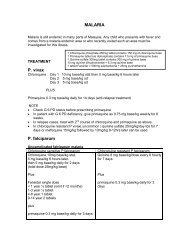
Chapter 79: Malaria 413<br />
Chapter 80: Tuberculosis 419<br />
Chapter 81: BCG Lymphadenitis 425<br />
Chapter 82: Dengue and Dengue Haemorrhagic Fever with Shock 427<br />
Chapter 83: Diphteria 439<br />
Section 11 Dermatology<br />
Chapter 84: Atopic Dermatitis 445<br />
Chapter 85: Infantile Hemangioma 451<br />
Chapter 86: Scabies 455<br />
Chapter 87: Steven Johnson Syndrome 457<br />
Section 12 Metabolic Disorders<br />
Chapter 88: Inborn errors metabolism (IEM): Approach to<br />
Diagnosis and Early Management in a Sick Child 461<br />
Chapter 89: Investigating Inborn errors metabolism (IEM)<br />
in a Child with Chronic Symptoms 471<br />
Chapter 90: Approach to Recurrent Hypoglycemia 483<br />
Chapter 91: Down Syndrome 489<br />
x
TABLE OF CONTENTS<br />
Section 13 <strong>Paediatric</strong> Surgery<br />
Chapter 92: Appendicitis 495<br />
Chapter 93: Vomiting in the Neonate and Child 497<br />
Chapter 94: Intussusception 507<br />
Chapter 95: Inguinal hernias, Hydrocoele 511<br />
Chapter 96: Undescended Testis 513<br />
Chapter 97: The Acute Scrotum 515<br />
Chapter 98: Penile Conditions 519<br />
Chapter 99: Neonatal Surgery 521<br />
Section 14 Rheumatology<br />
Chapter 100: Juvenile Idiopathic Arthritis (JIA) 535<br />
Section 15 Poisons and Toxins<br />
Chapter 101: Snake Bite 543<br />
Chapter 102: Common Poisons 549<br />
Chapter 103: Anaphylaxis 559<br />
Section 16 Sedation and Procedures<br />
Chapter 104: Recognition and Assessment of Pain 565<br />
Chapter 105: Sedation and Analgesia <strong>for</strong> Diagnostic<br />
and Therapeutic Procedures 567<br />
Chapter 108: Practical Procedures 571<br />
xi
Acknowledgements<br />
Again, to Dr Koh Chong Tuan, Consultant <strong>Paediatric</strong>ian at Island Hospital, Penang<br />
<strong>for</strong> his excellent work in proof reading the manuscript.<br />
xii
Chapter 1: Normal Values in Children<br />
Respiratory (Breath) Rate<br />
VITAL SIGNS<br />
Normal, Breath rate at rest Abnormal<br />
Age (years) Rate/min These values define Tachypnoea<br />
60<br />
2-5 25-30 2 mths - 1 year > 50<br />
5-12 20-25 1-5 years > 40<br />
Heart (Pulse) Rate<br />
Abnormal Normal Abnormal<br />
Age (years) Low (Bradycardia) Average High (Tachycardia)<br />
Newborn < 70/min 125/min > 190/min<br />
1-11 months < 80/min 120/min > 160/min<br />
2 years < 80/min 110/min > 130/min<br />
4 years < 80/min 100/min > 120/min<br />
6 years < 75/min 100/min > 115/min<br />
8 years < 70/min 90/min > 110/min<br />
10 years < 70/min 90/min > 110/min<br />
Ref: Nelson Textbook of Pediatrics, 18th <strong>Edition</strong><br />
Blood Pressure<br />
Hypotension if below Normal (average)<br />
Age (years) 5th centile <strong>for</strong> age 50th centile <strong>for</strong> age<br />
< 1 year 65 - 75 mmHg 80 - 90 mmHg<br />
1-2 years 70 - 75 mmHg 85 - 95 mmHg<br />
2-5 years 70 - 80 mmHg 85 - 100 mmHg<br />
5-12 years 80 - 90 mmHg 90 - 110 mmHg<br />
> 12 years 90 - 105 mmHg 100-120 mmHg<br />
Calculation <strong>for</strong> Expected Systolic Blood Pressure<br />
= 85 + (2 x age in years) mmHg <strong>for</strong> 50th centile - Median Blood Pressure<br />
= 65 + (2 x age in years) mmHg <strong>for</strong> 5th centile - Hypotension if below this value<br />
Ref: Advanced <strong>Paediatric</strong> Life Support: The Practical Approach, Fifth <strong>Edition</strong> 2011<br />
1<br />
GENERAL PAEDIATRICS
GENERAL PAEDIATRICS<br />
Blood Pressure in Hypertension<br />
Age Significant Hypertension Severe Hypertension<br />
1 week Systolic 96 mmHg Systolic 106 mmHg<br />
1 wk - 1 mth Systolic 104 mHg Systolic 110 mmHg<br />
Infant Systolic 112 mmHg Systolic 118 mmHg<br />
Diastolic 74 mmHg Diastolic 82 mmHg<br />
3-5 years Systolic 116 mmHg Systolic 124 mmHg<br />
Diastolic 76 mmHg Diastolic 86 mmHg<br />
6-9 years Systolic 122 mmHg Systolic 130 mmHg<br />
Diastolic 78 mmHg Diastolic 86 mmHg<br />
10-12 years Systolic 126 mmHg Systolic 134 mmHg<br />
Diastolic 82 mmHg Diastolic 90 mmHg<br />
13-15 years Systolic 136 mmHg Systolic 144 mmHg<br />
Diastolic 86 mmHg Diastolic 92 mmHg<br />
16-18 years Systolic 142 mmHg Systolic 150 mmHg<br />
Diastolic 92 mmHg Diastolic 98 mmHg<br />
2
ANTHROPOMETRIC MEASUREMENTS<br />
Age Weight Height Head size<br />
birth 3.5 kg 50 cm 35 cm<br />
6 months 7 kg 68 cm 42 cm<br />
1 year 10 kg 75 cm 47 cm<br />
2 years 12 kg 85 cm 49 cm<br />
3 years 14 kg 95 cm 49.5 cm<br />
4 years 100 cm 50 cm<br />
5-12 years 5 cm/year 0.33 cm/year<br />
Points to Note<br />
Weight<br />
• In the first 7 - 10 days of life, babies lose 10 - 15% of their birth weight.<br />
• In the first 3 months of life, the rate of weight gain is 25 gm/day<br />
• Babies regain their birth weight by the 2nd week, double this by 5 months<br />
age, and triple the birth weight by 1 year of age<br />
• Weight estimation <strong>for</strong> children (in Kg):<br />
Infants: (Age in months X 0.5) + 4<br />
Children 1 – 10 years: (Age in yrs + 4) X 2<br />
Head circumference<br />
• Rate of growth in preterm infants is 1 cm/week, but reduces with age.<br />
Head growth follows that of term infants when chronological age reaches term<br />
• Head circumference increases by 12 cm in the 1st year of life (6 cm in first 3<br />
months, then 3 cm in second 3 months, and 3 cm in last 6 months)<br />
Other normal values are found in the relevant chapters of the book.<br />
References:<br />
1. Advanced <strong>Paediatric</strong> Life Support: The Practical Approach Textbook,<br />
5th <strong>Edition</strong> 2011<br />
2. Nelson Textbook of Pediatrics, 18th <strong>Edition</strong>.<br />
3<br />
GENERAL PAEDIATRICS
HAEMATOLOGICAL PARAMETERS<br />
Age Hb PCV Retics MCV fl MCH pg TWBC Neutrophil Lymphocyte<br />
g/dL % % Lowest Lowest x1000 Mean Mean<br />
Cord Blood 13-7-20.1 45-65 5.0 110 - 9-30 61 31<br />
2 weeks 13.0-20.0 42-66 1.0 - 29 5-21 40 63<br />
3 months 9.5-14.5 31-41 1.0 - 27 6-18 30 48<br />
6 mths - 6 yrs 10.5-14.0 33-42 1.0 70-74 25-31 6-15 45 38<br />
7 - 12 years 11.0-16.0 34-40 1.0 76-80 26-32 4.5-13.5 55 38<br />
Adult male 14.0-18.0 42-52 1.6 80 27-32 5-10 55 35<br />
4<br />
Adult female 12.0-16.0 37-47 1.6 80 26-34 5-10 55 35<br />
Differential counts Points to note<br />
< 7 days age neutrophils > lymphocytes • Differential WBC: eosinophils: 2-3%; monocytes: 6-9 %<br />
• Platelets counts are lower in first months of age;<br />
but normal range by 6 months<br />
• Erythrocyte sedimentation rate (ESR) is < 16 mm/hr in<br />
children, provided PCV is at least 35%.<br />
1 wk - 4 years lymphocytes > neutrophils<br />
4 - 7 years neutrophils = lymphocytes<br />
> 7 years neutrophils > lymphocytes<br />
GENERAL PAEDIATRICS
Chapter 2: Immunisations<br />
National Immunisation Schedule <strong>for</strong> Malaysia (Ministry of Health, Malaysia)<br />
Age (months) School years<br />
Vaccine birth 1 2 3 5 6 9 10 12 18 7 yrs 13 yrs 15 yrs<br />
BCG 1 if no scar<br />
Hepatitis B 1 2 3<br />
DTaP 1 2 3 DT B + T B +<br />
IPV 1 2 3 B +<br />
Hib 1 2 3 B +<br />
5<br />
Measles Sabah<br />
MMR 1<br />
JE (Sarawak) 1 2 B*<br />
HPV 3 doses<br />
Legend: B + , Booster doses; B*, Booster at 4 years age; BCG, Bacille Calmette-Guerin; DTaP, Diphhteria, Tetanus, acellular Pertussis;<br />
DT, Diphtheria, Tetanus; T, Tetanus IPV, Inactivated Polio Vaccine; Hib, Haemophilus influenzae type B;<br />
MMR, Measles, Mumps, Rubella; JE, Japanese Encephalitis, HPV, Human Papilloma Virus;<br />
GENERAL PAEDIATRICS
GENERAL PAEDIATRICS<br />
General Notes<br />
• Many vaccines (inactivated or live) can be given together simultaneously (does<br />
not impair antibody response or increase adverse effect). But they are to be<br />
given at different sites unless given in combined preparations. Vaccines are now<br />
packaged in combinations to avoid multiple injections to the child.<br />
• sites of administration<br />
- oral – rotavirus, live typhoid vaccines<br />
- intradermal (ID) - BCG. Left deltoid area (proximal to insertion deltoid muscle)<br />
- deep SC, IM injections. (ALL vaccines except the above)<br />
• anterolateral aspect of thigh – preferred site in children<br />
• upper arm – preferred site in adults<br />
• upper outer quadrant of buttock - associated with lower antibody level production<br />
Immunisation : General contraindications<br />
• Absolute contraindication <strong>for</strong> any vaccine: severe anaphylaxis reactions to<br />
previous dose of the vaccine or to a component of the vaccine.<br />
• Postponement during acute febrile illness: Minor infection without fever or<br />
systemic upset is NOT a contraindication.<br />
• A relative contraindication: avoid a vaccine within 2 weeks of elective surgery.<br />
• Live vaccine: Absolute contraindications<br />
- Immunosuppressed children -malignancy; irradiation, leukaemia, lymphoma,<br />
primary immunodeficiency syndromes (but NOT asymptomatic HIV).<br />
- On chemotherapy or < 6 months after last dose.<br />
- On High dose steroids, i.e. Prednisolone ≥ 2 mg/kg/day <strong>for</strong> > 7 days or low<br />
dose systemic > 2 weeks: delay vaccination <strong>for</strong> 3 months.<br />
- If topical or inhaled steroids OR low dose systemic < 2 weeks or EOD <strong>for</strong> > 2<br />
weeks, can administer live vaccine.<br />
- If given another LIVE vaccine including BCG < 4 weeks ago.<br />
(Give live vaccines simultaneously. If unable to then give separately with<br />
a 4 week interval).<br />
- Within 3 months following IV Immunoglobulin (11 months if given high<br />
dose IV Immunoglobulins, e.g. in Kawasaki disease).<br />
3 weeks 3 months<br />
Live Vaccine HNIG Live vaccine<br />
(Human Normal Immunoglobulin)<br />
- Pregnancy (live vaccine - theoretical risk to foetus) UNLESS there is<br />
significant exposure to serious conditions like polio or yellow fever in<br />
which case the importance of vaccination outweighs the risk to the foetus.<br />
• Killed vaccines are generally safe. The only absolute contraindications are<br />
SEVERE local (induration involving > 2/3 of the limbs) or severe generalised<br />
reactions in the previous dose.<br />
6
The following are not contraindications to vaccination<br />
• Mild illness without fever e.g. mild diarrhoea, cough, runny nose.<br />
• Asthma, eczema, hay fever, impetigo, heat rash (avoid injection in affected area).<br />
• Treatment with antibiotics or locally acting steroids.<br />
• Child’s mother is pregnant.<br />
• Breastfed child (does not affect polio uptake).<br />
• Neonatal jaundice.<br />
• Underweight or malnourished.<br />
• Over the recommended age.<br />
• Past history of pertussis, measles or rubella (unless confirmed medically)<br />
• Non progressive, stable neurological conditions like cerebral palsy, Down<br />
syndrome, simple febrile convulsions, controlled epilepsy, mental retardation.<br />
• Family history of convulsions.<br />
• History of heart disease, acquired or congenital.<br />
• Prematurity (immunise according to schedule irrespective of gestational age)<br />
Vaccination: Special Circumstances<br />
• Measures to protect inpatients exposed to another inpatient with measles:<br />
- Protect all immunocompromised children with Immunoglobulin (HNIG)<br />
0.25-0.5 mls/kg. (Measles may be fatal in children in remission from<br />
leukaemia)<br />
- Check status of measles immunisation in the other children. Give measles<br />
monocomponent vaccine to unimmunised children within 24 hrs of exposure.<br />
Vaccination within 72 hours aborts clinical measles in 75% of contacts<br />
- Discharge the inpatient child with uncomplicated measles.<br />
- Do not <strong>for</strong>get to notify the Health Office.<br />
• Immunisation in children with HIV (Please refer to <strong>Paediatric</strong> HIV section)<br />
• In patients with past history or family history of febrile seizures, neurological<br />
or developmental abnormalities that would predispose to febrile seizures:-<br />
- Febrile seizures may occur 5 – 10 days after measles (or MMR) vaccination<br />
or within the first 72 hours following pertussis immunisation.<br />
- Give Paracetamol (120 mg or ¼ tablet) prophylaxis after immunisation<br />
(esp. DPT) 4-6 hourly <strong>for</strong> 48 hours regardless of whether the child is febrile.<br />
This reduces the incidence of high fever, fretfulness, crying, anorexia and<br />
local inflammation.<br />
• Maternal Chicken Pox during perinatal period. (Please refer to Perinatally<br />
acquired varicella section)<br />
• Close contacts of immunodeficient children and adults must be immunized,<br />
particularly against measles and polio (use IPV).<br />
• In contacts of a patient with invasive Haemophilus influenzae B disease:<br />
- Immunise all household, nursery or kindergarden contacts < 4 years of age.<br />
- Household contacts should receive Rifampicin prophylaxis at 20 mg/kg<br />
once daily (Maximum 600 mg) <strong>for</strong> 4 days (except pregnant women<br />
- give one IM dose of ceftriaxone )<br />
- Index case should be immunised irrespective of age.<br />
7<br />
GENERAL PAEDIATRICS
GENERAL PAEDIATRICS<br />
• Children with Asplenia (Elective or emergency splenectomy; asplenic<br />
syndromes; sickle cell anaemia) are susceptible to encapsulated bacteria<br />
and malaria.<br />
- Pneumococcal, Meningococcal A, C, Y & W-135, Haemophilus influenza b<br />
vaccines should be given.<br />
- For elective splenectomy (and also chemotherapy or radiotherapy): give<br />
the vaccines preferably 2 or more weeks be<strong>for</strong>e the procedure. However,<br />
they can be given even after the procedure.<br />
- Penicillin prophylaxis should continue ideally <strong>for</strong> life. If not until 16 years<br />
old <strong>for</strong> children or 5 years post splenectomy in adults.<br />
• Babies born to mothers who are HbeAg OR HbsAg positive should be given<br />
Hepatitis B immunoglobulin (200 IU) and vaccinated with the Hepatitis B<br />
vaccine within 12 hours and not later than 48 hours. Given in different<br />
syringes and at different sites.<br />
• Premature infants may be immunised at the same chronological age as term<br />
infants. (Please refer section on The premature infants <strong>for</strong> more discussion)<br />
8
Vaccines, indications, contraindications, doses and side effects<br />
Vaccine Indication/Dose Contraindication Possible Side Effects Notes<br />
BCG adenitis may occur. Intradermal.<br />
Local reaction: a papule at<br />
vaccination site may occur<br />
in 2 - 6 weeks. This grows<br />
and flattens with scaling<br />
and crusting. Occasionally a<br />
discharging ulcer may occur.<br />
This heals leaving a scar of<br />
at least 4 mm in successful<br />
vaccination.<br />
Not to be given to symptomatic<br />
HIV infected children.<br />
Can be given to newborns<br />
of HIV infected mother as<br />
the infant is usually asymptomatic<br />
at birth.<br />
BCG To be given at birth<br />
and to be repeated<br />
if no scar is present<br />
Intramuscular.<br />
Give with Hep B immunoglobulin<br />
<strong>for</strong> infants of HBsAg<br />
positive mothers.<br />
Local reactions. Fever and<br />
flu-like symptoms in first<br />
48 hours. Rarely, erythema<br />
multi<strong>for</strong>me or urticaria.<br />
Severe hypersensitivity to<br />
aluminium. The vaccine is<br />
also not indicated <strong>for</strong> HBV<br />
carrier or immuned patient<br />
( i.e. HBsAg or Ab positive)<br />
Hepatitis B All infants,<br />
including those born<br />
to HBsAg positive<br />
mothers<br />
All health care<br />
personnel.<br />
9<br />
Intramuscular<br />
Swelling, redness and pain<br />
A small painless nodule may<br />
develop at injection site –<br />
harmless.<br />
Transient fever, headaches,<br />
malaise, rarely anaphylaxis.<br />
Neurological reactions rare.<br />
Severe hypersensitivity to<br />
aluminium and thiomersal<br />
All infants should<br />
receive 5 doses<br />
including booster<br />
doses at 18 months<br />
and Standard 1<br />
Diphtheria,<br />
Tetanus<br />
(DT)<br />
GENERAL PAEDIATRICS
GENERAL PAEDIATRICS<br />
Vaccine Indication/Dose Contraindication Possible Side Effects Notes<br />
Pertussis All infants should Anaphylaxis to previous Local reaction. Severe if Intramuscular.<br />
receive 4 doses dose; encephalopathy involve 2/3 limbs<br />
including booster at develops within 7 days of Severe systemic reaction: Static neurological diseases,<br />
18 months<br />
vaccination<br />
Anaphylaxis (2 per 100 000 developmental delay, personal<br />
doses), encephalopathy (0 – or family history of fits are<br />
Precautions: severe reaction 10.5 per million doses), high NOT contraindications.<br />
to previous dose (systemic fever (fever>40.5), fits within<br />
or local) and progressive 72 hours, persistent incon-<br />
neurological diseases. solable crying (0.1 to 6%),<br />
hyporesponsive state.<br />
Acellular Pertussis vaccine<br />
associated with less side<br />
It is recommended<br />
that booster doses<br />
be given at Std 1<br />
and at Form 3 due<br />
to increased cases<br />
of Pertussis amongst<br />
adolescents in<br />
recent years<br />
10<br />
effects<br />
Local reactions. Intramuscular.<br />
Allergies to neomycin, polymyxin<br />
and streptomycin<br />
Previous severe anaphylactic<br />
reaction<br />
All infants to be<br />
given 4 doses<br />
including booster at<br />
18 months.<br />
Inactivated<br />
Polio Vaccine<br />
(IPV)<br />
Intramuscular<br />
Local swelling, redness and<br />
pain soon after vaccination<br />
and last up to 24 hours in<br />
10% of vaccinees<br />
Malaise, headaches, fever, irritability,<br />
inconsolable crying.<br />
Very rarely seizures.<br />
Confirmed anaphylaxis to<br />
previous Hib and allergies<br />
to neomycin, polymyxin and<br />
streptomycin<br />
All infants should<br />
receive 4 doses<br />
including booster at<br />
18 months.<br />
Patients with splenic<br />
dysfunction, and<br />
post splenectomy.<br />
Haemophilus<br />
Influenzae<br />
type B (Hib)
Vaccine Indication/Dose Contraindication Possible Side Effects Notes<br />
Intramuscular.<br />
** Long term prospective studies<br />
have found no association<br />
between measles or MMR vaccine<br />
and inflammatory bowel<br />
diseases, autism or SSPE.<br />
Transient rash in 5%.<br />
May have fever between D5-<br />
D12 post vaccination.<br />
URTI symptoms.<br />
Febrile convulsions (D6-D14)<br />
in 1:1000 – 9000 doses of vaccine.<br />
(Natural infection 1:200)<br />
Encephalopathy within 30 days<br />
in 1:1,000,000 doses. (Natural<br />
infection 1:1000 - 5000)<br />
Avoid in patients with<br />
hypersensitivity to eggs,<br />
neomycin and polymyxin.<br />
Pregnancy.<br />
Children with untreated<br />
leukemia, TB and other<br />
cancers.<br />
Immunodeficiency.<br />
Measles Sabah, Orang Asli<br />
population at 6 mths.<br />
Not usually given to<br />
children 3 weeks after vaccination.<br />
Orchitis and retro bulbar<br />
neuritis very rare.<br />
Meningoencephalitis is mild<br />
and rare. (1:800,000 doses).<br />
(natural infection 1:400).<br />
GENERAL PAEDIATRICS
GENERAL PAEDIATRICS<br />
Vaccine Indication/Dose Contraindication Possible Side Effects Notes<br />
Rubella Rash, fever, lymphadenopa- Given as MMR<br />
thy, thrombocytopenia,<br />
transient peripheral neuritis.<br />
Arthritis and arthralgia occurs<br />
in up to 3% of children<br />
and 20% of adults.<br />
Japanese Given in Sarawak at Immunodeficiency and Local redness, swelling, Inactivated vaccine.<br />
Encephalitis 9, 10 and 18 months malignancy, diabetes , acute pain, fever, chills, headache, Subcutaneous.<br />
(JE) Booster at 4 years. exacerbation of cardiac, lassitude..<br />
Protective efficacy > 95%.<br />
hepatic and renal conditions<br />
Human Pap- Indicated <strong>for</strong> Not recommended in Headache, myalgia, injec- 2 vaccines available:<br />
illoma Virus females aged 9-45 pregnant patients.<br />
tion site reactions, fatigue, Cervarix (GSK): bivalent.<br />
(HPV) years.<br />
nausea, vomiting, diarrhoea, Gardasil (MSD): quadrivalent.<br />
abdominal pain, pruritus, - 3 dose schedule IM (0,<br />
rash, urticaria, myalgia, 1-2month, 6 month).<br />
arthralgia, fever.<br />
Recombinant vaccine.<br />
Protective efficacy almost<br />
100% in preventing vaccine<br />
type cervical cancer in first<br />
5 years.<br />
12
Vaccine Indication/Dose Contraindication Possible Side Effects Notes<br />
Not in Blue Book<br />
Immunogenic in children<br />
< 2 years<br />
Children who have severe<br />
allergic reaction to previous<br />
pneumococcal vaccine<br />
Inactivated vaccine.<br />
Intramuscular<br />
Decreased appetite,<br />
irritability, drowsiness,<br />
restless sleep, fever, inj site<br />
erythema, induration or<br />
pain, rash.<br />
Dosage:<br />
Infants 2-6 mth age.<br />
3-dose primary<br />
series at least 1 mth<br />
apart from 6 wks<br />
of age.<br />
Booster: 1 dose<br />
between 12-15 mths<br />
of age.<br />
Unvaccinated:<br />
infants 7-11 mths<br />
2 doses 1 month<br />
apart, followed by a<br />
<strong>3rd</strong> dose at 12- 15<br />
months; children 12-<br />
23 months 2 doses<br />
at least 2 months<br />
apart; healthy<br />
children 2 - 5 years:<br />
Single dose<br />
Pneumococcal<br />
(conjugate)<br />
vaccine: PCV<br />
13/ PCV 7<br />
Healthy children under 6<br />
weeks and more than 59<br />
months of age<br />
High risk children:<br />
immunosuppression (including<br />
asymptomatic HIV),<br />
asplenia, nephrotic syndrome<br />
and chronic lung or heart<br />
disease.<br />
13<br />
Unvaccinated high<br />
risk children 2-5 yrs<br />
age may be given<br />
2 doses (6-8 wks<br />
apart)<br />
GENERAL PAEDIATRICS
GENERAL PAEDIATRICS<br />
Vaccine Indication/Dose Contraindication Possible Side Effects Notes<br />
Hypersensitivity reactions. Listed in Blue Book.<br />
Intramuscular, Subcutaneous<br />
Immunogenic in children ≥2<br />
yrs. Against 23 serotypes.<br />
High risk: immunosuppression,<br />
asymptomatic HIV, asplenia,<br />
nephrotic syndrome, chronic<br />
lung disease. If these children<br />
are 2<br />
yrs, then the polysaccharide<br />
vaccine is used.<br />
Age < 2 years old.<br />
Revaccination within 3 years<br />
has high risk of adverse<br />
reaction;<br />
Avoid during chemotherapy<br />
or radiotherapy and less<br />
than 10 days prior to commencement<br />
of such therapy<br />
– antibody response is poor.<br />
Pregnancy.<br />
Recommended <strong>for</strong><br />
children at high risk.<br />
> 2 years old.<br />
Single dose.<br />
Booster at 3-5 years<br />
only <strong>for</strong> high risk<br />
patients.<br />
Pneumococcal(polysaccharide<br />
vaccine)<br />
14<br />
Oral live-attenuated vaccine.<br />
Protective efficacy 88-91%<br />
<strong>for</strong> any rotavirus gastroenteritis<br />
episode; 63-79% <strong>for</strong> all<br />
causes of gastroenteritis.<br />
Loss of appetite, irritability,<br />
fever, fatigue, diarrhoea,<br />
vomiting, flatulence, abdominal<br />
pain, regurgitation<br />
of food.<br />
Prior hypersensitivity to any<br />
vaccine component.<br />
Uncorrected congenital GIT<br />
mal<strong>for</strong>mation, e.g. Meckel’s<br />
diverticulum<br />
Severe combined immunodeficiency<br />
disease (reported<br />
prolonged shedding of vaccine<br />
virus reported in infants<br />
who had live Rotavirus<br />
vaccine)<br />
Rotavirus First dose given to<br />
infants ≥ 6 wks old.<br />
Rotateq (3 doses)<br />
Subsequent doses<br />
given at 4-10 wks interval.<br />
<strong>3rd</strong> dose given<br />
≤ 32 weeks age.<br />
Rotarix (2 doses). 2nd<br />
dose to be given by<br />
24 weeks age. Interval<br />
between doses<br />
should be > 4 wks.
Vaccine Indication/Dose Contraindication Possible Side Effects Notes<br />
Live attenuated vaccine.<br />
Subcutaneous.<br />
70 – 90% effectiveness.<br />
Occasionally, papulovesicular<br />
eruptions, injection site<br />
reactions, headache, fever,<br />
paresthesia, fatigue<br />
Pregnant patients.<br />
Patients receiving high dose<br />
systemic immunosuppression<br />
therapy.<br />
Patients with malignancy<br />
especially haematological<br />
malignancies or blood<br />
dyscrasias.<br />
Hypersensitivity to neomycin.<br />
12 mths to 12 yrs:<br />
Single dose<br />
> 12 yrs:<br />
2 doses ≥4 wks apart.<br />
Varicella<br />
Zoster<br />
Non immune susceptible<br />
health care<br />
workers who regularly<br />
come in contact<br />
with VZV infection<br />
Asymptomatic/mildly<br />
symptomatic children<br />
with HIV (with CD4%<br />
> 15%); 2 doses at 3<br />
mths interval.<br />
Children in remission<br />
from leukemia <strong>for</strong> ≥1<br />
yr, have >700/ml circulating<br />
lymphocytes<br />
may receive vaccine<br />
under paediatrician<br />
supervision (2doses).<br />
15<br />
Intramuscular.<br />
Inactivated vaccine.<br />
Protective efficacy 94%.<br />
Local reactions. Flu-like<br />
symptoms lasting 2 days in<br />
10% of recipients<br />
Severe hypersensitivity to<br />
aluminium hydroxide, phenoxyethanol,<br />
neomycin<br />
Hepatitis A For children >1 yr.<br />
2 doses., given 6-12<br />
months apart.<br />
GENERAL PAEDIATRICS
GENERAL PAEDIATRICS<br />
Vaccine Indication/Dose Contraindication Possible Side Effects Notes<br />
Gastroenteritis Oral inactivated vaccine.<br />
Protective efficacy 80-90%<br />
after 6 mths waning to 60%<br />
after 3 yrs.<br />
Cholera Children 2-6 yrs:<br />
3 doses at 1-6 wk<br />
interval.<br />
Children > 6 yrs:<br />
2 doses at 1-6 wks<br />
interval.<br />
Booster dose >2 yrs.<br />
Intramuscular.<br />
Inactivated vaccine.<br />
Protective efficacy 70-90%<br />
Require yearly revaccination<br />
<strong>for</strong> continuing protection.<br />
Transient swelling, redness,<br />
pain and induration locally.<br />
Myalgia, malaise and<br />
fever <strong>for</strong> 1 – 2 days starting<br />
within a few hours post<br />
vaccination. Very rarely,<br />
neurological (Guillain-Barre),<br />
glomerulonephritis, ITP or<br />
anaphylactic reaction occurs.<br />
Hypersensitivity to egg or<br />
chicken protein, neomycin,<br />
<strong>for</strong>maldehyde.<br />
Febrile illness, acute infection.<br />
Influenza Single dose.<br />
Min age 6 mths.<br />
Unprimed individuals<br />
require 2nd dose 4 -<br />
6 wks after 1st dose.<br />
Recommended <strong>for</strong><br />
children with:<br />
chronic decompensated<br />
respiratory or<br />
cardiac disorders,<br />
e.g. cyanotic heart<br />
diseases chronic lung<br />
disease, HIV infection.<br />
In advanced disease,<br />
vaccination may not<br />
induce protective<br />
antibody levels.<br />
16
Vaccine Indication/Dose Contraindication Possible Side Effects Notes<br />
Inactivated vaccine.<br />
(Available in Malaysia<br />
as Purified Vero<br />
Cell Rabies Vaccine<br />
(PVRV).<br />
Intramuscular.<br />
Headache, dizziness, malaise,<br />
abdominal pain, nausea, myalgia.<br />
Injection site reactions<br />
such as itching, swelling, pain.<br />
Rabies Pre-exposure: 3 doses at Day 0,<br />
7, 28. Booster every 2-3 yrs.<br />
Post-exposure treatment:<br />
Fully immunised: 2 doses at<br />
Day 0, Day 3. Rabies Immune<br />
Globulin (RIG) unnecessary.<br />
Unimmunised: 5 doses at Day<br />
0, 3, 7, 14 and 28. RIG (20 IU/<br />
kg given half around the wound<br />
and the rest IM.<br />
Intramuscular.<br />
Local reactions. Irritability,<br />
fever and rigors <strong>for</strong> 1-2 days.<br />
Very rarely, anaphylaxis.<br />
Single dose.<br />
Immunity up to 3 yrs.<br />
Meningococcus<br />
A, C, Y &<br />
W-135<br />
17<br />
Intramuscular.<br />
Polysaccharide<br />
vaccine<br />
Local reactions. Myalgia,<br />
malaise, nausea, headaches<br />
and fever in 3% of recipients.<br />
Children < 2yrs.<br />
(Immunogenicity < 2 yrs of<br />
age has not been established)<br />
Single dose. Seroconversion in<br />
85-95% of recipients; confers<br />
60-80% protection beginning<br />
2 wks after vaccination.<br />
Boosters every 3 yrs.<br />
Typhoid<br />
(Typhim Vi)<br />
Oral. Live attenuated<br />
vaccine.<br />
Very rarely: mild GIT<br />
disturbances or a transitory<br />
exanthema.<br />
Infant
GENERAL PAEDIATRICS<br />
Recommended Immunisation Schedule <strong>for</strong> Infants and Children<br />
Not Immunised at the Recommended Time<br />
Time of Immunisation Age at first visit<br />
1st visit BCG, DPT/DTaP, Hib1,<br />
IPV1, HBV1<br />
Between 6 wks -12 mths 12 months and older<br />
18<br />
BCG, DPT/DTaP1, Hib1,<br />
IPV1, HBV1, measles<br />
(footnote 2) at 6 or 9<br />
mths,<br />
MMR at 12 mths of age<br />
2nd visit (1 mth later) DPT/DTaP2, IPV2, HBV2,<br />
Hib2<br />
<strong>3rd</strong> visit (1 mth later) DPT/DTaP2,Hib2, IPV2,<br />
HBV2<br />
4th visit (4 mths after<br />
<strong>3rd</strong> visit)<br />
2-8 mths later HBV3, DTaP4, Hib4 &<br />
IPV4 (booster), measles<br />
in Sabah at 9 mths age,<br />
MMR at 12 mths age<br />
DPT/DTaP3, IPV3,<br />
DPT/DTaP3,Hib3, IPV3, HBV3, DPT/DTaP4, IPV4,<br />
Polio, DT/DTaP, MMR (at<br />
school entry)<br />
Footnotes:<br />
1. For infants < 6 wks age, use “Recommended Immunisation Schedule <strong>for</strong><br />
Infants & Children”.<br />
2. Measles vaccine should be given only after 9 mths. (exception - given at 6<br />
months in Sabah)<br />
3. For special groups of children with no regular contact with Health Services<br />
and with no immunisation records, BCG, HBV, DTaP- Hib-IPV and MMR can<br />
be given simultaneously at different sites at first contact.<br />
4. It is not necessary to restart a primary course of immunisation<br />
regardless of the period that has elapsed since the last dose was given.<br />
Only the subsequent course that has been missed need be given. (Example.<br />
An infant who has been given IPV1 and then 9 months later comes <strong>for</strong><br />
follow-up, the IPV1 need not be repeated. Go on to IPV2.). Only exception<br />
is Hepatitis A vaccine.
Chapter 3: <strong>Paediatric</strong> Fluid and Electrolyte Guidelines<br />
Well children with Normal hydration<br />
Very few well children require intravenous fluids (IV). Whenever possible use<br />
an enteral (oral) route <strong>for</strong> fluids.<br />
These guidelines apply to children who are unable to tolerate enteral fluids.<br />
The safe use of IV fluid therapy in children requires accurate prescribing of<br />
fluids and careful monitoring because incorrectly prescribed or administered<br />
fluids are hazardous.<br />
If IV fluid therapy is required then maintenance fluid requirements should be<br />
calculated using the Holliday and Segar <strong>for</strong>mula based on weight.<br />
However this should be only be used as a starting point and the individuals’<br />
response to fluid therapy should be monitored closely by clinical observation,<br />
fluid balance, weight and a minimum daily electrolyte profile.<br />
Prescribing Intravenous fluids<br />
Fluids are given intravenously <strong>for</strong> the following reasons:<br />
• Circulatory support in resuscitating vascular collapse.<br />
• Replacement of previous fluid and electrolyte deficit.<br />
• Maintenance of daily fluid requirement.<br />
• Replacement of ongoing losses.<br />
• Severe dehydration with failed nasogastric tube fluid replacement<br />
(e.g. on-going profuse losses, diarrhoea or abdominal pain).<br />
• Certain co-morbidities, particularly GIT conditions (e.g. short gut or<br />
previous gut surgery)<br />
Resuscitation<br />
Fluids appropriate <strong>for</strong> bolus administration are:<br />
Crystalloids 0.9% Normal Saline<br />
Ringer’s Lactate @ Hartmann’s solution<br />
Colloids Gelafundin, Voluven<br />
4.5% albumin solution<br />
Blood products Whole blood, blood components<br />
• Fluid deficit sufficient cause impaired tissue oxygenation (i.e. clinical shock)<br />
should be corrected with a fluid bolus of 10-20mls/kg.<br />
• Always reassess circulation - give repeat boluses as necessary.<br />
• Look <strong>for</strong> the cause of circulatory collapse - blood loss, sepsis, etc.<br />
This helps decide on the appropriate alternative resuscitation fluid.<br />
• Fluid boluses of 10mls/kg in selected situations - e.g. diabetic ketoacidosis,<br />
intracranial pathology or trauma.<br />
• Avoid low sodium-containing (hypotonic) solutions <strong>for</strong> resuscitation as this<br />
may cause hyponatremia.<br />
• Check blood glucose: treat hypoglycemia with 2mls/kg of 10% Dextrose<br />
solution.<br />
19<br />
GENERAL PAEDIATRICS
GENERAL PAEDIATRICS<br />
• Measure Na, K and glucose at the outset and at least 24hourly from then on.<br />
More frequent testing is indicated in ill patients or those with co-morbidities.<br />
Rapid results of electrolytes can be done with blood gases measurements.<br />
• Consider septic work-up or surgical consult in severely unwell patients with<br />
abdominal symptoms (i.e. gastroenteritis).<br />
Maintenance<br />
• Maintenance fluid is the volume of daily fluid intake. It includes insensible<br />
losses (from breathing, perspiration, and in the stool), and allows <strong>for</strong><br />
excretion of the daily production of excess solute load (urea, creatinine,<br />
electrolytes) in the urine.<br />
• Most children can safely be managed with solution of 0.45% saline with<br />
added glucose (i.e. 0.45% saline in 5% glucose or 0.45% saline in<br />
10% glucose) depending on glucose requirement.<br />
• Sodium chloride 0.18 saline with glucose 5% should not be used as a<br />
maintenance fluid and is restricted to specialist area to replace ongoing<br />
loses of hypotonic fluids. These areas include high dependency, renal, liver<br />
and intensive care.<br />
• Most children will tolerate standard fluid requirements. However some<br />
acutely ill children with inappropriately increased anti-diuretic hormone<br />
secretion (SIADH) may benefit from their maintenance fluid requirement<br />
being restricted to two-thirds of the normal recommended volume.<br />
• Children who are at high risk of hyponatremia should be given isotonic<br />
solutions (i.e. 0.9% saline ± glucose) with careful monitoring to avoid<br />
iatrogenic hyponatremia in hospital.<br />
These include children with the following conditions:<br />
• Peri-or post-operative<br />
• Require replacement of ongoing losses<br />
• A plasma Na at lower normal range of normal (definitely if < 135mmol/L)<br />
• Intravascular volume depletion<br />
• Hypotension<br />
• Central nervous system (CNS) infection<br />
• Head injury<br />
• Bronchiolitis<br />
• Sepsis<br />
• Excessive gastric or diarrhoeal losses<br />
• Salt-wasting syndromes<br />
• Chronic conditions such as diabetes, cystic fibrosis and pituitary deficits.<br />
20
Calculation of Maintanence Fluid Requirements<br />
The following calculations approximate the maintenance fluid requirement of<br />
well children according to weight in kg. (Holliday-Segar calculator)<br />
Weight Total fluids Infusion rate<br />
First 10 Kgs 100 ml/kg 4 mls/kg/hour<br />
Subsequent 10 Kgs 50 ml/kg 2 mls/kg/hour<br />
All additional Kg 20 ml/kg 1 mls/kg/hour<br />
Example: A Child of 29 kg will require:<br />
100mls/kg <strong>for</strong> first 10kg of weight 10 x 100 = 1000 mls<br />
50mls/kg <strong>for</strong> second 10kg of weight 10 x 50 = 500 mls<br />
20mls/kg <strong>for</strong> all additional weight 9 x 20 = 180 mls<br />
Total = 1680 mls<br />
= 1680/24<br />
Rate = 70mls/hour<br />
Composition of commonly used intravenous solution<br />
Osmolality Na content Osmolality Tonicity<br />
Fluid (mOsm/l) (mmol/l) compared to<br />
plasma<br />
with ref to cell<br />
membrane<br />
Na chloride 0.9% 308 154 IsoOsmolar Isotonic<br />
Na chloride 0.45% 154 77 HypoOsmolar Hypotonic<br />
Na chloride 0.9%<br />
+ Glucose 5%<br />
586 150 HyperOsmolar Isotonic<br />
Na chloride 0.45%<br />
+ Glucose 5%<br />
432 75 HyperOsmolar Hypotonic<br />
Na chloride 0.18%<br />
+ Glucose 5%<br />
284 31 IsoOsmolar Hypotonic<br />
Dextrose 5% 278 Nil IsoOsmolar Hypotonic<br />
Dextrose 10% 555 Nil HyperOsmolar Hypotonic<br />
Hartmann’s 278 131 IsoOsmolar Isotonic<br />
21<br />
GENERAL PAEDIATRICS
GENERAL PAEDIATRICS<br />
Deficit<br />
• A child’s water deficit in mls can be calculated following an estimation of the<br />
degree of dehydration expressed as % of body weight.<br />
Example: A 10kg child who is 5% dehydration has a water deficit of 500mls.<br />
Maintenance<br />
100mls/kg <strong>for</strong> first 10 kg = 10 × 100 = 1000mls<br />
Infusion rate/hour<br />
Deficit (give over 24hours)<br />
= 1000mls/24 hr = 42mls/hr<br />
5% dehydration (5% of body water): 5/100 × 10kg × 1000mls = 500mls<br />
Infusion rate/hour (given over 24 hrs) = 500mls/24 hr = 21mls/hr<br />
• The deficit is replaced over a time period that varies according to the<br />
child’s condition. Precise calculations (e.g. 4.5%) are not necessary.<br />
The rate of rehydration should be adjusted with ongoing clinical assessment.<br />
• Use an isotonic solution <strong>for</strong> replacement of the deficit, e.g. 0.9% saline.<br />
• Reassess clinical status and weight at 4-6hours, and if satisfactory continue.<br />
If child is losing weight, increase the fluid and if weight gain is excessive<br />
decrease the fluid rate.<br />
• Replacement may be rapid in most cases of gastroenteritis (best achieved<br />
by oral or nasogastric fluids), but should be slower in diabetic ketoacidosis<br />
and meningitis, and much slower in hypernatremic states (aim to rehydrate<br />
over 48-72 hours, the serum Na should not fall by >0.5mmol/l/hr).<br />
Ongoing losses (e.g. from drains, ileostomy, profuse diarrhoea)<br />
• These are best measured and replaced. Any fluid losses > 0.5ml/kg/hr needs<br />
to be replaced.<br />
• Calculation may be based on each previous hour, or each 4 hour period<br />
depending on the situation. For example; a 200mls loss over the previous 4<br />
hours will be replaced with a rate of 50mls/hr <strong>for</strong> the next 4 hours).<br />
• Ongoing losses can be replaced with 0.9% Normal Saline or Hartmann’s<br />
solution. Fluid loss with high protein content leading to low serum albumin<br />
(e.g. burns) can be replaced with 5% Human Albumin.<br />
22
SODIUM DISORDERS<br />
• The daily sodium requirement is 2-3mmol/kg/day.<br />
• Normal serum sodium is between 135-145mmol/l.<br />
Hypernatremia<br />
• Hypernatremia is defined as serum Na + > 150mmol/l,<br />
moderate hypernatremia is when serum Na + is 150-160mmol/l, and<br />
severe hypernatremia is when serum Na + > 160mmol/l.<br />
• It can be due to:<br />
• water loss in excess of sodium<br />
(e.g. diarrhoea)<br />
• water deficit<br />
(e.g. diabetes insipidus)<br />
• sodium gain<br />
(e.g. large amount of NaHCO3<br />
infusion or salt poisoning).<br />
• If the cause of the hypernatremia is central diabetes insipidus, it is advisable<br />
to consult Endocrinology team regarding management.<br />
• In hypernatremia the child appears sicker than expected <strong>for</strong> the degree of<br />
dehydration.<br />
• Shock occurs late because intravascular volume is relatively preserved.<br />
Signs of hypernatremic dehydration tend to be predominantly that of<br />
intracellular dehydration and neurological dysfunction.<br />
Management<br />
This will depend on the cause of hypernatremia.<br />
For hypernatremic dehydration with Na + Clinical signs of Hypernatremic dehydration<br />
Irritability<br />
Skin feels “doughy”<br />
Ataxia, tremor, hyperreflexia<br />
Seizure<br />
Reduced awareness, coma<br />
> 150mmol/l<br />
• If the patient is in shock, give volume resuscitation with 0.9% Normal saline<br />
as required with bolus/es.<br />
• Avoid rapid correction as this may cause cerebral oedema, convulsion and<br />
death.<br />
• Aim <strong>for</strong> correction of deficit over 48-72 hours and a fall of serum sodium<br />
concentration not more than 0.5mmol/l/hour.<br />
• Give 0.9% saline to ensure the drop in sodium is not too rapid.<br />
• Remember to also give maintenance and replace ongoing losses following<br />
the recommendation above.<br />
• Repeat blood urea and electrolytes every 6 hours until stable.<br />
Special considerations<br />
• A slower rate will be required <strong>for</strong> children with chronic hypernatremia<br />
(present <strong>for</strong> more than 5 days).<br />
• Calcium and glucose need to be checked as hypernatremia can be<br />
associated with hypocalcaemia and hyperglycemia, these conditions need to<br />
be corrected concurrently.<br />
23<br />
GENERAL PAEDIATRICS
GENERAL PAEDIATRICS<br />
Hyponatremia<br />
• Hyponatremia is defined when serum Na + < 135mmol/l.<br />
• Hyponatremic encephalopathy is a medical emergency that requires rapid<br />
recognition and treatment to prevent poor outcome.<br />
• As part of the general resuscitative measures, bolus of 4ml/kg of 3% sodium<br />
chloride should be administered over 30 minutes. This will raised the serum<br />
sodium by 3mmol/l and will usually help stop hyponatremic seizures.<br />
• Gradual serum sodium correction should not be more than 8mmol/day to<br />
prevent osmotic demyelination syndrome.<br />
Calculating sodium correction in acute hyponatremia<br />
mmol of sodium required = (135-present Na level)× 0.6 × weight(kg)<br />
The calculated requirements can then be given from the following available<br />
solutions dependent on the availability and hydration status:<br />
0.9% sodium chloride contains 154 mmol/l<br />
3% sodium chloride contains 513mmol/l<br />
• Children with asymptomatic hyponatremia do not require 3% sodium<br />
chloride treatment and if dehydrated may be managed with oral fluids or<br />
intravenous rehydration with 0.9% sodium chloride.<br />
• Children who are hyponatremic and have a normal or raised volume status<br />
should be managed with fluid restriction.<br />
• For Hyponatremia secondary to diabetic ketoacidosis; refer DKA protocol.<br />
POTASSIUM DISORDERS<br />
• The daily potassium requirement is 1-2mmol/kg/day.<br />
• Normal values of potassium are:<br />
• Birth - 2 weeks: 3.7 - 6.0mmol/l<br />
• 2 weeks – 3 months: 3.7 - 5.7mmol/l<br />
• 3 months and above: 3.5 - 5.0mmol/l<br />
Hyperkalemia<br />
• Causes are:<br />
• Dehydration<br />
• Acute renal failure<br />
• Diabetic ketoacidosis<br />
• Adrenal insufficiency<br />
• Tumour lysis syndrome<br />
• Drugs e.g. oral potassium supplement, K + sparing diuretics, ACE inhibitors.<br />
Treatment: see algorithm on next page<br />
24
Hyperkalemia Treatment Algorithm<br />
Hyperkalemia K + > 5.5 mmol/l<br />
Stop all K + supplementation<br />
Stop medication causing hyperK +<br />
Cardiac monitoring<br />
Exclude pseudo hyperkalemia<br />
Recheck with venous sample<br />
Child unstable<br />
or symptomatic<br />
Abnormal ECG<br />
K + > 7.0 mmol/l<br />
Transfer to<br />
tertiary centre?<br />
Discuss <strong>for</strong> dialysis<br />
IV Calcium<br />
Neb Salbutamol<br />
IV Insulin<br />
with glucose<br />
IV Bicarbonate<br />
± PR/PO Resonium<br />
Child stable,<br />
asymptomatic<br />
Normal ECG<br />
K + >6, ≤ 7 mmol/L<br />
Neb Salbutamol<br />
IV Insulin<br />
with glucose<br />
± IV Bicarbonate<br />
if acidosis<br />
± PR/PO Resonium<br />
Drug doses:<br />
• IV Calcium 0.1 mmol/kg.<br />
• Nebulised Salbutamol:<br />
Age ≤2.5 yrs: 2.5 mg; Age 2.5-7.5 yrs: 5 mg; >7.5 yrs: 10 mg<br />
• IV Insulin with Glucose:<br />
Start with IV Glucose 10% 5ml/kg/hr (or 20% at 2.5 ml/kg/hr).<br />
Once Blood sugar level >10mmol/l and the K + level is not falling,<br />
add IV Insulin 0.05 units/kg/hr and titrate according to glucose level.<br />
• IV Sodium Bicarbonate: 1-2 mmol/kg.<br />
• PO or Rectal Resonium : 1Gm/kg.<br />
25<br />
ECG changes in Hyperkalemia<br />
Tall, tented T waves<br />
Prolonged PR interval<br />
Prolonged QRS complex<br />
Loss of P wave,<br />
wide biphasic QRS<br />
Child stable,<br />
asymptomatic<br />
Normal ECG<br />
K + ≥ 5.5, ≤ 6.0 mmol/L<br />
Consider treatment ?<br />
± Neb Salbutamol<br />
± IV Bicarbonate<br />
if acidosis<br />
± PR/PO Resonium<br />
GENERAL PAEDIATRICS
GENERAL PAEDIATRICS<br />
Hypokalemia<br />
• Hypokalemia is defined as serum Na + > 3.4 mmol/l<br />
(Treat if < 3.0mmol/l or Clinically Symptomatic < 3.4 mmol/l)<br />
• Causes are:<br />
• Sepsis<br />
• GIT losses - diarrhoea, vomiting<br />
• Iatrogenic- e.g. diuretic therapy,<br />
salbutamol, amphotericin B.<br />
• Diabetic ketoacidosis<br />
• Renal tubular acidosis<br />
• Hypokalaemia is often seen<br />
with chloride depletion<br />
and metabolic alkalosis.<br />
• Refractory hypokalaemia may occur with hypomagnesaemia.<br />
Treatment<br />
• Identify and treat the underlying condition.<br />
• Unless symptomatic, a potassium level of 3.0 and 3.4 mmol/l is generally<br />
not supplemented but rather monitored in the first instance.<br />
• The treatment of hypokalaemia does not lend itself to be incorporated into a<br />
protocol and as a result each patient will need to be treated individually.<br />
Oral Supplementation<br />
• Oral Potassium Chloride (KCL), to a maximum of 2 mmol/kg/day in divided<br />
doses is common but more may be required in practice.<br />
Intravenous Supplementation (1gram KCL = 13.3 mmol KCL)<br />
• Potassium chloride is always given by IV infusion, NEVER by bolus injection.<br />
• Maximum concentration via a peripheral vein is 40 mmol/l (concentrations<br />
of up to 60 mmol/l can be used after discussion with senior medical staff).<br />
• Maximum infusion rate is 0.2mmol/kg/hr (in non-intensive care setting).<br />
Intravenous Correction (1gram KCL = 13.3 mmol KCL)<br />
• K + < 2.5 mmol/L may be associated with significant cardiovascular<br />
compromise. In the emergency situation, an IV infusion KCL may be given<br />
• Dose: initially 0.4 mmol/kg/hr into a central vein, until K + ECG changes of Hypokalemia<br />
These occur when K<br />
level is restored.<br />
• Ideally this should occur in an intensive care setting.<br />
+ < 2.5mmol/l<br />
Prominent U wave<br />
ST segment depression<br />
Flat, low or diphasic T waves<br />
Prolonged PR interval (severe hypoK + )<br />
Sinoatrial block (severe hypoK + )<br />
26
Chapter 4: Developmental Milestones in Normal Children<br />
Age Gross Motor Fine Motor Speech/Language Social<br />
6 wks Pull to sit: Head lag, rounded Fixates and follows to 90 Vocalising by 8 weeks. Quiets Smiles responsively.<br />
back<br />
degrees<br />
to sound. Startles to sound.<br />
Ventral Suspension: Head<br />
briefly in same plane as body.<br />
Prone: Pelvis high, knees no<br />
longer under abdomen. Chin<br />
raised occasionally.<br />
Laughs.<br />
Squeals with delight.<br />
Turns head to sound.<br />
Hand regard. Follows object<br />
from side to side (180°). Hands<br />
held loosely. Grasps object<br />
placed in hand.<br />
Not reaching out.<br />
3 mths Pull to sit: Slight head lag. Head<br />
occasionally bobs <strong>for</strong>ward.<br />
Ventral Suspension: Head above<br />
plane of body.<br />
Prone: Pelvis flat. Lifts head up<br />
27<br />
45°- 90°.<br />
Mouthing.<br />
Reaches <strong>for</strong> objects.<br />
Plays with toes.<br />
5 mths Pull to sit: No head lag and sits<br />
with straight back.<br />
Lying supine: Feet to mouth.<br />
Palmar grasp of cube, ulnar<br />
approach. Moves head, eyes in<br />
all directions. No squint (after<br />
4 months).<br />
6 mths Pulls to sit: Lifts head in anticipation.<br />
Sits with support.<br />
Bears weight on legs. Prone:<br />
Supports weight on hands;<br />
chest, upper abdomen off<br />
couch. Rolls prone to supine.<br />
GENERAL PAEDIATRICS
GENERAL PAEDIATRICS<br />
Age Gross Motor Fine Motor Speech/Language Social<br />
Imitates housework.<br />
Toilet trained. Uses spoon<br />
well. Casting stops.<br />
Points to 2 - 3 body parts.<br />
Picture Cards - identify one.<br />
Tower of 3 cubes.<br />
Scribbles spontaneously.<br />
Visual test: Picture charts.<br />
Handedness<br />
18 mths Gets up and down stairs<br />
holding on to rail or with<br />
one hand held. Pulls toy<br />
or carries doll. Throws ball<br />
without falling.<br />
Sits on a chair.<br />
Puts on shoes, socks, pants.<br />
Dry by day.<br />
Play near other children but<br />
not with them.<br />
2-3 word sentences. Uses<br />
‘you’ ‘me’ ‘I’. names 3<br />
objects. Obeys 4 simple<br />
commands. Points to 4 body<br />
parts.<br />
Tower of 6 cubes.<br />
Imitates cubes of train with<br />
no chimney.<br />
Imitates straight line.<br />
Visual test: Snellen’s chart.<br />
2 yrs Goes up and down stairs<br />
alone, 2 feet per step. Walks<br />
backwards (21 months)<br />
Runs. Picks up toy without<br />
falling.<br />
Throws, kick ball without<br />
falling.<br />
28<br />
Knows full name and gender.<br />
Names one colour.<br />
Tower of 8.<br />
Imitates train with<br />
chimney.<br />
Holds pencil well.<br />
Imitates and<br />
Tower of 9.<br />
Imitates bridge with cubes:<br />
2.5 yrs Jumps on both feet.<br />
Walks on tip toes.<br />
Dresses, undresses with<br />
help. Dry by night.<br />
Plays with others.<br />
Can count to 10. Names 2<br />
colours. Nursery rhymes.<br />
Understands “on”, “in”,<br />
“under”.<br />
O Copies Imitates<br />
Draw a man test. (3 - 10y)<br />
3 yrs Goes up stairs one foot per<br />
step.<br />
Down stairs 2 feet per step.<br />
Jumps off bottom step.<br />
Stands on 1 foot <strong>for</strong><br />
seconds.<br />
Rides tricycle.
Age Gross Motor Fine Motor Speech/Language Social<br />
4 yrs Goes up and down stairs Imitates gate with cubes. Names 3 colours. Fluent Buttons clothes fully. Attends<br />
one foot per step.<br />
conversation. Understands to own toilet needs.<br />
Skips on one foot.<br />
Copies<br />
“in front of”, “between”,<br />
Hops on one foot. Goodenough test 4. behind”.<br />
4.5 yrs Copies gate with cubes.<br />
Copies square.<br />
Draws recognisable man and<br />
house.<br />
Ties shoelaces.<br />
Dresses and undresses<br />
alone.<br />
Knows AGE. Names 4<br />
colours.<br />
Triple order preposition.<br />
Tell’s the time.<br />
Copies ‘X’ (5 years)<br />
Copies (5½ years) triangle.<br />
5 yrs Skips on both feet.<br />
Runs on toes.<br />
29<br />
Goodenough test 8.<br />
Copies:<br />
6 yrs Walks heel to toe<br />
Kicking, throwing, climbing.<br />
Goodenough test 12.<br />
Imitates or copies steps with 10<br />
cubes<br />
Note: Goodenough test: 3 + a/4 years (a = each feature recorded in his picture).<br />
GENERAL PAEDIATRICS
GENERAL PAEDIATRICS<br />
Age Gross Motor Fine Motor Speech/Language Social<br />
7 mths Sits with hands on couch <strong>for</strong> Feeds self with biscuits. Babbling in single syllables. Stranger anxiety.<br />
support.<br />
Transfers objects - hand to (combined syllables at 8<br />
Rolls from supine to prone. hand.<br />
months). Distraction Test.<br />
Rakes at pea.<br />
9 mths Sits steadily. Leans <strong>for</strong>ward Inferior pincer grasp Localises sound at 3 feet, Feeds with spoon occasion-<br />
but cannot pivot.<br />
(Scissors grasp)<br />
above and below the ear ally.<br />
Stands holding on.<br />
level.<br />
Looks <strong>for</strong> fallen toys.<br />
Pulls self to sit.<br />
Understands “NO!”<br />
10 mths Crawls on abdomen. Index approach. Uses index<br />
Waves “Bye bye”<br />
Pull self to stand.<br />
finger to poke at pea.<br />
Plays “Pat-a-Cake”<br />
Lets go of cube in hand.<br />
30<br />
One word with meaning. Plays “peek-a-boo”<br />
11 mths Creeping on all fours.<br />
Pivoting. Cruising. Walks with<br />
both hands held.<br />
Casting (13 months)<br />
Less mouthing.<br />
Shy.<br />
Understands phases; 2 - 3<br />
words with meaning. Localising<br />
sound above head.<br />
Neat pincer grasp. Bangs 2<br />
cubes.<br />
Sees and picks up hundreds<br />
and thousands.<br />
1 year Gets from lying to sitting to<br />
crawling to standing.<br />
Walks like a bear. Walks with<br />
one hand held.<br />
Walks well (13 months).<br />
Stands alone<br />
Takes off shoe.<br />
Feeds self with cup and spoon<br />
(but spills). Mouthing stops<br />
More words.<br />
Points to objects he wants.<br />
Continual jabber and jargon.<br />
Tower of 2 cubes.<br />
Scribbles spontaneously (15-<br />
18 months)<br />
13 mths Creeps upstairs.<br />
Stoops <strong>for</strong> toy and stands up<br />
without support. (best at 18<br />
months)
Chapter 5: Developmental Assessment<br />
Development is the progressive, orderly, acquisition of skills and abilities as a<br />
child grows. It is influenced by genetic, neurological, physical, environmental<br />
and emotional factors.<br />
Important points to note:<br />
• child must be co-operative, not tired, fretful, hungry nor sick.<br />
Remember that a child may behave differently in an unfamiliar environment<br />
• allowance must be made <strong>for</strong> prematurity up to two years.<br />
• take note of parental account of what child can or cannot do.<br />
Note parental comments on abnormal gait, speech defects, etc.<br />
• normal development is dependent on integrity of child’s hearing and vision.<br />
• normal pattern of speech and language development is essential <strong>for</strong> a<br />
normal social, intellectual and emotional development.<br />
• delay in development may be global i.e. affecting all areas equally, or specific<br />
areas only e.g. oro-motor dysfunction causing speech delay.<br />
Key Developmental Warning Signs<br />
1 Discrepant head size or crossing centile lines (too large or too small).<br />
2 Persistence of primitive reflexes > 6 months of age<br />
3 No response to environment or parent by 12 months<br />
4 Not walking by 18 months<br />
5 No clear spoken words by 18 months<br />
6 No two word sentences by 2 years<br />
7 Problems with social interaction at 3 years<br />
8 Congenital anomalies, odd facies<br />
9 Any delay or failure to reach normal milestones<br />
Note: Parental concerns must always be taken seriously<br />
Assessment of children with suspected developmental delay<br />
History<br />
• consanguinity<br />
• family history of developmental delay<br />
• maternal drugs, alcohol, illness and infection in pregnancy<br />
• prematurity, perinatal asphyxia<br />
• severe neonatal jaundice, hypoglycaemia or seizures<br />
• serious childhood infections, hospital admissions or trauma<br />
• home environment conditions (environmental deprivation)<br />
Physical examination<br />
• head circumference, dysmorphic features<br />
• neurocutaneous markers<br />
• neurological abnormalities<br />
• full developmental assessment<br />
31<br />
GENERAL PAEDIATRICS
GENERAL PAEDIATRICS<br />
Investigations<br />
(individualised according to<br />
history and physical findings)<br />
• Visual and auditory testing<br />
• T4, TSH<br />
• Chromosomal Analysis<br />
• Consider<br />
- Creatine kinase in boys<br />
- MRI Brain<br />
- Metabolic screen<br />
- Specific genetic studies<br />
(Fragile X, Prader Willi<br />
or Angelman syndrome)<br />
- Refer to a geneticist<br />
- EEG if history of seizures<br />
Assessment of Children with Suspected Hearing Impairment<br />
or Speech Delay<br />
History<br />
• Congenital infection<br />
• Perinatal medications<br />
• Severe neonatal jaundice<br />
• Family history of deafness<br />
or speech delay<br />
• Chronic ear infections<br />
• Quality, quantity of speech<br />
Physical examination<br />
• Examine ears<br />
• Dysmorphic features<br />
• Distraction Test<br />
• Assess expressive, receptive<br />
speech<br />
• Neurological / development<br />
assessment<br />
Management<br />
• Formal hearing assessment<br />
• Speech-language assessment and interventions<br />
Consider<br />
Hypothyroidism<br />
Chromosomal anomaly<br />
Cerebral palsy<br />
Congenital intrauterine infection<br />
Congenital brain mal<strong>for</strong>mations<br />
Inborn errors of metabolism<br />
Autistic spectrum disorder<br />
Attention deficit hyperactivity disorder<br />
Prior brain injury, brain infections<br />
Neuroctaneous disorders<br />
Duchenne’s muscular dystrophy<br />
Warning Signs <strong>for</strong> Hearing Impairment<br />
1 Child appears not to hear<br />
2 Child makes no attempt to listen.<br />
3 Does not respond to name, “No” or<br />
clue words e.g. “Shoe”, by 1 yr age<br />
4 Any speech/language milestone delay<br />
Consider<br />
Congenital sensorineural deafness<br />
Familial, genetic deafness<br />
Congenital rubella infection<br />
Oro-motor dysfunction<br />
32
Hearing Tests at different ages<br />
Age Test Comments<br />
Newborn Automated Otoacoustic Determines cochlear function.<br />
screening Emission (OAE) test Negative test in conductive<br />
hearing loss, middle ear<br />
infections, or in moderate to<br />
severe sensorineural hearing<br />
loss.<br />
Any age Brainstem Auditory Measures brainstem responses<br />
Evoked Responses to sound.<br />
(BAER)<br />
Negative test in sensorineural<br />
hearing loss<br />
7-9 months Infant Distraction Test Determines response to<br />
(IDT)<br />
sound whilst presented during<br />
a visual distraction.<br />
Infants Behavioural Observation Audiologist identifies bod-<br />
Assessment (BOA) test ily reactions to sound, i.e.<br />
cessation of activity, body<br />
movement, eye widening and<br />
opening suckling rate.<br />
> 2.5 years Conditioned Play Earphones placed on child and<br />
Audiometry<br />
various games are done when<br />
test tone is heard.<br />
Older Children Pure Tone Audiometry Patient presses a response<br />
button or raises a hand when<br />
the test tone is heard<br />
Assessment of Children with Suspected Visual Impairment<br />
Children at risk<br />
• Prematurity.<br />
• Intrauterine Infection (TORCHES)<br />
• Family history of cataracts,<br />
retinoblastoma or squint.<br />
Warning Signs <strong>for</strong> Visual Impairment<br />
• Previous meningitis, asphyxia 1 Does not fix on mother’s face by 6 wks<br />
• Dysmorphic babies<br />
2 Wandering or roving eyes after 6 wks<br />
3 Abnormal head postures<br />
4 Leukocoria (white eye reflex)<br />
5 Holds objects very close to eye.<br />
6 Squint after 6 months of age.<br />
33<br />
GENERAL PAEDIATRICS
GENERAL PAEDIATRICS<br />
Assessment of Children with Suspected Learning Difficulties<br />
It is sometimes a challenge to identify the primary cause of the learning<br />
difficulty as conditions like dyslexia , ADHD and intellectual impairment share<br />
common symptoms.<br />
A. History (A thorough history is important)<br />
• Antenatal perinatal and postnatal complications<br />
• High risk behaviour like substance abuse in mother<br />
• Family history of development delay, learning difficulties etc.<br />
• Detailed developmental milestones<br />
• When learning problems were first noted (preschool achievement, etc. as<br />
children with dyslexia or ADHD will have symptoms in early childhood)<br />
• Past and current education per<strong>for</strong>mance<br />
• Areas of learning difficulties<br />
• Specific: e.g. reading difficulties (dyslexia) , writing difficulties (dysgraphia)<br />
but extremely good in tasks that require visual stimulation, e.g. art, music<br />
• General : more commonly seen in children with some degree of intellectual<br />
impairment or from an extremely understimulated environment<br />
• Strength of the child perceived by parents and teachers<br />
• Who is the main caregiver at home ?<br />
• Social background of the family<br />
B1. Review School concerns with the patient, parents & teachers<br />
(always ask <strong>for</strong> teachers report). Common symptoms<br />
• Apathy towards school<br />
• Avoidance or poor per<strong>for</strong>mance in specific subject areas<br />
• Disruptive or negative behaviour in certain classes<br />
B2. Review all school workbooks (not only report card)<br />
C. Basic Cognitive (intellectual functioning) screening tool in a Pediatric Clinic:<br />
• Ask child to tell about a recent event : birthday, visit to grandparents etc.<br />
(note whether language is fluent, coherent, organized)<br />
• Ask parents whether child has difficulty taking retaining classroom<br />
instructions or instructions at home (short term memory)<br />
• Observe the child using a pencil to copy symbols and words<br />
(visual perceptual motor disorder characterized by confusing symbol,<br />
easy distractibility , inability to copy in<strong>for</strong>mation)<br />
• Ask the child to per<strong>for</strong>m a 3-step command<br />
(sequencing ability to communicate and understand in<strong>for</strong>mation in a orderly<br />
and meaningful manner)<br />
• Ask the child to repeat four words , remember them and repeat them again<br />
when asked in 5 -10 minutes (memory , attention)<br />
• Ask the child to repeat three, then four digits <strong>for</strong>ward then repeat three,<br />
then four digits backward (concentration)<br />
34
D. Physical Examination<br />
• Anthropometric measurement<br />
• General alertness and response to surrounding<br />
(Children with dyslexia will be very alert and usually very enterprising)<br />
• Dysmorphism<br />
• Look <strong>for</strong> neurocutaneous stigmata<br />
• Complete CNS examination including hand eye coordination<br />
as children may have a associated motor difficulties like dyspraxia<br />
• Complete developmental assessment.<br />
• Ask child to draw something he or she likes (this can help to get a clearer<br />
picture about intellect of the child)<br />
Block and Pencil test (From Parry TS: Modern Medicine, 1998)<br />
Age<br />
3 - 3.5 yrs<br />
Block Test<br />
Build a bridge<br />
Pencil Test<br />
Draw a circle O<br />
3.5 - 4 yrs Draw a cross<br />
3 - 4.5 yrs Build a gate Draw a square<br />
5 - 6 yrs Build steps Draw a triangle<br />
This test screens cognitive and perceptual development <strong>for</strong> age.<br />
Block test: build the structure without child observing then ask the<br />
child to copy the structure.<br />
Pencil test: Draw the object without child observing then ask the<br />
child to copy it.<br />
E. Differential Diagnosis that need to be ruled out:<br />
Common causes<br />
• Autism<br />
• ADHD or combination of both<br />
• Specific learning difficulty like Dyslexia<br />
• Limited environmental stimulation<br />
Genetic or a chromosomal disorder<br />
• Fragile X<br />
• Hypothyroidism<br />
• Intellectual impairment<br />
• Tourette<br />
• Neurofibromatosis<br />
Neurological<br />
• Seizures<br />
• Neurodegenerative condition<br />
35<br />
GENERAL PAEDIATRICS
GENERAL PAEDIATRICS<br />
Miscellanous causes<br />
• Anaemia<br />
• Auditory or visual impairment<br />
• Toxins (fetal alcohol syndrome, prenatal cocaine exposure, lead poisoning)<br />
F. Plan of Management<br />
Dependent on the primary cause <strong>for</strong> learning difficulties<br />
• Dyslexia screening test if available<br />
• DSM 1V <strong>for</strong> ADHD or Autistic Spectrum Disorder<br />
(Refer CPG <strong>for</strong> management of children and adolescents with ADHD :2008)<br />
• Refer Occupational therapist <strong>for</strong> school preparedness (pencil grip, attention<br />
span etc) or <strong>for</strong> associated problems like dyspraxia<br />
• Refer speech therapist if indicated<br />
• Assess vision and hearing as indicated by history and clinical examination<br />
• Targeted and realistic goals set with child and parents<br />
• One-to-one learning may be beneficial<br />
• Registration as a Child with Special Needs as per clinical indication and after<br />
discussion with parents<br />
G. Investigations<br />
Clinical impressions guides choice. Consider:<br />
• DNA analysis <strong>for</strong> Fragile X syndrome <strong>for</strong> males with Intellectual impairment<br />
• Genetic tests, e.g. Prader Willi, Angelman, DiGeorge, Williams syndromes<br />
• Inborn errors of metabolism<br />
• TSH if clinically indicated<br />
• Creatine Kinase if clinically indicated<br />
• MRI brain study abnormal neurological examination<br />
• EEG only if clinically indicated<br />
When is IQ Testing Indicated?<br />
When diagnosis is unclear and there is a need to determine options <strong>for</strong> school<br />
placement.<br />
If unsure of diagnosis refer patient to a Pediatrician, Developmental Pediatrician,<br />
Pediatric Neurologist, Child Psychiatrist and Child Psychologist depending<br />
on availability of services in your area.<br />
36
Chapter 6: Developmental Dyslexia<br />
Some first signs suggestibe of dyslexia<br />
Preschool and Kindergarten<br />
Language • May have difficulty pronouncing words and<br />
slow to add new vocabulary words<br />
• May be unable to recall the right word<br />
• Trouble learning nursery rhymes or playing rhyming<br />
• Trouble learning to recognize letters of the alphabet<br />
(important predictor of later reading skills: recognition<br />
of letters of alphabets starts be<strong>for</strong>e decoding )<br />
Memory • Difficulty remembering rote in<strong>for</strong>mation (name,<br />
phone number, address)<br />
Fine motor skills • Fine motor skills may develop more slowly than in<br />
other children<br />
Lower Grades in School<br />
Language • Delayed decoding abilities <strong>for</strong> reading<br />
• Trouble following directions<br />
• Poor spelling and using of pronouns, verbs<br />
Memory • Slow recall of facts<br />
• Organizational problems<br />
• Slow acquisition of new skills<br />
Attention • Impulsive, easily distractible and careless errors<br />
Fine motor skills • Unstable pencil grip<br />
• Trouble with letter <strong>for</strong>mation<br />
Visual skills • Confuses words, e.g. at –to, does –goes, etc<br />
• Consistent reading and spelling errors<br />
• Transposes number sequence, maths signs (+,- X/=)<br />
Middle Grades of School<br />
Language • Poor reading comprehension<br />
• Trouble with word problems<br />
• Lack of verbal participation in class<br />
Memory • Slow or poor recall of math facts and failure of<br />
automatic recall<br />
Attention • Inconsistency and poor ability to discern relevant<br />
details<br />
Fine motor skills • Fist-like or tight pencil grip<br />
• illegible, slow or inconsistent writing<br />
Visual skills • May reverse sequences (e.g.: soiled <strong>for</strong> solid )<br />
37<br />
GENERAL PAEDIATRICS
GENERAL PAEDIATRICS<br />
Higher Grades in School<br />
Language • Weak grasp of explanation<br />
• Poor written expressions<br />
• Trouble summarizing<br />
Memory • Trouble studying <strong>for</strong> test<br />
• Slow work pace<br />
Attention • Memory problems due to poor attention<br />
• Mental fatigue<br />
Fine motor skills • Less significant<br />
Visual skills • Misreads in<strong>for</strong>mation<br />
• Trouble taking multiple choice questions<br />
• Difficulty with sequencing (maths, music and<br />
science: physics)<br />
Essentials in making a diagnosis of dyslexia<br />
History (A thorough history is important)<br />
• When reading problems was first noted<br />
• What were the problems?<br />
• What is the current reading problem faced by the child at school<br />
• Neurodevelopment (esp speech delay)<br />
• Family history (esp. speech delay and learning disability)<br />
• Significant birth and medical history<br />
• Assessment of school work (esp. exam papers and teacher’s report) .<br />
• Strength of the child<br />
• Any educational interventions or others attempted be<strong>for</strong>e<br />
Physical Examination<br />
Look <strong>for</strong> this, as some of these findings may be associated with features of<br />
dyslexia:<br />
• Microcephaly<br />
• Vision and Hearing problems<br />
• Syndromic facies<br />
• Neurocutaneous stigmata<br />
• Neurodevelopmental examination<br />
Neurodevelopmental assessment<br />
Look specifically <strong>for</strong> problems in the following areas:<br />
Gross motor<br />
• Coordination (some can be “clumsy”)<br />
• Motor planning<br />
• Visual motor & spatial functioning.<br />
38
Fine motor<br />
• Small muscle manipulation (dyspraxia)<br />
Visual motor integration<br />
• Spatial relationship<br />
• Patterns in written material<br />
• Meaning of maths symbols<br />
Temporal sequential organization<br />
• Auditory sequencing<br />
• Understanding time<br />
• Organization & planning<br />
Language<br />
• Receptive and Expressive language.<br />
• Comprehension.<br />
• Grammar<br />
• Spoken and written instructions<br />
Behavior<br />
• Attention<br />
• Adaptation<br />
• Self monitoring<br />
Investigations<br />
Tailored to patients needs.<br />
• IQ testing <strong>for</strong> those where diagnosis or underlying cause is unclear<br />
e.g. Borderline intellectual impairment with dyslexic features.<br />
Differential Diagnosis<br />
• Hearing and visual problems.<br />
• Attention deficit hyperactivity disorder (ADHD)<br />
• Global developmental delay<br />
• Intellectual impairment<br />
Minimum Interventions that can be done:<br />
• Advocate <strong>for</strong> the educational needs of the child<br />
• Network <strong>for</strong> services that child may need out of the school,<br />
e.g. one-to-one tuition<br />
• Discuss with parents how to tackle child’s difficulties, best school<br />
placements, registration as a child with special needs, etc.<br />
• Refer to other disciplines e.g. Dyspraxia and Pencil grip: Occupational<br />
therapist<br />
• May need referral to speech therapist<br />
39<br />
GENERAL PAEDIATRICS
GENERAL PAEDIATRICS<br />
Suggestions <strong>for</strong> School Based Interventions<br />
• A phonics-based reading program that teaches the link between spoken and<br />
written sounds<br />
• A multi-sensory approach to learning, which means using as many different<br />
senses as possible such as seeing, listening, doing and speaking<br />
• Arrangements with the child’s school - <strong>for</strong> example, <strong>for</strong> them to take oral<br />
instead of written tests<br />
• Learning via audiotape or videotape<br />
• Arrange <strong>for</strong> extra time <strong>for</strong> exams<br />
• Arrange <strong>for</strong> readers <strong>for</strong> UPSR students (need to write to JPN one year ahead<br />
of the UPSR exams)<br />
Features of Dyslexia that can be elicited in the General Pediatric Clinic Setting<br />
(tables in following pages)<br />
• Assessment needs to be done in accordance to the child’s cooperativeness<br />
level (may require 2-3 visits <strong>for</strong> a thorough assessment)<br />
• This is not a validated assessment checklist, when in doubt refer to a<br />
Pediatrician, Developmental Pediatrician or an Educational Psychologist,<br />
depending on services available at your hospital.<br />
At the end of the assessment, please answer these 2 questions below, and tick<br />
the appropriate column.<br />
Question<br />
1. Does the limitation in reading, spelling and writing cause<br />
significant learning difficulty in school?<br />
2. From your clinical assessment do you agree that the IQ of<br />
the child is appropriate <strong>for</strong> age?<br />
Yes No<br />
If the answer to the both the above questions is “yes” then the probable<br />
diagnosis is Dyslexia.<br />
If unsure about diagnosis please refer to Pediatrician, Developmental Pediatrician<br />
or Educational Psychologist depending on services available in your area.<br />
References<br />
1.Shaywitz, NEJM 1998<br />
2.Kenneth L.Grizzle Pedia N Am 54; 2007<br />
3. Center <strong>for</strong> community child health<br />
4. Dyslexia screening Test<br />
5. Dr Khoo Teik Beng’s Dyslexia Clinic<br />
40
Skill Features Examples How to Test in Clinic<br />
Listen to the child read aloud from his<br />
or her own grade level reader. (Keep<br />
a set of graded readers available in<br />
your clinic)<br />
Reading Unable to read appropriately <strong>for</strong> age Give age appropriate passage<br />
or books<br />
Child may appear visibly tired after<br />
reading <strong>for</strong> only a short time<br />
Show single words as suggested and<br />
ask child to read.<br />
Single Word Reading<br />
• Boy<br />
• Chair<br />
• Kite<br />
• Hope<br />
Reading will be slow, labored, inaccurate<br />
reading of even single words<br />
( ensure that there is no visual cues<br />
while doing this)<br />
• Pilau = Pulau<br />
• Karusi = Kerusi<br />
• Maja = Meja<br />
Unable to read unfamiliar words or<br />
pseudo words and usually will try to<br />
guess or make up words because of<br />
41<br />
some familiarity.<br />
Consider the educational background<br />
of the child<br />
• Mana<br />
• Nama<br />
• Mama<br />
• Dapat<br />
Difficulty in differentiating words that<br />
sounds alike<br />
Phonological<br />
processing /<br />
awareness<br />
• Padat<br />
A, B, C, D, E . . . Prepare a table of alphabets and ask<br />
child to read out (ensure you point to<br />
the alphabets that you want the child<br />
to read). Take note that child maybe<br />
able to recite from memory<br />
Difficulty to name letters of the<br />
alphabet<br />
Letter<br />
Indentification<br />
GENERAL PAEDIATRICS
GENERAL PAEDIATRICS<br />
Skill Features Examples How to Test in Clinic<br />
Letter-Sound Difficulty identifying words beginning • Doll, Dog, etc<br />
Association with the same letter<br />
• Buku, buka, etc<br />
• What remains if the /k/<br />
sound was taken away<br />
from “cat” = at<br />
• What remains if the /Ta/<br />
sound taken away from<br />
“table”= ble<br />
• What remains if the /p/<br />
sound was taken away<br />
from “paku” = aku<br />
• What remains if the sound<br />
/ma/ sound taken away<br />
from “mata” = mata<br />
Segmentation Difficulty in identifying word that<br />
would remain if a particular sound<br />
were removed<br />
42<br />
Have a short story which goes like<br />
this: “Ali ada seekor kuching bernama<br />
Tompok. Tompok suka makan ikan. Ali<br />
pergi memancing ikan di sungai dan<br />
memberikan ikan itu kepada Tompok.”<br />
Narrate story to the child<br />
then ask questions like:<br />
• Apa nama kuching Ali?<br />
• Tompok suka makan apa?<br />
• Di mana Ali pergi<br />
memancing?<br />
Difficulty recalling a sentence or a<br />
story that was just told<br />
Short term<br />
Verbal memory<br />
(eg, recalling a<br />
sentence or a<br />
story that was<br />
just told)
Skill Features Examples How to Test in Clinic<br />
Can use numbers <strong>for</strong> rapid naming or<br />
to test ability of remembering numbers<br />
in a reverse order.<br />
Use flash cards with<br />
pictures only, colours or<br />
numbers<br />
Rapid Naming Difficulty in rapidly naming a continuous<br />
series of familiar objects, digits,<br />
letters, or colors<br />
Ask child to name colours. If child not<br />
be able to do so ask child to point to a<br />
particular colour in a book. Usually the<br />
child will not have difficulty in doing so.<br />
Give me the names of animals you<br />
know<br />
Difficulty in listing out name of animals<br />
or objects<br />
Expressive<br />
vocabulary or<br />
word retrieval<br />
Ask child to recite simple multiplication<br />
table or to say out days of the<br />
week or months of the year in order.<br />
• Multiplication tables<br />
• Days of the week or<br />
months of the year in<br />
order<br />
Rote memory Difficulty in memorizing non-meaningful<br />
facts (facts that are not personally<br />
interesting and personally relevant)<br />
43<br />
• Tying shoelaces<br />
• Printing letters: can’t<br />
remember the sequence<br />
of pencil strokes<br />
necessary to <strong>for</strong>m that<br />
letter. May write a in an<br />
odd way<br />
Difficulty in per<strong>for</strong>ming task that needs<br />
sequencing<br />
Sequencing<br />
steps in a task<br />
GENERAL PAEDIATRICS
GENERAL PAEDIATRICS<br />
Skill Features Examples How to Test in Clinic<br />
Ask child to do simple spelling with 2<br />
syllables first if able to do then<br />
proceed to multisyllable words<br />
• Buku, meja, mata,<br />
sekolah, etc<br />
Spelling Difficulty in spelling even simple words<br />
that is age appropriate<br />
• Substitution : b-p or d-q,<br />
n-u, and m-w<br />
• Confusion about<br />
directionality words:<br />
First-last, be<strong>for</strong>e-after,<br />
next-previous, over-under<br />
Directionality Left-Right confusion<br />
Up-Down confusion<br />
Observe school workbook <strong>for</strong> writing<br />
problems.<br />
• Words may be widely<br />
spaced or tightly pushed<br />
together.<br />
• Margins are often<br />
ignored.<br />
Dysgraphia Poor, nearly illegible handwriting or<br />
difficulty in writing on a straight line.<br />
Difficulty in differentiating small or big<br />
letters.<br />
Unusual spatial organization of the<br />
44<br />
page.<br />
Observe school workbook which<br />
needs copying<br />
• Tying shoelaces<br />
• Printing letters: can’t<br />
remember the sequence<br />
of pencil strokes<br />
necessary to <strong>for</strong>m that<br />
letter. May write a in an<br />
odd way<br />
Copying Difficulty in copying from blackboard<br />
Takes a long time to copy and copied<br />
work will have a lot of mistakes
Chapter 7: The H.E.A.D.S.S. Assessment<br />
A Psychosocial Interview <strong>for</strong> Adolescents<br />
Introduction<br />
Adolescence is the developmental phase between childhood and adulthood and<br />
is marked by rapid changes in physical, psychosocial, sexual, moral and cognitive<br />
growth.<br />
Dr. Cohen refined a system <strong>for</strong> organizing the developmentally-appropriate<br />
psychosocial history that was developed in 1972 by Dr. Harvey Berman.<br />
The approach is known by the acronym HEADSS (Home, Education /employment,<br />
peer group Activities, Drugs, Sexuality, and Suicide/depression). It was<br />
subsequently expanded to HEEADSSS by adding Eating and Safety.<br />
Preparing <strong>for</strong> the Interview<br />
Parents, family members, or other adults should not be present during the<br />
HEADSS assessment unless the adolescent specifically gives permission, or asks<br />
<strong>for</strong> it.<br />
Starting the interview<br />
1. Introduction<br />
Set the stage by introducing yourself to the adolescent and parents. If the<br />
parents are present be<strong>for</strong>e the interview, always introduce yourself to the<br />
adolescent first.<br />
2. Understanding of Confidentiality<br />
Ask the adolescent to explain their understanding of confidentiality.<br />
3. Confidentiality Statement<br />
After the adolescent has given you his/her views, acknowledge his/her<br />
response and add your views accordingly (confidentiality statement),<br />
based on the particular situation.<br />
The HEADSS assessment Items are in listed in the following pages<br />
Suggestions <strong>for</strong> ending interviews with adolescents<br />
• give them an opportunity to express any concerns you have not covered,<br />
and ask <strong>for</strong> feedback about the interview.<br />
• ask if there is any in<strong>for</strong>mation you can provide on any of the topics you have<br />
discussed. Try to provide whatever educational materials young people are<br />
interested in.<br />
45<br />
GENERAL PAEDIATRICS
GENERAL PAEDIATRICS<br />
Item Examples of Questions<br />
Home • Who lives at home with you? Where do you live? Do you have your own room?<br />
• How many brothers and sisters do you have and what are their ages?<br />
• Are your brothers and sisters healthy?<br />
• Are your parents healthy? What do your parents do <strong>for</strong> a living?<br />
• How do you get along with your parents, your siblings?<br />
• Is there anything you would like to change about your family?<br />
Education • Which school do you go to? What grade are you in? Any recent changes in schools?<br />
• What do you like best and least about school? Favourite subjects? Worst subjects?<br />
• What were your most recent grades? Are these the same or different from the past?<br />
• How much school did you miss last/this year? Do you skip classes? Have you ever<br />
been suspended?<br />
• What do you want to do when you finish school?<br />
• How do you get along with teachers? How do you get along with your peers?<br />
• Inquire about “bullying”.<br />
46<br />
Employment • Are you in any full time or part time job?<br />
Eating • What do you like and not like about your body?<br />
• Has there been any recent change in your weight?<br />
• Have you dieted in the last one year? How? How often?<br />
• How much exercise do you get on an average day ?Week?<br />
• Do you worry about your weight? How often?<br />
• Does it ever seem as though your eating is out of control?<br />
• Have you ever made yourself throw-up on purpose to control your weight?
Item Examples of Questions<br />
Activities • Are most of your friends from school or somewhere else? Are they the same age as you?<br />
• Do you hang out with mainly people of your same sex or a mixed crowd?<br />
• Do you have a lot of friends?<br />
• Do you see your friends at school and on weekends, too?<br />
• Do you do any regular sport or exercise? Hobbies or interests?<br />
• How much TV do you watch? What are your favourite shows?<br />
• Dave you ever been involved with the police? Do you belong to a group or gang?<br />
Drugs • When you go out with your friends, do most of the people that you hang out with drink or smoke?<br />
Do you? How much and how often?<br />
• Have you or your friends ever tried any other drugs? Specifically, what?<br />
• Do you regularly use other drugs? How much and how often?<br />
47<br />
Sexuality • Have you ever been in a relationship? When?<br />
• Have you had sex? Number of partners? Using contraception?<br />
• Have you ever been pregnant or had an abortion?<br />
• Have you ever been checked <strong>for</strong> a sexually transmitted infection (STI)?<br />
• Knowledge about STIs and prevention?<br />
• For females: Ask about menarche, last menstrual period (LMP), and menstrual<br />
cycles. Also inquire about breast self examination (BSE) practices.<br />
• For males: Ask about testicular self-examination (TSE) practices.<br />
GENERAL PAEDIATRICS
GENERAL PAEDIATRICS<br />
Item Examples of Questions<br />
• Do you have difficulties to sleep? Has there been any change in your appetite recently?<br />
• Do you mix around well others? Do you have hopeless or helpless feelings?<br />
• Have you ever attempted suicide?<br />
Suicide,<br />
Depression<br />
Safety • Have you ever been seriously injured? Do you always wear a seatbelt in the car?<br />
• Do you use safety equipment <strong>for</strong> sports and or other physical activities (<strong>for</strong> example, helmets <strong>for</strong> biking)?<br />
• Is there any violence in your home? Does the violence ever get physical?<br />
• Have you ever been physically or sexually abused?<br />
• Have you ever been bullied? Is that still a problem?<br />
• Have you gotten into physical fights in school or your neighborhood? Are you still getting into fights?<br />
48
Chapter 8: End of Life Care in Children<br />
Introduction<br />
<strong>Paediatric</strong> palliative care has been defined as ‘an active and total approach to<br />
care embracing physical, emotional and spiritual elements. It focuses on quality<br />
of life <strong>for</strong> the child and support <strong>for</strong> the family and includes management<br />
of distressing symptoms, provision of respite and care through death and<br />
bereavement’. 1<br />
Causes of <strong>Paediatric</strong> Mortality (<strong>Malaysian</strong> Public <strong>Hospitals</strong>)<br />
• In paediatric departments at <strong>Malaysian</strong> public hospitals, 70% of deaths occur<br />
in neonates and 30% are in older children. 2<br />
• Under Five Mortality Study data shows that 76% are hospital deaths; 24%<br />
are non hospital deaths.<br />
• A third (33%) of hospital deaths were congenital mal<strong>for</strong>mations,<br />
de<strong>for</strong>mations and chromosomal abnormalities; 5% had oncology disorders.<br />
It is difficult to ascertain the exact percentage who require palliative care in<br />
the latter group.<br />
The data suggests that there is plenty of work to be done in paediatric palliative<br />
medicine and end of life care. Why is this important?<br />
Impact of the lost of a child<br />
• The care of dying children is different from adults as the dying process of a<br />
child affects many individuals with grief over the loss that is more intense,<br />
long lasting and complicated. 3 This is because children are generally expected<br />
to outlive their parents.<br />
• Parental grief is the most severe <strong>for</strong>m of grief 4 ; with an associated increase<br />
in morbidity and mortality. 5 It often intensifies in 2nd or <strong>3rd</strong> year (when<br />
friends and relatives expect them to be ‘over it’).<br />
• For parents who have lost a child , there is an increased risk of psychiatric<br />
hospitalisation. 6 This risk is higher in bereaved mothers than bereaved<br />
fathers, the risk is highest in the 1st year following their child’s death, and<br />
remains elevated <strong>for</strong> ≥ 5 years. 7<br />
• Care-related factors may influence parents’ psychological outcomes. 8<br />
Among factors that continued to affect parents 4-9 years following their<br />
child’s death was the memory of the child having had unrelieved pain and<br />
experienced a ‘difficult moment of death’. Interviews with 449 bereaved<br />
parents suggest that the child’s physical pain and circumstances at the<br />
moment of death contributed to parents’ long term distress. 9<br />
Quality of End of Life Care<br />
Parents associated quality end of life care with physicians. 10<br />
• Giving clear in<strong>for</strong>mation about what to expect in the End of Life period<br />
• Communicating with care and sensitivity<br />
• Communicating directly with child where appropriate<br />
• Preparing the parent <strong>for</strong> circumstances surrounding the child’s death<br />
As healthcare providers we have the unique opportunity to contribute towards<br />
quality end of life care. A bereavement clinic follow up, or home visit, can be<br />
arranged in 6-12 weeks after death.<br />
49<br />
GENERAL PAEDIATRICS
GENERAL PAEDIATRICS<br />
End of life Care <strong>for</strong> <strong>Paediatric</strong> Patients<br />
When the disease trajectory of a patient has reached the final days, and the<br />
family or caregivers understand the situation, the following steps can be taken<br />
to help the patient and family. Existing medical orders and management strategies<br />
should be reviewed with the goal of enhancing com<strong>for</strong>t and decreasing<br />
noxious and invasive interventions.<br />
Aspects of care that should be addressed are<br />
• Discontinue parenteral nutrition. Enteral feeding reduced, discontinued or<br />
offered as a com<strong>for</strong>t measure; breastfeeding may be offered if desired by<br />
mother and baby; a lactation referral <strong>for</strong> breastfeeding mothers to stop milk<br />
production.<br />
• Discontinue tests and treatments to minimize noxious or painful procedures.<br />
• Intravenous access maintained <strong>for</strong> medications to decrease pain, anxiety or<br />
seizures. Alternatives to IV access are oral, sublingual or rectal medications.<br />
• Discontinue antibiotics.<br />
• Discontinue cardiac sustaining medications e.g. dopamine, adrenaline.<br />
• Ventilator support: parents must be included in the decision to disconitnue<br />
mechanical ventilation support and should be provided with in<strong>for</strong>mation<br />
about the expected sequence of events surrounding disconnection from the<br />
ventilator as well as the infant’s physical response, including the possibility<br />
that the infant may not die immediately. 12<br />
• Moral and ethical issues e.g. do not resuscitate status; Do not resuscitate<br />
(DNR) orders should be explicit and developed collaboratively with the family.<br />
• Pain management; com<strong>for</strong>t measures e.g. discontinue non essential<br />
investigations, observations <strong>for</strong> pain, agitation, nausea and vomiting;<br />
appropriate management to improve the quality of life; give additional<br />
morphine <strong>for</strong> breakthrough pain.<br />
• Communication with care givers; their understanding of what to expect,<br />
choice of place where they prefer the child to die; how the rest of the family<br />
is coping or understands; patient’s desire or wish list ; organ donation.<br />
• Religious and spiritual needs of the parents and family.<br />
• For the child dying in hospital, whether the family wants to take the body<br />
home, how will the body be transported; are there any specific religious<br />
requirements, and does the family want symbolic memorials (e.g. hand<br />
prints, hair lock).<br />
• Transitional care, family support, sibling support, staff support, organ<br />
donation, follow up support <strong>for</strong> family.<br />
50
Neonatal Palliative Care Plan <strong>for</strong> the Infant with Lethal Anomalies<br />
“The goal of palliative care is the best quality of life <strong>for</strong> patients and their<br />
families”<br />
The following is a list of lethal congenital anomalies:<br />
• Genetic<br />
Trisomy 13 or 18, triploidy, thanatophoric dwarfism or lethal <strong>for</strong>ms of<br />
osteogenesis imperfecta; inborn errors of metabolism that are lethal even<br />
with available therapy.<br />
• Renal (with oligo/anhydramnios and pulmonary hypoplasia)<br />
Potter’s syndrome, renal agenesis, multicystic or dysplastic kidneys,<br />
polycystic kidney disease, renal failure that requires dialysis.<br />
• Central nervous system<br />
Anencephaly, holoprosencephaly, complex, severe meningomyelocele, large<br />
encephaloceles, hydranencephaly, congenital severe hydrocephalus with<br />
absent or minimal brain growth; neurodegenerative diseases, e.g. spinal<br />
muscular atrophy type 1.<br />
• Cardiac<br />
Acardia, Inoperable heart anomalies, hypoplastic left heart syndrome,<br />
pentalogy of Cantrell (ectopia cordis).<br />
• Other structural anomalies<br />
Certain cases of giant omphalocoele, severe congenital diaphragmatic hernia<br />
with hypoplastic lungs; inoperable conjoined twins.<br />
Some of these conditions may be prenatally diagnosed – thus allowing the<br />
paediatric palliative care team to be activated early.<br />
Others may need further evaluation to ensure certainty – in these cases it is<br />
advisable to do what is medically necessary to support the baby. The life<br />
sustaining medical support can be withdrawn once a definitive diagnosis or<br />
prognosis is established.<br />
51<br />
GENERAL PAEDIATRICS
GENERAL PAEDIATRICS<br />
Neonatal Palliative Care Plan <strong>for</strong> the Infant with Lethal Anomalies<br />
Com<strong>for</strong>t measures <strong>for</strong> babies:<br />
• dry and warm baby, provide warm blankets.<br />
• provide a hat.<br />
• allow mothers to room-in.<br />
• minimize disruptions within medically safe practice <strong>for</strong> mother<br />
• lower lights if desired.<br />
• allow presence of parents and extended family as much as possible<br />
without disruption to work flow in the unit.<br />
• make siblings com<strong>for</strong>table; they may wish to write letters or draw <strong>for</strong> the<br />
baby.<br />
• begin bereavement preparation and memory building, if indicated, to<br />
include hand and footprints, pictures, videos, locks of hair.<br />
• encourage parent/child bonding and interaction: bathe, dress baby; feeds,<br />
diaper change.<br />
Selected medical interventions<br />
• Humidified oxygen ( _____________ % )<br />
• Nasal cannula oxygen ( _____________ L/min)<br />
• Suctioning of airway and secretions.<br />
• Morphine sublingual 0.15 mg/kg or IV 0.05 mg/kg, as needed.<br />
• Buccal midazolam or oral clonazepam as needed.<br />
• Artificial hydration or nutrition : ________________________________<br />
• natural hydration or nutrition : _________________________________<br />
Note: Avoid distressing delays in treating symptoms by making medications<br />
available in all available concentrations and doses.<br />
Spiritual care<br />
• religious preference: __________________________________<br />
• identified religious leader: __________________________________<br />
• religious ritual desired at or near time of death:<br />
__________________________________<br />
In the event of child’s death in hospital<br />
• Diagnostic procedures: __________________________________<br />
• Autopsy preference: __________________________________<br />
• Tissue/organ procurement preferences:<br />
__________________________________<br />
• Funeral home chosen by family:__________________________________<br />
• Rituals required <strong>for</strong> body care: __________________________________<br />
Please notify:__________________________________________________<br />
52
References<br />
Section 1 General <strong>Paediatric</strong>s<br />
Chapter 1 Normal Values<br />
1.Advanced <strong>Paediatric</strong> Life Support: The Practical Approach Textbook, Fifth<br />
<strong>Edition</strong> 2011.<br />
2.Nelson Textbook of Pediatrics, 18th <strong>Edition</strong><br />
Chapter 2 Immunisations<br />
1.Ministry Of Health Malaysia.<br />
2.Health Technology Assessment Expert Committee report on immunisation<br />
(MOH Malaysia).<br />
3.<strong>Malaysian</strong> Immunisation Manual 2nd <strong>Edition</strong>. College of <strong>Paediatric</strong>s,<br />
Academy of Medicine of Malaysia. 2008.<br />
4.AAP Red Book 2009.<br />
5.Advisory Committee on Immunisation Practices (ACIP).<br />
Chapter 3 Fluid and Electrolytes<br />
1.Mohammed A et al. Normal saline is a safe rehydration fluid in children<br />
with diarrhea-related hypernatremia. Eur J Pediatric 2012 171; 383-388<br />
2.Advanced <strong>Paediatric</strong> Life Support: The practical approach 5th <strong>Edition</strong> 2011<br />
Wiley- Blackwell; 279-289<br />
3.Manish Kori, Nameet Jerath. Choosing the right maintenance intravenous<br />
fluid in children, Apollo Medicine 2011 December Volume 8, Number 4;<br />
pp. 294-296<br />
4.Corsino Rey, Marta Los-Arcos, Arturo Hernandez, Amelia Sanchez, Juan<br />
Jse Diaz, Jesus Lopez Herce. Hypotonic versus isotonic maintenance fluids<br />
in critically ill children: a multicenter prospective randomized study, Acta<br />
<strong>Paediatric</strong>a 2011, 100; pp.1138-1143<br />
5.Mark Terris, Peter Crean. Fluid and electrolyte balance in children, Anaesthesia<br />
and intensive care medicine 13.1 2011 Elsevier; pp. 15-19<br />
6.Michael L. Moritz, Juan C Ayus. Intravenous fluid management <strong>for</strong> the<br />
acutely ill child, Current opinion in Pediatrics 2011, 23; pp.186-193<br />
7.Davinia EW. Perioperative Fluid Management.Basics. Anaesthesia, Intensive<br />
Care and Pain in Neonates and Children Springer-Verlag Italia 2009; 135-<br />
147<br />
8.Michael Y, Steve K. Randomised controlled trial of intravenous maintenance<br />
fluid. Journal of <strong>Paediatric</strong> and Child Health 2009 45; 9-14<br />
9.Malcolm A Holliday, Patricio E ray, Aaron L Friedman. Fluid therapy <strong>for</strong> children:<br />
facts, fashion and questions, Arch Dis Child 2007, 92; pp.546-550<br />
10.B Wilkins. Fluid therapy in acute paediatric: a physiological approach. Current<br />
<strong>Paediatric</strong>s 1999 9; 51-56<br />
11.Anthony L. <strong>Paediatric</strong> fluid and electrolytes therapy guidelines. Surgery<br />
2010 28. 8 369-372<br />
12.Clinical practice guideline RCH. Intravenous fluid therapy.<br />
53<br />
GENERAL PAEDIATRICS
GENERAL PAEDIATRICS<br />
Chapters 4 and 5 Developmental Milestones and Assessment<br />
1.RS. Illingworth. The Development of the Infant and Young Child.<br />
2.First L, Palfrey J. The infant or young child with developmental delay. NEJM<br />
1994;330:478-483<br />
3.Shevell M, et al. Practice parameter: Evaluation of the child with global developmental<br />
delay. Neurology 2003;60:367-380<br />
4.Joint Committee on Infant Hearing Year 2000 Position Statement: Principles<br />
and Guidelines <strong>for</strong> Early Hearing Detection and Intervention Programs. Pediatrics.<br />
2000; 106:798-817. http://www.infanthearing.org/jcih/<br />
5.R.R. Anand: Neuropsychiatry of Learning Disabilities, 2007<br />
6.Trevor S Parry Assessment of developmental learning and behavioural problems<br />
in children and young people. MJA 2005<br />
7.Assessment and investigation of the child with disordered development .<br />
Arch Dis Child Edu Practice 2011.<br />
Chapter 6 Developmental Dyslexia<br />
1.Shaywitz SE. Dyslexia. N Engl J Med. 1998;338:307-12.<br />
2.Dyslexia screening Test<br />
Chapter 7 HEADSS Assessment<br />
1.Goldenring J, Cohen, E. Getting into adolescents heads. Contemporary Pediatrics<br />
1988: 75-80.<br />
2.Goldenring JM, Rosen DS. Getting into Adolescent Heads: An essential update.<br />
Contemporary <strong>Paediatric</strong>s 2004 21:64.<br />
Chapter 8 End of Life Care<br />
1.A Guide to the Development of Chidren’s Palliative Care Services, Update<br />
of a Report by THE ASSOCIATION FOR CHILDREN WITH LIFE THREATENING<br />
OR TERMINAL CONDITIONS AND THEIR FAMILIES. THE ROYAL COLLEGE OF<br />
PAEDIATRICS AND CHILD HEALTH. 2nd edition Sept 2003. ACT Promoting<br />
Palliative Care <strong>for</strong> Children.<br />
2.A study on the Under Five Mortality in Malaysia in the Year 2006 , Ministry of<br />
Health Malaysia. Dr Wong Swee Lan et al p37/8<br />
3.Papadatou D (1997) Training health professionals in caring <strong>for</strong> dying children<br />
and grieving families. Death studies.21;6,575-600.<br />
4.Li J, Laursen TM Precht DH et al Hospitalisation <strong>for</strong> mental illness among<br />
parents after the death of a child. N E J Med 2005;352:1190-6<br />
5.Li J, Precht DH, Mortenson PB et al Mortality in parents after death of a child<br />
in Denmark: a nationwide follow up study. Lancet 2003; 361:363-7<br />
6.Mack JW, Hilden JM, Watterson J et al. Parent and Physician perspective<br />
on quality of life at the end of life care in children with cancer J Clin Oncol<br />
2005;23:9155-61<br />
7.Gay Gale, Alison Brooks Implementing a palliative care program in a newborn<br />
intensive care unit. Advances in Neonatal Care; 2006;6(1):37.e1-37.e21<br />
8.<strong>Malaysian</strong> CPG on withdrawal and withholding care in children.<br />
9.Steven R Leuthner. Palliative Care of the infant with lethal anomalies. Pediatric<br />
Clinics of North America 51(2004)749-759.<br />
54
Chapter 9: Principles of Transport of the Sick Newborn<br />
Introduction<br />
• Transport of neonates involves pre-transport intensive care level<br />
resuscitation and stabilisation and continuing intra-transport care to ensure<br />
that the infant arrives in a stable state.<br />
• Organized neonatal transport teams bring the intensive care environment to<br />
critically ill infant be<strong>for</strong>e and during transport.<br />
• Good communication and coordination between the referring and receiving<br />
hospital is essential.<br />
• There is rarely a need <strong>for</strong> haste.<br />
• However, there must be a balance between the benefits of further<br />
stabilization versus anticipated clinical complications that may arise due to<br />
delay in the transport.<br />
Special Considerations in Neonates<br />
Apnoea<br />
Premature and septic babies are especially prone to apnoea<br />
Bradycardia<br />
Hypoxia causes bradycardia followed by heart block and asystole<br />
Oxygen toxicity to the lungs and retina<br />
especially important in the premature infant<br />
Reversal to fetal circulation (Persistent pulmonary hypertension of the neonate, PPHN)<br />
Precipitating factors: hypoxia, hypercarbia, acidosis and sepsis<br />
Hypothermia<br />
Thermoregulation is less developed, infant has a larger body surface area<br />
to mass ratio. If bowels are exposed, heat and fluid loss are compounded<br />
by evaporation. The effects of hypothermia are acidosis and subsequent<br />
PPHN, impaired immune function and delayed wound healing.<br />
Hypoglycemia<br />
The neonate lacks glycogen stores in liver and fat deposits.<br />
Mode of transport<br />
Careful consideration must be made as to the mode of transport.<br />
• The best mode of transfer is “in utero”, e.g. a mother in premature labour<br />
should be managed in a centre with NICU facilities or <strong>for</strong> an antenatally<br />
detected surgical, the mother should be advised to deliver at a centre with<br />
paediatric surgical facilities.<br />
• The advantages and disadvantages of road, air (helicopter / commercial<br />
airlines) and riverine transport must be considered in each child<br />
• Transport incubators with monitors, ventilators, oxygen and suction<br />
equipment are ideal.<br />
55<br />
NEONATOLOGY
NEONATALOGY<br />
Air Transport<br />
Patients can be transported by either commercial airlines with pressurised<br />
cabins or by helicopters flying without pressurised cabins at lower altitudes.<br />
There are special problems associated with air transport:<br />
• Changes in altitude – Reduced atmospheric pressure causes decreased<br />
oxygen concentration and expansion of gases. This may be important in<br />
infants with pneumothorax, pneumoperitoneum, volvulus and intestinal<br />
obstruction. These must be drained be<strong>for</strong>e setting off as the gases will<br />
expand and cause respiratory distress. Infants requiring oxygen may have<br />
increased requirements and become more tachypnoeic at the higher altitude<br />
in non-pressurised cabins.<br />
• Poor lighting - Can make assessment of child difficult .<br />
• Noise and Vibration – May stress the infant and transport team; May also<br />
cause interference with the monitors, e.g. pulse oximeters. Use ear muffs if<br />
available. It is also impossible to per<strong>for</strong>m any procedures during transport.<br />
• Limited cabin space – Limits access to the infant especially in helicopters.<br />
Commercial aircraft and helicopters are unable to accommodate transport<br />
incubators. The infant is thus held in the arms of a team member.<br />
• Weather conditions and availability of aircraft – Speed of transfer may be<br />
compromised by unavailability of aircraft/flight or weather conditions.<br />
Stress and safety to the infant and team during poor weather conditions<br />
needs to be considered.<br />
• Take off and landing areas – special areas are required and there will be<br />
multiple transfers: hospital – ambulance – helicopter – ambulance - hospital.<br />
• Finances – Air transport is costly but essential where time is of essence.<br />
Pre-transport Stabilisation<br />
Transport is a significant stress and the infant may easily deteriorate during the<br />
journey. Hypothermia, hypotension and metabolic acidosis has a significant<br />
negative impact on the eventual outcome. Procedures are difficult to do during<br />
the actual transport. There<strong>for</strong>e, pre-transport stabilization is critical.<br />
The principles of initial stabilisation of the neonate<br />
(see tables on following pages)<br />
Airway<br />
Breathing<br />
Circulation, Communication<br />
Drugs, Documentation<br />
Environment, Equipment<br />
Fluids – electrolytes, glucose<br />
Gastric decompression<br />
56
The principles of initial stabilisation of the neonate<br />
Airway<br />
Establish a patent airway<br />
Evaluate the need <strong>for</strong> oxygen, frequent suction (Oesophageal atresia) or<br />
an artificial airway (potential splinting of diaphragm).<br />
Security of the airway – The endotracheal tubes (ETT) must be secure to<br />
prevent intra-transport dislodgement<br />
Chest X-ray – to check position of the ETT<br />
Breathing<br />
Assess the need <strong>for</strong> intra-transport ventilation. Does the infant have:<br />
• Requirement of FiO2 60% to maintain adequate oxygenation.<br />
• ABG – PaCO2 > 60mmHg.<br />
• Tachypnoea and expected respiratory fatigue.<br />
• Recurrent apnoeic episodes.<br />
• Expected increased abdominal/bowel distension during air transport.<br />
If there is a possibility that the infant needs mechanical ventilation<br />
during the transfer, it is safer to electively intubate and ventilate be<strong>for</strong>e<br />
transport. Check the position of the Endotracheal tube be<strong>for</strong>e setting off.<br />
In certain conditions it may be preferable not to ventilate, e.g. tracheooesophageal<br />
fistula with distal obstruction. If in doubt, the receiving<br />
surgeon/paediatrician should be consulted. If manual ventilation is to be<br />
per<strong>for</strong>med throughout the journey, possible fatigue and the erratic nature<br />
of ventilation must be considered.<br />
Circulation<br />
Assess:<br />
• Heart rate, Urine output, Current weight compared to birth weight - are<br />
good indicators of hydration status of the newborn infant.<br />
Also note that:<br />
• Blood pressure in infants drops just be<strong>for</strong>e the infant decompensates.<br />
• Minimum urine output should be 1-2 mls/kg /hr.<br />
• The infant can be catheterised or the nappies weighed (1g = 1 ml urine)<br />
• Ensure reliable intravenous access (at least 2 cannulae) be<strong>for</strong>e transport.<br />
• If the infant is dehydrated, the infant must be rehydrated be<strong>for</strong>e leaving.<br />
57<br />
NEONATOLOGY
NEONATALOGY<br />
The principles of initial stabilisation of the neonate<br />
Communication<br />
Good communication between referring doctor, transport team and<br />
neonatologist / paediatric surgeon aids proper pre-transfer stabilization,<br />
coordination, timing of transfer, and preparedness of receiving hospital.<br />
• In<strong>for</strong>m receiving specialist, emergency department of receiving hospital.<br />
• Provide Name and telephone contact of referring doctor and hospital<br />
• Provide patient details<br />
• Give a clear history, physical findings, provisional diagnosis, investigations<br />
• Detail current management and status of the infant<br />
• Discuss mode of transport, expected departure time, arrival at referral centre<br />
• Decide on destination of the patient (e.g. A&E, NICU, Ward)<br />
Drugs as required<br />
• Antibiotics – needed in most sick neonates<br />
• Analgesia or Sedation – if infant has peritonitis or is intubated<br />
• Inotropes<br />
• Vitamin K<br />
• Sodium bicarbonate<br />
Documentation<br />
• History including antenatal and birth history, physical findings, diagnosis<br />
• Previous and current management<br />
• Previous operative and histopathology notes, if any<br />
• Input/output charts<br />
• Investigation results, X-rays<br />
• Consent – in<strong>for</strong>med and signed by parents <strong>for</strong> high risk infants and<br />
especially if parents are not accompanying child.<br />
• Parents’ contact address, telephone numbers, if not accompanying infant.<br />
• 10 mls of Mother’s blood <strong>for</strong> cross match, if she is not accompanying infant.<br />
Environment<br />
Maintain a Neutral Thermal Environment<br />
Optimal temperature <strong>for</strong> the neonate (axilla) – 36.5 0C– 37.0 0C. Prevention of heat loss involves maintaining an optimal ambient<br />
temperature as well as covering the exposed surfaces.<br />
• Transport Incubator – would be ideal.<br />
• Wrap limbs of the infant with cotton, metal foil or plastic.<br />
• Do not <strong>for</strong>get a cotton-lined cap <strong>for</strong> the head.<br />
• Remove all wet linen as soon as possible.<br />
• Care of exposed membranes. (See section on Abdominal Wall Defects)<br />
• Warm intravenous fluids.<br />
• ELBW placed in polyethylene bags <strong>for</strong> newborn infants to prevent heat<br />
loss by evaporation.<br />
58
The principles of initial stabilisation of the neonate<br />
Environment (continued)<br />
Special Consideration.<br />
In Hypoxic Ischaemic Encephalopathy, therapeutic hypothermia may be<br />
indicated. Please discuss with receiving neonatal team prior to transfer.<br />
Equipment (see Table at end of chapter)<br />
Check all equipment: completeness and function be<strong>for</strong>e leaving hospital.<br />
• Monitors- Cardiorespiratory monitor/ Pulse oximeter <strong>for</strong> transport.<br />
If unavailable or affected by vibration, a praecordial stethoscope and a<br />
finger on the pulse and perfusion will be adequate.<br />
• Syringe and/or infusion pumps with adequately charged batteries.<br />
If unavailable, intravenous fluids prepared into 20 or 50ml syringes can<br />
be administered manually during the journey via a long extension tubing<br />
connected to the intravenous cannulae.<br />
• Intubation and ventilation equipment; Endotracheal tubes of varying sizes.<br />
• Oxygen tanks – ensure adequacy <strong>for</strong> the whole journey.<br />
• Suction apparatus , catheters and tubings.<br />
• Anticipated medication and water <strong>for</strong> dilution and injection.<br />
• Intravenous fluids and tubings. Pre-draw fluids, medication into syringes<br />
if required during the journey.<br />
Fluid therapy<br />
Resuscitation Fluid<br />
• Give boluses of 10 - 20 mls/kg over up to 2 hours as per clinical status<br />
• Use Normal Saline or Hartmann’s solution.<br />
• If blood loss then use whole blood or pack red cells.<br />
This fluid is also used to correct ongoing measured (e.g. orogastric) or<br />
third space losses as required. The perfusion and heart rates are reliable<br />
indicators of the hydration.<br />
• If ongoing or anticipated losses in surgical neonates, e.g. gastroschisis,<br />
intestinal obstruction, , then use 0.45% Saline + 10% Dextrose<br />
• Watch out <strong>for</strong> hyponatraemia and hypoglycemia.<br />
Gastric decompression<br />
• An orogastric tube is required in most surgical neonates, especially in<br />
intestinal obstruction, congenital diaphragmatic hernia or abdominal<br />
wall defects.<br />
• The oral route is preferred as a larger bore tube can be used without<br />
compromising nasal passages (neonates are obligatory nasal breathers).<br />
• As an orogastric tube is easily dislodged, check the position regularly.<br />
• 4 hourly aspiration and free flow of gastric contents is recommended.<br />
59<br />
NEONATOLOGY
NEONATALOGY<br />
Immediately be<strong>for</strong>e Departure<br />
• Check vital signs and condition of the infant.<br />
• Check and secure all tubes.<br />
• Check the equipment.<br />
• Re-communicate with receiving doctor about current status<br />
and expected time of arrival.<br />
Intra-transport Care<br />
• Transport Team. Ideally, there should be a specialised neonatal transport<br />
team. Otherwise, a neonatal-trained doctor with/without a neonatal-trained<br />
staff nurse should escort the infant. A minimum of 2 escorts will be required<br />
<strong>for</strong> a ventilated/critically ill infant. The team should be familiar with<br />
resuscitation and care of a neonate.<br />
• Safety of the team must be a priority.<br />
Insurance, life jackets and survival equipment should be available.<br />
• Monitoring. Regular monitoring of vital signs, oxygenation and perfusion of<br />
the infant should be per<strong>for</strong>med.<br />
• Fluids. Intravenous fluids must be given to the ill infant to prevent<br />
dehydration and acidosis during the transport. Boluses need to be given as<br />
necessary depending on the haemodynamic assessment . If catheterised, the<br />
urine output can be monitored. The orogastric tube should be aspirated and<br />
kept on free drainage. Losses are replaced as required.<br />
• Temperature Regulation. Check temperature intermittently. Wet clothes<br />
should be changed especially in the infant with abdominal wall defects.<br />
Disposable diapers and one way nappy liners are useful.<br />
Arrival at the Receiving Hospital<br />
• Reassessment of the infant<br />
• Handover to the resident team<br />
Intrahospital Transport<br />
• Use transport incubator if available.<br />
• Ensure all parties concerned are ready be<strong>for</strong>e transfer.<br />
• Send team member ahead to commandeer lifts, clear corridors.<br />
• Ensure patient is stable be<strong>for</strong>e transport.<br />
• Skilled medical and nursing staff should accompany patient.<br />
• Ensure adequate supply of oxygen.<br />
• Prepare essential equipment and monitors <strong>for</strong> transport.<br />
• Ensure venous lines are patent, well secured.<br />
• Infusion pumps should have charged batteries. To decrease bulk of<br />
equipment, consider cessation of non-essential infusions.<br />
60
Pre-Departure Checklist<br />
Equipment<br />
Transport incubator (if available)<br />
Airway and intubation equipment are all available and working<br />
(ET tubes of appropriate size, laryngoscope, Magill <strong>for</strong>ceps)<br />
Batteries with spares<br />
Manual resuscitation (Ambu) bags, masks of appropriate size<br />
Suction apparatus<br />
Oxygen cylinders-full and with a spare<br />
Oxygen tubing<br />
Nasal oxygen catheters and masks, including high-flow masks<br />
Infusion pumps<br />
Intravenous cannulae of various sizes<br />
Needles of different sizes<br />
Syringes and extension tubings<br />
Suture material<br />
Adhesive tape, scissors<br />
Gloves, gauze, swabs (alcohol and dry)<br />
Stethoscope, thermometer<br />
Nasogastric tube of different sizes<br />
Pulse oximeter<br />
Cardiac monitor if indicated<br />
Portable Ventilator if indicated<br />
Patient Status<br />
Airway is secured and patent (do a post-intubation chest X-ray be<strong>for</strong>e<br />
departure to make sure ET tube is at correct position.)<br />
Venous access is adequate and patent (at least 2 IV lines ) and fluid is<br />
flowing well.<br />
Patient is safely secured in transport incubator or trolley.<br />
Vital signs are charted.<br />
Tubes - all drains (if present) are functioning and secured .<br />
61<br />
NEONATOLOGY
NEONATALOGY<br />
Pre-Departure Checklist (continued)<br />
Medications<br />
Intravenous fluids<br />
• 0.9% Normal Saline<br />
• Hartmann’s solution<br />
• 5% Albumin in Normal Saline<br />
• 0.18% Saline with 10% Dextrose<br />
• 0.45% Saline with10% Dextrose<br />
• 10% Dextrose water<br />
Inotropes<br />
• Dopamine<br />
• Dobutamine<br />
• Adrenaline<br />
Sedative/ Analgesia<br />
• Morphine<br />
• Midazolam<br />
Blood product if indicated<br />
Others<br />
• Atropine<br />
• Sodium bicarbonate<br />
• Sterile water <strong>for</strong> injection<br />
• Normal saline <strong>for</strong> injection<br />
• Antibiotics if indicated<br />
Documentation<br />
Patient notes, referral letter<br />
X-rays<br />
Consent <strong>for</strong>m<br />
Vital signs chart<br />
Input, Output charts<br />
Maternal blood (<strong>for</strong> infant less than 6 months)<br />
62
Chapter 10: The Premature Infant<br />
Introduction<br />
• The Premature infant: < 37 weeks gestation<br />
• Low Birth Weight (LBW): < 2500 g<br />
• Very Low Birth Weight (VLBW): < 1500 g<br />
• Extremely Low Birth Weight (ELBW): < 1000 g<br />
• Small <strong>for</strong> Gestational Age: < 10th centile of birth weight <strong>for</strong> age.<br />
Early and Late Complications in premature infants<br />
Hypothermia<br />
Respiratory distress syndrome, Apnoea<br />
Hypotension, Patent ductus arteriosus<br />
Intraventricular haemorrhage, Periventricular leukomalacia<br />
Gastrointestinal: Paralytic ileus, Necrotizing enterocolitis<br />
Hypoglycaemia, Hyperglycaemia<br />
Neonatal Jaundice<br />
Hypoprothrombinaemia<br />
Fluid and Electrolyte disorders:<br />
hyponatraemia, hyperkalemia, metabolic acidosis<br />
Septicaemia<br />
Anaemia<br />
Osteopaenia of prematurity<br />
Retinopathy of prematurity<br />
Chronic lung disease<br />
Neuro-developmental disability<br />
Psychosocial problems<br />
Management<br />
Be<strong>for</strong>e and During Labour<br />
• Prewarmed incubator and appropriate equipment <strong>for</strong> neonatal intensive<br />
care should always be kept ready in the labour room or operating theatre.<br />
Adequate Resuscitation<br />
Transfer from Labour Room (LR) to NNU (Neonatal Unit)<br />
• Use prewarmed transport incubator if available. If not the baby must be<br />
wiped dry and wrapped in dry linen be<strong>for</strong>e transfer. For extremely low<br />
birth weight infant, from birth, the infant should be wrapped up to the<br />
neck with polyethylene plastic wrap or food plastic bag to prevent<br />
evaporative heat loss.<br />
63
• If infant’s respiration is inadequate, keep the infant intubated with<br />
manual bag ventilation with oxygen during the transfer.<br />
• For those with mild respiratory distress, preferably initiate CPAP in labour<br />
room, and if tolerated CPAP during transport. Use a pulse oxymeter where<br />
available.<br />
Admission Routine<br />
• Ensure thermoneutral temperature <strong>for</strong> infant. An incubator or radiant<br />
warmer is necessary <strong>for</strong> more premature and ill infants.<br />
• Ventilation in NICU is often necessary if ventilated during transfer.<br />
However, some infants take longer to adapt to extrauterine life and may<br />
not require ventilation especially those with no risk factors and given a full<br />
course of antenatal steroids. For the larger preterm infants above 1250<br />
grams, review the required ventilation to maintain a satisfactory blood gas<br />
and consider extubation if the ventilator requirements are low, patient has<br />
good tone and good spontaneous respiration.<br />
• Maintain SaO₂ between 89-92% <strong>for</strong> ELBW; 90-94% <strong>for</strong> the larger preterm<br />
• Head circumference (OFC), length measurements, bathing can be omitted.<br />
• Quickly and accurately examine and weigh the infant.<br />
• Assess the gestational age with Dubowitz or Ballard score when stable<br />
(see end of this section <strong>for</strong> score).<br />
• Monitor temp, HR, RR, BP and SaO₂.<br />
Immediate Care <strong>for</strong> Symptomatic infants<br />
• Investigations are necessary as indicated and include:<br />
• Blood gases.<br />
• Blood glucose (dextrostix)<br />
• Full blood count with differential WBC and IT ratio (if possible)<br />
• Blood culture.<br />
• CXR (if respiratory signs and symptoms are present)<br />
• Start on 10% dextrose drip.<br />
• Correct anaemia.<br />
• Correct hypotension (keep mean arterial pressure (MAP) > gestational age<br />
(GA) in wks). Ensure hyperventilation is not present (a cause of hypotension).<br />
If the baby has good tone and is active, observe first as the BP may rise after<br />
first few hours of life towards a MAP approximating GA in weeks.<br />
• Correct hypovolaemia: Give 10 ml/kg of Normal Saline over 20-30 mins, or<br />
packed cells if anaemic. Avoid repeat fluid boluses unless there is volume loss.<br />
• Start inotrope infusion if hypotension persists after volume correction.<br />
• Start antibiotics after taking cultures e.g. Penicillin and Gentamycin<br />
• Start IV Aminophylline or caffeine in premature infants
General Measures <strong>for</strong> Premature infants<br />
• Monitor vitals signs (colour, temperature, apex beat, respiratory rate).<br />
Look <strong>for</strong> signs of respiratory distress (cyanosis, grunting, tachypnoea, nasal<br />
flaring, chest recessions, apnoea). In VLBL and ill infants pulse oximetry<br />
and blood pressure monitoring are necessary.<br />
• Check Blood Sugar (see Hypoglycaemia protocol).<br />
• Keep warm in incubator at thermoneutral temperature <strong>for</strong> age and birth<br />
weight. ELBW should preferably have humidified environment at least <strong>for</strong><br />
the first 3 days.<br />
• Ensure adequate nutrition.<br />
• Provide parental counselling and allow free parental access.<br />
• Infection control: observe strict hand washing practices.<br />
• Immunisation:<br />
• Hep B vaccine at birth if infant stable and BW is >1.8 kg.<br />
Otherwise give be<strong>for</strong>e discharge.<br />
• Ensure BCG vaccine is given on discharge.<br />
• For long stayers other immunisation should generally follow the schedule<br />
according to chronological rather than corrected age.<br />
• Defer immunisation in the presence of acute illnesses.<br />
• Supplements:<br />
• At birth: IM Vitamin K (0.5 mg <strong>for</strong> BW
Prognosis<br />
• Mortality and morbidity are inversely related to gestation and birth weight.<br />
• Complications include retinopathy of prematurity, chronic lung disease,<br />
neurodevelopmental delay, growth failure, cerebral palsy, mental<br />
retardation, epilepsy, blindness and deafness.<br />
66
Chapter 11: Enteral Feeding in Neonates<br />
Introduction<br />
• The goal of nutrition is to achieve as near to normal weight gain and<br />
growth as possible.<br />
• Enteral feeding should be introduced as soon as possible. This means<br />
starting in the labour room itself <strong>for</strong> the well infant.<br />
• Breast milk is the milk of choice. All mothers should be encouraged to give<br />
breast milk to their newborn babies.<br />
• Normal caloric requirements in: Term infants: 110 kcal/kg/day<br />
Preterm infants : 120 – 140 kcal/kg/day<br />
• Babies who have had a more eventful course need up to 180kcal/kg/day<br />
to have adequate weight gain.<br />
Types of milk <strong>for</strong> Newborn feeding<br />
There are three choices:<br />
• expressed breast milk<br />
• normal infant <strong>for</strong>mula<br />
• preterm infant <strong>for</strong>mula<br />
Breast Milk<br />
Breast milk is preferred as studies have shown that breast fed babies had<br />
low risk <strong>for</strong> necrotising enterocolitis and had better development quotients.<br />
However, expressed breast milk (EBM) alone is not adequate <strong>for</strong> the<br />
nutritional needs of the very preterm infant as it:<br />
• Has insufficient calories and protein to <strong>for</strong> optimal early growth at<br />
20 kcal/30mls.<br />
• Has insufficient sodium to compensate <strong>for</strong> high renal sodium losses.<br />
• Has insufficient calcium or phosphate - predisposes to osteopenia of<br />
prematurity.<br />
• Is low in vitamins and iron relative to the needs of a preterm infant.<br />
Human Milk Fortifier (HMF)<br />
• It is recommended to add HMF to EBM in babies < 32 wks or < 1500 grams.<br />
• HMF will give extra calories, vitamins, calcium and phosphate.<br />
• HMF should be added to EBM when the baby is feeding at 75 mls/kg/day.<br />
• VLBW infants on exclusive breastmilk may require sodium<br />
supplementation until 32-34 weeks corrected age.<br />
Infant Formula<br />
Infant <strong>for</strong>mula should only be given if there is no supply of EBM. There are 2<br />
types of infant <strong>for</strong>mula: Preterm <strong>for</strong>mula and Normal Term Formula.<br />
• Preterm <strong>for</strong>mula : <strong>for</strong> babies born < 32 weeks or < 1500 grams.<br />
• Normal infant <strong>for</strong>mula : <strong>for</strong> babies born ≥ 32 weeks or > 1500 grams.<br />
67<br />
NEONATOLOGY
NEONATALOGY<br />
Strategies of administering enteral feeding<br />
Orogastric Route<br />
• Neonates are obligate nose breathers thus nasogastric tubes can obstruct<br />
the nasal passage and compromise breathing. Thus the orogastric route is<br />
preferable.<br />
Continuous vs. intermittent bolus feeding<br />
• Bolus fed babies tolerate feeds better and gained weight faster. Babies on<br />
continuous feeding have been shown to take longer to reach full feeding<br />
but there is no difference in days to discharge, somatic growth and<br />
incidence of necrotising enterocolitis (NEC).<br />
Cup feeding<br />
• If the baby is able to suckle and mother is not with the baby, cup feeding is<br />
preferable to bottle feeding to prevent nipple confusion.<br />
When to start milk?<br />
• As soon as possible <strong>for</strong> the well term babies<br />
• However, in very preterm infants there may be an increased risk <strong>for</strong> NEC if<br />
feeding is advanced too rapidly, although early feeds with EBM is to be<br />
encouraged. Studies suggest that rapid increments in feeds has a higher<br />
risk <strong>for</strong> NEC than the time at which feeding was started.<br />
• Minimal enteral feeding (MEF) is recommended in very preterm infants.<br />
The principle is to commence very low volume enteral feeds on day 1 - 3 of<br />
life (i.e. 5 - 25 mls/kg/day) <strong>for</strong> both EBM and <strong>for</strong>mula milk. MEF enhances<br />
gut DNA synthesis hence promotes gastrointestinal growth. This approach<br />
allows earlier establishment of full enteral feeds and shorter hospital<br />
stays, without any concomitant increase in NEC.<br />
How much to increase?<br />
• Generally the rate of increment is about 20 to 30 mls/kg/day.<br />
• Well term babies should be given breastfeeding on demand.<br />
• Milk requirements <strong>for</strong> babies on full enteral feed from birth:<br />
Day 1 60 mls/kg/day<br />
Day 2 – 3 90 mls/kg/day<br />
Day 4 – 6 120 mls/kg/day<br />
Day 7 onwards 150 mls/kg/day<br />
Add 15% if the babies is under phototherapy<br />
• In babies requiring IV fluids at birth: The rate of increment need to be<br />
individualized to that baby. Babies should be observed <strong>for</strong> feeding<br />
intolerance (vomit or large aspirate) and observe <strong>for</strong> any abdominal<br />
distention be<strong>for</strong>e increasing the feed.<br />
68
What is the maximum volume?<br />
• Target weight gain should be around 15g/kg/day (range 10-25g/kg/day).<br />
Less weight gain than this suggests a need to increase calories especially<br />
protein calories.. More weight gain than 30g/kg/day should raise the<br />
possibility of fluid overload particularly in babies with chronic lung disease.<br />
• Preterm infants<br />
• Increase feed accordingly to 180 to 200 mls/kg/day. (This should only be<br />
achieved by Day 10 to Day 14 respectively if baby had tolerated feeds<br />
well from Day 1)<br />
• If on EBM, when volume reaches 75 mls/kg/day: add HMF.<br />
• Term infants: allow feeding on demand.<br />
When to stop HMF or Preterm Formula?<br />
• Consider changing preterm to standard <strong>for</strong>mula and stop adding HMF<br />
to EBM when babies are breastfeeding on demand or have reached their<br />
expected growth curve.<br />
• Preterm with poor weight gain can be given specially <strong>for</strong>mulated post<br />
discharge <strong>for</strong>mula <strong>for</strong> preterm infants. Preterm <strong>for</strong>mula meant <strong>for</strong><br />
newborn preterm infants should not be given to infants > 2 months<br />
post conceptual age in view of potential Vitamin A and D toxicity.<br />
Vitamin and mineral supplementation<br />
• Vitamins: a premature infant’s daily breast milk/ breast milk substitute<br />
intake will not supply the daily vitamin requirement. Multivitamins can be<br />
given after day 14 of life when on feeding of 150 ml/s kg/day.<br />
Vitamin supplements at 0.5 mls daily to be continued <strong>for</strong> 3-4 months post<br />
discharge.<br />
• Iron: Premature infants have reduced intra uterine iron accumulation and<br />
can become rapidly depleted of iron when active erythropoiesis resumes.<br />
There<strong>for</strong>e babies of birth weight < 2000g should receive iron supplements.<br />
Iron is given at a dose of 3 mg/kg elemental iron per day.<br />
• Ferric Ammonium Citrate (400mg/5mls) contains 86 mg/5 mls of<br />
elemental iron.<br />
• Start on day 42, continue until 3-4 months post discharge or until review.<br />
• Babies who have received multiple blood transfusions may not require as<br />
much iron supplementation.<br />
Special Cases<br />
• IUGR babies with reversed end-diastolic flow on antenatal Doppler: Studies<br />
have show that these babies are at risk of NEC. Thus feeds should be<br />
introduced slowly and initially use only EBM.<br />
69<br />
NEONATOLOGY
NEONATALOGY<br />
Composition of Various Milk<br />
Component Cow’s milk Standard <strong>for</strong>mula Mature breastmilk Preterm <strong>for</strong>mula Preterm breastmilk<br />
Carbohydrate g/100ml 4.6 7.5 7.4 8.6 6.4<br />
Fat g/100ml 3.9 3.6 4.2 4.4 3.1<br />
Protein g/100ml 3.4 1.5 1.1 2.0 2.7<br />
Casein : Lactalbumin ratio 4:1 2:3 2:3 2:3 2.3<br />
Calories KCal/100ml 67 67 70 80 74<br />
Sodium mmol/l 23 6.4 6.4 14 17<br />
Potassium mmol/l 40 14 15 19 17<br />
70<br />
Calcium mg% 124 46 35 77 29<br />
Phosphate mg% 98 33 15 41 13<br />
Iron mg% 0.05 0.8 0.08 0.67
Chapter 12: Total Parenteral Nutrition <strong>for</strong> Neonates<br />
Introduction<br />
• Total parenteral nutrition (TPN) is the intravenous infusion of all nutrients<br />
necessary <strong>for</strong> metabolic requirements and growth.<br />
• Earlier introduction and more aggressive advancement of TPN is safe<br />
and effective, even in the smallest and most immature infants.<br />
• Premature infants tolerate TPN from day 1 of post-natal life.<br />
The goal of TPN is to<br />
• Provide sufficient nutrients to prevent negative energy and nitrogen<br />
balance and essential fatty acid deficiency.<br />
• Support normal growth rates without increased significant morbidity.<br />
Indication <strong>for</strong> TPN<br />
• Birth weight < 1000 gm<br />
• Birth weight 1000-1500 gm and anticipated to be not on significant feeds<br />
<strong>for</strong> 3 or more days.<br />
• Birth weight > 1500 gm and anticipated to be not on significant feeds <strong>for</strong><br />
5 or more days.<br />
• Surgical conditions in neonates: necrotizing enterocolitis, gastroschisis,<br />
omphalocoele, tracheo-esophageal fistula, intestinal atresia, malrotation,<br />
short bowel syndrome, meconium ileus and diaphragmatic hernia.<br />
Components of TPN<br />
The essential components of parenteral nutrition are:<br />
• Fluids<br />
• Carbohydrate<br />
• Protein<br />
• Lipids<br />
• Electrolytes<br />
• Vitamins<br />
• Trace minerals<br />
Goal is to provide 120-130 KCal/kg/day.<br />
• 10% dextrose solution provides 0.34 KCal/ml.<br />
• 10% lipid solution gives 0.9 KCal/ml; 20% lipid solution gives 1.1 KCal/ml.<br />
• Protein/Energy ratio: 3-4 gm/100 KCal is needed to promote protein<br />
accretion. A baby given only glucose will lose 1.5 grams body protein/day.<br />
Thus it is important to start TPN within the first 24 hours of life in the smaller<br />
preterm infants
NEONATALOGY<br />
Amino acids<br />
• Amino acids prevents catabolism; prompt introduction via TPN achieves an<br />
early positive nitrogen balance.<br />
• Decreases frequency and severity of neonatal hyperglycaemia by stimulating<br />
endogenous insulin secretion and stimulates growth by enhancing the<br />
secretion of insulin and insulin-like growth factors.<br />
• Protein is usually started at 2g/kg/day of crystalline amino acids and<br />
subsequently advanced, by <strong>3rd</strong> to 4th postnatal day, to 3.0 g/kg/day of<br />
protein in term and by 5th day 3.7 to 4.0 g/kg/day in the extremely low<br />
birthweight (ELBW) infants.<br />
• Reduction in dosage may be needed in critically ill, significant hypoxaemia,<br />
suspected or proven infection and high dose steroids.<br />
• Adverse effects of excess protein include a rise in urea and ammonia and<br />
high levels of potentially toxic amino acids such as phenylalanine.<br />
Glucose<br />
• There is a relatively high energy requirement in the ELBW and continuous<br />
source of glucose is required <strong>for</strong> energy metabolism.<br />
• In the ELBW minimum supply rate is 6 mg/kg/min to maintain adequate<br />
energy <strong>for</strong> cerebral function; additional 2-3 mg/kg/min (25 cal/kg) of<br />
glucose per gram of protein intake is needed to support protein deposition.<br />
Maximum rate: 12 - 13 mg/kg/min (lower if lipid also administered) but in<br />
practice often limited by hyperglycaemia.<br />
• Hyperglycaemia occurs in 20-80% of ELBW as a result of decreased insulin<br />
secretion and insulin resistance, presumably due to to glucagon,<br />
catecholamine and cortisol release.<br />
• Hyperglycaemia in the ELBW managed by decreasing glucose administration,<br />
administering intravenous amino acids and/or infusing exogenous insulin.<br />
• Glucose administration is started at 6 mg/kg/min, advancing to<br />
12-14 mg/kg/min and adjusted to maintain euglycaemia.<br />
• If hyperglycaemia develops glucose infusion is decreased. Insulin infusion is<br />
generally not required if sufficient proteins are given and less glucose is<br />
administered during the often transient hyperglycaemia. Insulin infusion,<br />
if used <strong>for</strong> persistent hyperglycaemia with glycosuria, should be titrated to<br />
reduce risk of hypoglycaemia.<br />
Lipid<br />
• Lipids prevent essential fatty acid deficiency, provide energy substrates and<br />
improve delivery of fat soluble vitamins.<br />
• LBW infants may have immature mechanisms <strong>for</strong> fat metabolism. Some<br />
conditions inhibit lipid clearance e.g. infection, stress, malnutrition.<br />
• Start lipids at 1g/kg/day, at the same time as amino acids are started, to<br />
prevent essential fatty acid deficiency; gradually increase dose up to<br />
3 g/kg/day (3.5g/kg/day in ELBW infants). Use smaller doses in sepsis,<br />
compromised pulmonary function, hyperbilirubinaemia.<br />
• It is infused continuously over as much of the 24 hour period as practical.<br />
• Avoid concentrations >2g/kg/day if infant has jaundice requiring<br />
phototherapy.<br />
72
• Preparation of 20% emulsion is better than 10% as 20% solutions require<br />
less fluid volume and provide a lower phospholipid-to-triglyceride ratio.<br />
10% solution interferes with triglyceride (TG) clearance leading to higher<br />
TG and cholesterol values. Use of preparations containing lipids from fish<br />
oil and olive oil may reduce the risk of cholestasis with prolonged TPN.<br />
• Heparin at 0.5 to 1 units/mL of TPN solutions (max 137 units/day) can<br />
facilitate lipoprotein lipase activity to stabilize serum triglyceride values.<br />
• Lipid clearance monitored by plasma triglyceride (TG) levels.<br />
(Max TG concentration ranges from 150 mg/dl to 200 mg/dl).<br />
• Exogenous lipid may interfere with respiratory function. Suggested<br />
mechanisms include impaired gas exchange from pulmonary intravascular<br />
accumulation or impaired lymph drainage resulting in oedema. Lipid may<br />
also aggravate pulmonary hypertension in susceptible individuals.<br />
• The syringe and infusion line should be shielded from ambient light.<br />
Electrolytes<br />
• The usual sodium need of the newborn infant is 2-3 mEq /kg/day in<br />
term and 3-5 mEq/kg/day in preterm infants after the initial diuretic<br />
phase(first 3-5 days). Sodium supplementation should be started after<br />
initial diuresis(usually after the 48 hours), when serum sodium starts to<br />
drop or at least at 5-6% weight loss. Failure to provide sufficient sodium<br />
may be associated with poor weight gain.<br />
• Potassium needs are 2-3 mEq/kg/day in both term and preterm infants.<br />
Start when urine output improves after the first 2-3 days of life.<br />
Minerals, Calcium (Ca), Phosphorus (P) And Magnesium<br />
• In extrauterine conditions, intrauterine calcium accretion rates is difficult<br />
to attain. Considering long-term appropriate mineralization and the fact<br />
that calcium retention between 60 to 90 mg/kg/d suppresses the risk of<br />
fracture and clinical symptoms of osteopenia, a mineral intake between<br />
100 to 160 mg/kg/d of highly-absorbed calcium and 60 to 75 mg/kg/d of<br />
phosphorus could be recommended.<br />
• Monitoring <strong>for</strong> osteopaenia of prematurity is important especially if<br />
prolonged PN.<br />
• A normal magnesium level is a prerequisite <strong>for</strong> a normal calcaemia. In well<br />
balanced <strong>for</strong>mulations, however, magnesium level does not give rise to<br />
major problems.<br />
Trace Elements<br />
• Indicated if PN is administered <strong>for</strong> ≥ 1 week. Commercial preparations are<br />
available.<br />
Vitamins<br />
• Both fat and water soluble vitamins are essential. It should be added to<br />
the fat infusion instead of amino-acid glucose mixture to reduce loss<br />
during administration.<br />
73<br />
NEONATOLOGY
NEONATALOGY<br />
Administration<br />
• TPN should be delivered where possible through central lines.<br />
• Peripheral lines are only suitable <strong>for</strong> TPN ≤ 3 days duration and dextrose<br />
concentration ≤ 12.5%.<br />
• Peripheral lines are also limited by osmolality (
While on TPN, monitoring required :<br />
Laboratory<br />
• Full blood count, plasma sodium, potassium and creatinine.<br />
Daily <strong>for</strong> 1 week then 2-3 times a week until stable.<br />
• Plasma calcium, magnesium, phosphate. Twice/wk until stable then weekly.<br />
• Triglyceride levels. After dose changes then weekly.<br />
• Liver function test: If long term TPN (> 2 weeks duration).<br />
Clinical<br />
• Blood sugar / dextrostix, 4-6 hrly first 3 days, twice a day once stable.<br />
• Daily weight<br />
• Meticulous care of the catheter site and monitoring <strong>for</strong> infection.<br />
Prevention of hospital acquired infection<br />
• Aseptic precautions during preparation of PN.<br />
• Use of laminar air flow.<br />
• No compromise on disposables.<br />
• Trained staff.<br />
• No reuse of the PN solutions.<br />
• No interruption of the venous line carrying PN.<br />
• Use of bacterial filter in AA-glucose line.<br />
75<br />
NEONATOLOGY
NEONATALOGY<br />
76
Chapter 13: NICU - General Pointers <strong>for</strong> Care and Review<br />
of Newborn Infants<br />
Checklist <strong>for</strong> Review of an infant in Intensive Care<br />
• Age of infant<br />
If 72 hours : 150 ml/kg/day<br />
• Slower rates of increment <strong>for</strong> preterm infants, i.e. of 20 mls/kg/day.<br />
More increments may be needed if evidence of dehydration,<br />
i.e. excessive weight loss and hypernatraemia >145 mmol/l.<br />
77<br />
NEONATOLOGY
NEONATALOGY<br />
• Generally, 10% Dextrose fluid is given on the 1st day; and<br />
Sodium and Potassium added on the second/third day.<br />
• Total parenteral nutrition should be started as soon as possible <strong>for</strong> the<br />
infant below 1000 -1250 grams, preferably within the first day of life.<br />
Larger preterm infants may be started on parenteral nutrition if expected<br />
to not able to be fed enterally <strong>for</strong> 5 or more days ( <strong>for</strong> eg congenital<br />
diaphragmatic hernia, omphalocele/gastrochiasis) .<br />
• Empirically:<br />
- A preterm infant need 4-5 mmol/kg/day of sodium and 2-3 mmol/kg/day<br />
of potassium, after the first few days of life.<br />
- ELBW infants are prone <strong>for</strong> hyperkalaemia and adjustments should be<br />
made based on serum electrolytes.<br />
- Term infants need 2-3 mmol/kg/day of both sodium and potassium.<br />
• Fluid and electrolyte therapy are influenced by underlying illness,<br />
complications: make neccesary adjustments based on these conditions,<br />
intake/output, weight, blood urea and electrolytes (BUSE).<br />
- Monitor BUSE; correct any imbalances after considering underlying cause.<br />
- Ensure the urine output is > 1 ml/kg/hr after the first day of life.5<br />
• Infection<br />
• Is there a possibility of infection? Is the child on antibiotics?<br />
• Fungal infection should be considered if the infant is a preterm infant<br />
who has been on several courses of broad spectrum antibiotics and on<br />
total parenteral nutrition. Consider discontinuing antibiotics if the blood<br />
culture is negative and the patient improved “too quickly” after starting<br />
antibiotics, probably responding to other measures to improve dehydration<br />
or inadequate ventilatory support.<br />
• Feeding<br />
• Enteral feeds can be given via oro or nasogastric tube. Orogastric tube is<br />
preferred in small infants as it prevents blockage of airway.<br />
• Encourage expressed breast milk to be started within the first 2 days of life.<br />
• Temperature Control<br />
• Use of cling wrap/plastic wrap with cap <strong>for</strong> preterm infants soon after<br />
delivery will help maintain normothermia.<br />
• Under the radiant warmer, covering the open area of open hoods with<br />
cling wrap and increasing water content with a humidifier will help in<br />
temperature control and fluid regulation of the ELBW infant. Transfer<br />
to a closed humidified incubator as soon as possible. Ensure thermoneutral<br />
environment. Humidity is essential to maintain temperature in the<br />
extremely preterm infants and reduce excessive weight loss in the first few<br />
weeks of life. Below is a humidification guide <strong>for</strong> preterm infants.<br />
78
26 weeks gestation and below 27-30 weeks gestation<br />
80% Humidity <strong>for</strong> at least 4 wks<br />
(may require higher % to cope with<br />
increased sodium)<br />
79<br />
80% Humidity <strong>for</strong> at least 2 wks<br />
The infant’s skin should have keratinised fully at the end of this period,<br />
there<strong>for</strong>e the humidity can be gradually reduced, as tolerated, to maintain<br />
a satisfactory axillary temperature<br />
Reduce the humidity gradually according to the infant’s temperature (70%<br />
- 60% - 50%) until 20-30% is reached be<strong>for</strong>e discontinuing.<br />
• Skin care<br />
• A vital component of care especially <strong>for</strong> the premature infants.<br />
• Avoid direct plastering onto skin and excessive punctures <strong>for</strong> blood taking<br />
and setting up of infusion lines.<br />
• Meticulous attention must be given to avoid extravasation of infusion fluid<br />
and medication which can lead to phlebitis, ulceration and septicaemia.<br />
• Group your blood taking together to minimise skin breaks/ breakage of<br />
indwelling arterial lines.<br />
• Observe limbs and buttocks prior to insertion of umbilical lines and at<br />
regular intervals afterwards to look <strong>for</strong> areas of pallor or poor perfusion<br />
due to vascular spasm.<br />
• Central nervous system<br />
• Check fontanelle tension and size, condition of sutures i.e. overriding or<br />
separated, half-hourly to hourly head circumference monitoring (when<br />
indicated e.g. infants with subaponeurotic haemorrhage).<br />
• Sensorium, tone, movement, responses to procedures e.g. oral suctioning,<br />
and presence or absence of seizure should be noted.<br />
• Ventilation<br />
• Check if ventilation is adequate. Is the child maintaining the optimum<br />
blood gases? Can we start weaning the child off the ventilator?<br />
• Overventilation is to be avoided as it may worsen the infant’s condition.<br />
NEONATOLOGY
NEONATALOGY<br />
Endotracheal tube (ETT) Care<br />
Infant weight ETT size ETT position (oral) 1,2,3<br />
3000g 3.5-4.0 9 cm<br />
Footnotes:<br />
1. oral ETT “tip-to-lip” distance; 2. or weight in kg + 6<br />
3. <strong>for</strong> nasal ETT: add 2 cm respectively; For 1 kg and below - add 1.5 cm<br />
Note:<br />
The length of ETT beyond the lips should be checked as to be just sufficient <strong>for</strong><br />
com<strong>for</strong>table anchoring and not excessively long so as to reduce dead space.<br />
Suction of ETT<br />
• Per<strong>for</strong>med only when needed, as it may be associated with desaturation and<br />
bradycardia.<br />
• During suctioning, the FiO2 may need to be increased as guided by the SaO₂<br />
monitor during suctioning.<br />
• Remember to reduce to the level needed to keep SaO₂ 89-95%.<br />
Umbilical Arterial Catheter (UAC) and Umbilical Venous Catheter (UVC) care<br />
• Do not use iodine to prepare the skin <strong>for</strong> UAC or UVC placement .<br />
• Do not allow the solution to pool under the infant as it may burn the skin<br />
particularly in the very low birthweight infant.<br />
• Change any damp or wet linen under the infant immediately following the<br />
procedure.<br />
• Sterile procedure is required <strong>for</strong> inserting the lines.<br />
• For other than the time of insertion, wash hands or use alcohol rub be<strong>for</strong>e<br />
taking blood from the UAC.<br />
• Ensure aseptic procedure when handling the hub or 3 way tap of the line to<br />
withdraw blood.<br />
• UAC position<br />
• Length to be inserted measured from the abdominal wall is:<br />
3 X BW(kg) + 9 cm.<br />
• Confirm with X-ray to ensure that the tip of the UAC is between T6 to T9 or<br />
between L3-L4.<br />
• Reposition promptly if the tip is not in the appropriate position. The high<br />
positioning of the UAC is associated with less thrombotic events than the<br />
low position.<br />
• The UAC is kept patent with a heparin infusion (1U/ml) at 1 ml/hr and can<br />
be attached to the intra-arterial blood pressure monitor.<br />
80
• UVC position<br />
• Length to be inserted measured from the abdominal wall is:<br />
½ UAC length as calculated above +1 cm.<br />
• This usually put the tip above the diaphragm. However, this <strong>for</strong>mula is<br />
not as accurate as using catheter length based on shoulder umbilical<br />
length. (Check available graph) . The shoulder umbilical length is taken as a<br />
perpendicular line dropped from the shoulder to the level of the umbilicus.<br />
• Placement of the catheter tip in the portal circulation or liver is not<br />
acceptable and catheter should be removed and a new catheter inserted<br />
under sterile technique. In an emergency situation, it can be withdrawn to<br />
the level of the umbilical vein to be used <strong>for</strong> a short period until an<br />
alternative venous access is available.<br />
• Remember to add on the length of the umbilical stump <strong>for</strong> calculating the<br />
length of both UAC and UVC.<br />
Ventilation<br />
• Initial ventilator setting (in most situations):<br />
Total Flow: 8 - 10 litres/min<br />
Peak Inspiratory Pressure (PIP): 20-25 mmHg (lower in ELBW infants<br />
and those ventilated <strong>for</strong><br />
non-pulmonary cause,<br />
i. e normal lungs)<br />
Positive End Expiratory Pressure (PEEP): 4 - 5 mmHg<br />
Inspiration Time: 0.3- 0.35 sec<br />
Ventilation rate: 40- 60 / min<br />
FiO₂: 60 to 70% or based on initial oxygen<br />
requirement on manual positive<br />
pressure ventilation.<br />
When Volume Guarantee is used: VG = 4 – 6 ml/kg<br />
• The ventilator setting is then adjusted according to the clinical picture, pulse<br />
oximetry reading and ABG which is usually done within the 1st hour.<br />
• Note:<br />
• The I:E ratio should not be inverted (i.e. > 1) unless requested specifically<br />
by a specialist.<br />
• Tailor the ventilation settings to the baby’s ABG.<br />
Keep: pH 7.25 - 7.40<br />
PaO₂ 50 - 70 mmHg <strong>for</strong> premature infants<br />
60 - 80 mm Hg <strong>for</strong> term infants<br />
PaCO₂ 40 - 60 (NB. the trend is not to ‘chase’ the<br />
PaCO₂ by increasing ventilator settings<br />
unless there is respiratory acidosis).<br />
SaO₂ 89 - 92% <strong>for</strong> preterm infants.<br />
81<br />
NEONATOLOGY
NEONATALOGY<br />
• Changing of ventilator settings:<br />
• To produce an increase in PaO₂ either: -<br />
- Increase FiO2 concentration.<br />
- Increase PEEP.<br />
- Increase PIP (increases minute volume).<br />
- rarely, increase I/E ratio (prolong inspiration).<br />
• To produce a decrease in PaCO₂ either: -<br />
- Increase Rate (increases minute volume).<br />
- Decrease I/E ratio (prolong expiration).<br />
- Increase PEEP in worsening lung disease.<br />
- Decrease PEEP in recovery phase.<br />
- Increase Targeted Volume in Ventilation<br />
• Do the opposite to decrease PaO₂ or to increase PaCO₂.<br />
• Minute volume = tidal volume (volume per breath) x rate per minute.<br />
Minute volume should be about 0.1 – 0.3L/kg/min<br />
• With volume-limited settings, minute volume can be calculated<br />
(use tidal volume = 4-6 ml/kg).<br />
• With pressure-limited mode - increasing peak inspiratory pressure<br />
results in increased minute volume.<br />
Sedation and Ventilation<br />
• Avoid the use of paralysing agents as far as possible. Paralysis has been shown<br />
to result in poorer lung function, more dependent oedema and longer duration<br />
of ventilation.<br />
• Use morphine infusion as an analgesia and sedative, if required.<br />
82
Consider the following if the child deteriorates on ventilation:<br />
Worsening of primary condition, e.g. RDS or congenital pneumonia<br />
Mechanical problems :<br />
• ETT Dislodged or Obstructed<br />
• ETT displaced/ too deep<br />
• Pneumothorax<br />
• Ventilator tubes disconnected<br />
• Ventilator malfunction<br />
Overventilation of the lung<br />
Pneumonia such as nosocomial pneumonia<br />
PDA or heart failure<br />
Persistent pulmonary hypertension<br />
High Frequency Oscillatory Ventilation (HFOV)<br />
Indications<br />
• When conventional ventilation fails HFOV should be considered. This is to be<br />
discussed with the specialist.<br />
• Care should be taken not to overinflate the lungs as this can lead to further<br />
deterioration of child’s condition – i.e. worsening saturation, hypotension.<br />
Practical management<br />
• Switching from conventional ventilation to HFOV :<br />
- Initial setting<br />
• Leave FiO₂ level at the same level as that on conventional ventilation.<br />
• MAP - For RDS, start at 2 cmH2O above the MAP of conventional<br />
ventilation. In cases of air trapping, start MAP at same level as<br />
conventional ventilation and adjust according to CXR and blood gas.<br />
• Amplitude - 50-100% (Draeger Babylog 8000), Amplitude in Sensor Medic<br />
(start with twice MAP value); adjust until chest and upper abdomen<br />
vibrates but not whole abdomen.<br />
• Frequency - 10Hz.<br />
• Tidal volume - about 2 to 2.5ml/kg. (VThf on Draeger Babylog 8000)<br />
- Continuation of HFOV<br />
• Chest X-ray after 30-60 minutes, aim <strong>for</strong> lung expansion to 8-9th rib level<br />
• Hypoxia - increase MAP or FiO₂ if not already on FiO₂ of 1.0<br />
• Hyperoxia - reduce FiO₂ or decrease MAP (MAP to be reduced first if CXR<br />
shows diaphragm to be below T9 or flattened or hyperinflated lung fields)<br />
• Hypercapnia<br />
- Increase amplitude<br />
- Decrease frequency<br />
- Increase MAP (if persistent or lung volume still poor)<br />
83<br />
NEONATOLOGY
NEONATALOGY<br />
• Hypocapnia<br />
- Decrease amplitude.<br />
- Increase frequency.<br />
- Decrease MAP.<br />
• Overinflation<br />
- Reduce MAP.<br />
- Consider discontinuing HFOV.<br />
- Weaning<br />
• Reduce FiO2 to 0.3-0.5.<br />
• Reduce MAP by 1 to 2 mbar per hour until 8 to 9 mbar.<br />
• Reduce amplitude.<br />
• Extubate to head box/CPAP or change to conventional ventilation.<br />
Guidelines <strong>for</strong> packed red blood cells (PRBCs) transfusion thresholds <strong>for</strong><br />
preterm neonates.<br />
< 28 days age, and • Assisted ventilation with FiO 2 > 0.3: Hb 12.0 gm/dL<br />
or PCV < 40%<br />
• Assisted ventilation with FiO 2 < 0.3: Hb 11.0 g/dL<br />
or PCV < 35%<br />
• CPAP: Hb < 10 gm/dL or PCV 28 days age, and • Assisted ventilation: Hb < 10 gm/dL or PCV < 30%<br />
• CPAP: Hb < 8 gm/dL or PCV < 25%<br />
Any age, breathing<br />
spontaneously,<br />
and<br />
• On FiO 2 > 0.21: Hb < 8 gm/dL or PCV < 25%*<br />
• On Room Air: Hb < 7 gm/dL or PCV < 20%*<br />
*Consider transfusion if there is poor weight gain or<br />
metabolic acidosis as an indication of tissue hypoxia.<br />
Guidelines <strong>for</strong> platelet transfusions in non-immune thrombocytopaenic<br />
neonates<br />
Platelet count<br />
< 30,000/mm 3<br />
Platelet count<br />
30,000/mm 3 -<br />
50,000/mm 3<br />
Platelet count<br />
30,000/mm 3 -<br />
99,000/mm 3<br />
• Transfuse all neonates, even if asymptomatic<br />
Consider transfusion in<br />
• Sick or bleeding newborns<br />
• Newborns
Chapter 14: Vascular Spasm and Thrombosis<br />
Thromboembolism (TE) is being increasingly recognised as a significant<br />
complication of intravascular catheters in sick newborn infants. Many factors<br />
contribute to neonatal catheter-related thrombosis, including the small caliber<br />
of the vessel, endothelial damage, abnormal blood flow, design and site,<br />
duration of catheterisation and composition of the infusate, in addition to the<br />
increased risk of thrombus <strong>for</strong>mation in sick infants. Sepsis and catheters are<br />
the most common correlates of thrombosis in the NICU.<br />
Definitions<br />
• Vascular spasm – transient, reversible arterial constriction, triggered by<br />
intravascular catheterisation or arterial blood sampling. The clinical effects of<br />
vascular spasm usually last < 4 hours from onset, but the condition may be<br />
difficult to differentiate from the more serious TE. The diagnosis of vascular<br />
spasm may thus only be made retrospectively on documenting the transient<br />
nature of the ischaemic changes and complete recovery of the circulation.<br />
• Thrombosis – complete or partial occlusion of arteries or veins by blood<br />
clot(s).<br />
Assessment<br />
Clinical diagnosis<br />
• Peripheral arterial thrombosis/ vasospasm – pallor or cyanosis of the<br />
involved extremity with diminished pulses or perfusion.<br />
• Central venous line (CVL) associated venous thrombosis – CVL malfunction,<br />
superior vena cava (SVC) syndrome, chylothorax, swelling and livid<br />
discolouration of extremity.<br />
• Aortic or renal artery thrombosis – systemic hypertension, haematuria,<br />
oliguria.<br />
Diagnostic imaging<br />
• Contrast angiography is the “gold standard”, but difficult to per<strong>for</strong>m in<br />
critically ill neonates and requires infusion of radiocontrast material that may<br />
be hypertonic or cause undesired increase in vascular volume.<br />
• Doppler ultrasonagraphy – portable, non-invasive, useful to monitor<br />
progress over time. False positive and false negative results may occur, as<br />
compared to contrast angiography.<br />
Additional diagnostic tests<br />
• Obtain detailed family history in all cases of unusual or extensive TE.<br />
• In the absence of predisposing risk factors <strong>for</strong> TE, consider investigations <strong>for</strong><br />
thrombophilic disorders: anticardiolipin, antithrombin III, protein C, protein<br />
S deficiency.<br />
Management of vascular spasm<br />
• Immediate measures to be taken:<br />
- Lie the affected limb in horizontal position<br />
- If only one limb is affected, warm (using towel) opposite unaffected leg to<br />
induce reflex vasodilatation of the affected leg.<br />
- Maintain neutral thermal environment <strong>for</strong> the affected extremity, i.e. keep<br />
heat lamps away from the area.<br />
85<br />
NEONATOLOGY
NEONATALOGY<br />
• In<strong>for</strong>m the paediatrician immediately.<br />
• Consider removing the catheter. If mild cyanosis of the fingers or toes is<br />
noted after insertion of an arterial catheter, but peripheral pulses are still<br />
palpable, a trial of reflex vasodilatation with close observation is reasonable<br />
– check continuously to see that the cyanosis is improving within a few<br />
minutes. A white or “blanched” appearing extremity is an indication <strong>for</strong><br />
immediate removal of the catheter.<br />
• Other risk factors contributing to thrombosis includes dehydration, sepsis,<br />
and polycythaemia. These factors may need to be corrected immediately.<br />
• Maintain good circulatory volume. If there is no immediate improvement<br />
with removal of catheter, try volume expansion 10 mls/kg of normal saline.<br />
• Topical nitroglycerine – using patch or topical 2% ointment at a dose of<br />
4 mm/kg body weight, applied as a thin film over the affected body area;<br />
may be repeated after 8 hours. Monitor <strong>for</strong> hypotension and be prepared to<br />
treat immediately.<br />
• If the limb ischaemia persists <strong>for</strong> > 1 hour without any improvement, refer<br />
urgently to the radiologist if available. An urgent doppler ultrasound scan is<br />
needed to ascertain whether the limb ischaemia is caused by vasospasm or<br />
thrombosis.<br />
Management of catheter-related thromboembolism<br />
• Management of vascular TE may involve one or more of the following:<br />
supportive care, anticoagulation, fibrinolytic therapy, surgical intervention.<br />
• Treatment <strong>for</strong> neonates is highly individualised and is determined by the<br />
extent of thrombosis and the degree to which diminished perfusion to the<br />
affected extremity or organ affects function.<br />
• Consultation with a paediatric haematologist, orthopaedic or vascular<br />
surgeon may be required.<br />
• Initial management<br />
• As <strong>for</strong> vascular spasm <strong>for</strong> peripheral arterial ischaemia<br />
• Removal of catheter as soon as blanching is seen.<br />
• Supportive care – correct volume depletion, electrolyte abnormalities,<br />
anaemia and thrombocytopaenia; treat sepsis.<br />
• Anticoagulant/ thrombolytic therapy<br />
• The risk of serious bleeding associated with antithrombotic therapy in<br />
neonates must be balanced against the possibility of organ or limb loss or<br />
death without appropriate treatment. Adequate randomised trials to guide<br />
therapy in neonates are not available.<br />
• Contraindications:<br />
- Major surgery within the preceding 10 days.<br />
- Major bleeding: intracranial, pulmonary, gastrointestinal.<br />
- Pre-existing cerebral ischaemic lesions.<br />
- Known history of heparin induced thrombocytopaenia or allergy to<br />
heparin.<br />
86
• Relative contraindications –<br />
- Platelet count < 50,000 x 10⁹ /L.<br />
- Fibrinogen levels < 100mg/dL.<br />
- Severe coagulation factor deficiency.<br />
- Hypertension.<br />
Note: anticoagulation/thrombolytic therapy can be given after correcting<br />
these abnormalities.<br />
• Precautions:<br />
- no arterial punctures<br />
- no subcutaneous or IM injections<br />
- no urinary catheterisations<br />
- avoid aspirin or other antiplatelet drugs<br />
- monitor serial ultrasound scans <strong>for</strong> intracranial haemorrhage<br />
• Anticoagulants<br />
• Standard or unfractionated heparin (UFH)<br />
- Anticoagulant, antithrombotic effect limited by low plasma levels of<br />
antithrombin in neonates. For dosage see Table below.<br />
- Optimal duration is unknown but therapy is usually given <strong>for</strong> 5-14 days<br />
- Monitor thrombus closely during and following treatment.<br />
- Anti- Factor X activity (if available) aimed at 0.3-0.7 U/mL.<br />
- Baseline aPTT is prolonged at birth and aPTT prolongation is not linear<br />
with heparin anticoagulant effect. There<strong>for</strong>e Anti factor X activity more<br />
effectively monitors UFH use in newborn infants.<br />
Stage Description aPTT Bolus Hold % Rate Repeat<br />
(s) (U/kg) (min) change aPTT<br />
I Loading dose 75 IV over 10 mins<br />
II Initial maintainence dose 28/h<br />
III Adjustment 120 0 60 -15 4 hrs<br />
• A loading dose of 75 U/kg over 10 min followed by a maintainence dose of<br />
28 units/kg (infants < 1 year) is recommended.<br />
• An aPTT should be checked 4h after the heparin loading dose and 4h after<br />
every change in infusion rate. Once aPTT is in therapeutic range, a complete<br />
blood count and aPTT should be checked daily or as clinically indicated.<br />
• For preterm infants, loading dose is 50U/kg.<br />
• Initial maintenance dose <strong>for</strong> newborn < 28 weeks: 15U/kg/hr, newborn<br />
28-36 weeks : 20U/kg/hr<br />
Abbreviations: aPTT, activated partial thromboplastin time.<br />
87<br />
NEONATOLOGY
NEONATALOGY<br />
- Complications: bleeding, heparin-induced thrombocytopaenia.<br />
- Antidote: Protamine sulphate – see Table below <strong>for</strong> dosage.<br />
Heparin: Time since last dosing Protamine dose<br />
< 30 min 1 mg/100 u heparin received<br />
30-60 min 0.5 - 0.75 mg/100 u heparin received<br />
60-120 min 0.375 - 0.5 mg/100 u heparin received<br />
>120 min 0.25 - 0.375 mg/100 u heparin received<br />
Maximum dose 50 mg<br />
Infusion rate 10 mg/ml solution; rate < 5 mg/min<br />
• Low molecular weight heparin (LMWH)<br />
- Advantages: Subcutaneous administration. Heparin induced<br />
thrombocytopaenia is rarely associated with LMWH.<br />
- Antidote: Omit 2 doses if an invasive procedure is required. Protamine is<br />
partially effective, dosage 1mg/100U heparin given within the last 3-4 hrs.<br />
Age Initial treatment dose Prophylactic dose<br />
< 2 months 1.5 mg/kg q12h 0.75 mg/kg q12h<br />
> 2 months 1 mg/kg q12h 0.5 mg/kg q12h<br />
Therapeutic dose range may vary from 0.95-3.5mg/kg/q12h.<br />
Note :<br />
- LMWH has specific anti-factor Xa activity.<br />
- Therapy is monitored using anti-Factor Xa and not APTT<br />
(aim <strong>for</strong> anti-Factor Xa levels of 0.5-1U/mL), monitoring 4 hours after<br />
dosage adjustment; weekly once therapeutic level attained.<br />
- Monitoring of anti-FXa levels may not be available in some laboratories.<br />
• Thrombolytic agents<br />
• Consider thrombolytic agents (r-tPA: recombinant tissue plasminogen<br />
activator, streptokinase) if there is major vessel occlusion causing critical<br />
compromise of organs or limbs.<br />
• Supplemental plasminogen (in the <strong>for</strong>m of FFP) enhances thrombolytic effect.<br />
• Thrombi already present <strong>for</strong> several days may be resistant to thrombolysis<br />
(failure rates ≈ 50%).<br />
• Monitoring<br />
- Monitor fibrinogen levels, thrombin time, plasminogen levels be<strong>for</strong>e<br />
starting, 3-4 hrs after starting and 3-4 times daily thereafter. Stop if<br />
fibrinogen < 100 mg/dL.<br />
- Imaging studies q4-12 hr to allow discontinuing treatment as soon as clot<br />
lysis achieved.<br />
- Complications: bleeding, embolisation.<br />
88
Thrombolytic regimen in neonates<br />
Drug IV bolus dose IV Maintenance dose<br />
Streptokinase 1000 units/kg 1000 units/kg/hr<br />
Urokinase 4,400 U/kg over 20 mins 4,400 units/kg/hr <strong>for</strong> 6-12 hrs<br />
Tissue plasminogen<br />
activator<br />
(dose <strong>for</strong> direct infusion<br />
into thrombus)<br />
0.5 mg/kg over 10 mins 0.015-0.2 mg/kg/hr<br />
Recommendations <strong>for</strong> management of thrombolytic therapy<br />
Be<strong>for</strong>e initiating therapy:<br />
• Exclude contraindications.<br />
• Monitor full blood count, including platelets, fibrinogen.<br />
• Obtain blood type, cross match.<br />
• Ensure adequate supply of blood products, cryoprecipitate,<br />
aminocaproic acid.<br />
• Obtain cranial ultrasound.<br />
• Ensure adequate venous access <strong>for</strong> infusion and monitoring.<br />
• Have compresses and topical thrombin available in case of localised bleeding.<br />
During therapy:<br />
• Post sign on bed that patient is receiving thrombolytic therapy.<br />
• Monitor PT, PTT, fibrinogen level every 4 h during infusion and 4h and 12h<br />
after infusion.<br />
• Daily cranial ultrasound.<br />
• Maintain fibrinogen > 150 mg/dlL with cryoprecipitate (1 unit/5 kg);<br />
expect 20-50% decrease.<br />
• Maintain platelet count > 100,000/ml.<br />
• No IM injections.<br />
• No urinary catheterisation, rectal temperatures or arterial puncture.<br />
• Minimal manipulation of patient.<br />
• Avoid warfarin, antiplatelet agents.<br />
89<br />
NEONATOLOGY
NEONATALOGY<br />
90
Chapter 15: Guidelines <strong>for</strong> the Use of Surfactant<br />
• Surfactant therapy <strong>for</strong> respiratory distress syndrome (RDS) is standard care<br />
<strong>for</strong> preterm infants, based on numerous randomised controlled trials<br />
demonstrating decreased mortality.<br />
• Surfactant therapy reduces mortality rates most effectively in infants < 30<br />
weeks and those of birthweight < 1250 gm.<br />
• The guideline below is to address how to optimally use surfactant and in<br />
which subpopulation of preterm infants.<br />
• The approach should be an individualised one based on clinical appraisal as<br />
given in the guideline below.<br />
• Not all preterm infants have RDS and many of them initially have sufficient<br />
surfactant to establish relatively normal ventilation be<strong>for</strong>e other factors such<br />
as hypothermia, atelectasis or ventilation trauma inactivates the surfactant.<br />
• The use of antenatal steroids has also reduced the incidence of RDS.<br />
Who to give surfactant to?<br />
• Depressed preterm infants who have no spontaneous respiration after 30<br />
seconds of ventilation with T-piece resuscitator or resuscitation bag with<br />
CPAP attachment and pressure manometers, and thus require positive<br />
pressure ventilation (PPV).<br />
• Preterm infants below 28 weeks gestation who are given only CPAP from<br />
birth in delivery room, i.e. the infant has spontaneous respiration and good<br />
tone at birth. Surfactant to be given within 30 minutes after birth. Decision<br />
as to whether to leave the patient intubated after surfactant depends on the<br />
lung compliance, severity of RDS and degree of prematurity<br />
• Preterm infants between 28-32 weeks – to have CPAP from birth in delivery<br />
room. To assess requirement <strong>for</strong> surfactant in NICU based on oxygen<br />
requirement of FiO2 > 30% and respiratory distress.<br />
To consider INSURE technique – INtubate, SURfactant, Extubate to CPAP<br />
• More mature or larger infants should also be given surfactant if the RDS<br />
is severe i.e. arterial alveolar (a/A) PO2 ratio of 0.5<br />
PaO2 (mmHg)<br />
Calculation <strong>for</strong> a/A PO2 ratio :<br />
(760-47)FiO2 –PaCO2 (mmHg)<br />
• To be considered in severe meconium aspiration syndrome with type II<br />
respiratory failure – to be used prior to high frequency oscillatory ventilation<br />
and nitric oxide to allow the lungs to “open” optimally.<br />
Timing of therapy<br />
• Attempts to treat with surfactant be<strong>for</strong>e the infant can breathe resulted in<br />
more bronchopulmonary dysplasia than early treatment in delivery room<br />
because it interferes with initial stabilisation of the infant. There<strong>for</strong>e<br />
surfactant delivery within the first minute of life is not indicated.<br />
91<br />
NEONATOLOGY
NEONATALOGY<br />
• The first dose has to be given as early as possible to the preterm infants<br />
requiring mechanical ventilation <strong>for</strong> RDS. The repeat dose is given 4-6 hours<br />
later if FiO is still > 0.30 with optimal tidal volume settings <strong>for</strong>those below<br />
2<br />
32 weeks and if FiO > 0.40 and CXR still shows moderate to severe RDS<br />
2<br />
(“white” CXR) <strong>for</strong> those infants > 32 weeks gestational age.<br />
Types of surfactant and dosage<br />
There are two types of surfactant currently available in Malaysia<br />
• Survanta , a natural surfactant, bovine derived<br />
Dose : 4 ml/kg per dose.<br />
• Curosurf , a natural surfactant, porcine derived (not in Blue Book)<br />
Dose: 1.25 mls/kg per dose.<br />
Method of administration<br />
• Insert a 5 Fr feeding tube that has been cut to a suitable length so as not<br />
to protrude beyond the tip of the ETT on insertion, through the ETT. If the<br />
surfactant is given soon after birth, it will mix with foetal lung fluid and<br />
gravity will not be a factor. There<strong>for</strong>e no positional changes are required <strong>for</strong><br />
surfactant given in delivery room.<br />
• Surfactant is delivered as a bolus as fast as it can be easily be pushed through<br />
the catheter. Usually this takes 2 aliquots over a total of a few minutes.<br />
Continue PPV in between doses and wait <strong>for</strong> recovery be<strong>for</strong>e the next<br />
aliquot, with adjustments to settings if there is bradycardia or<br />
desaturation. Administration over 15 minutes has been shown to have poor<br />
surfactant distribution in the lung fields.<br />
• Alternatively the surfactant can be delivered through the side port on ETT<br />
adaptor without disconnecting the infant from the ventilator. There will be<br />
more reflux of surfactant with this method.<br />
Monitoring<br />
• Infants should be monitored closely with a pulse oximeter and regular blood<br />
gas measurements. An indwelling intra-arterial line wiould be useful.<br />
Ventilator settings must be promptly wound down to reduce the risk of<br />
pneumothorax and ventilator induced lung injury. Consider extubation to<br />
CPAP if the oxygen requirement is less than 30% and there are minimal<br />
pressure requirements.<br />
92
Chapter 16: The Newborn and Acid Base Balance<br />
The rate of metabolism in infants is twice as great in relation to body mass as<br />
in adults, which means twice as much acid is <strong>for</strong>med which leads to a tendency<br />
toward acidosis. Functional development of kidneys is not complete till the end<br />
of the first month and hence renal regulation of acid base may not be optimal.<br />
Causes of Acidosis<br />
Metabolic acidosis Respiratory acidosis<br />
Renal failure Asphyxia<br />
Septicaemia<br />
(injury to respiratory centre)<br />
Hypoxia Obstruction to respiratory tract e.g.<br />
Hypothermia<br />
secretions, blocked endotracheal tube<br />
Hypotension Respiratory distress syndrome (RDS)<br />
Cardiac failure Pneumonia<br />
Dehydration Pulmonary oedema<br />
Hyperkalaemia<br />
Hyperglycaemia<br />
Anaemia<br />
Intraventricular haemorrhage<br />
Drugs (e.g. acetazolamide)<br />
Metabolic disorders<br />
Causes of Alkalosis<br />
Apnoea<br />
Metabolic alkalosis Respiratory alkalosis<br />
Sodium bicarbonate Asphyxia<br />
Pyloric stenosis<br />
(overstimulation of respiratory centre)<br />
Hypokalaemia Over-ventilation while on mechanical<br />
Drugs (e.g.thiazides and frusemide)<br />
ventilation<br />
Effects of acidosis and alkalosis in the body<br />
• Acidosis<br />
- Depression of central nervous system (CNS)<br />
- Disorientation and coma.<br />
- Increased depth and rate of respiration in metabolic acidosis and depressed<br />
respiration in respiratory acidosis.<br />
- High PaCO₂ in respiratory acidosis increases cerebral blood flow and risk of<br />
intraventricular haemorrhage.<br />
93<br />
NEONATOLOGY
NEONATALOGY<br />
• Alkalosis<br />
- Over-excitability of the central nervous system.<br />
- Decreased cerebral blood flow - causing cerebral ischaemia, convulsions<br />
Measurement of Acid Base Status<br />
• Done by analyzing following parameters in an arterial blood gas sample:<br />
Normal values:<br />
pH 7.34-7.45<br />
PaCO 5.3-6.0 kpa (40-45 mmHg)<br />
2<br />
HCO3 - 20-25 mmol/L<br />
PaO 8-10 kpa (60-75 mmHg)<br />
2<br />
BE ± 5 mmol/L<br />
Interpretation of Blood Gases<br />
• pH < 7.34 : acidosis<br />
- If PaCO₂ and HCO₃ are low and base deficit is high: metabolic acidosis.<br />
- If PaCO₂ and HCO₃ are high and base excess is high: respiratory acidosis.<br />
- If both PaCO₂ and base deficit are high: mixed metabolic and<br />
repiratory acidosis.<br />
• pH > 7.45: alkalosis<br />
- If PaCO₂ is low: respiratory alkalosis<br />
- If HCO₃ and base excess are high: metabolic alkalosis<br />
Acidosis and alkalosis may be partially or fully compensated by the opposite<br />
mechanism.<br />
• Low PaCO₂: hypocarbia; high PaCO₂: hypercarbia<br />
Permissive hypercapnia (PCO 45-55 mmHg) is an important ventilation<br />
2<br />
technique to reduce the risk of volume trauma and chronic lung disease.<br />
• Low PaO₂: hypoxaemia; high PaO₂: hyperoxaemia<br />
Management of Metabolic Acidosis and Alkalosis<br />
• Treat underlying cause when possible.<br />
• Do not treat acute metabolic acidosis by hyperventilation or by giving<br />
bicarbonate. This may correct pH but has deleterious effects on cardiac<br />
output and pulmonary blood flow. The use of sodium bicarbonate in acute<br />
resuscitative conditions is not advocated by the current body of evidence.<br />
• Volume expansion (i.e., bolus 10 mL/kg of 0.9% Normal Saline) should not<br />
be used to treat acidosis unless there are signs of hypovolemia. A volume<br />
load is poorly tolerated in severe acidosis because of decreased myocardial<br />
contractility.<br />
• NaHCO₃ should be used only in the bicarbonate-losing metabolic acidoses<br />
such as diarrhea or renal tubular acidosis.<br />
• Dose of NaHCO₃ <strong>for</strong> treatment of metabolic acidosis can be calculated by:<br />
Dose in mmol of NaHCO 3 = Base deficit (mEq) x Body weight (kg) x 0.3<br />
• Do not give NaHCO₃ unless infant is receiving assisted ventilation that is<br />
adequate. With inadequate ventilation, NaHCO₃ will worsen acidosis from<br />
liberation of CO₂.<br />
94
• For chronic mild metabolic acidosis in small premature infants on<br />
hyperalimentation, maximize acetate and minimize chloride in the solution.<br />
• Metabolic alkalosis: usually iatrogenic in premature infants - diuretic use,<br />
gastrointestinal losses, and occurs in combination with contracted<br />
intravascular and ECF volumes.<br />
Treatment of respiratory acidosis and alkalosis<br />
• A steadily rising PaCO₂ at any stage in the disease is an indication that<br />
ventilatory assistance is likely to be needed.<br />
• A sudden rise may be an indication of acute changes in the infant’s condition<br />
e.g. pneumothorax, collapsed lobes, misplaced endotracheal tube. .<br />
(DOPE mnemonic: Displacement, Obstruction, Pneumothorax and<br />
Equipment Failure)<br />
• A swift rise in PaCO₂ often accompanied by hypoxia following weaning is<br />
often an indication that the infant is not ready <strong>for</strong> weaning.<br />
• A gradual rise in PaCO₂ at the end of the first week in a LBW infant on<br />
ventilator may be an indicator of the presence of a patent ductus arteriosus.<br />
• Low PaCO₂ in a infant on a ventilator means overventilation, hence<br />
treatment is to wean down the ventilation settings.<br />
Interpretation of Blood Gases<br />
Examples of Arterial Blood Gas (ABG) Interpretation<br />
1. A 29 weeks’ gestation and 1.1 kg BW infant has RDS. He is 20 hours old and<br />
is being nursed on nasal CPAP.<br />
His ABG shows:<br />
Question (Q): What does the ABG show?<br />
pH 7.21<br />
Answer (A): Mild respiratory acidosis due to<br />
PaCO₂ 6.6 kPa<br />
worsening Respiratory Distress Syndrome.<br />
PaO₂ 7.5 kPa Q: What is the next appropriate mode of therapy?<br />
A: Mechanical ventilation<br />
HCO₃ 20 mmol/L<br />
BE -4 mmol/L<br />
2. Below is the ABG of a 10 hour old 28 weeks’ gestation infant :<br />
pH 7.22<br />
PaCO₂ 7.0 kPa<br />
PaO₂ 10.0 kPa<br />
HCO₃ 17 mmol/L<br />
BE -8 mmol/L<br />
Q: What does the ABG show?<br />
A: Mixed respiratory and metabolic acidosis<br />
Q: Name a likely diagnosis<br />
A: Respiratory distress syndrome<br />
95<br />
NEONATOLOGY
NEONATALOGY<br />
3. The following is the ABG of a 40 day old 26 weeks’ gestation baby:<br />
pH 7.38<br />
PaCO₂ 8.0 kPa<br />
PaO₂ 8.0 kPa<br />
HCO₃ 35 mmol/L<br />
BE +10 mmol/L<br />
Q: What does the ABG show?<br />
A: Compensated respiratory acidosis<br />
Q: What is a likely diagnosis?<br />
A: Chronic lung disease.<br />
4. An infant of 30 weeks’ gestation and BW 1.3 kg is on a ventilator.<br />
ABG shows:<br />
pH 7.35<br />
Q: Interpret the ABG<br />
A: Compensated metabolic acidosis by<br />
PaCO₂ 3.0 kPa respiratory alkalosis and hyperoxaemia<br />
PaO₂ 15.0 kPa<br />
HCO₃ 12 mmol/L<br />
BE -12 mmol/L<br />
Q: What is your next course of action?<br />
A: Reduce FiO₂, treat any contributory cause of<br />
acidosis and wean down ventilation settings.<br />
5. A term infant is being ventilated <strong>for</strong> meconium aspiration.<br />
His ABG is as follows :<br />
pH 7.16<br />
PaCO₂ 10.0 kPa<br />
PaO₂ 6.0 kPa<br />
HCO₃ 16 mmol/L<br />
BE -10 mmol/L<br />
Q: What is likely to have happened?<br />
A: Pneumothorax<br />
Q: What is your interpretation of the ABG<br />
A: Mixed respiratory and metabolic acidosis<br />
with hypoxaemia.<br />
6. A 6 day old infant is being ventilated <strong>for</strong> a cyanotic heart disease.<br />
ABG shows :<br />
Q: What does the ABG show?<br />
pH 7.2<br />
A: Metabolic acidosis with severe hypoxaemia.<br />
PaCO₂ 4.5 kPa<br />
PaO₂ 3.0 kPa<br />
HCO₃ 8 mmol/L<br />
BE -15 mmol/L<br />
Pearls<br />
Conversion of kPa to mmHg is a factor of 7.5.<br />
Q: What is your next course of action ?<br />
A: Consider prostaglandin infusion, confirm heart<br />
defect by Echocardiography, consider reducing<br />
ventilation.<br />
96
Chapter 17: Neonatal Encephalopathy<br />
• Neonatal Encephalopathy (NE) is a clinical syndrome of disturbed<br />
neurological function, caused by failure to make a successful transition to<br />
extrauterine gas exchange<br />
• Manifests in a difficulty in initiating and maintaining spontaneous<br />
respiration, depression of muscle tone and reflexes, depressed<br />
consciousness and often seizures.<br />
• Occurs in 3.5 - 6/1000 live births; usually affects full term infants.<br />
• The terminology NE is preferred to Hypoxic Ischemic Encephalopathy (HIE)<br />
as it is not always possible to document a significant hypoxic-ischemic insult<br />
and there are other aetiologies such as CNS mal<strong>for</strong>mation, infection, multiple<br />
gestation, IUGR, maternal autoimmune disorders, metabolic disorders, drug<br />
exposure, and neonatal stroke as possible causes of the encephalopathy.<br />
• Risk factors <strong>for</strong> neonatal encephalopathy were mainly seen in the antenatal<br />
period (69%) as compared to the intrapartum period (25%) in a large<br />
Western Australian study. Only 4% were due to intrapartum hypoxia.<br />
HIE in newborn requires the presence of all 3 of the following criteria:<br />
1. Presence of a clinically recognized encephalopathy within 72 hrs of birth.<br />
AND<br />
2. Three or more supporting findings from the following list:<br />
• Arterial cord pH < 7.00<br />
• Apgar score at 5 minutes of 3 or less<br />
• Evidence of multiorgan system dysfunction within 72 hours of birth<br />
• Evidence of foetal distress on antepartum monitoring: persistent late<br />
decelerations, reversal of end-diastolic flow on Doppler flow studies of<br />
the umbilical artery or a biophysical profile of 2 or less<br />
• Evidence of CT, MRI, technetium or ultrasound brain scan per<strong>for</strong>med<br />
within 7 days of birth of diffuse or multifocal ischaemia or of cerebral<br />
oedema.<br />
• Abnormal EEG: low amplitude and frequency, periodic, paroxysmal or<br />
isoelectric.<br />
AND<br />
3. The absence of an infectious cause, a congenital mal<strong>for</strong>mation of the<br />
brain, an inborn error of metabolism or other condition, which could<br />
explain the encephalopathy.<br />
• In HIE, the brain injury is caused by a deficit in oxygen supply.<br />
• This can occur by<br />
• Hypoxemia - a decrease in oxygen saturation in the blood supply, or<br />
• Ischaemia - a decrease in the amount of blood perfusing the brain<br />
or both processes.<br />
97<br />
NEONATOLOGY
NEONATALOGY<br />
Staging of Neonatal Hypoxic Ischaemic Encephalopathy (HIE)<br />
This done using the Sarnat and Sarnat Staging system (facing page). This is<br />
mainly used in term infants or infants > 35 weeks gestation. It is not useful in<br />
premature infants.<br />
Management<br />
• Adequate and effective resuscitation.<br />
• Commence cooling therapy within 6 hours of life <strong>for</strong> moderate to severe<br />
HIE in those more than or equal to 35 weeks gestation.<br />
• Vital sign monitoring. Monitoring of blood gases, urine output, blood sugar<br />
and electrolytes.<br />
• Management is supportive.<br />
• Avoid hyperthermia that may be associated with adverse outcome<br />
• Maintain normoglycaemia, both hypo- and hyperglycemia can be harmful.<br />
• Review infection risk and cover with antibiotics if necessary<br />
• Maintain adequate hydration (do not dehydrate or over hydrate).<br />
• Cerebral protection measures<br />
• Maintain normal Blood Pressure. If necessary, consider use of inotrope<br />
infusion rather volume expander unless there is hypovolaemia.<br />
• Treat seizures (see chapter on Neonatal Seizures)<br />
• Mechanical ventilation to maintain normocarbia.<br />
• Treat other systemic complications that arise:<br />
• Renal. Acute tubular necrosis.<br />
If oliguria with urine output < 1ml/kg/hr, check <strong>for</strong> prerenal cause and<br />
treat accordingly. If in established renal failure, restrict fluid and maintain<br />
normal electrolyte levels.<br />
• Cardiac. Hypoxic damage to myocardium with cardiogenic shock and failure.<br />
Use of inotropes and careful fluid balance.<br />
• Lungs. Persistent Pulmonary Hypertension (PPHN).<br />
See relevant chapter on PPHN<br />
• Gastrointestinal. Stress ulcers, feed intolerance, necrotizing enterocolitis.<br />
Enteral feeding is preferable to parenteral but avoid rapid increase in<br />
volume of feeds to decrease risk of necrotizing enterocolitis.<br />
• Haematology. Disseminated Intravascular Coagulation.<br />
Correct coagulopathy as indicated.<br />
• Others. SIADH, hypoglycaemia, hypocalcaemia, and hypomagnesaemia<br />
Restrict fluids in SIADH. Correct hypoglycaemia and electrolyte<br />
imbalances.<br />
98
Staging of Hypoxic Ischaemic Encephalopathy (HIE)<br />
Only <strong>for</strong> term infants or > 35 weeks gestation. Not <strong>for</strong> use in premature infants.<br />
Variable Stage I Stage II Stage III<br />
Level of consciousness Alert Lethargy Coma<br />
Muscle tone Normal or<br />
hypertonia<br />
Hypotonia Flaccidity<br />
Tendon reflexes Increased Increased Depressed or<br />
absent<br />
Myoclonus Present Present Absent<br />
Seizures<br />
Complex reflexes<br />
Absent Frequent Frequent,<br />
then subsides<br />
Suck<br />
Poor<br />
Weak<br />
Absent<br />
Moro<br />
Exaggerated Incomplete Absent<br />
Grasp<br />
Normal or<br />
exaggerated<br />
Exaggerated Absent<br />
Doll’s eyes<br />
Autonomic function<br />
Normal Overactive Reduced<br />
or absent<br />
Pupils<br />
Dilated, reactive Constrictive, Variable or<br />
reactive fixed<br />
Respirations Regular Variation in rate,<br />
depth; Periodic<br />
Ataxic, apneic<br />
Heart rate Normal<br />
or tachycardia<br />
Bradycardia Bradycardia<br />
Salivation Sparse Profuse Variable<br />
Electroencephalogram Normal Early<br />
Early<br />
Low voltage- Periodic, Burst<br />
continuous, suppression<br />
Later<br />
Later<br />
Periodic,<br />
paroxysmal<br />
Isoelectric<br />
Outcome No impairment 25% Impaired 92% Impaired<br />
99<br />
NEONATOLOGY
NEONATALOGY<br />
Investigations<br />
Investigation Indication<br />
Cranial Ultrasound To exclude haemorrhage and other intracerebral<br />
abnormalities.<br />
Doppler studies (done after 24 hours of life)<br />
suggest that a resistive index of less than 0.5-0.6 is<br />
consistent with the diagnosis of HIE.<br />
Brain CT scan To exclude haemorrhage, cerebral oedema and<br />
other intracerebral abnormalities. May assist with<br />
prognosis. Extensive areas of low attenuation with<br />
apparent brightness of basal ganglia are associated<br />
with very poor prognosis (done after 1st week of<br />
life).<br />
Brain MRI MRI may provide prognostic in<strong>for</strong>mation.<br />
Thalamic, basal ganglia abnormalities are<br />
associated with a risk of abnormal<br />
neuro-developmental outcome. Superior to CT<br />
scans.<br />
Amplitude intergrated<br />
Electroencephalogram<br />
(aEEG)<br />
Overall risks <strong>for</strong> death or disability were 95% <strong>for</strong><br />
a severely abnormal aEEG, 64% <strong>for</strong> a moderately<br />
abnormal aEEG and 3 % <strong>for</strong> a normal or mildly<br />
abnormal aEEG.<br />
Follow up<br />
• All infants with NE should be followed up to look <strong>for</strong> development and<br />
neurological problems.<br />
• Manage epilepsy (see Ch 44: Epilepsy), developmental delay, cerebral palsy,<br />
learning difficulty as appropriate.<br />
• To evaluate hearing and vision on follow-up and manage appropriately.<br />
100
Chapter 18: Neonatal Seizures<br />
Seizures are the most frequent manifestation of neonatal neurological diseases.<br />
It is important to recognize seizures, determine aetiology and treat them as:<br />
1. The seizures may be related to diseases that require specific treatment.<br />
2. The seizures may interfere with supportive measures e.g. feeding and<br />
assisted respiration <strong>for</strong> associated disorders.<br />
3. The seizures per se may lead to brain injury.<br />
Etiology<br />
Determination of etiology is critical because it gives the opportunity to treat<br />
specifically and also to make a meaningful prognosis.<br />
Onset<br />
Etiology<br />
1 Frequency2 0-3 days >3 days Preterm Term<br />
Hypoxic - ischemic encephalopathy + +++ +++<br />
Intracranial hemorrhage + + ++ +<br />
Intracranial infections + + ++ ++<br />
Brain mal<strong>for</strong>mations + + + ++<br />
Hypoglycaemia + + +<br />
Hypocalcaemia + + + +<br />
Metabolic disturbances, inborn errors + +<br />
Epileptic Syndromes + + +<br />
Footnote: 1, Postnatal age; 2, Relative frequency of seizures among all etiologies:<br />
+++ most common, ++ less common, + least common.<br />
From JJ Volpe: Neurology of the Newborn 4th edition. Page 190<br />
Notes:<br />
• Hypoxic ischaemic encephalopathy<br />
• Usually secondary to perinatal asphyxia.<br />
• Most common cause of neonatal seizures (preterm and term)<br />
• Seizures occur in the first day of life (DOL)<br />
• Presents with subtle seizures; multifocal clonic or focal clonic seizures<br />
• If focal clonic seizures may indicate associated focal cerebral infarction<br />
• Intracranial haemorrhage (ICH)<br />
• Principally germinal matrix-intraventricular (GM-IVH), often with<br />
periventricular haemorrhagic (PVH) infarction in the premature infant<br />
• Severe GM-IVH: onset of seizures in first 3 DOL (usually generalized tonic<br />
type with subtle seizures).<br />
• With associated PVH usually develop seizures after 3 DOL.<br />
• In term infants ICH are principally subarachnoid (may occur with HIE) and<br />
subdural (often associated with a traumatic event, usually presenting with<br />
focal seizures in the first 2 DOL).<br />
101<br />
NEONATOLOGY
NEONATALOGY<br />
Classification of Neonatal Seizures<br />
Clinical Seizure EEG seizure Manifestation<br />
Subtle Common • Ocular phenomena<br />
• Tonic horizontal deviation of eyes common in term infants.<br />
• Sustained eye opening with fixation common in preterm infants.<br />
• Blinking.<br />
• Oral-buccal-lingual movements<br />
• Chewing common in preterm infants.<br />
• Lip smacking, cry-grimace.<br />
• Limb movements<br />
• Pedaling, stepping, rotary arm movements<br />
• Apnoeic spells common in term infants<br />
102<br />
Well localized clonic jerking, infant usually not unconscious<br />
Multifocal clonic movements; simultaneous, in sequence or non-ordered (non-Jacksonian) migration<br />
Common<br />
Common<br />
Clonic<br />
Focal<br />
Multifocal<br />
Sustained posturing of a limb, asymmetrical posturing of trunk or neck<br />
• Tonic extension of upper and lower limbs (mimic decerebrate posturing)<br />
• Tonic flexion of upper limbs and extension of lower limbs (mimic decorticate posturing)<br />
• Those with EEG correlates; autonomic phenomena, e.g. increased BP are prominent features.<br />
Common<br />
Uncommon<br />
Tonic<br />
Focal<br />
Generalized<br />
Well localized, single or multiple, migrating jerks usually of limbs<br />
Single/several bilateral synchronous jerks or flexion movement more in upper than lower limbs.<br />
Uncommon<br />
Common<br />
Myoclonic<br />
Focal, Multifocal<br />
Generalized
• Intracranial Infection<br />
• Common organisms are group B streptococci, E. coli., toxoplasmosis, herpes<br />
simplex, coxsackie B, rubella and cytomegalovirus.<br />
• Mal<strong>for</strong>mations of cortical development<br />
• Neuronal migration disorder resulting in cerebral cortical dysgenesis<br />
• Metabolic disorder<br />
• Hypoglycemia. It may be difficult to establish hypoglycemia as the cause of<br />
seizures because of associated hypoxic-ischemic encephalopathy,<br />
hypocalcaemia or hemorrhage.<br />
• Hypocalcaemia has 2 major peaks of incidences:<br />
- First 2 - 3 days of life, in low birth weight infants, infant of a diabetic<br />
mother or history of hypoxic-ischemic encephalopathy. A therapeutic<br />
response to IV calcium will help in determe if low serum calcium is the<br />
cause of the seizures. Early hypocalcaemia is more commonly an<br />
associated factor rather than the cause of seizures.<br />
- Later-onset hypocalcaemia is associated with endocrinopathy (maternal<br />
hypoparathyroidism, neonatal hypoparathyroidism) and heart disease<br />
(+/- Di George Syndrome); rarely with nutritional disorders (cow’s milk, high<br />
phosphorus synthetic milk). Hypomagnesemia is a frequent accompaniment.<br />
• Other metabolic disorders, e.g. intoxication with lidocaine, hypo- and<br />
hyper-natraemia, hyperammonemia amino acidopathy, organic acidopathy,<br />
non-ketotic hyperglycinemia, mitochondrial diosrders, pyridoxine<br />
dependency (recalcitrant seizures cease with IV pyridoxine) and glucose<br />
transporter defect (GLUT1 deficiency: low CSF glucose but normal blood<br />
glucose - treated with a ketogenic diet).<br />
Seizures versus Jitteriness and Other Non-epileptic Movements<br />
Jitteriness and other normal movement during sleep (Myoclonic jerks as infant<br />
wakes from sleep) or when awake/ drowsy (roving sometimes dysconjugate<br />
eye movements, sucking not accompanied by ocular fixation or deviation) in<br />
newborns may be mistaken <strong>for</strong> seizures.<br />
Clinical Manifestation Jitteriness Seizure<br />
Abnormality of gaze or eye movement 0 +<br />
Movements exquisitely stimulus sensitive + 0<br />
Predominant movement Tremors1 Clonic,<br />
jerking2 Movements stop with passive flexion of affected limb + 0<br />
Autonomic changes (tachycardia, high BP, apnoea,<br />
salivation, cutaneous vasomotor phenomena)<br />
0 +<br />
Footnote: 1, Tremors – alternating movements are rhythmical and of equal rate and<br />
amplitude; 2, Clonic, jerking – movements with a fast and slow component<br />
Adapted from JJ Volpe: Neurology in the Newborn 4th <strong>Edition</strong>. Page 188<br />
103<br />
NEONATOLOGY
NEONATALOGY<br />
Management<br />
• Ensure adequate respiratory ef<strong>for</strong>t and perfusion.<br />
• Correct metabolic and electrolyte disorders.<br />
• No consensus on the treatment of minimal or absent clinical manifestations.<br />
• Anticonvulsant treatment prevents potential adverse effects on ventilatory<br />
function, circulation and cerebral metabolism (threat of brain injury).<br />
• Little evidence <strong>for</strong> use of any anticonvulsant drugs currently prescribed in<br />
the neonatal period. Also lack of consensus on optimal treatment protocol.<br />
• Controversy regarding identification of adequacy of treatment, elimination<br />
of clinical seizures or electrophysiology seizures. Generally majority attempt<br />
to eliminate all or nearly all clinical seizures.<br />
• Anticonvulsant drugs may not treat electroencephalographic seizures even if<br />
they are effective in reducing or eliminating the clinical manifestations<br />
(electro-clinical dissociation).<br />
• Uncertainty exists over when to commence anticonvulsant drugs.<br />
Consider anticonvulsant drugs to treat seizures when seizures:<br />
• Are prolonged – greater than 2-3 minutes.<br />
• Are frequent– greater than 2-3 per hour.<br />
• Disrupt of ventilation and/or blood pressure homeostasis.<br />
• Administer anti convulsant drugs:<br />
• Intravenously to achieve rapid onset of action and predictable blood levels.<br />
• To achieve serum levels in the high therapeutic range.<br />
• To maximum dosage be<strong>for</strong>e introducing a second drug.<br />
• Requirement <strong>for</strong> maintenance and duration of therapy is not well defined.<br />
Keep duration of anti convulsant drug treatment as short as possible.<br />
However, this depends on diagnosis and likelihood of seizure recurrence.<br />
• Maintenance therapy may not be required if loading doses of anticonvulsant<br />
drugs control clinical seizures.<br />
• Babies with prolonged or difficult to treat seizures and those with<br />
abnormality on EEG may benefit from continuing anticonvulsant treatment.<br />
Duration of Anticonvulsant Therapy- Guidelines<br />
Duration of therapy depends on the probability of recurrence of seizures if<br />
the drugs are discontinued and the risk of subsequent epilepsy. This can be<br />
determined by considering the neonatal neurological examination, cause of the<br />
seizure and the EEG.<br />
Neonatal Period<br />
• If neonatal neurological examination becomes normal, discontinue therapy<br />
• If neonatal neurological examination is persistently abnormal,<br />
• Consider etiology and obtain electroencephalogram (EEG).<br />
• In most cases – continue phenobarbitone, discontinue phenytoin.<br />
• And re-evaluate in one month.<br />
104
One Month after Discharge<br />
• If neurological examination has become normal, discontinue<br />
phenobarbitone over 2 weeks.<br />
• If neurological examination is persistently abnormal, obtain EEG.<br />
• If no seizure activity or not overtly paroxysmal on EEG, discontinue<br />
phenobarbitone over 2 weeks.<br />
• If seizure activity is overtly paroxysmal continue phenobarbitone until<br />
3 months of age and reassess in the same manner.<br />
Prognosis<br />
Prognosis according to aetiology of neonatal seizures<br />
Neurological disorder Normal Development (%) 1<br />
Hypoxic Ischemic Encephalopathy 50<br />
Severe Intraventricular Haemorrhage with<br />
10<br />
periventricular hemorrhagic infarction<br />
Hypocalcaemia<br />
Early onset<br />
50<br />
(depends on prognosis of complicating<br />
illness, if no neurological illness present<br />
prognosis approaches that of later onset )<br />
Later onset (nutritional type)<br />
100<br />
Hypoglycemia 50<br />
Bacterial meningitis 50<br />
Mal<strong>for</strong>mation of Cortical Development 0<br />
Footnote:1, Prognosis based cases with the stated neurological disease when<br />
seizures are a manifestation. This will differ from overall prognosis of the disease.<br />
From JJ Volpe: Neurology in the Newborn:4th edition. Page 202<br />
105<br />
NEONATOLOGY
NEONATALOGY<br />
Infant with Clinical Seizures<br />
Assess ABCs.<br />
Ensure adequate<br />
ventilation, perfusion<br />
Capillary Blood Sugar STAT<br />
No hypoglycemia<br />
IV Phenobarbitone 20mg/kg<br />
slow infusion over 30 mins<br />
If seizures continue,<br />
Give another 5-10mg/kg until<br />
either seizures stop or a total<br />
dose of 40mg/kg<br />
Seizures still !<br />
IV Phenytoin 10mg/kg slow<br />
infusion (max rate 0.5mg/kg/min)<br />
Repeat a 2nd loading dose if<br />
fits recur.<br />
Monitor Cardiac rate and<br />
rhythm during infusion<br />
Seizures still !<br />
IV Diazepam 0.3mg/kg/h<br />
infusion, OR<br />
IV Midazolam 0.15mg/kg over<br />
minimum of 5 minutes or as<br />
an infusion 1-4mcg/kg/min<br />
Seizures still !<br />
Management of Neonatal Seizures<br />
106<br />
Investigations to consider:<br />
• Blood sugar, Ca, Mg, electrolytes<br />
• Septic screen: FBC, Blood C&S,<br />
LP, TORCHES<br />
• Metabolic screen: ABG, ammonia,<br />
amino acids, organic acids<br />
• Neuroimaging: US, CT, MRI Brain<br />
• Electroencephalography (EEG)<br />
Hypoglycemia !<br />
Consider:<br />
IV Calcium Gluconate,10% solution: 0.5 ml/kg<br />
IV Magnesium sulfate, 50% solution: 0.2 ml/kg<br />
IV Pyridoxine 50-100mg<br />
IV 10% Dextrose at 2 ml/kg<br />
(200mg/kg)<br />
Then IV Glucose infusion at<br />
8 mg/kg/min infusion<br />
Maintenance therapy:<br />
IV or PO 3-5mg/kg/d q12h,<br />
Given 12-24 h after loading<br />
AE : Respiratory depression,<br />
hypotension<br />
Maintenance therapy:<br />
4-8mg/kg/d q12h, IV<br />
Given 12-24 h after loading<br />
AE: Heart block, hypotension<br />
Note: oral absorption erratic<br />
Alert:<br />
AE: Respiratory depression,<br />
hypotension (if injected rapidly or<br />
concomitant narcotic treatment),<br />
myoclonus in preterm, urinary<br />
retention
Chapter 19: Neonatal Hypoglycemia<br />
Introduction<br />
The authors of several literature reviews have concluded that there is not a<br />
specific plasma glucose concentration or duration of hypoglycemia that can<br />
predict permanent neurologic injury in high-risk infants.<br />
• Neonatal glucose concentrations decrease after birth, to as low as<br />
30 mg/dL (1.7 mmol/dL) during the first 1 to 2 hours after birth, and then<br />
increase to higher and relatively more stable concentrations, generally above<br />
45 mg/dL (2.5 mmol/L) by 12 hours after birth.<br />
• From birth to 4 hours, glucose level of above 25 mg/dL (1.5 mmol/L) is<br />
acceptable if the infant is asymptomatic.<br />
• Hypoglycaemia is defined as < 2.6 mmol/L after first 4 hours of life.<br />
• There is no specific plasma glucose concentration or duration of<br />
hypoglycemia that predicts permanent neurologic injury in high-risk infants.<br />
High Risk Infants<br />
• Infants of Diabetic Mothers.<br />
• Small <strong>for</strong> Gestational Age infants.<br />
• Preterm infants including late preterm infants.<br />
• Macrosomic infants / Large <strong>for</strong> gestational age infants > 4.0kg.<br />
• Ill infants including those with:<br />
• Hypoxic-ischemic encephalopathy.<br />
• Rhesus disease.<br />
• Polycythaemia.<br />
• Sepsis.<br />
• Hypothermia.<br />
Clinical Features<br />
Symptoms of hypoglycaemia include:<br />
• Jitteriness and irritability.<br />
• Apnoea and cyanosis.<br />
• Hypotonia and poor feeding.<br />
• Convulsions.<br />
Note: Hypoglycaemia may be asymptomatic there<strong>for</strong>e monitoring is important<br />
<strong>for</strong> high risk cases.<br />
Management<br />
Prevention and Early Detection – at birth.<br />
• Identify at risk infants.<br />
• Well infants who are at risk:<br />
• Immediate feeding – first feed can be given in Labour Room.<br />
• Supplement feeding until breastfeeding established.<br />
• Unwell infants:<br />
• Set up dextrose 10% drip.<br />
• Regular glucometer monitoring:<br />
• On admission and at 1, 2 and 4 hours after admission.<br />
• 3 -6 hourly just prior to feeding once stable <strong>for</strong> 24-48 hours.<br />
107<br />
NEONATOLOGY
NEONATALOGY<br />
Hypoglycaemia<br />
• Repeat the capillary blood sugar sampling and send RBS stat.<br />
• Examine and document any symptoms.<br />
• Note when the last feeding was given.<br />
• If on IV drip, check that IV infusion of glucose is adequate and running well.<br />
• Blood Sugar Level 2.6 mmol/L.<br />
• Glucose monitoring (capillary blood sugar - dextrostix, glucometer):<br />
• If blood sugar is < 2.6 mmol/l, re-check glucometer 1 / hourly.<br />
2<br />
• If blood sugar > 2.6 mmol/l <strong>for</strong> 2 readings: Monitor hourly x 2,<br />
Then 2 hourly X 2, Then to 4-6 hourly if blood sugar remains normal.<br />
• Start feeding when capillary blood sugar remains stable and increase as<br />
tolerated. Reduce the IV infusion rate one hour after feeding increment.<br />
Persistent Hypoglycaemia<br />
If hypoglycaemia persists despite intravenous dextrose, consult MO/ specialist<br />
and <strong>for</strong> district hospitals, consider early referral.<br />
• Re-evaluate the infant<br />
• Confirm hypoglycaemia with RBS but treat as such while awaiting RBS result.<br />
• Increase volume by 30ml/kg/day and/or increase dextrose concentration to<br />
12.5% or 15% . Concentrations >12.5% must be infused through a central line.<br />
• If hypoglycaemia still persists despite glucose delivery >8-10 mg/kg/min,<br />
consider glucagon 40 mcg/kg stat then 10-50mcg/kg/h. Glucagon is only<br />
useful where there is sufficient liver stores, thus should not be used <strong>for</strong> SGA<br />
babies or in adrenal insufficiency.<br />
• In others especially SGA, give IV Hydrocortisone 2.5 -5 mg/kg /dose bd.<br />
There may be hyperinsulinaemia in growth retarded babies as well.<br />
Prescription to make up a 50mL solution of various dextrose infusions :<br />
Infusion concentration Volume of 10% Dextrose Volume of 50% Dextrose<br />
12.5 % 46.5 ml 3.5 ml<br />
15 % 44.0 ml 6.0 ml<br />
Glucose requirement (mg/kg/min) = % of dextrose x rate (ml/hr)<br />
weight (kg) x 6<br />
108
Recurrent or resistant hypoglycaemia<br />
• Consider this if failure to maintain normal blood sugar levels despite a<br />
glucose infusion of 15 mg/kg/min, or<br />
• When stabilization is not achieved by 7 days of life. High levels of glucose<br />
infusion may be needed in the infants to achieve euglycemia.<br />
Differential diagnoses include:<br />
• Hyperinsulinaemic states (e.g.Beckwith-Wiedemann syndr, Nesidioblastosis)<br />
• Adrenal insufficiency.<br />
• Galactosaemia.<br />
• Metabolic (e.g. fatty acid oxidation disorders) and mitochondrial disorders.<br />
Investigations<br />
• Insulin , cortisol, growth hormone levels<br />
• Serum ketones<br />
• Urine <strong>for</strong> organic acids<br />
Take blood investigations be<strong>for</strong>e an increase in rate of glucose infusion when<br />
hypoglycaemia persists despite glucose infusion. Further investigation is<br />
directed by the results of these tests and the differential diagnosis above.<br />
Medical treatment<br />
• As per protocol <strong>for</strong> Management of Persistent Hypoglycaemia.<br />
• PO Diazoxide 10-25mg/kg/day in three divided doses<br />
- Reduces insulin secretion, there<strong>for</strong>e useful in hyperinsulinaemia.<br />
- Not to be used in SGA infants.<br />
• SC Octreotide (synthetic somatostatin) 2-10 μg/kg/day bd/tds or as infusion.<br />
Pearls and Pitfalls in Management<br />
• Depending on severity of hypoglycaemia, maintain some oral feeds as milk<br />
has more calories than 10% dextrose. Breastfeeding should be encouraged<br />
as it is more ketogenic.<br />
• Feed baby with as much milk as tolerated and infuse glucose at a sufficient<br />
rate to prevent hypoglycaemia. The glucose infusion is then reduced slowly<br />
while milk feeds is maintained or increased.<br />
• Avoid giving multiple boluses as they can cause a rapid rise in blood glucose<br />
concentration which may be harmful to neurological function and may be<br />
followed by rebound hypoglycaemia.<br />
• Any bolus given must be followed by a continuous infusion of glucose,<br />
initially providing 4-8 mg/kg/ min. There is no place <strong>for</strong> treatment with<br />
intermittent glucose boluses alone.<br />
• Ensure volume of IV fluid is appropriate <strong>for</strong> patient, taking into consideration<br />
concomitant problems like cardiac failure, cerebral oedema and renal failure.<br />
If unable to increase volume further, increase dextrose concentration.<br />
• RBS should be taken to correlate with low capillary blood sugar level as some<br />
glucose monitors are not as accurate <strong>for</strong> neonatal blood which has a higher<br />
haematocrit. Management can be instituted first whilst waiting <strong>for</strong> RBS<br />
results to be available.<br />
109<br />
NEONATOLOGY
NEONATALOGY<br />
Management of Persistent Hypoglycaemia<br />
BG < 1.5 mmol/L<br />
or symptomatic<br />
IV 10% Dextrose 2-3 ml/kg bolus<br />
IV Dextrose10% drip at 60 to<br />
90 ml/kg/day<br />
Hypoglycaemia<br />
Blood Glucose (BG) < 2.6 mmol/L<br />
Repeat BG in 30 minutes<br />
if still Hypoglycaemia:<br />
Re-evaluate *<br />
Increase Volume by 30ml/kg/day<br />
Repeat BG in 30 minutes<br />
if still Hypoglycaemia:<br />
Re-evaluate *<br />
Increase Concentration to D12.5%-D15% #<br />
Repeat BG in 30 minutes<br />
If glucose delivery > 8 -10mg/kg/min and Persistent Hypoglycaemia:<br />
• IV Glucagon 40 mcg/kg stat then 10-50mcg/kg/h.<br />
Not to be used in SGA or adrenal insufficiency.<br />
• IV Hydrocortisone 2.5 - 5 mg/kg/dose bd in others, esp. SGA.<br />
• PO Diazoxide 10 – 25 mg/kg/day in 3 divided doses<br />
Useful in hyperinsulinaemia, not to be used in SGA.<br />
• SC Octreotide 2 – 10 mcg/kg/day 2 - 3 times/day or as infusion.<br />
110<br />
BG 1.5 – < 2.6mmol/L<br />
and asymptomatic<br />
(0-4 hours of life)<br />
Give supplement feeding ASAP<br />
If refuses to feed,<br />
IV Dextrose10% drip 60ml/kg/day<br />
* If BG < 1.5mmol/L<br />
or symptomatic :<br />
Give IV D10% 2-3ml/kg bolus,<br />
then proceed with flowchart.<br />
# Give via a<br />
Central Line<br />
Consider further workup in Recurrent or Persistent Hypoglycaemia if:<br />
• Failure to maintain normal BG despite Glucose infusion rate of<br />
15mg/kg/min, or<br />
• When stabilization is not achieved in 7 days of life.<br />
Note: Once Blood Glucose level > 2.6mmol/L <strong>for</strong> 2 readings,<br />
monitor hourly x 2, then 2 hourly x 2, then 4 – 6 hourly.
Chapter 20: Neonatal Jaundice<br />
Introduction<br />
Jaundice can be detected clinically when the level of bilirubin in the serum rises<br />
above 85 μmol/l (5mg/dl).<br />
Risk factors <strong>for</strong> Bilirubin Encephalopathy<br />
Causes of neonatal jaundice<br />
• Haemolysis due to ABO or<br />
Preterm infants<br />
Rh-isoimmunisation, G6PD deficiency, Small <strong>for</strong> gestational age<br />
microspherocytosis, drugs.<br />
Sepsis<br />
• Physiological jaundice.<br />
• Cephalhaematoma, subaponeurotic Acidosis<br />
haemorrhage.<br />
Hypoxic-ischemic encephalopathy<br />
• Polycythaemia.<br />
• Sepsis septicaemia, meningitis, urinary Hypoalbuminaemia<br />
tract infection, intra-uterine infection. Jaundice < 24 hours of age<br />
• Breastfeeding and breastmilk jaundice.<br />
• Gastrointestinal tract obstruction: increase in enterohepatic circulation.<br />
Approach to an infant with jaundice<br />
History<br />
• Age of onset.<br />
• Previous infants with NNJ, kernicterus, neonatal death, G6PD deficiency.<br />
• Mother’s blood group (from antenatal history).<br />
• Gestation: the incidence of hyperbilirubinaemia increases with prematurity.<br />
• Presence of abnormal symptoms such as apnoea, difficulty in feeding, feed<br />
intolerance and temperature instability.<br />
Physical examination<br />
• General condition, gestation and weight, signs of sepsis, hydration status.<br />
• Signs of kernicterus: lethargy, hypotonia, seizure, opisthotonus, high pitch cry.<br />
• Pallor, plethora, cephalhaematoma, subaponeurotic haemorrhage.<br />
• Signs of intrauterine infection e.g. petechiae, hepatosplenomegaly.<br />
• Cephalo-caudal progression of severity of jaundice.<br />
Management<br />
Indications <strong>for</strong> referral to hospital:<br />
• Jaundice within 24 hours of life.<br />
• Jaundice below umbilicus (corresponds to serum bilirubin 200-250 μmol/L).<br />
• Jaundice extending to soles of feet:<br />
Urgent referral, may need exchange transfusion !<br />
• Family history of significant haemolytic disease or kernicterus.<br />
• Any unwell infant with jaundice.<br />
• Prolonged Jaundice of >14 days.<br />
- Refer infants with conjugated hyperbilirubinaemia urgently to a hospital.<br />
- Infants with unconjugated hyperbilirubinaemia can be investigated and<br />
referred only if the jaundice does not resolve or a definitive cause found.<br />
(ref Ch 22: Prolonged Jaundice in the Newborn).<br />
111<br />
NEONATOLOGY
NEONATALOGY<br />
Investigations<br />
• Total serum bilirubin<br />
• G6PD status<br />
• Others as indicated:<br />
• Infant’s blood group, maternal blood group, Direct Coombs’ test (indicated<br />
in Day 1 jaundice and severe jaundice).<br />
• Full blood count, reticulocyte count, peripheral blood film<br />
• Blood culture, urine microscopy and culture (if infection is suspected)<br />
Clinical Assessment of Neonatal Jaundice<br />
Zone Jaundice<br />
Estimated Serum<br />
(detected by blanching the skin with finger pressure) Bilirubin (µmol/L)<br />
1 Head and neck 68 -135<br />
2 Over upper trunk above umbilicus 85 - 204<br />
3 Lower trunk and thighs 136 - 272<br />
4 Over arms, legs and below knee 187 - 306<br />
5 Hands, feet > 306<br />
Note: This may be difficult in dark skinned infants<br />
DO NOT rely on Visual Assessment of Skin alone to Estimate the Bilirubin Level<br />
Treatment<br />
Avoid sunlight exposure due to risk of dehydration and sunburn.<br />
Phototherapy<br />
• Phototherapy lights should have a minimum irradiance of 15 µW/cm2 /nm.<br />
Measure intensity of phototherapy light periodically using irradiance meters.<br />
“Intensive phototherapy” implies irradiance in the blue-green spectrum of<br />
at least 30 μW/cm2 /nm measured at the infant’s skin directly below the<br />
center of the phototherapy unit.<br />
• Position light source 35-50 cm from top surface of the infant<br />
(when conventional fluorescent photolights are used).<br />
• Expose infant adequately; Cover infant’s eyes.<br />
• Monitor serum bilirubin levels as indicated.<br />
• Monitor infant’s temperature 4 hourly to avoid chilling or overheating.<br />
• Ensure adequate hydration and good urine output. Monitor <strong>for</strong> weight loss.<br />
Adjust fluid intake (preferably oral feeds) accordingly. Routine fluid<br />
supplementation is not required with good temperature homeostasis.<br />
• Allow parental-infant interaction.<br />
• Discontinue phototherapy when serum bilirubin is below phototherapy level.<br />
• Turn off photolights and remove eyepads during feeding and blood taking.<br />
• Once the baby is on phototherapy, visual observation as a means of<br />
monitoring is unreliable. Serum bilirubin levels must guide the management.<br />
112
• In infants without haemolytic disease, the average increase of bilirubin<br />
level in rebound jaundice after phototherapy is < 1 mg/dl (17 µmol/L).<br />
Hospital discharge need not be delayed to observe <strong>for</strong> rebound jaundice, and<br />
in most cases, no further measurement of bilirubin is necessary.<br />
Intensive phototherapy (KIV Exchange transfusion) indications:<br />
Total bilirubin >300 umol/L<br />
Early onset jaundice (First 24 hours)<br />
Rapidly rising jaundice (more than 8.5µmol/L/hr)<br />
If the total serum bilirubin does not decrease or continues to rise in an infant<br />
receiving intensive phototherapy, this strongly suggests hemolysis.<br />
Guidelines <strong>for</strong> Phototherapy and Exchange Transfusion (ET) in hospitalized<br />
infants of ≥ 35 weeks’ gestation (derived from fig 1, next page)<br />
Hours<br />
of Life<br />
< 24 *<br />
Low risk<br />
≥ 38 wk and well<br />
Intensive<br />
phototherapy<br />
Total Serum Bilirubin levels mg/dL (μmol/L)<br />
ET<br />
Medium risk<br />
≥ 38 wk + risk factors or<br />
35 to
NEONATALOGY<br />
Fig 1: Guidelines <strong>for</strong> Intensive Phototherapy in infants ≥ 35 wks gestation<br />
Fig 2: Guidelines <strong>for</strong> Exchange Transfusion in infants ≥ 35 wks gestation<br />
infants at lower risk ( ≥ 38 week and well)<br />
infants at medium risk ( ≥ 38 week + risk factors or 35-37 completed weeks)<br />
infants at higher risk ( 35-37 completed weeks)<br />
birth 24 hr 48 hr 72 hr 96 hr 5 days 6 days 7 days<br />
Age<br />
Notes:<br />
1. The dashed lines <strong>for</strong> the first 24 hours indicate uncertainty due to a wide range<br />
of clinical circumstances and a range of responses to phototherapy.<br />
2. Do an Immediate exchange transfusion if infant shows signs of acute bilirubin<br />
encephalopathy (hypertonia, retrocollis, ophisthotonus, fever, high pitched cry)<br />
or if total serum bilirubin is ≥ 5 mg/dL (85 μmol/L) above these lines<br />
3. For infants < 35 weeks gestational age- refer NICE (National Institute <strong>for</strong> Clinical<br />
Excellence) Guidelines 2010 <strong>for</strong> recommended levels of phototherapy and ET.<br />
Figures 1 & 2 adapted from American Academy of <strong>Paediatric</strong>s. Pediatrics, 2004. 114:297-316<br />
114<br />
513 μmol/L<br />
428 μmol/L<br />
342 μmol/L<br />
257 μmol/L<br />
171 μmol/L
Additional Notes<br />
• Failure of phototherapy has been defined as an inability to observe a decline<br />
in bilirubin of 1-2 mg/dl (17-34 µmol/L) after 4-6 hours and/or to keep the<br />
bilirubin below the exchange transfusion level.<br />
• Do an immediate exchange transfusion if infant shows signs of acute<br />
bilirubin encephalopathy (hypertonia, retrocollis, opisthotonus, fever, high<br />
pitch cry) or if TSB is ≥5 mg/dL (85 umol/L) above exchange levels stated above.<br />
• Use total bilirubin level. Do not subtract direct or conjugated bilirubin.<br />
• During birth hospitalisation, ET is recommended if the TSB rises to these<br />
levels despite intensive phototherapy.<br />
• Infants who are of lower gestation will require phototherapy and ET at lower<br />
levels, (please check with your specialist).<br />
Intravenous Immunoglobulins (IVIG)<br />
• High dose intravenous immunoglobulin (IVIG) (0.5 - 1 gm/kg over 2 hours)<br />
reduces the need <strong>for</strong> exchange transfusions in Rh and ABO hemolytic disease.<br />
• Give as early as possible in hemolytic disease with positive Coombs test or<br />
where the serum total bilirubin is increasing despite intensive phototherapy.<br />
• Dose can be repeated in 12 hours if necessary. If exchange transfusion is<br />
already indicated, IVIG should be given after ET.<br />
Measures to prevent severe neonatal jaundice<br />
• Inadequate breast milk flow in the first week may aggravate jaundice.<br />
Supportive measures should be there to promote successful breastfeeding.<br />
Supplementary feeds may be given to ensure adequate hydration, especially<br />
if there is more than 10% weight loss from birth weight.<br />
• Interruption of breastfeeding in healthy term newborns is discouraged and<br />
frequent breast-feeding (at least 8-10 times/24 hours) should be continued.<br />
Supplementing with water or dextrose water does not lower bilirubin level.<br />
• G6PD status should be known be<strong>for</strong>e discharge. Observe infants with G6PD<br />
deficiency, <strong>for</strong> 5 days if not jaundiced and longer with moderate jaundice.<br />
• Infants of mothers with blood group “O” and with a sibling who had severe<br />
neonatal jaundice should be observed <strong>for</strong> at least the first 24 hours of life.<br />
• If phototherapy in infants with hemolytic diseases is initiated early and<br />
discontinued be<strong>for</strong>e the infant is 3 - 4 days old, monitor <strong>for</strong> rebound jaundice<br />
and adequacy of breast feeding within the next 24-48 hours.<br />
Follow-up<br />
• All infants discharged < 48 hours after birth should be seen by a healthcare<br />
professional in an ambulatory setting, or at home within 2-3 days of discharge.<br />
• For infants with risk factors <strong>for</strong> severe neonatal jaundice, early follow up to<br />
be arranged to detect rebound jaundice after discharge.<br />
• Infants with serum bilirubin > 20 mg/dl (340 µmol/L) and those who require<br />
exchange transfusion should be followed <strong>for</strong> neurodevelopmental outcome.<br />
Do a Hearing assessment (using BAER, not OAE) at 0-3 months of corrected age.<br />
• Infants with hemolytic diseases not requiring ET should be closely followed<br />
up <strong>for</strong> anaemia until the risk of ongoing hemolysis is minimal.<br />
115<br />
NEONATOLOGY
NEONATALOGY<br />
Agents to be avoided in Infants with G6PD Deficiency<br />
Foods and Herbs to be avoided<br />
Fava Beans (Kacang Parang)<br />
Chinese herbs/medicine: Chuen Lin, San Chi, 13 herbs, 12 herbs<br />
Avoid Other traditional herbs/medications unless with medical advice<br />
Other Chemicals to be avoided<br />
Naphthalene (moth balls)<br />
Mosquito coils and insect repellants which contains pyrethium<br />
Drugs to be avoided or contraindicated<br />
Acetanilide Doxorubicin<br />
Furazolidene Methylene Blue<br />
Nalidixic acid Niridazole<br />
Nitrofurantoin Phenozopyridine<br />
Promaquine Sulfamethoxazole<br />
Bactrim<br />
Drugs that can be safely given in therapeutic doses<br />
Paracetamol Ascorbic Acid<br />
Aspirin Chloramphenicol<br />
Chloroquine Colchicine<br />
Diphendramine Isoniazid<br />
Phenacetin Phenylbutazone<br />
Phenytoin Probenecid<br />
Procainamide Pyrimethamine<br />
Quinidine Streptomycin<br />
Sulfisoxazole Trimethoprim<br />
Tripelennamine Vitamin K<br />
Mefloquine<br />
116
Chapter 21: Exchange Transfusion<br />
Introduction<br />
• Exchange transfusion (ET) is indicated <strong>for</strong> severe hyperbilirubinaemia.<br />
• Kernicterus has a 10% mortality and 70% long term morbidity.<br />
• Neonates with significant neonatal jaundice should be monitored closely and<br />
treated with intensive phototherapy.<br />
• Mortality within 6 hours of ET ranged from zero death to 3 - 4 per 1000<br />
exchanged term infants. Causes of death includes kernicterus itself,<br />
necrotising enterocolitis, infection and procedure related events.<br />
Indications<br />
• Double volume exchange<br />
• Blood exchange transfusion to lower serum bilirubin level and reduce the<br />
risk of brain damage associated with kernicterus.<br />
• Hyperammonimia<br />
• To remove bacterial toxins in septicaemia.<br />
• To correct life-threatening electrolyte and fluid disorders in acute renal failure.<br />
• Partial exchange transfusion<br />
• To correct polycythaemia with hyperviscosity.<br />
• To correct severe anaemia without hypovolaemia.<br />
Preparation of infant<br />
• Signed In<strong>for</strong>med Consent from parent.<br />
• Ensure resuscitation equipment is ready and available.<br />
• Stabilise and maintain temperature, pulse and respiration.<br />
• Obtain peripheral venous access <strong>for</strong> maintenance IV fluids.<br />
• Proper gentle restraint.<br />
• Continue feeding the child; Omit only the LAST feed be<strong>for</strong>e ET.<br />
If < 4 hours from last feed, empty gastric contents by NG aspiration be<strong>for</strong>e ET.<br />
Type of Blood to be used<br />
• Rh isoimmunisation: ABO compatible, Rh negative blood.<br />
• Other conditions: Cross-match with baby and mother’s blood.<br />
• In Emergencies if Blood type unkown (rarely): ‘O’ Rh negative blood.<br />
Procedure (Exchange Transfusion)<br />
• Volume to be exchanged is 2x the infant’s total blood volume (2x80mls/kg).<br />
• Use (preferably irradiated) Fresh Whole Blood preferably < 5 days old or<br />
reconstituted Packed Red Blood Cells and FFP in a ratio of 3:1.<br />
• Connect baby to a cardiac monitor.<br />
• Take baseline observations (either via monitor or manually) and record down<br />
on the Neonatal Exchange Blood Transfusion Sheet.<br />
The following observations are recorded every 15 minutes:<br />
apex beat, respiration, oxygen saturation.<br />
• Doctor per<strong>for</strong>ms the ET under aseptic technique using a gown and mask.<br />
117<br />
NEONATOLOGY
NEONATALOGY<br />
• Cannulate the umbilical vein to a depth of NOT > 5-7cm in a term infant<br />
<strong>for</strong> catheter tip to be proximal to the portal sinus (<strong>for</strong> push-pull technique ET<br />
through UVC). Refer to section on procedure <strong>for</strong> umbilical vein cannulation.<br />
• Aliquot <strong>for</strong> removal and replacement – 5-6 mls/ kg (Not more than 5-8%<br />
of blood volume) Maximum volume per cycle - 20 mls <strong>for</strong> term infants, not<br />
to exceed 5 ml/kg <strong>for</strong> ill or preterm infants.<br />
• At the same time the nurse keeps a record of the amount of blood given or<br />
withdrawn, and medications given (see below).<br />
Isovolumetric or continuous technique<br />
• Indication: where UVC cannulation is not possible e.g. umbilical sepsis,<br />
failed cannulation.<br />
• Blood is replaced as a continuous infusion into a large peripheral vein while<br />
simlutaneously removing small amount blood from an arterial catheter at<br />
regular intervals, matching the rate of the infusion closely<br />
- e.g. in a 1.5 kg baby, total volume to be exchanged is 240 mls.<br />
Delivering 120mls an hour allowing 10 ml of blood to be removed<br />
every 5 mins <strong>for</strong> 2 hours.<br />
• Care and observation <strong>for</strong> good perfusion of the limb distal to the arterial<br />
catheter should be per<strong>for</strong>med as per arterial line care<br />
Points to note<br />
• Pre-warm blood to body temperature using a water bath.<br />
Avoid other methods, e.g. placing under radiant warmer, massaging between<br />
hands or placing under running hot water, to minimise preprocedure<br />
hemolysis of donor blood. Shake blood bag gently every 5-10 cycles to<br />
prevent settling of red blood cells.<br />
• Rate of exchange: 3 -4 minutes per cycle (1 minute ‘out’, 1 minute ‘in’, 1-2<br />
minute ‘pause’ excluding time to discard blood and draw from blood bag).<br />
• Syringe should be held vertical during infusion ‘in’ to prevent air embolism.<br />
• Total exchange duration should be 90-120 minutes utilising 30-35 cycles.<br />
• Begin the Exchange with an initial removal of blood, so that there is always a<br />
deficit to avoid cardiac overload.<br />
• Routine administration of calcium gluconate is not recommended.<br />
• Remove the UVC after procedure unless a second ET is anticipated and there<br />
was difficulty inserting the UVC.<br />
• Continue intensive phototherapy after the procedure.<br />
• Repeat ET may be required in 6 hours <strong>for</strong> infants with high rebound SB.<br />
• Feed after 4 hours if patient is well and a repeat ET not required.<br />
• If child is anemic (pre-exchange Hb
Investigations<br />
• Pre-exchange (1st volume of blood removed)<br />
• Serum Bilirubin.<br />
• FBC.<br />
• Blood C&S (via peripheral venous blood; UVC to reduce contamination).<br />
• HIV, Hepatitis B (baseline).<br />
• Others as indicated.<br />
Complications of ET<br />
• Post-exchange<br />
Catheter related<br />
(Discard initial blood remaining in<br />
• Infection<br />
UVC be<strong>for</strong>e sampling)<br />
• Serum Bilirubin.<br />
• Haemorrhage<br />
• FBC.<br />
• Necrotising enterocolitis<br />
• Capillary blood sugar.<br />
• Serum electrolytes and Calcium. • Air embolism<br />
• Others as indicated.<br />
• Vascular events<br />
Post ET Management<br />
• Maintain intensive phototherapy.<br />
• Monitor vital signs:<br />
Hourly <strong>for</strong> 4 - 6 hours, and<br />
4 hourly subsequently.<br />
• Monitor capillary blood sugar:<br />
Hourly <strong>for</strong> 2 hours following ET.<br />
• Check serum Bilirubin:<br />
4 - 6 hours after ET.<br />
Follow-up<br />
• Long term follow-up to monitor<br />
hearing and neurodevelopmental<br />
assessment.<br />
Partial Exchange Transfusion<br />
• To correct hyperviscosity due to polycythaemia.<br />
Assuming whole blood volume is approximately 80 ml/kg<br />
(Initial PCV – Desired PCV)<br />
Volume exchanged (mL) = Blood volume x<br />
Initial PCV<br />
• To correct severe anemia without hypovolaemia<br />
Packed Cell vol (ml)<br />
required<br />
= 80 ml x Bwt(kg) x<br />
119<br />
• Portal, Splenic vein thrombosis (late)<br />
Haemodynamic problems<br />
• Overload cardiac failure<br />
• Hypovolaemic shock<br />
• Arrhythmia (catheter tip near sinus<br />
node in right atrium)<br />
Electrolyte/Metabolic disorders<br />
Hyperkalemia<br />
Hypocalcemia<br />
Hypoglycaemia or Hyperglycaemia<br />
[Desired Hb – Initial Hb]<br />
22g/dL – Hbw<br />
Where Hbw is reflection of the Hb removed during partial exchange transfusion:<br />
Hbw = [Hb desired + Hb initial]/2<br />
NEONATOLOGY
NEONATALOGY<br />
120
Chapter 22: Prolonged Jaundice in Newborn Infants<br />
Definition<br />
• Visible jaundice (or serum bilirubin SB >85 µmol/L) that persists beyond 14<br />
days of life in a term infant or 21 days in a preterm infant.<br />
Causes of Prolonged Jaundice<br />
Unconjugated Hyperbilirubinaemia Conjugated Hyperbilirubinaemia<br />
Septicaemia Biliary tree abnormalities:<br />
Urinary tract infection • Biliary atresia - extra, intra-hepatic<br />
Breast milk jaundice • Choledochal cyst<br />
Hypothyroidism • Paucity of bile ducts<br />
Hemolysis: • Alagille syndrome, non-syndromic<br />
• G6PD deficiency Idiopathic neonatal hepatitis syndrome<br />
• Congenital spherocytosis Septicaemia<br />
Galactosaemia Urinary tract infection<br />
Gilbert syndrome Congenital infection (TORCHES)<br />
Metabolic disorders<br />
• Citrin deficiency<br />
• Galactosaemia<br />
• Progressive familial intrahepatic<br />
cholestasis (PFIC)<br />
• Alpha-1 antitrypsin deficiency<br />
Total Parenteral Nutrition<br />
The early diagnosis and treatment of biliary atresia and hypothyroidism is important<br />
<strong>for</strong> favourable long-term outcome of the patient.<br />
Unconjugated hyperbilirubinaemia<br />
• Admit if infant is unwell. Otherwise follow-up until jaundice resolves.<br />
• Important investigations: Thyroid function, urine FEME and C&S, urine <strong>for</strong><br />
reducing sugar, FBC, reticulocyte count, peripheral blood film, G6PD screening.<br />
• Exclude urinary tract infection and hypothyroidism.<br />
• Congenital hypothyroidism is a Neonatal Emergency. (Check Screening TSH<br />
result if done at birth). See Ch 54 Congenital Hypothyroidism.<br />
• Where indicated, investigate <strong>for</strong> galactosaemia<br />
• Breast milk Jaundice is a diagnosis of exclusion. Infant must be well, gaining<br />
weight appropriately, breast-feeds well and stool is yellow. Management is<br />
to continue breast-feeding.<br />
121<br />
NEONATOLOGY
NEONATALOGY<br />
Conjugated hyperbilirubinaemia<br />
• Defined as conjugated bilirubin > 25 µmol/dL.<br />
• Investigate <strong>for</strong> biliary atresia and neonatal hepatitis syndrome.<br />
• Other tests : LFT, coagulation profile, lipid profile, Hepatitis B and C virus<br />
status, TORCHES, VDRL tests, alpha-1 antitrypsin level.<br />
• Admit and observe stool colour, <strong>for</strong> 3 consecutive days. If pale, biliary atresia<br />
is a high possibility: consider an urgent referral to <strong>Paediatric</strong> Surgery.<br />
• Other helpful investigations are:<br />
• Serum gamma glutamyl transpeptidase (GGT) – Good discriminating test<br />
between non obstructive and obstructive causes of neonatal hepatitis.<br />
A significantly elevated GGT (few hundreds) with a pale stool strongly<br />
favours biliary obstruction whereas, a low/normal GGT with significant<br />
cholestasis suggests non obstructive causes of neonatal hepatitis.<br />
• Ultrasound of liver – Must be done after at least 4 hours of fasting.<br />
Dilated intrahepatic bile ducts (poor sensitivity <strong>for</strong> biliary atresia) and an<br />
absent, small or contracted gall bladder even without dilated intrahepatic<br />
ducts is highly suspicious of extra hepatic biliary atresia in combination<br />
with elevated GGT and pale stool. A normal gall bladder usually excludes<br />
biliary atresia BUT if in the presence of elevated GGT and pale stool, biliary<br />
atresia is still a possibility. An experience sonographer would be able to<br />
pick up Choledochal Cyst, another important cause of cholestasis.<br />
Biliary atresia<br />
• Biliary atresia can be treated successfully by the Kasai Procedure.<br />
This procedure must be per<strong>for</strong>med within the first 2 months of life.<br />
• With early diagnosis and biliary drainage through a Kasai procedure by<br />
4-6 weeks of age, successful long-term biliary drainage is achieved in >80%<br />
of children. In later surgery good bile flow is achieved only in 20-30%.<br />
• Liver transplantation is indicated if there is failure to achieve or maintain bile<br />
drainage.<br />
Further investigations<br />
Aim to exclude other (especially treatable) causes of a Neonatal Hepatitis<br />
Syndrome early which include:<br />
Metabolic causes (see also Ch 88 Inborn Errors of Metabolism)<br />
• Classical Galactosaemia<br />
• Dried blood spots <strong>for</strong> total blood galactose and galactose-1-uridyl<br />
transferase level (GALT). Usually sent in combination with acylcarnitine<br />
profile in a single filter paper to IMR biochemistry.<br />
• Urine reducing sugar may be positive in infants who are on lactose<br />
containing <strong>for</strong>mula or breastfeeding.<br />
• A recent blood transfusion will affect GALT assay accuracy (false negative)<br />
but not so much on the total blood galactose and urine reducing sugar.<br />
• Treatable with lactose free <strong>for</strong>mula.<br />
122
• Citrin deficiency<br />
• An important treatable cause of neonatal hepatitis among Asians.<br />
• Investigations MAY yield elevated total blood galactose but normal<br />
galactose-1-uridyl transferase (GALT) (i.e. secondary Galactosemia).<br />
• Elevated plasma citrulline (in plasma amino acids & acylcarnitine profile).<br />
• Treatable with lactose free <strong>for</strong>mula with medium chain triglyceride (MCT)<br />
supplementation.<br />
• Note: Use lithium heparin container to send plasma amino acids.<br />
• Tyrosinaemia type I<br />
• Treatable with NTBC (2-(2-nitro-4-trifluoromethylbenzoyl)-1,3-cyclohexanedione).<br />
• Urine organic acids specifically looking <strong>for</strong> presence of succinylacetone is<br />
highly specific. Take particular attention of sending urine organic acids<br />
frozen and protected from light (i.e. covered plain urine container) to<br />
maintain the accuracy of the test.<br />
• Neonatal Haemochromatosis<br />
• This needs to be excluded in infants presenting with liver failure within<br />
first weeks of life.<br />
• Significantly elevated serum ferritin (few thousands) is characteristic.<br />
• Diagnosis is confirmed by presence of iron deposits in extra hepatic tissue,<br />
e.g. lip tissue (iron deposits in minor salivary glands). Lip biopsy can be<br />
safely per<strong>for</strong>med even in severely coagulopathic infants where liver biopsy<br />
is contraindicated.<br />
• Treatment with combination of immunoglobulins, desferral and antioxidant<br />
cocktails is potentially life saving (avoid liver transplant which at<br />
present not an option <strong>for</strong> neonatal onset liver failure).<br />
• Antenatal intravenous immunoglobulin prevents recurrence in subsequent<br />
children.<br />
• Primary bile acid synthesis disorder<br />
• Suspect if cholestasis, low GGT and low cholesterol.<br />
• Serum bile acids is a good screening tool (ensure patient is not on<br />
ursodeoxycholic acid < 1 week prior to sampling).<br />
• Definite diagnosis requires urine bile acids analysis (available at specialized<br />
laboratory in UK).<br />
• Treatment with cholic acid (not ursodeoxycholic acid) confers excellent<br />
outcome on all subtypes.<br />
• Peroxisomal biogenesis disorders<br />
• Cholestasis may be part of the manifestation.<br />
• Plasma very long chain fatty acids (VLCFA) is elevated.<br />
• Mitochondrial depletion syndrome<br />
• Suspect in presence of other neurological signs e.g. rotatory nystagmus,<br />
hypotonia and elevated blood lactate. Metabolic/genetic consult <strong>for</strong><br />
further diagnostic evaluation.<br />
123<br />
NEONATOLOGY
NEONATALOGY<br />
Infective causes<br />
• Septicaemia<br />
• Urinary tract infection<br />
• Herpes simplex virus infection<br />
• Consider in infants with liver failure within first few weeks of life.<br />
• IV Acyclovir therapy while waiting <strong>for</strong> Herpes IgM results in affected<br />
infants may be justified.<br />
• Hepatitis B virus infection<br />
• Can potentially present as early infantile liver failure but incidence is rare.<br />
• Presence of positive Hepatitis B surface antigen, positive Hepatitis B virus<br />
envelope antigen and high viral load confirms the diagnosis.<br />
Alagille syndrome<br />
• Consider in infants who have cardiac murmurs or dysmorphism.<br />
• One of the parents is usually affected (AD inheritance, variable penetrance)<br />
• Affected infants might not have typical dysmorphic features at birth due to<br />
evolving nature of the syndrome.<br />
• Important screening tests include :<br />
• Slit eye lamp examination: look <strong>for</strong> posterior embryotoxon.<br />
May also help to rule out other aetiologies in neonatal hepatitis syndrome,<br />
e.g. retinitis in congenital infection, cataract in galactosaemia.<br />
• Vertebral x ray: To look <strong>for</strong> butterfly vertebrae.<br />
• Echocardiography: look <strong>for</strong> branched pulmonary artery stenosis.<br />
Other known abnormalities - ASD, valvular pulmonary stenosis.<br />
• Gene test: JAG1 gene mutation which can be done at IMR (EDTA container)<br />
(Consult Geneticist Prior to Testing)<br />
Idiopathic Neonatal Hepatitis Syndrome<br />
• Follow up with LFT <strong>for</strong>tnightly.<br />
• Watch out <strong>for</strong> liver failure and bleeding tendency (vitamin K deficiency).<br />
• Repeat Hepatitis B and C virus screening at 6 weeks.<br />
• Most infants with idiopathic neonatal hepatitis in the absence of physical<br />
signs of chronic liver disease usually make a complete recovery.<br />
124
Chapter 23: Apnoea in the Newborn<br />
Definition<br />
• Apnea of prematurity is defined as sudden cessation of breathing that lasts<br />
<strong>for</strong> at least 20 seconds or is accompanied by bradycardia or oxygen<br />
desaturation (cyanosis) in an infant younger than 37 weeks’ gestational age.<br />
• It usually ceases by 43 weeks’ postmenstrual age but may persist <strong>for</strong> several<br />
weeks beyond term, especially in infants born be<strong>for</strong>e 28 weeks’ gestation<br />
with this risk decreasing with time.<br />
Classification<br />
Types:<br />
• Central: absence of respiratory ef<strong>for</strong>t with no gas flow and no evidence of<br />
obstruction.<br />
• Obstructive: continued ineffective respiratory ef<strong>for</strong>t with no gas flow<br />
• Mixed central and obstructive: most common type<br />
Aetiology<br />
Symptomatic of underlying problems, commoner ones of which are:<br />
• Respiratory conditions (RDS, pulmonary haemorrhage, pneumothorax,<br />
upper airway obstruction, respiratory depression due to drugs).<br />
• Sepsis<br />
• Hypoxaemia<br />
• Hypothermia<br />
• CNS abnormality (e.g. IVH, asphyxia, increased ICP, seizures)<br />
• Metabolic disturbances (hypoglycaemia, hyponatraemia, hypocalcaemia)<br />
• Cardiac failure, congenital heart disease, anaemia<br />
• Aspiration/ Gastro-oesophageal reflux<br />
• Necrotising ennterocolitis, Abdominal distension<br />
• Vagal reflex: Nasogastric tube insertion, suctioning, feeding<br />
Differentiate from Periodic breathing<br />
• Regular sequence of respiratory pauses of 10-20 sec interspersed with<br />
periods of hyperventilation (4-15 sec) and occurring at least 3x/ minute,<br />
not associated with cyanosis or bradycardia.<br />
• Benign respiratory pattern <strong>for</strong> which no treatment is required.<br />
• Respiratory pauses appear self-limited, and ventilation continues cyclically.<br />
• Periodic breathing typically does not occur in neonates in the first 2 days of life<br />
125<br />
NEONATOLOGY
NEONATALOGY<br />
Management<br />
• Immediate resuscitation.<br />
Surface stimulation<br />
(Flick soles, touch baby)<br />
Gentle nasopharyngeal suction<br />
(Be careful: may prolong apnoea)<br />
Ventilate with bag and mask on previous FiO2.<br />
Be careful not to use supplementary oxygen if infant has been in air<br />
as child’s lungs are likely normal and a high PaO may result in ROP<br />
2<br />
Intubate, IPPV if child cyanosed or apnoea is recurrent/persistent<br />
Try CPAP in the milder cases<br />
• Review possible causes (as above) and institute specific therapy,<br />
e.g. septic workup if sepsis suspected and commence antibiotics<br />
Remember to check blood glucose via glucometer.<br />
• Management to prevent recurrence.<br />
• Nurse baby in thermoneutral environment.<br />
• Nursing prone can improve thoraco-abdominal wall synchrony and reduce<br />
apnoea.<br />
• Variable flow NCPAP or synchronised NIPPV can reduce work of breathing<br />
and reduce risk of apnoea.<br />
• Monitoring:<br />
- Pulse Oximeter<br />
- Cardio-respiratory monitor<br />
• Drug therapy<br />
- Methylxanthine compounds:<br />
- Caffeine citrate (preferred if available)<br />
- IV Aminophylline or Theophylline.<br />
• Start methylxanthines prophylactically <strong>for</strong> babies < 32 weeks gestation.<br />
For those > 32 weeks of gestation, give methylxanthines if babies have apnoea.<br />
To stop methylxanthines if :<br />
• gestation > 34 weeks<br />
• Apnoea free <strong>for</strong> 1 week when the patient is no longer on NCPAP<br />
• Monitor <strong>for</strong> at least 1 week once the methylxanthines are stopped.<br />
After discharge , parents should be given advice <strong>for</strong> prevention of SIDS:<br />
• Supine sleep position.<br />
• Safe sleeping environments.<br />
• Elimination of prenatal and postnatal exposure to tobacco smoke.<br />
126
Chapter 24: Neonatal Sepsis<br />
Definition<br />
Neonatal sepsis generally falls into two main categories:<br />
• Early onset: usually acquired from mother with ≥ 1 obstetric complications.<br />
• Late onset: sepsis occurring > 72hours after birth.<br />
Usually acquired from the ward environment or from the community.<br />
Clinical Features<br />
Risk Factors of Infants and Mother<br />
• Any stage<br />
• Prematurity, low birth weight.<br />
• Male gender.<br />
• Neutropenia due to other causes.<br />
• Early Onset Sepsis<br />
• Maternal GBS (Group B Streptococcus) carrier (high vaginal swab [HVS],<br />
urine culture, previous pregnancy of baby with GBS sepsis).<br />
• Prolonged rupture of membranes (PROM) (>18 hours).<br />
• Preterm labour/PPROM.<br />
• Maternal pyrexia > 38˚ C, maternal peripartum infection, clinical<br />
chorioamnionitis, discoloured or foul-smelling liquor, maternal urinary<br />
tract infection.<br />
• Septic or traumatic delivery, fetal hypoxia.<br />
• Infant with galactosaemia (increased susceptibility to E. coli).<br />
• Late Onset Sepsis<br />
• Hospital acquired (nosocomial) sepsis.<br />
- Overcrowded nursery.<br />
- Poor hand hygiene.<br />
- Central lines, peripheral venous catheters, umbilical catheters.<br />
- Mechanical ventilation.<br />
- Association with indomethacin <strong>for</strong> closure of PDA, IV lipid administration<br />
with coagulase-negative Staphylococcal (CoNS) bacteriemia.<br />
• Colonization of patients by certain organisms.<br />
• Infection from family members or contacts.<br />
• Cultural practices, housing and socioeconomic status.<br />
Signs and symptoms of Sepsis<br />
• Temperature instability: hypo or hyperthermia<br />
• Change in behaviour : lethargy, irritability or change in tone<br />
(‘baby just doesn’t seem right or doesn’t look well)<br />
• Skin: poor perfusion, mottling, pallor, jaundice, scleraema, petechiae<br />
• Feeding problems: poor feeding, vomiting, diarrhea, abdominal distension<br />
• Cardiovascular: tachycardia, hypotension,<br />
• Respiratory: apnoea, tachypnoea,cyanosis, respiratory distress,<br />
• Metabolic: hypo or hyperglycaemia, metabolic acidosis<br />
• Evaluate neonate (late onset sepsis) carefully <strong>for</strong> primary or secondary foci,<br />
e.g. meningitis, pneumonia, urinary tract infection, septic arthritis,<br />
osteomyelitis, peritonitis, omphalitis or soft tissue infection.<br />
127<br />
NEONATOLOGY
NEONATALOGY<br />
Investigations<br />
• FBC: Hb, TWBC with differential, platelets, Blood culture (>1ml of blood).<br />
• Where available :<br />
• Serial CRP 24 hours apart<br />
• Ratio of immature <strong>for</strong>ms over total of neutrophils + immature <strong>for</strong>ms:<br />
IT ratio > 0.2 is an early predictor of infection during first 2 weeks of life.<br />
• Where indicated:<br />
• Lumbar puncture, CXR, AXR, Urine Culture.<br />
• Culture of ETT aspirate (Cultures of the trachea do not predict the<br />
causative organism in the blood of the neonate with clinical sepsis.)<br />
Management<br />
• Empirical antibiotics<br />
• Start immediately when diagnosis is suspected and after all appropriate<br />
specimens taken. Do not wait <strong>for</strong> culture results.<br />
• Trace culture results after 48 - 72 hours. Adjust antibiotics according to<br />
results. Stop antibiotics if cultures are sterile, infection is clinically unlikely.<br />
• Empirical antibiotic treatment (Early Onset)<br />
• IV C.Penicillin/Ampicillin and Gentamicin<br />
• Specific choice when specific organisms suspected/confirmed.<br />
• Change antibiotics according to culture and sensitivity results<br />
• Empirical antibiotic treatment – (Late Onset)<br />
• For community acquired infection, start on<br />
- Cloxacillin/Ampicillin and Gentamicin <strong>for</strong> non-CNS infection, and<br />
- C.Penicillin and Cefotaxime <strong>for</strong> CNS infection<br />
• For hospital acquired (nosocomial) sepsis<br />
- Choice depends on prevalent organisms in the nursery and its sensitivity.<br />
- For nursery where MRCoNS/ MRSA are common, consider Vancomycin;<br />
<strong>for</strong> non-ESBL gram negative rods, consider cephalosporin; <strong>for</strong> ESBLs<br />
consider carbapenams; <strong>for</strong> Pseudomonas consider Ceftazidime.<br />
- Anaerobic infections (e.g. Intraabdominal sepsis), consider Metronidazole.<br />
- Consider fungal sepsis if patient not responding to antibiotics<br />
especially if preterm/ VLBW or with indwelling long lines.<br />
• Duration of Antibiotics<br />
• 7-10 days <strong>for</strong> pneumonia or proven neonatal sepsis<br />
• 14 days <strong>for</strong> GBS meningitis<br />
• At least 21 days <strong>for</strong> Gram-negative meningitis<br />
• Consider removing central lines<br />
• Complications and Supportive Therapy<br />
• Respiratory: ensure adequate oxygenation (give oxygen, ventilator support)<br />
• Cardiovascular: support BP and perfusion to prevent shock.<br />
• Hematological: monitor <strong>for</strong> DIVC<br />
• CNS: seizure control and monitor <strong>for</strong> SIADH<br />
• Metabolic: look <strong>for</strong> hypo/hyperglycaemia, electrolyte, acid-base disorder<br />
• Therapy with IV immune globulin had no effect on the outcomes of<br />
suspected or proven neonatal sepsis.<br />
128
Chapter 25: Congenital Syphilis<br />
Guidelines <strong>for</strong> Management of Infants with Congenital Syphilis<br />
Maternal VDRL/RPR Reactive<br />
No treatment<br />
Not infected<br />
NOT<br />
DETECTED<br />
Maternal TPHA<br />
• Do VDRL/RPR titre monthly:<br />
treat if increasing trend.<br />
• If risk of defaulting follow-up:<br />
Option to give single dose of<br />
IM Benzathine Penicillin G 50,000 units/kg<br />
• Baby has normal physical examination<br />
• VDRL/RPR1 titre negative, or<br />
DETECTED<br />
• < 4-fold maternal titre<br />
Mother completed treatment:<br />
• With adequate penicillin regime<br />
• Baby has normal physical examination<br />
YES<br />
• 30 days prior delivery and no<br />
possibility of reinfection, and<br />
• IV C Penicillin G 50,000u/kg/dose BD<br />
<strong>for</strong> first 7 days then TDS<br />
- <strong>for</strong> 10 days if CSF normal<br />
- 14 days if CSF abnormal, OR<br />
• VDRL/RPR ≥ 4-fold maternal titre<br />
• Do FBC and CSF analysis2 <strong>for</strong> VDRL,<br />
cell count and protein<br />
• With documented 4-fold drop in<br />
VDRL/RPR titre<br />
129<br />
NO<br />
• IM Procaine Penicillin G 50,000u/kg/dose<br />
daily <strong>for</strong> 10 – 14 days<br />
• Baby has evidence of congenital syphilis3 • Do FBC and CSF analysis <strong>for</strong> VDRL, cell<br />
count and protein<br />
• Other tests, as clinically indicated<br />
(long-bone x-ray, CXR, LFT, cranial ultrasound,<br />
ophthalmologic examination and<br />
auditory brainstem response)<br />
Presence of any one risk below:<br />
• No or inadequate treatment<br />
• Notification<br />
• Refer parents to STD Clinic<br />
• Treatment with non-penicillin<br />
regime<br />
• VDRL/TPHA at follow up at<br />
3 and 6 months old<br />
Note: If > 1 day of treatment is missed,<br />
the entire course shoudl be restarted.<br />
YES<br />
• Treatment not completed 30 days<br />
be<strong>for</strong>e delivery<br />
Baby has normal physical examination<br />
regardless of non treponemal test result<br />
*Consider LP after discussion with specialist.<br />
• Treated but no documented 4<br />
fold decrease in VDRL/RPR titre<br />
• High likelihood of reinfection<br />
NEONATOLOGY
NEONATALOGY<br />
Footnotes to algorithm on previous page:<br />
1. VDRL/RPR test on venous blood sample as umbilical cord may be<br />
contaminated with maternal blood and could yield a false-positive result.<br />
2. Clinical features of congenital syphilis: non-immune hydrops, IUGR, jaundice,<br />
hepatosplenomegaly, rhinitis, skin rash, pseudoparalysis of extremity.<br />
3. Recommended value of 5 WBCs/mm3 and protein of 40mg/dL as the upper<br />
limits of normal <strong>for</strong> “non traumatic tap”.<br />
Follow up of patients<br />
• All sero-reactive infants should receive careful follow up examination and<br />
serologic testing (VDRL/RPR) every 2-3 month until the test becomes nonreactive<br />
or the titre has decreased 4-fold.<br />
• VDRL/RPR titre should decline by age of 3 month and should be<br />
non-reactive by age of 6 month if the infants was not infected or was<br />
infected but adequately treated.<br />
• If the VDRL/RPR titre are stable or increase after 6-12 month, the child<br />
should be evaluated and treated with a 10-day course of parenteral<br />
Penicillin G.<br />
• For infants whose initial CSF evaluations are abnormal should undergo a<br />
repeat lumbar puncture in 6 months. A reactive CSF VDRL test or abnormal<br />
CSF indices that cannot be attributed to other ongoing illness required<br />
re-treatment <strong>for</strong> possible neurosyphilis. If CSF is improving, monitor with<br />
follow-up serology.<br />
Additional Notes:<br />
• Tetracycline, doxycycline or erythromycin does not have an established and<br />
well-evaluated high rate of success as injection penicillin in the treatment of<br />
syphilis.<br />
• Penetration of tetracycline, doxycycline and erythromycin into CSF is poor.<br />
130
Chapter 26: Ophthalmia Neonatorum<br />
Definition<br />
Conjunctivitis occurring in newborn during 1st 4 weeks of life with clinical signs<br />
of erythema and oedema of the eyelids and palpebral conjunctivae, purulent<br />
eye discharge with one or more polymorph nuclear per oil immersion field on a<br />
Gram stained conjunctival smear.<br />
Diagnosis<br />
• Essentially a clinical diagnosis<br />
• Laboratory diagnosis to determine aetiology<br />
• Eye swab <strong>for</strong> Gram stain (fresh specimen to reach laboratory in 30 mins)<br />
• Gram stain of intracellular gram negative diplococci - high sensitivity and<br />
specificity <strong>for</strong> Neisseria gonorrhoea.<br />
• Eye swab <strong>for</strong> culture and sensitivity.<br />
• Conjunctival scrapping <strong>for</strong> indirect fluorescent antibody identification <strong>for</strong><br />
Chlamydia.<br />
Aetiology<br />
Bacterial<br />
Gonococcal<br />
• Most important bacteria by its potential to damage vision.<br />
• Bilateral purulent conjunctival discharge within first few days of life.<br />
Treatment:<br />
• Systemic:<br />
- Ceftriaxone 25-50mg/kg (max. 125mg) IV or IM single dose. or<br />
- Cefotaxime 100 mg/kg IV or IM single dose.<br />
(preferred if premature or hyperbilirubinaemia present)<br />
• Disseminated infections :<br />
- Ceftriaxone 25-50mg/kg/day IV or IM in single daily dose <strong>for</strong> 7days, or<br />
- Cefotaxime 25mg/kg/dose every 12 hours <strong>for</strong> 7 days.<br />
• Documented meningitis : 10-14 days<br />
• Local: Irrigate eyes with sterile normal saline initially every 15 mins and<br />
then at least hourly as long as necessary to eliminate discharge. Frequency<br />
can be reduced as discharge decreases. Topical antibiotics optional.<br />
Non- Gonococcal<br />
• Includes Coagulase negative staphylococci, Staphylococcus aureus,<br />
Streptococcus viridans, Haemophilus, E.coli, Klebsiella species and<br />
Pseudomonas. Most are hospital acquired conjuctivitis.<br />
Treatment:<br />
• Local: Chloramphenicol, gentamicin eye ointment 0.5%, both eyes<br />
(Change according to sensitivity ,duration according to response), or<br />
In non- responsive cases refer to ophthalmologist and consider<br />
Fucithalmic, Ceftazidime 5% ointment bd to qid <strong>for</strong> a week.<br />
• Eye toilet (refer as above).<br />
131<br />
NEONATOLOGY
NEONATALOGY<br />
Chlamydial<br />
• Replaced N. gonorrhoea as most common aetiology associated with sexually<br />
transmitted infections (STI).<br />
• Unilateral or bilateral conjunctivitis with peak incidence at 2 weeks of life<br />
Treatment:<br />
• Erythromycin 50mg/kg/d in 4 divided doses <strong>for</strong> 2 weeks<br />
Caution - association with hypertrophic pyloric stenosis<br />
May need to repeat course of erythromycin <strong>for</strong> further 2 weeks if poor<br />
response as elimination after first course ranges from 80-100%<br />
• If subsequent failure of treatment, use Trimethoprim-sulfamethazole<br />
0.5ml/kg/d in 2 doses <strong>for</strong> 2 wks (Dilution 200mg SMZ /40mg TM in 5 mls).<br />
• Systemic treatment is essential. Local treatment may be unnecessary if<br />
systemic treatment is given.<br />
Herpes simplex virus<br />
• Herpes simplex keratoconjunctivitis usually presents with generalized<br />
infection with skin, eyes and mucosal involvement.<br />
• May have vesicles around the eye and corneal involvement<br />
Systemic treatment<br />
• IV acyclovir 30mg/kg/d divided tds <strong>for</strong> 2 weeks.<br />
Important Notes<br />
• Refer patients to an ophthalmologist <strong>for</strong> assessment.<br />
• Ophthalmia neonatorum (all <strong>for</strong>ms) is a notifiable disease<br />
• Check VDRL of the infant to exclude associated congenital syphilis and screen<br />
<strong>for</strong> C. trachomatis and HIV.<br />
• Screen both parents <strong>for</strong> Gonococcal infections, syphilis and HIV.<br />
Parents should be referred to STD clinic <strong>for</strong> further management.<br />
• On discharge, infants should be seen in 2 weeks with a repeat eye swab gram<br />
stain and C&S.<br />
132
Chapter 27: Patent Ductus Arteriosus in the Preterm<br />
Introduction<br />
Gestational age is the most important determinant of the incidence of patent<br />
ductus arteriosus (PDA). The other risk factors <strong>for</strong> PDA are lack of antenatal<br />
steroids, respiratory distress syndrome (RDS) and need <strong>for</strong> ventilation.<br />
Clinical Features<br />
• Wide pulse pressure/ bounding pulses<br />
• Systolic or continuous murmur<br />
• Tachycardia<br />
• Lifting of xiphisternum with heart beat<br />
• Hyperactive precordium<br />
• Apnoea<br />
• Increase in ventilatory requirements<br />
Complications<br />
• Congestive cardiac failure<br />
• Intraventricular haemorrhage (IVH)<br />
• Pulmonary haemorrhage<br />
• Renal impairment<br />
• Necrotising enterocolitis<br />
• Chronic lung disease<br />
Management<br />
• Confirm PDA with cardiac ECHO if available<br />
• Medical<br />
• Fluid restriction. Care with fluid balance to avoid dehydration<br />
• No role <strong>for</strong> diuretics<br />
• IV or oral Indomethacin 0.2mg/kg/day daily dose <strong>for</strong> 3 days<br />
or<br />
IV or oral Ibuprofen 10mg/kg first dose, 5mg/kg second and third doses,<br />
administered by syringe pump over 15 minutes at 24 hour intervals.<br />
• Indomethacin or ibuprofen is contraindicated if<br />
- Infant is proven or suspected to have infection that is untreated.<br />
- Bleeding, especially active gastrointestinal or intracranial.<br />
- Platelet count < 60 x 109 /L<br />
- NEC or suspected NEC<br />
- Duct dependant congenital heart disease<br />
- Impaired renal function: creatinine > 140 µmol/L, blood urea >14 mmol/L.<br />
• Monitor urine output and renal function. If urine output < 0.6 ml/kg/hr<br />
after a dose given, withhold next dose until output back to normal.<br />
• Monitor <strong>for</strong> GIT complications e.g. gastric bleeding, per<strong>for</strong>ation.<br />
• Surgical ligation<br />
• Persistence of a symptomatic PDA and failed 2 courses of Indomethacin<br />
• If medical treatment fails or contraindicated<br />
133<br />
NEONATOLOGY
NEONATALOGY<br />
• In older preterm infant who is asymptomatic, i.e. only cardiac murmur<br />
present in an otherwise well baby – no treatment required. Follow-up as<br />
necessary. Most PDA in this group will close spontaneously.<br />
Pearls and Pitfalls in Management<br />
• There is a higher success rate in closure of PDA if indomethacin is given in<br />
the first two weeks of life.<br />
• When using oral indomethacin, ensure that suspension is freshly prepared<br />
and well mixed be<strong>for</strong>e serving.<br />
• IV indomethacin is unstable once the vial is opened.<br />
134
Chapter 28: Persistent Pulmonary Hypertension of the Newborn<br />
Definition<br />
Persistent pulmonary hypertension (PPHN) of the newborn is defined as a<br />
failure of normal pulmonary vasculature relaxation at or shortly after birth,<br />
resulting in impedance to pulmonary blood flow which exceeds systemic<br />
vascular resistance, such that unoxygenated blood is shunted to the systemic<br />
circulation.<br />
PPHN can be:<br />
• Idiopathic - 20%<br />
• Associated with a variety of lung diseases:<br />
• Meconium aspiration syndrome (50%)<br />
• Pneumonia/sepsis (20%)<br />
• RDS (5%)<br />
• Congenital diaphragmatic hernia (CDH)<br />
• Others: Asphyxia, Maternal diabetes, Polycythemia<br />
Diagnosis<br />
• History<br />
- Precipitating factors during antenatal, intrapartum, postnatal periods<br />
• Respiratory signs<br />
- Signs of respiratory distress (tachypnoea, grunting, nasal flaring, chest<br />
retractions)<br />
- Onset at birth or within the first 4 to 8 hours of life<br />
- Marked lability in pulse oximetry<br />
• Cardiac signs<br />
- Central cyanosis (differential cyanosis between the upper and lower body<br />
may be noted clinically, by pulse oximetry and blood gasses)<br />
- Prominent praecordial impulse<br />
- Low parasternal murmur of tricuspid incompetence<br />
• Radiography<br />
- Lung fields<br />
Normal, parenchymal lesions if lung disease is present, or oligaemia<br />
- Cardiac shadow<br />
Normal sized-heart, or cardiomegaly (usually right atrial or ventricular<br />
enlargement).<br />
• Echocardiography - important to:<br />
- Exclude congenital heart disease.<br />
- Define pulmonary artery pressure using tricuspid incompetence, ductal<br />
shunt velocities.<br />
- Define the presence, degree, direction of shunt through the duct / <strong>for</strong>amen<br />
ovale.<br />
- Define the ventricular output.<br />
• The Hyperoxic test may play a role in diagnosis if 2D echocardiography is not<br />
available. However, severe PPHN is likely to produce a similar result to<br />
cyanotic CHD.<br />
135<br />
NEONATOLOGY
NEONATALOGY<br />
Differential Diagnosis<br />
In centres where there is a lack of readily available echocardiography and/or<br />
<strong>Paediatric</strong> Cardiology services, the challenge is to differentiate PPHN from<br />
Congenital Cyanotic Cardiac diseases.<br />
Differentiating points between the two are:<br />
• Babies with congenital cyanotic heart diseases are seldom critically ill at<br />
delivery.<br />
• Bradycardia is almost always due to hypoxia, not a primary cardiac problem<br />
• Infants with cyanotic lesions usually do not have respiratory distress.<br />
• Infants with PPHN usually had some perinatal hypoxia and handles poorly.<br />
• The cyanosed cardiac baby is usually pretty happy, but blue.<br />
Management<br />
• General measures:<br />
• Preventing and treating<br />
- Hypothermia<br />
- Hypoglycaemia<br />
- Hypocalcaemia<br />
- Hypovolaemia<br />
- Anaemia<br />
• Avoid excessive noise, discom<strong>for</strong>t and agitation.<br />
• Minimal handling<br />
• Sedation<br />
• Morphine – given as an infusion at 10-20 mcg/kg/hr. Morphine is a safe<br />
sedative and analgesic even in the preterm infants.<br />
• Midazolam – not recommended <strong>for</strong> preterms < 34 weeks gestational age,<br />
assocated with adverse long term neurodevelopmental outcomes.<br />
• Ventilation<br />
• Conventional ventilation – adopt ‘gentle ventilatory’ approach by<br />
- Avoidance of hyperventilation (i.e. hypocarbia and hyperoxia).<br />
Hypocarbia is associated with periventricular leukomalacia. Aim <strong>for</strong> a<br />
PCO of 45-55mmHg.<br />
2<br />
Hyperoxia leads to chronic oxygen dependency and bronchopulmonary<br />
dysplasia. Keep PO within normal limits of 60 -80 mmHg.<br />
2<br />
- Ventilating to achieve a tidal volume of 3 to 5mls/kg.<br />
- Short inspiratory time (0.2-0.3 sec) to prevent alveolar overdistension<br />
- Inadvertently increasing ventilatory settings may lead to overdistention<br />
of the lungs and high mean airway pressures compromising venous<br />
return to the heart which further aggravates systemic hypotension as<br />
cardiac output is compromised.<br />
• High Frequency Oscillatory ventilation / High Frequency Jet Ventilation<br />
Effective in newborns with PPHN secondary to a pulmonary pathology.<br />
136
• Circulatory support<br />
Inotropes <strong>for</strong> circulatory support improve cardiac output and enhances<br />
systemic oxygenation. Its use is poorly substantiated in PPHN, especially with<br />
the use of inhaled nitric oxide (iNO), which through its pulmonary<br />
vasodilating effect helps to improve cardiac output and the systemic blood<br />
pressure. Aim to keep the mean arterial pressure > 50 mmHg in term infants.<br />
However, inotropes are still recommended in institutions without facilities<br />
<strong>for</strong> iNO:<br />
Dopamine 5 – 20 mcg/kg/min<br />
Dobutamine 5 – 20 mcg/kg/min<br />
Adrenaline 0.1 – 1.0 mcg/kg/min<br />
• Vasodilators<br />
• Inhaled nitric oxide (iNO)- selective pulmonary vasodilator.<br />
- In term and near term infants (>34 weeks gestational age) reduces need<br />
<strong>for</strong> Extra Corporeal Membrane Oxygenation (ECMO)(Dose: 5-20 ppm).<br />
- insufficient evidence to support use in preterm infants < 34 weeks age.<br />
• Prostacycline and Sildenafil. These are not recommended <strong>for</strong> routine use<br />
as their safety and efficacy had not been tested in large randomized trials.<br />
Sildenafil in the treatment of PPHN has significant potential especially in<br />
resource limited settings.<br />
• Extracorporeal membrane oxygenation (ECMO)<br />
ECMO is effective in PPHN. It is expensive as it requires trained personnel,<br />
specialized equipment and a good nursing-cot ratio.<br />
• Practices not recommended <strong>for</strong> routine use:<br />
• Sodium bicarbonate.<br />
There is lack of sufficient evidence to recommend its routine use<br />
• Magnesium sulphate<br />
Anecdotal efficacy in PPHN but not yet been tested in large randomized<br />
trials to justify its use routinely.<br />
• Muscle relaxant agents<br />
No evidence <strong>for</strong> use in PPHN. Use had been known to increase mortality<br />
rates.<br />
137<br />
NEONATOLOGY
NEONATALOGY<br />
138
Chapter 29: Perinatally Acquired Varicella<br />
Introduction<br />
• In maternal infection (onset of rash) within 5 days be<strong>for</strong>e and 2 days after<br />
delivery 17-30% infants develop neonatal varicella with lesions appearing at<br />
5-10 days of life.<br />
• Mortality is high (20%-50%), as these infants have not acquired maternal<br />
protecting antibodies. Cause of death is due to severe pulmonary<br />
disease or widespread necrotic lesions of viscera.<br />
• When maternal varicella occurs 5-21 days be<strong>for</strong>e delivery, lesions typically<br />
appear in the first 4 days of life and prognosis is good with no associated<br />
mortality. The mild course is probably due to the production and<br />
transplacental passage of maternal antibodies that modify the course of<br />
illness in new-borns.<br />
• Infants born to mothers who develop varicella between 7 days antenatally<br />
and 14 days postnatally should receive as prophylaxis:<br />
• Varicella Zoster immunoglobulin (VZIG) as soon as possible within 96<br />
hours of initial exposure (to reduce the occurrence of complications and<br />
fatal outcomes). Attenuation of disease might still be achieved with<br />
administration of VZIG up to 10 days.<br />
• If Zoster immunoglobulin is not available give IV Immunoglobulin<br />
400 mg/kg ( this is less effective), AND<br />
• IV Acyclovir 15 mg/kg/dose over 1 hour every 8hrly (total 45 mg /kg/day)<br />
<strong>for</strong> 5 days.<br />
• On sending home, warn parents to look out <strong>for</strong> new vesicles or baby being<br />
unwell <strong>for</strong> 28 days after exposure. If so, parents to bring the infant to the<br />
nearest hospital as soon as possible (62% of healthy such neonates given<br />
VZIG after birth)<br />
• If vesicles develop to give IV Acyclovir 10-15 mg/kg/dose over 1 hour every<br />
8hrly (total 30-45 mg /kg/day) <strong>for</strong> 7-10 days.<br />
• Women with varicella at time of delivery should be isolated from their<br />
newborns, breast-feeding is contraindicated. Mother should express breast<br />
milk in the mean time and commence breast-feeding when all the lesions<br />
have crusted.<br />
• Neonates with varicella lesions should be isolated from other infants but<br />
not from their mothers.<br />
• It has been generally accepted that passive immunization of the neonate<br />
can modify the clinical course of neonatal varicella but it does not prevent<br />
the disease and, although decreased, the risk of death is not completely<br />
eliminated.<br />
• Infants whose mothers develop Zoster be<strong>for</strong>e or after delivery have<br />
maternal antibodies and they will not need ZIG.<br />
• Recommend immunisation of family members who are not immune.<br />
139<br />
NEONATOLOGY
NEONATALOGY<br />
Postnatal exposure to varicella in the hospital<br />
• Give VZIG within 96 hours to those who have been exposed if they fit the<br />
following criteria:<br />
• All babies born at < 28 weeks gestation or who weighed < 1000g at birth<br />
irrespective of maternal history of chickenpox.<br />
• All preterm babies born at ≥28 weeks gestation whose mothers have not<br />
had chickenpox or whose status is unknown.<br />
• All immunocompromised patients, e.g. immunosuppressive therapy,<br />
have malignant disease or are immunodeficient.<br />
• Isolate patient who has varicella infection and susceptible patients who have<br />
been exposed to the virus. Treatment of symptomatic patients with acyclovir<br />
as above.<br />
• Screen exposed, susceptible hospital staff <strong>for</strong> skin lesions, fever, headache<br />
and systemic symptoms. They are potentially infective 10-21 days after<br />
exposure and should be placed on sick leave immediately should any<br />
symptoms or skin lesion arise. If possible they can also be reassigned during<br />
the incubation period to areas where the patients are not as susceptible or<br />
non-patient care areas.<br />
Other notes<br />
• In hospitals, airborne transmission of VZV has been demonstrated when<br />
varicella has occurred in susceptible persons who have had no direct contact<br />
with the index case-patient.<br />
• Incubators are not positive pressure air flow & there<strong>for</strong>e do not provide<br />
isolation. Neonates may not be protected given that they are frequently<br />
open <strong>for</strong> nursing purposes.<br />
• All staff should preferably be screened and susceptible staff vaccinated <strong>for</strong><br />
varicella be<strong>for</strong>e commencing work in neonatal, oncology and ICU wards.<br />
If not,they should receive post exposure vaccination as soon as possible<br />
unless contraindications exist such as pregnancy. VZIG is an option <strong>for</strong><br />
exposed susceptible pregnant staff to prevent complications in the mother<br />
rather than to protect the foetus.<br />
• The use of VZIG following exposure does not necessarily prevent varicella<br />
and may prolong the incubation period by > 1 week and hence signs or<br />
symptoms should be observed <strong>for</strong> 28 days post exposure.<br />
• VZIG is not presently recommended <strong>for</strong> healthy full term infants who are<br />
exposed postnatally, even if their mothers have no history of varicella<br />
infection. To emphasise to parents to bring back early <strong>for</strong> treatment with<br />
acyclovir if any skin lesion appear within the next 3 weeks.<br />
140
References<br />
Section 2 Neonatology<br />
Chapter 9 Transport of a Sick Newborn<br />
1.Hatch D, Sumner E and Hellmann J: The Surgical Neonate: Anaesthesia and<br />
Intensive Care, Edward Arnold, 1995<br />
2.McCloskey K, Orr R: Pediatric Transport Medicine, Mosby 1995<br />
3.Chance GW, O’ Brien MJ, Swyer PR: Transportation of sick neonates 1972: An<br />
unsatisfactory aspect of medical care. Can Med Assoc J 109:847-852, 1973<br />
4.Chance GW, Matthew JD, Gash J et al: Neonatal Transport: A controlled study<br />
of skilled assisstance J Pediatrics 93: 662-666,1978<br />
5.Vilela PC, et al: Risk Factors <strong>for</strong> Adverse Outcome of Newborns with Gastroschisis<br />
in a Brazilian hospital. J Pediatr Surg 36: 559-564, 2001<br />
6.Pierro A: Metabolism and Nutritional Support in the Surgical neonate. J<br />
Pediatr Surg 37: 811-822, 2002<br />
7.Lupton BA, Pendray MR: Regionalized neonatal emergency transport. Seminars<br />
in Neonatology 9:125-133, 2004<br />
8.Insoft RM: Essentials of neonatal transport<br />
9.Holbrook PR: Textbook of <strong>Paediatric</strong> Critical Care, Saunders, 1993<br />
10. B.L Ohning : Transport of the critically ill newborn introduction and historical<br />
perspective. 2011<br />
11.Fairchild K, Sokora D, Scott J, Zanelli S. Therapeutic hypothermia on<br />
neonatal transport: 4-year experience in a single NICU. J Perinatol. May<br />
2010;30(5):324-9.<br />
Chapter 11 Enteral Feeding in Neonates<br />
1.Schandler RJ, Shulman RJ, LauC, Smith EO, Heitkemper MM. Feeding strategies<br />
<strong>for</strong> premature infants: randomized trial of gastrointestinal priming and<br />
tube feeding method. Pediatrics 1999; 103: 492-493.<br />
2.Premji S. & Chessel L. Continuous nasogastric milk feeding versus intermittent<br />
bolus milk feeding <strong>for</strong> premature infants less than 1500 grams.<br />
Cochrane Database of Systematic Reviews. Issue 1, 2002<br />
3.Tyson JE, Kennedy KA. Minimal enteral nutrition to promote feeding tolerance<br />
and prevent morbidity in parenterally fed neonates (Cochrane Review).<br />
In: The Cochrane Library, Issue 1, 1999. Ox<strong>for</strong>d: Update Software.<br />
4.Kuschel C, Evans N, Askie L, Bredermeyer S, Nash J, Polverino J. A Randomised<br />
trial of enteral feeding volumes in infants born be<strong>for</strong>e 30 weeks.<br />
Arch Dis Child<br />
5.McDonnell M, Serra-Serra V, Gaffney G, Redman CW, Hope PL. Neonatal<br />
outcome after pregnancy complicated by abnormal velocity wave<strong>for</strong>ms in<br />
the umbilical artery. Arch Dis Child 1994; 70: F84-9.<br />
6.Malcolm G, Ellwood D, Devonald K, Beilby R, Henderson-Smart D. Absent or<br />
reversed end diastolic flow velocity in the umbilical artery and necrotising<br />
enterocolitis. Arch Dis Child 1991; 66: 805-7.<br />
7.Patricia W. Lin, MD, Tala R. Nasr, MD, and Barbara J. Stoll. Necrotizing Enterocolitis:<br />
Recent Scientific Advances in Pathophysiology and Prevention. Semin<br />
Perinatol . 2008, 32:70-82.<br />
141<br />
NEONATOLOGY
NEONATALOGY<br />
Chapter 12 Total Parental Nutrition <strong>for</strong> Neonates<br />
1.Jacques Rigo, Jacques Senterre, Nutritional needs of premature infants:<br />
Current issues. J. Pediatrics, 2006 149:S80-S88<br />
2.Anna M. Dusick, Brenda B. Poindexter, Richard A. Ehrenkranz, and<br />
3.James A. Lemons. Growth failure in preterm infants – Can we catch up?<br />
Seminars in Perinatology, 2003 Vol 27 (4):302-310<br />
4.Spear et al Effect of heparin dose and infusion rate on lipid clearance and<br />
bilirubin binding in premature infants receiving intravenous fat emulsions J<br />
Pediatr 1988; 112: 94-98<br />
5.Paisley JE, Thureen PJ, Baron KA, Hay WW. Safety and efficacy of low vs high<br />
parenteral amino acid intakes in extremely low birth weight neonates immediately<br />
after birth. Pediatr Res 2000; 47:293A, Abstr no: 1732<br />
6.Porcelli PJ, Sisk PM. Increased parenteral amino acid administration to<br />
extremely low birth weight infants during early postnatal life. Journal of<br />
Pediatric Gastroenterology and Nutrition 2002;34:174-9<br />
7.Collins et al. A controlled trial of insulin infusion and parenteral nutrition<br />
in extremely low birth-weight infants with glucose intolerance. J Pediatr<br />
1991; 118: 921-27<br />
8.Ziegler EE, Thureen PJ, Carlson SJ. Aggressive nutrition of the very low<br />
birthweight infant. Clin Perinatol 2002; 29(2):225-44.<br />
9.Hulzebos CV, Sauer PJ. Energy requirements. Semin Fetal Neonatal Med<br />
2007; 12:2-10.<br />
10.Thureen P, Melara D, Fennessey P. Effect of low versus high intravenous<br />
amino acid intake.<br />
Chapter 13 NICU: General Pointers <strong>for</strong> Care and Review of Newborn Infants<br />
1.Murray NA, Roberts IAG. Neonatal transfusion practice. Arch Dis Child FN<br />
2004;89:101-107.<br />
2.Neonatal Benchmarking Group UK.<br />
Chapter 14 Vascular Spasm and Thrombosis<br />
1.Schmidt B, Andrew M. Report of the Scientific and Standardization Subcommittee<br />
on Neonatal Haemostatsis. Diagnosis and treatment of neonatal<br />
thrombosis. Throm Hemostat. 1992; 67: 381-382<br />
2.Baserga MC,Puri A, Sola A. The use of topical nitroglycerine ointment to<br />
treat peripheral tissue ischaemia secondary to aterial line complication in<br />
neonates. J. Perinatol. 2002; 22:416-419<br />
3.Monagle P, Chan A, Chalmers E, Michelson AD. Antithrombin therapy in<br />
children. The 7th ACCP conference on antithrombotic and thrombolytic<br />
therapy. Chest. 2004;126:645S-687S<br />
4.Williams MD, Chalmers EA, Gibson BE. Haemostasis and thrombosis task<br />
<strong>for</strong>ce, British Committee <strong>for</strong> standards in haematology. The investigation<br />
and management of neonatal haemostasis and thrombosis. Br. J Haematology.<br />
2002;119:295-309<br />
5.John CM, Harkensee C. Thrombolytic agents <strong>for</strong> arterial and venous thrombosis<br />
in neonates. Cochrane Database Sys Rev. 2005; 25:CD004242<br />
142
Chapter 15 Guidelines <strong>for</strong> the Use of Surfactant<br />
1.Horbar JD, Wright EC, Onstad L et al, Decreasing mortality associated with<br />
the introduction of surfactant therapy: an observed study of neonates<br />
weighing 601 to 1300 grams at birth. Pediatrics. 1993;92<br />
2.Schwartz RM, Luby AM, Scanlon JW et al. Effect of surfactant on morbidity,<br />
mortality and resource use in newborn infants weighing 500-1500 grams.<br />
NEJM 1994; 330:1476-1480<br />
3.Jobe A. Surfactant: the basis <strong>for</strong> clinical treatment strategies. Chapter 4<br />
from The Newborn Lung, Neonatology questions and controversies. Ed.<br />
Bancalari E, Polin RA. Publisher Saunders, Elsevier. 2008<br />
4.Kendig JW, Ryan RM, Sinkin RA et al. Comparison of two startegies <strong>for</strong> surfactant<br />
prophylaxis in very premature infants : a multicenter randomised<br />
controlled trial. Pediatrics. 1998;101:1006-1012.<br />
Chapter 16 The Newborn and Acid Base Balance<br />
1.Aschner J.L., Poland R.L. Sodium Bicarbonate: Basically useless therapy.<br />
Pediatrics 2008; 122: 831-835<br />
2.Forsythe S.M., Schmidt G.A. Socium bicarbonate <strong>for</strong> the treatment of lactic<br />
acidosis. Chest 2000; 117:260-267<br />
Chapter 17 Neonatal Encephalopathy<br />
1.Nelson KB, Leviton A. How much of neonatal encephalopathy is due to<br />
birth asphyxia? Am J Dis Child 1991;145(11):1325-31<br />
2.N. Badawi et al. Antepartum risk factors <strong>for</strong> newborn encephalopathy: the<br />
Western Australia case-control study, BMJ 317 (1998) 1549– 1553<br />
3.N. Badawi, et al., Intrapartum risk factors <strong>for</strong> newborn encephalopathy: the<br />
Western Australian case-control study, BMJ 317 (1998): 1554– 1558<br />
4.G.D.V. Hankins, M. Speer, Defining the pathogenesis and pathophysiology<br />
of neonatal encephalopathy and cerebral palsy, Obstet. Gynecol. 2003;<br />
(102) :628– 636<br />
5.Holme G ,Rowe J, Haf<strong>for</strong>d J, Schmidt R. Prognostic value of EEG in neonatal<br />
seizures. Electroenceph Clin Neurophysiol 1982;53: 60-72<br />
6.Helllstorm-Westas L, Rosen I, Svenningsen NW. Predictive value of early<br />
continuous amplitude integrated EEG recording on outcome after severe<br />
birth asphyxia in full term infants. Arch Dis Child 1995; 72: F34-8<br />
7. Low JA, Panayiotopoulos C, Derick EJ. Newborn complications after<br />
intrapartum asphyxia with metabolic acidosis in term fetus. Am J Obstet<br />
Gynecol 1994;170:1081-7<br />
8. Levene ML. Management of asphyxiated full term infant. Arch Dis Child<br />
1993;68:612-6<br />
9. Evan D, Levene M. Neonatal seizures. Arch Dis Child 1998;78:F70-5<br />
10. Jacobs S, hunt R, Tarnow-Mordi W et al. cooling <strong>for</strong> newborns with<br />
hypoxic ischaemic encephalopathy (review). CochraneDatabase of<br />
Systematic Reviews 2007, Issue 4. Art. No.: CD003311.<br />
11. Barks J.D.E Current controversies in hypothermic protection. Sem Fetal &<br />
Neonatal Medicine 2008; (13):30-34.<br />
143<br />
NEONATOLOGY
NEONATALOGY<br />
Chapter 18 Neonatal Seizures<br />
1.JJ Volpe: Neurology in the Newborn: Fourth <strong>Edition</strong><br />
2.Klauss & Fanaroff: Care of The High Risk Neonate: Fifth <strong>Edition</strong><br />
3.MaytalJ, Novak GP, King KC: Lorazepam in the treatment of refractory<br />
neonatal seizures:J Child Neuro6;319-323,1991<br />
4.Hu KC, Chiu NC, Ho CS, Lee ST, Shen EY Continuous midazolam infusion in<br />
the treatment of uncontrollable neonatal seizures. Acta Paediatr Taiwan.<br />
2003 Sep-Oct;44(5):279-81.<br />
5.Ng E, Klinger G, Shah V, Taddio A.Safety of benzodiazepines in Newborns.<br />
Ann Pharmacother. 2002 Jul-Aug;36(7-8):1150-5.<br />
6.Gamstorp I, SedinG; Neonatal convulsions treated with continuous intravenous<br />
infusion of diazepam:Ups J Med Sci87: 143-149, 1982<br />
7.Evans D, Levene M. Neonatal seizures. Archives of Diseases in Childhood.<br />
Fetal and Neonatal <strong>Edition</strong>. 1998; 78(1):F70-5.<br />
8.Levene M. Recognition and management of neonatal seizures. <strong>Paediatric</strong>s<br />
and Child Health. 2008; 18(4):178-182.<br />
9.Booth D, Evans DJ. Anticonvulsants <strong>for</strong> neonates with seizures. Cochrane<br />
Database Syst Rev. 2004; (4):CD004218<br />
10.Glass HC, Wirrell E. Controversies in Neonatal Seizure Management. Journal<br />
of Child Neurology. 2009; 24(5):591-599<br />
11.Thomson Reuters. NeoFax®: a manual of drugs used in neonatal care 24th<br />
ed: Thomson Reuters; 2011.<br />
Chapter 19 Neonatal Hypoglycemia<br />
1.Davis H Adamkin, MD and COMMITTEE ON FETUS AND NEWBORN. Clinical<br />
Report –Postnatal Glucose Homeostasis in Late Preterm and Term infants.<br />
Pediatrics 2011;127(3):575-579<br />
2.Mehta A. Prevention and managemant of neonatal hypoglycaemia. Arch<br />
Dis Child 1994; 70: 54-59<br />
3.Cornblath M, Hawdon JM, Williams AF, Aynsley-Green A, Ward-Platt MP,<br />
Schwartz R, Kalhan SC. Controversies regarding definition of neonatal hypoglycemia:<br />
suggested operational thresholds. Pediatrics 2000;105:1141-5<br />
4.Cornblath M, Ichord R. Hypoglycemia in the neonate. Semin Perinatol<br />
2000;24:136-49<br />
5. Lilien LD, Pildes RS, Srinivasan G. Treatment of neonatal hypoglycemia with<br />
minibolus and intravenous glucose infusion. J Pediatr. 1980;97:295-98<br />
6. McGowan J.E. Neonatal hypoglycemia. Pediatrics in Review 1999;20;e6<br />
7. Miralles R.E., Lodha A, Perlman M, Moore A.m., Experience with IV<br />
Glucagon infusions as a treatment <strong>for</strong> resistant neonatal hypoglycaemia.<br />
Arch Pediatr Adolesc Med. 2002;156:999-1004<br />
8. Collins JE, Leonard JV, Teale D, Marks V, Williams DM, Kennedy CR, Hall<br />
MA. Hyperinsulinaemic hypoglycaemia in small <strong>for</strong> dates babies. Arch Dis<br />
Child 1990; 65: 1118-20<br />
144
Chapter 20 Neonatal Jaundice<br />
1.Integrated Plan <strong>for</strong> Detection and Management of Neonatal Jaundice,<br />
Ministry of Health, 2009<br />
2.Guideline on Screening and Management of NNJ with Special Emphasis on<br />
G6PD Deficiency, MOH 1998<br />
3.American Academy of <strong>Paediatric</strong>s . Provisonal Committee <strong>for</strong> Quality Improvement<br />
and Subcommittee on Hyperbilrubinemia. Practice Parameter:<br />
management of hyperbilirubinemia in the healthy term newborn. Pediatrics,<br />
2004. 114:297-316<br />
4.Gartner LM, Herschel M. Jaundice and breast-feeding. Pediatr Clin North<br />
Am 2001;48:389-99.<br />
5.Maisels MJ, Baltz RD, Bhutani V, et al. AAP Guidelines -Management of<br />
hyperbilirubinemia in the newborn infant 35 or more weeks of gestation.<br />
Pediatrics. 2004;114 :297 –316.<br />
6.Madan A, Mac Mohan JR, Stevenson DK.Neonatal Hyperbilrubinemia. In<br />
Avery’s Diseases of the Newborn. Eds: Taeush HW, Ballard RA, Gleason CA.<br />
8th edn; WB Saunders., Philadelphia, 2005: pp 1226-56.<br />
7.NICE clinical guidelines on Neonatal jaundice. RCOG, UK. May 2010<br />
Chapter 21 Exchange Transfusion<br />
1.Ip S, Chung M, Kulig J et al. An evidence-based review of important issues<br />
concerning neonatal hyperbilirubinemia. Pediatrics:113(6) www.pediatrics.<br />
org/cgi/content/full/113/6/e644.<br />
2.AAP Subcommittee on hyperbilirubinemia. Clinical Practice Guideline:<br />
Management of hyperbilirubinemia in the newborn infant 35 or more<br />
weeks of gestation. Pediatrics 2004; 114 (1): 297-316.<br />
3.Murki S, Kumar P. Blood exchange transfusion <strong>for</strong> infants with severe neonatal<br />
hyperbilurubinaemia. Seminars in Perinatology. 2011. 35:175-184<br />
4.Steiner LA, Bizzaro MJ, Ehrenkranz RA, Gallagher PG. A decline in the<br />
frequency of neonatal exchange transfusions and its effect on exchangerelated<br />
morbidity and mortality. Pediatrics 2007; 120 (1): 27-32.<br />
5.Narang A, Gathwala G, Kumar P. Neonatal jaundice: an analysis of 551<br />
cases. Indian Pediatr 1997: 34: 429 – 432.<br />
6.Madan A, Mac Mohan JR, Stevenson DK.Neonatal Hyperbilrubinemia. In<br />
Avery’s Diseases of the Newborn. Eds: Taeush HW, Ballard RA, Gleason CA.<br />
8th edn; WB Saunders., Philadelphia, 2005: pp 1226-56.<br />
Chapter 22 Prolonged Jaundice<br />
1.Madan A, Mac Mohan JR, Stevenson DK.Neonatal Hyperbilrubinemia. In<br />
Avery’s Diseases of the Newborn. Eds: Taeush HW, Ballard RA, Gleason CA.<br />
8th edn; WB Saunders., Philadelphia, 2005: pp 1226-56.<br />
2.Winfield CR, MacFaul R. Clinical study of prolonged jaundice in breast and<br />
bottle-fed babies. Arch Dis Child 1978;53:506-7.<br />
3.NICE clinical guidelines <strong>for</strong> neonatal jaundice. RCOG, UK. May 2010.<br />
145<br />
NEONATOLOGY
NEONATALOGY<br />
Chapter 23 Apnoea in the Newborn<br />
1.Apnea, Sudden Infant Death Syndrome, and Home Monitoring Committee<br />
on Fetus and Newborn; Pediatrics Vol. 111 No. 4 April 2003, pp. 914-917<br />
2.William J. R. et al , Apnes in Premature Infants: Monitoring, Incidence,<br />
Heart Rate changes, and an Effects of Environmental Temperature;. Pediatrics<br />
Vol. 43 No. 4 April 1, 1969 pp. 510 -518<br />
3.Eric C. et al, Apnea Frequently Persists Beyond Term Gestation in Infants<br />
Delivered at 24 to 28 Weeks; Pediatrics Vol. 100 No. 3 September 1, 1997<br />
pp. 354 -359<br />
4.Hoffman HJ, Damus K, Hillman L, Krongrad E. Risk factors <strong>for</strong> SIDS.Results<br />
of the National Institute of Child Health and Human Development SIDS<br />
Cooperative Epidemiological Study. Ann N Y Acad Sci.1988;533:13–30<br />
5.Bhat RY, Hannam S, Pressler R et al (2006) Effect of prone and supine<br />
position on sleep, apneas, and arousal in preterm infants. Pediatrics<br />
118(1):101–107<br />
6.Pantalitschka T, Sievers J, Urschitz MS et al (2009) Randomized crossover<br />
trial of four nasal respiratory support systems on apnoea of prematurity in<br />
very low birth weight infants. Arch Dis Child Fetal Neonatal Ed 94(4):245–<br />
248<br />
7.American Academy of Pediatrics: Committee on Fetus and Newborn: Apnea,<br />
Sudden Infant Death Syndrome, and Home Monitoring. Pediatrics,Vol.<br />
111 No. 4 April 2003.<br />
Chapter 24 Neonatal Sepsis<br />
1. R.C. Roberton. Textbook of Neonatology. Churchill Livingstone<br />
2.Ohlsson A, Lacy JB. Intravenous immunoglobulin <strong>for</strong> suspected or subsequently<br />
proven infection in neonates. The Cochrane Library 2007; Issue 4<br />
3.D Isaacs. Unnatural selection: reducing antibiotic resistance in neonatal<br />
units Arch. Dis. Child. Fetal Neonatal Ed., January 1, 2006; 91(1): F72 - F74.<br />
4.U K Mishra, S E Jacobs, L W Doyle, and S M Garland. Newer approaches to<br />
the diagnosis of early onset neonatal sepsis. Arch. Dis. Child. Fetal Neonatal<br />
Ed., May 1, 2006; 91(3): F208 - F212.<br />
5. S Vergnano, M Sharland, P Kazembe, C Mwansambo, and P T Heath<br />
Neonatal sepsis: an international perspective. Arch. Dis. Child. Fetal<br />
Neonatal Ed., May 1, 2005; 90(3): F220 - f224.<br />
6. Remington and KleinTextbook of Infectious Diseases of the Fetus and<br />
Newborn Infant. 6th <strong>Edition</strong><br />
7. Treatment of of Neonatal Sepsis with Intravenous Immune Globulin. The<br />
INIS Collaborative Group N Engl J Med 2011: 365:1201-1211.<br />
146
Chapter 25 Congenital Syphilis<br />
1.Centers <strong>for</strong> Disease Control. Sexually transmitted diseases treatment guidelines,<br />
2010. MMWR 2010;59(No. RR-12).<br />
2.Centers <strong>for</strong> Disease Control Northern Territory. Guidelines <strong>for</strong> the investigation<br />
and treatment of infants at risk of congenital syphilis in the Northern<br />
Territory, 2005.<br />
Chapter 26 Ophthalmia Neonatorum<br />
1.Fransen L, Klauss V. Neonatal ophthalmia in the developing world. Epidemiology,<br />
etiology, management and control. Int Ophthalmol. 1988;11(3):189-96<br />
2.PS Mallika et al. Neonatal Conjunctivitis- A Review. <strong>Malaysian</strong> Family Physician<br />
2008; Volume 3, Number 2<br />
3.MMWR Recomm Rep 2010; 59 (RR12): 53-55. Sexually Transmitted Diseases<br />
Treatment Guidelines, 2010.<br />
4.Palafox et al. Ophthalmia Neonatorum. J Clinic Exp Ophthalmol 2011, 2:1<br />
5.Input from Dr Joseph Alagaratnam, Consultant Opthalmologist HKL , is<br />
acknowledged.<br />
Chapter 27 Patent Ductus Arteriosus in the Preterm<br />
1.Raval M, Laughon M, Bose C, Phillips J. Patent ductus arteriosus ligation in<br />
premature infants: who really benefits, and at what cost? J Pediatr Surg<br />
2007; 42:69-75<br />
2.Patent ductus arteriosus. Royal Prince Alfred Hospital Department of Neonatal<br />
Medicine Protocol Book, 2001<br />
3.Knight D. The treatment of patent ductus arteriosus in preterm infants. A<br />
review and overview ofrandomized trials. Sem Neonatol 2000;6:63-74<br />
4.Cooke L, Steer P, Woodgate P. Indomethacin <strong>for</strong> asymptomatic patent ductus<br />
arteriosus in preterm infants. Cochrane databse of systematic reviews 2003<br />
5.Brion LP, Campbell, DE. Furosemide <strong>for</strong> prevention of morbidity in indomethacin-treated<br />
infants with patent ductus arteriosus. Cochrane database of<br />
systematic reviews 2001.<br />
6.Herrera CM, Holberton JR, Davis PG. Prolonged versus short course of indomethacin<br />
<strong>for</strong> the treatment of patent ductus arteriosus in preterm infants.<br />
Cochrane Database of Systematic Reviews 2007, Issue 2. Art. No.: CD003480.<br />
DOI: 10.1002/14651858.CD003480.pub3. Most recent review 2009.<br />
7.Shannon E.G. Hamrick and Georg Hansmann. Patent Ductus Arteriosus of the<br />
Preterm Infant. Pediatrics 2010;125;1020-1030<br />
147<br />
NEONATOLOGY
NEONATALOGY<br />
Ch 28 Persistent pulmonary hypertension of the newborn<br />
1.Abman SH. Neonatal pulmonary hypertension: a physiologic approach to<br />
treatment. Pediatr Pulmonol 2004;37 (suppl 26):127-8<br />
2.Ng E, Taddio A, Ohlsson A. Intravenous midazolam infusion <strong>for</strong> sedation of<br />
infants in the neonatal intensive care unit. Cochrane Database of Systematic<br />
Reviews 2003, Issue 1. Art. No.: CD002052.<br />
3.Keszler M, Abubakar K. Volume Guarantee: Stability of Tidal Volume and<br />
Incidence of Hypocarbia. Pediatr Pulmonol 2004;38:240-245<br />
4.Finer NN, Barrington KJ. Nitric oxide <strong>for</strong> respiratory failure in infants born at<br />
or near term. Cochrane Database of Systematic Reviews 2006, Issue 4. Art.<br />
No.: CD000399.<br />
5.Shah PS, Ohlsson A. Sildenafil <strong>for</strong> pulmonary hypertension in neonates.<br />
Cochrane Database Syst Rev. August 10, 2011<br />
6.Walsh-Sukys MC, Tyson JE, Wright LL et al. Persistent pulmonary hypertension<br />
of the newborn in the era be<strong>for</strong>e nitric oxide: practice variation and<br />
outcomes. Pediatrics 2000;105(1):14-21.<br />
Ch 29 Perinatally acquired varicella<br />
1.CDC Morbidity and Mortality Weekly Report (MMWR) Vol 61/No.12 March<br />
30, 2012<br />
2.Prevention of Varicella. Recommendations of National Advisory committee<br />
on Immunization practise (ACIP). MMWR 2007<br />
3.Hayakawa M et al. Varicella exposure in a neonatal medical centre: successful<br />
prophylaxis with oral acyclovir . Journal of Hospital Infection. 2003; (54):212-<br />
215<br />
4.Sauerbrei A. Review of varizella-zoster virus infections in pregnany women<br />
and neonates. Health. 2010; 2(2): 143-152<br />
148
Chapter 30: Asthma<br />
The International Studies on Asthma And Allergy (ISAAC) has shown that the<br />
prevalence of asthma among school age children is 10%.<br />
Definition<br />
• Chronic airway inflammation leading to increase airway responsiveness that<br />
leads to recurrent episodes of wheezing, breathlessness, chest tightness and<br />
coughing particularly at night or early morning.<br />
• Often associated with widespread but variable airflow obstruction that is<br />
often reversible either spontaneously or with treatment.<br />
• Reversible and variable airflow limitation as evidenced by >15% improvement<br />
in PEFR (Peak Expiratory Flow Rate), in response to administration of a<br />
bronchodilator.<br />
Important Points to Note in:<br />
Clinical History Physical Examination<br />
Current symptoms Signs of chronic illness<br />
Pattern of symptoms Harrison’s sulci<br />
Precipitating factors Hyperinflated chest<br />
Present treatment Eczema / dry skin<br />
Previous hospital admission Hypertrophied turbinates<br />
Typical exacerbations Signs in acute exacerbation<br />
Home/ school environment Tachypnoea<br />
Impact on life style Wheeze, rhonchi<br />
History of atopy Hyperinflated chest<br />
Response to prior treatment Accessory muscles<br />
Prolonged URTI symptoms Cyanosis<br />
Family history Drowsiness<br />
Tachycardia<br />
Note: Absence of Physical Signs Does Not Exclude Asthma!<br />
• In pre-school children, epidemiological studies have delineated children with<br />
wheezing into 3 different phenotypes:<br />
Transient wheezers, Persistent wheezers and Late-onset wheezers.<br />
• These phenotypes are only useful when applied retrospectively.<br />
149<br />
RESPIRATORY
RESPIRATORY<br />
• Hence, there are recommendations to define pre-school wheezing into two<br />
main categories:<br />
• Episodic (viral) wheeze. Children who only wheeze with viral infections and<br />
are well between episodes.<br />
• Multiple trigger wheezers are children who have discrete exacerbations<br />
and symptoms in between these episodes. Triggers are smoke, allergens,<br />
crying, laughing and exercise.<br />
• The presence of atopy (eczema, allergic rhinitis and conjunctivitis) in the<br />
child or family supports the diagnosis of asthma . However, the absence of<br />
these conditions does not exclude the diagnosis.<br />
• Thus, because of the difficulty to diagnose asthma in young children, an<br />
asthmatic predictive index can be helpful in predicting children who were<br />
going to be asthmatics. The possibility of those with negative index not<br />
becoming asthmatic by 6 years old was 95% whereas those with a positive<br />
index have a 65% chance of becoming asthmatic by 6 years old.<br />
A Clinical Index to define Risk of Asthma in young children with Recurrent Wheeze:<br />
Positive index (> 3 wheezing episodes / year during first 3 years)<br />
plus 1 Major criterion or 2 Minor criteria.<br />
Major criteria • Eczema1 • Parental asthma1 • Positive aeroallergen skin test1 Minor criteria • Positive skin test1 • Wheezing without upper respiratory tract infection<br />
• Eosinophilia (> 4%)<br />
Footnote: 1, Doctor Diagnosed<br />
• The child who presents with chronic cough alone (daily cough <strong>for</strong> > 4 weeks)<br />
and has never wheezed is unlikely to have asthma. These children require<br />
further evaluation <strong>for</strong> other illnesses that can cause chronic cough.<br />
150
MANAGEMENT OF CHRONIC ASTHMA<br />
Patients with a new diagnosis of asthma should be properly evaluated as to<br />
their degree of asthma severity:<br />
Evaluation of the background of newly diagnosed asthma<br />
Category Clinical Parameters<br />
Intermittent • Daytime symptoms less than once a week<br />
• Nocturnal symptoms less than once a month<br />
• No exercise induced symptoms<br />
• Brief exacerbations not affecting sleep and activity<br />
• Normal lung function<br />
Persistent (Threshold <strong>for</strong> preventive treatment)<br />
Mild Persistent • Daytime symptoms more than once a week<br />
• Nocturnal symptoms more than twice a month<br />
• Exercise induced symptoms<br />
• Exacerbations > 1x/month affecting sleep, activity<br />
• PEFR / FEV1 > 80%<br />
Moderate Persistent • Daytime symptoms daily<br />
• Nocturnal symptoms more than once a week<br />
• Exercise induced symptoms<br />
• Exacerbations > 2x/month affecting sleep, activity<br />
• PEFR / FEV1 60 - 80%<br />
Severe Persistent • Daytime symptoms daily<br />
• Daily nocturnal symptoms<br />
• Daily exercise induced symptoms<br />
• Frequent exacerbations > 2x/month affecting sleep,<br />
activity<br />
• PEFR / FEV1 < 60%<br />
Note<br />
• This division is arbitrary and the groupings may merge. An individual patient’s<br />
classification may change from time to time.<br />
• There are a few patients who have very infrequent but severe or life threatening<br />
attacks with completely normal lung function and no symptoms between<br />
episodes. This type of patient remains very difficult to manage.<br />
• PEFR = Peak Expiratory Flow Rate; FEV1 = Forced Expiratory Volume in One Second.<br />
151<br />
RESPIRATORY
RESPIRATORY<br />
• In 2006, the Global Initiatives on Asthma (GINA) has proposed the management<br />
of asthma from severity based to control based. The change is due to the fact<br />
that asthma management based on severity is on expert opinion rather than<br />
evidence based, with limitation in deciding treatment and it does not predict<br />
treatment response.<br />
• Asthma assessment based on levels of control is based on symptoms and<br />
the three levels of control are well controlled, partly control and<br />
uncontrolled.<br />
• Patients who are already on treatment should be assessed at every clinic<br />
visit on their control of asthma<br />
Levels of Asthma Control (GINA 2006)<br />
Characteristics Controlled Partly Controlled<br />
All of the following: Any measure present<br />
in any week:<br />
152<br />
Uncontrolled<br />
Daytime symptoms None > 2 per week ≥ 3 features of<br />
Limitation of<br />
activities<br />
Nocturnal symptoms<br />
or Awakenings<br />
None Any<br />
None Any<br />
Need <strong>for</strong> Reliever None > 2 per week<br />
Lung function test Normal < 80% predicted or<br />
personal best<br />
partly controlled<br />
asthma present<br />
in any week<br />
Exacerbations None ≥ 1 per year One in any week<br />
Prevention<br />
Identifying and avoiding the following common triggers may be useful<br />
• Environmental allergens<br />
• These include house dust mites, animal dander, insects like cockroach,<br />
mould and pollen.<br />
• Useful measures include damp dusting, frequent laundering of bedding<br />
with hot water, encasing pillow and mattresses with plastic/vinyl covers,<br />
removal of carpets from bedrooms, frequent vacuuming and removal of<br />
pets from the household.<br />
• Cigarette smoke<br />
• Respiratory tract infections - commonest trigger in children.<br />
• Food allergy - uncommon trigger, occurring in 1-2% of children<br />
• Exercise<br />
• Although it is a recognised trigger, activity should not be limited. Taking a<br />
β₂-agonist prior to strenuous exercise, as well as optimizing treatment, are<br />
usually helpful.
Drug Therapy<br />
Drug Therapy: Delivery systems available & recommendation <strong>for</strong> different ages.<br />
Age (years) Oral MDI +<br />
Spacer<br />
153<br />
MDI + Mask<br />
+ Spacer<br />
< 5 + + - -<br />
5 – 8 - + - -<br />
> 8<br />
Note:<br />
- + + +<br />
MDI = Meter dose inhaler<br />
Mask used should be applied firmly to the face of the child<br />
Dry Powder<br />
Inhaler<br />
Treatment of Chronic Asthma<br />
Asthma management based on levels of control is a step up and step down<br />
approach as shown in the table below:<br />
STEP 1<br />
Intermittent<br />
As needed<br />
rapid acting<br />
β₂-agonist<br />
Controller<br />
Options<br />
Management Based On Control<br />
Reduce Increase<br />
STEP 2<br />
Mild<br />
Persistent<br />
As needed<br />
rapid acting<br />
β₂-agonist<br />
STEP 3<br />
Moderate<br />
Persistent<br />
STEP 4<br />
Severe<br />
Persistent<br />
STEP 5<br />
Severe<br />
Persistent<br />
Select one Select One Add one / more Add one / both<br />
Low dose<br />
inhaled<br />
steroids<br />
Leukotriene<br />
modifier<br />
Low dose ICS<br />
+ long acting<br />
β₂-agonist<br />
Medium / High<br />
dose ICS<br />
Medium / High<br />
dose ICS<br />
+ long acting<br />
β₂-agonist<br />
Leukotriene<br />
modifier<br />
Low dose ICS SR Theophylline<br />
+ Leukotriene<br />
modifier<br />
Low dose ICS +<br />
SR Theophylline<br />
Footnote: ICS, Inhaled Corticosteroids; SR, Sustained Release.<br />
Oral<br />
Glucocorticoids<br />
lowest dose<br />
Anti-IgE<br />
RESPIRATORY
RESPIRATORY<br />
Drug Dosages <strong>for</strong> Medications used in Chronic Asthma<br />
Drug<br />
Relieving Drugs<br />
β₂-agonists<br />
Formulation Dosage<br />
• Salbutamol Oral<br />
0.15 mg/kg/dose TDS-QID/PRN<br />
Metered dose inhaler 100-200 mcg/dose QID/PRN<br />
Dry powder inhaler 100-200 mcg/dose QID/PRN<br />
• Terbutaline Oral 0.075 mg/kg/dose TDS-QID/PRN<br />
250-500 mcg/dose QID/PRN<br />
500-1000 mcg/dose QID/PRN<br />
(maximum 4000 mcg/daily)<br />
• Fenoterol Metered dose inhaler 200 mcg/dose QID/PRN<br />
Ipratropium Bromide Metered dose inhaler<br />
Preventive Drugs<br />
Corticosteroids<br />
40-60mcg /dose TDS/QID/PRN<br />
• Prednisolone Oral 1-2 mg/kg/day in divided doses<br />
• Beclomethasone Metered dose inhaler
Note:<br />
• Patients should commence treatment at the step most appropriate to the<br />
initial severity. A short rescue course of Prednisolone may help establish<br />
control promptly.<br />
• Explain to parents and patient about asthma and all therapy<br />
• Ensure both compliance and inhaler technique optimal be<strong>for</strong>e progression<br />
to next step.<br />
• Step-up; assess patient after 1 month of initiation of treatment and if control<br />
is not adequate, consider step-up after looking into factors as above.<br />
• Step-down; review treatment every 3 months and if control sustained <strong>for</strong> at<br />
least 4-6 months, consider gradual treatment reduction.<br />
Monitoring<br />
During each follow up visit, three issues need to be assessed. They are:<br />
• Assessment of asthma control based on:<br />
• Interval symptoms.<br />
• Frequency and severity of acute exacerbation.<br />
• Morbidity secondary to asthma.<br />
• Quality of life.<br />
• Peak Expiratory Flow Rate (PEFR) or FEV1 monitoring.<br />
• Compliance to asthma therapy:<br />
• Frequency.<br />
• Technique.<br />
• Asthma education:<br />
• Understanding asthma in childhood.<br />
• Reemphasize compliance to therapy.<br />
• Written asthma action plan.<br />
Patients with High Risk Asthma are at risk of developing near fatal asthma<br />
(NFA) or fatal asthma (FA) . This group of patients need to be identified and<br />
closely monitored which includes frequent medical review (at least 3 monthly),<br />
objective assessment of asthma control with lung function on each visit,<br />
review of asthma action plan and medication supply, identification of<br />
psychosocial issues and referral to a paediatrician or respiratory specialist.<br />
155<br />
RESPIRATORY
RESPIRATORY<br />
MANAGEMENT OF ACUTE ASTHMA<br />
Assessment of Severity<br />
Initial (Acute assessment)<br />
• Diagnosis<br />
- symptoms e.g. cough, wheezing. breathlessness , pneumonia<br />
• Triggering factors<br />
- food, weather, exercise, infection, emotion, drugs, aeroallergens<br />
• Severity<br />
- respiratory rate, colour, respiratory ef<strong>for</strong>t, conscious level<br />
Chest X Ray is rarely helpful in the initial assessment unless complications like<br />
pneumothorax, pneumonia or lung collapse are suspected.<br />
Initial ABG is indicated only in acute severe asthma.<br />
Management of acute asthma exacerbations<br />
• Mild attacks can be usually treated at home if the patient is prepared and<br />
has a personal asthma action plan.<br />
• Moderate and severe attacks require clinic or hospital attendance.<br />
• Asthma attacks require prompt treatment.<br />
• A patient who has brittle asthma, previous ICU admissions <strong>for</strong> asthma or<br />
with parents who are either uncom<strong>for</strong>table or judged unable to care <strong>for</strong> the<br />
child with an acute exacerbation should be admitted to hospital.<br />
Criteria <strong>for</strong> admission<br />
• Failure to respond to standard home treatment.<br />
• Failure of those with mild or moderate acute asthma to respond to nebulised<br />
β₂-agonists.<br />
• Relapse within 4 hours of nebulised β₂- agonists.<br />
• Severe acute asthma.<br />
156
The Initial Assessment is the First Step in the Management of Acute Asthma<br />
Severity of Acute Asthma Exacerbations<br />
Parameters Mild Moderate Severe Life Threatening<br />
Breathless When walk- When talking At rest<br />
ing Infant: Feeding Infant: Stops<br />
difficulties feeding<br />
Talks in Sentences Phrases Words Unable to speak<br />
Alertness Maybe Usually Usually Drowsy/ con-<br />
agitated agitated agitated fused/ coma<br />
Respiratory Normal to Increased Markedly Poor Respiratory<br />
rate Mildly Increased<br />
Increased Ef<strong>for</strong>t<br />
Accessory Absent Present - Present – Paradoxical<br />
Muscle usage /<br />
Moderate Severe thoraco-abdominal<br />
retractions<br />
movement<br />
Wheeze Moderate,<br />
often only<br />
end expiratory<br />
Loud Usually loud Silent chest<br />
SpO2 (on air) >95% 92-95% 5yrs)<br />
>160 (infants)<br />
Bradycardia<br />
PEFR1 Footnote:<br />
>80% 60-80%
RESPIRATORY<br />
Management of Acute Exarcerbation of Bronchial Asthma in Children<br />
Severity Treatment Observation<br />
MILD<br />
MODERATE<br />
SEVERE/LIFE THREATENING<br />
• Nebulised Salbutamol or<br />
MDI Salbutamol + spacer<br />
4-6 puffs (6 yrs)<br />
• Oral Prednisolone<br />
1 mg/kg/day (max 60 mg)<br />
x 3 - 5 days<br />
Review after 20 min,<br />
if No Improvement<br />
then treat as Moderate<br />
• Nebulised Salbutamol<br />
± Ipratopium Bromide<br />
(3 @ 20 min intervals)<br />
+ Oral Prednisolone<br />
1 mg/kg/day x 3-5 days<br />
+ Oxygen 8L/min by face mask<br />
Admission if No<br />
Improvement<br />
• Nebulised Salbutamol<br />
+ Ipratopium Bromide<br />
(3x @ 20 mins intervals/<br />
continuously)<br />
+ Oxygen 8L/min by face mask<br />
+ IV Corticosteroid<br />
+ IV Salbutamol continuous<br />
infusion 1- 5 mcg/kg/min<br />
± Loading 15 mcg/kg over<br />
10 minutes<br />
± SC Terbutaline/Adrenaline<br />
± IV Magnesium sulphate 50%<br />
bolus 0.1 mL/kg (50 mg/kg)<br />
over 20 mins<br />
Consider HDU/ICU admission<br />
± IV Aminophylline<br />
± Mechanical Ventilation<br />
Observe<br />
<strong>for</strong> 60 min<br />
after<br />
Last Dose<br />
Observe<br />
<strong>for</strong> 60 min<br />
after<br />
Last Dose<br />
Continous<br />
Obervation<br />
158<br />
• Discharged with<br />
Improved Long Term<br />
Treatment and<br />
Asthma Action Plan<br />
• Short course of Oral<br />
Steroid (3-5 days)<br />
• Regular Bronchodilators<br />
4-6Hly <strong>for</strong> a few days then<br />
given PRN.<br />
• Early Review in 2-4 weeks<br />
Review
Footnotes on Management of Acute Exacerbation of Asthma:<br />
1. Monitor pulse, colour, PEFR, ABG and O2 Saturation.<br />
Close monitoring <strong>for</strong> at least 4 hours.<br />
2. Hydration - give maintenance fluids.<br />
3. Role of Aminophylline debated due to its potential toxicity.<br />
To be used with caution, in a controlled environment like ICU.<br />
4. IV Magnesium Sulphate : Consider as an adjunct treatment in severe<br />
exacerbations unresponsive to the initial treatment. It is safe and beneficial<br />
in severe acute asthma.<br />
5. Avoid Chest physiotherapy as it may increase patient discom<strong>for</strong>t.<br />
6. Antibiotics indicated only if bacterial infection suspected.<br />
7. Avoid sedatives and mucolytics.<br />
8. Efficacy of prednisolone in the first year of life is poor.<br />
9. On discharge, patients must be provided with an Action Plan to assist<br />
parents or patients to prevent/terminate asthma attacks.<br />
The plan must include:<br />
a. How to recognize worsening asthma.<br />
b. How to treat worsening asthma.<br />
c. How and when to seek medical attention.<br />
• Salbutamol MDI vs nebulizer<br />
< 6 year old: 6 x 100 mcg puff = 2.5 mg Salbutamol nebules.<br />
> 6 year old: 12 x 100 mcg puff = 5.0 mg Salbutamol nebules.<br />
159<br />
RESPIRATORY
RESPIRATORY<br />
Drug Dosages <strong>for</strong> Medications used in Acute Asthma<br />
Drug Formulation Dosage<br />
β₂-agonists<br />
• Salbutamol Nebuliser solution<br />
5 mg/ml or 2.5 mg/ml<br />
nebule<br />
Intravenous<br />
• Terbutaline Nebuliser solution<br />
10 mg/ml, 2.5 mg/ml or<br />
5 mg/ml respule<br />
Parenteral<br />
160<br />
0.15 mg/kg/dose (max 5 mg) or<br />
< 2 years old : 2.5 mg/dose<br />
> 2 years old : 5.0 mg/dose<br />
Continuous : 500 mcg/kg/hr<br />
Bolus:<br />
5-10 mcg/kg over 10 min<br />
Infusion:<br />
Start 0.5-1.0 mcg/kg/min,<br />
increase by 1.0 mcg/kg/min every<br />
15 min to a max of 20 mcg/kg/min<br />
0.2-0.3 mg/kg/dose, or<br />
< 20 kg: 2.5 mg/dose<br />
> 20 kg: 5.0 mg/dose<br />
5-10 mcg/kg/dose<br />
• Fenoterol<br />
Corticosteroids<br />
Nebuliser solution 0.25-1.5 mg/dose<br />
• Prednisolone Oral 1-2 mg/kg/day in divided doses<br />
(<strong>for</strong> 3-7 days)<br />
• Hydrocortisone Intravenous 4-5 mg/kg/dose 6 hourly<br />
• Methylprednisolone Intravenous 1-2 mg/kg/dose 6-12 hourly<br />
Other agents<br />
Ipratropium bromide Nebuliser solution<br />
(250 mcg/ml)<br />
< 5 years old : 250 mcg 4-6 hourly<br />
> 5 years old : 500 mcg 4-6 hourly<br />
Aminophylline Intravenous 6 mg/kg slow bolus (if not previously<br />
on theophylline) followed by infusion<br />
0.5-1.0 mg/kg/hr<br />
Montelukast Oral 4 mg granules<br />
5mg/tablet on night chewable<br />
10mg/tablet ON
Chapter 31: Viral Bronchiolitis<br />
Aetiology and Epidemiology<br />
• A common respiratory illness especially in infants aged 1 to 6 months old<br />
• Respiratory Syncytial Virus (RSV) remains the commonest cause of acute<br />
bronchiolitis in Malaysia.<br />
• Although it is endemic throughout the year, cyclical periodicity with annual<br />
peaks occur, in the months of November, December and January.<br />
Clinical Features<br />
• Typically presents with a mild coryza, low grade fever and cough.<br />
• Tachypnoea, chest wall recession, wheeze and respiratory distress<br />
subsequently develop. The chest may be hyperinflated and auscultation<br />
usually reveals fine crepitations and sometimes rhonchi.<br />
• A majority of children with viral bronchiolitis has mild illness and about 1% of<br />
these children require hospital admission.<br />
Guidelines <strong>for</strong> Hospital Admission in Viral Bronchiolitis<br />
Home Management Hospital management<br />
Age < than 3 months No Yes<br />
Toxic – looking No Yes<br />
Chest recession Mild Moderate/Severe<br />
Central cyanosis No Yes<br />
Wheeze Yes Yes<br />
Crepitations on auscultation Yes Yes<br />
Feeding Well Difficult<br />
Apnoea No Yes<br />
Oxygen saturation > 95% < 93 %<br />
High risk group No Yes<br />
Chest X-ray<br />
• A wide range of radiological changes are seen in viral bronchiolitis:<br />
- Hyperinflation (most common).<br />
- Segmental collapse/consolidation.<br />
- Lobar collapse/consolidation.<br />
• A chest X-ray is not routinely required, but recommended <strong>for</strong> children with:<br />
- Severe respiratory distress.<br />
- Unusual clinical features.<br />
- An underlying cardiac or chronic respiratory disorder.<br />
- Admission to intensive care.<br />
161<br />
RESPIRATORY
RESPIRATORY<br />
Management<br />
General measures<br />
• Careful assessment of the respiratory status and oxygenation is critical.<br />
• Arterial oxygenation by pulse oximetry (SpO₂) should be per<strong>for</strong>med at<br />
presentation and maintained above 93%.<br />
- Administer supplemental humidified oxygen if necessary.<br />
• Monitor <strong>for</strong> signs of impending respiratory failure:<br />
- Inability to maintain satisfactory SpO₂ on inspired oxygen > 40%,<br />
or a rising pCO₂.<br />
• Very young infants who are at risk of apnoea require greater vigilance.<br />
• Blood gas analysis may have a role in the assessments of infants with severe<br />
respiratory distress or who are tiring and may be entering respiratory failure.<br />
• Routine full blood count and bacteriological testing (of blood and urine) is<br />
not indicated in the assessment and management of infants with typical<br />
acute bronchiolitis .<br />
Nutrition and Fluid therapy<br />
• Feeding. Infants admitted with viral bronchiolitis frequently have poor<br />
feeding, are at risk of aspiration and may be dehydrated. Small frequent<br />
feeds as tolerated can be allowed in children with moderate respiratory<br />
distress. Nasogastric feeding, although not universally practiced, may be<br />
useful in these children who refuse feeds and to empty the dilated stomach.<br />
• Intravenous fluids <strong>for</strong> children with severe respiratory distress, cyanosis and<br />
apnoea. Fluid therapy should be restricted to maintenance requirement of<br />
100 ml/kg/day <strong>for</strong> infants, in the absence of dehydration.<br />
Pharmacotherapy<br />
• The use of 3% saline solution via nebulizer has been shown to increase<br />
mucus clearance and significantly reduce hospital stay among non-severe<br />
acute bronchiolits. It improves clinical severity score in both outpatients and<br />
inpatients populations.<br />
• Inhaled β₂-agonists. Pooled data have indicated a modest clinical<br />
improvement with the use of β₂-agonist. A trial of nebulised β₂-agonist, given<br />
in oxygen, may be considered in infants with viral bronchiolitis. Vigilant and<br />
regular assessment of the child should be carried out.<br />
• Inhaled steroids. Randomised controlled trials of the use of inhaled or oral<br />
steroids <strong>for</strong> treatment of viral bronchiolitis show no meaningful benefit.<br />
• Antibiotics are recommended <strong>for</strong> all infants with<br />
- Recurrent apnoea and circulatory impairment.<br />
- Possibility of septicaemia.<br />
- Acute clinical deterioration.<br />
- High white cell count.<br />
- Progressive infiltrative changes on chest radiograph.<br />
• Chest physiotherapy using vibration and percussion is not recommended in<br />
infants hospitalized with acute bronchiolitis who are not admitted into<br />
intensive care unit.<br />
162
Chapter 32: Viral Croup<br />
Aetiology and epidemiology<br />
• A clinical syndrome characterised by barking cough, inspiratory stridor, hoarse<br />
voice and respiratory distress of varying severity.<br />
• A result of viral inflammation of the larynx, trachea and bronchi, hence the<br />
term laryngotracheobronchitis.<br />
• The most common pathogen is parainfluenza virus (74%), (types 1, 2 and 3).<br />
The others are Respiratory Syncytial Virus, Influenza virus types A and B,<br />
Adenovirus, Enterovirus, Measles, Mumps and Rhinoviruses and rarely<br />
Mycoplasma pneumoniae and Corynebacterium Diptheriae.<br />
Clinical Features<br />
• Low grade fever, cough and coryza <strong>for</strong> 12-72 hours, followed by:<br />
• Increasingly bark-like cough and hoarseness.<br />
• Stridor that may occur when excited, at rest or both.<br />
• Respiratory distress of varying degree.<br />
Diagnosis<br />
• Croup is a clinical diagnosis. Studies show that it is safe to visualise the<br />
pharynx to exclude acute epiglotitis, retropharyngeal abscess etc.<br />
• In severe croup, it is advisable to examine the pharynx under controlled<br />
conditions, i.e. in the ICU or Operation Theatre.<br />
• A neck Radiograph is not necessary, unless the diagnosis is in doubt, such as<br />
in the exclusion of a <strong>for</strong>eign body.<br />
Assessment of severity<br />
Clinical Assessment of Croup (Wagener)<br />
• Severity<br />
• Mild: Stridor with excitement or at rest, with no respiratory distress.<br />
• Moderate: Stridor at rest with intercostal, subcostal or sternal recession.<br />
• Severe: Stridor at rest with marked recession, decreased air entry and<br />
altered level of consciousness.<br />
• Pulse oximetry is helpful but not essential<br />
• Arterial blood gas is not helpful because the blood parameters may remain<br />
normal to the late stage. The process of blood taking may distress the child.<br />
Management<br />
Indications <strong>for</strong> Hospital admission<br />
• Moderate and severe viral croup.<br />
• Age less than 6 months.<br />
• Poor oral intake.<br />
• Toxic, sick appearance.<br />
• Family lives a long distance from hospital; lacks reliable transport.<br />
Treatment (ref Algorithm on next page)<br />
• The sustained action of steroids combined with the quick action of<br />
adrenaline may reduce the rate of intubation from 3% to nil.<br />
• Antibiotics are not recommended unless bacterial super-infection is<br />
strongly suspected or the patient is very ill.<br />
• IV fluids are not usually necessary except <strong>for</strong> those unable to drink.<br />
163<br />
RESPIRATORY
RESPIRATORY<br />
Algorithm <strong>for</strong> the Management of Viral Croup<br />
Moderate Severe<br />
Mild<br />
Outpatient Inpatient Inpatient<br />
• Nebulised Adrenaline<br />
0.5 mls/kg 1:1000 (Max dose 5 mls)<br />
AND<br />
• Dexamethasone<br />
Parenteral 0.3-0.6 mg/kg<br />
AND<br />
• Nebulised Budesonide<br />
2 mg stat, 1 mg 12 hrly<br />
AND<br />
• Oxygen<br />
• Dexamethasone<br />
Oral/Parenteral 0.3-0.6 mg/kg,<br />
AND single dose<br />
/OR<br />
• Nebulised Budesonide<br />
2 mg stat and 1 mg 12 hrly<br />
• Dexamethasone (Preferred)<br />
Oral/Parenteral 0.15 kg/single dose<br />
May repeat at 12 and 24 hours<br />
OR<br />
• Prednisolone<br />
1-2 mg/kg/stat<br />
OR<br />
• Nebulised Budesonide (if vomiting)<br />
2 mg single dose only<br />
164<br />
No Improvement<br />
or Deterioration<br />
Improvement<br />
No Improvement<br />
or Deterioration<br />
Intubate and Ventilate<br />
• Nebulised Adrenaline<br />
0.5 mls/kg 1:1000 (Max dose 5 mls)<br />
Home<br />
Footnote:<br />
• The decision to intubate under controlled conditions (in Operation Theatre or Intensive Care Unit, with standby <strong>for</strong> tracheostomy)<br />
is based on clinical criteria, often from increasing respiratory distress.<br />
• Indications <strong>for</strong> oxygen therapy include: 1. severe viral croup; 2. percutaneous SpO2 < 93%<br />
• With oxygen therapy, SpO2 may be normal despite progressive respiratory failure and a high PaCO2. Hence clinical assessment is important.
Chapter 33: Pneumonia<br />
Definition<br />
There are two clinical definitions of pneumonia:<br />
• Bronchopneumonia: a febrile illness with cough, respiratory distress with<br />
evidence of localised or generalised patchy infiltrates.<br />
• Lobar pneumonia: similar to bronchopneumonia except that the physical<br />
findings and radiographs indicate lobar consolidation.<br />
Aetiology<br />
• Specific aetiological agents are not identified in 40% to 60% of cases.<br />
• It is often difficult to distinguish viral from bacterial disease.<br />
• The majority of lower respiratory tract infections are viral in origin,<br />
e.g. Respiratory syncytial virus, Influenza A or B, Adenovirus, Parainfluenza virus.<br />
• A helpful indicator in predicting aetiological agents is the age group.<br />
The predominant bacterial pathogens are shown in the table below:<br />
Pathogens <strong>for</strong> Pneumonia<br />
Age Bacterial Pathogens<br />
Newborns Group B streptococcus, Escherichia coli, Klebsiella species,<br />
Enterobacteriaceae<br />
Infants 1- 3 months Chlamydia trachomatis<br />
Preschool age Streptococcus pneumoniae, Haemophilus influenzae type b,<br />
Staphylococcal aureus<br />
Less common: Group A Streptococcus,<br />
Moraxella catarrhalis, Pseudomonas aeruginosa<br />
School age Mycoplasma pneumoniae, Chlamydia pneumoniae<br />
Assessment of Severity in Pneumonia<br />
Age < 2 months Age 2 months - 5 years<br />
Severe Pneumonia Mild Pneumonia<br />
• Severe chest indrawing • Tachypnoea<br />
• Tachypnoea Severe Pneumonia<br />
• Chest indrawing<br />
Very Severe Pneumonia Very Severe Pneumonia<br />
• Not feeding • Not able to drink<br />
• Convulsions • Convulsions<br />
• Abnormally sleepy, difficult to wake • Drowsiness<br />
• Fever, or Hypothermia • Malnutrition<br />
165<br />
RESPIRATORY
RESPIRATORY<br />
Assessment of severity of pneumonia<br />
The predictive value of respiratory rate <strong>for</strong> the diagnosis of pneumonia may be<br />
improved by making it age specific. Tachypnoea is defined as follows :<br />
< 2 months age: > 60 /min<br />
2- 12 months age: > 50 /min<br />
12 months – 5 years age: > 40 /min<br />
Investigations and assessment<br />
Children with bacterial pneumonia cannot be reliably distinguished from those<br />
with viral disease on the basis of any single parameter: Clinical, laboratory or<br />
chest X-ray findings.<br />
• Chest radiograph<br />
• Indicated when clinical criteria suggest pneumonia.<br />
• Does not differentiate aetiological agents.<br />
• Not always necessary if facilities are not available or if pneumonia is mild.<br />
• White blood cell count<br />
• Increased counts with predominance of polymorphonuclear cells suggests<br />
bacterial cause.<br />
• Leucopenia suggests either a viral cause or severe overwhelming infection.<br />
• Blood culture<br />
• Non-invasive gold standard <strong>for</strong> determining the precise aetiology.<br />
• Sensitivity is low: Positive blood cultures only in 10%-30% of patients.<br />
• Do cultures in severe pneumonia or if poor response to first line antibiotics.<br />
• Pleural fluid analysis<br />
• If there is significant pleural effusion, a diagnostic pleural tap will be helpful.<br />
• Serological tests<br />
• Serology is per<strong>for</strong>med in patients with suspected atypical pneumonia,<br />
i.e. Mycoplasma pneumoniae, Chlamydia, Legionella, Moraxella catarrhalis<br />
• Acute phase serum titre > 1:160 or paired samples taken 2-4 weeks apart<br />
with a 4 fold rise is a good indicator of Mycoplasma pneumoniae infection.<br />
• This test should be considered <strong>for</strong> children aged five years or older.<br />
Assessment of oxygenation<br />
• Objective measurement of hypoxia by pulse oximetry avoids the need <strong>for</strong><br />
arterial blood gases. It is a good indicator of the severity of pneumonia.<br />
Criteria <strong>for</strong> hospitalization<br />
• Community acquired pneumonia can be treated at home<br />
• Identify indicators of severity in children who need admission, as pneumonia<br />
can be fatal. The following indicators can be used as a guide <strong>for</strong> admission:<br />
• Children aged 3 months and below, whatever the severity of pneumonia.<br />
• Fever ( more than 38.5 ⁰C ), refusal to feed and vomiting<br />
• Fast breathing with or without cyanosis<br />
• Associated systemic manifestation<br />
• Failure of previous antibiotic therapy<br />
• Recurrent pneumonia<br />
• Severe underlying disorder, e.g. Immunodeficiency<br />
166
Antibiotics<br />
• When treating pneumonia, consider clinical, laboratory, radiographic findings,<br />
as well as age of the child, and the local epidemiology of respiratory pathogens<br />
and resistance/sensitivity patterns to microbial agents.<br />
• Severity of the pneumonia and drug costs also impact on selection of therapy.<br />
• Majority of infections are caused by viruses and do not require antibiotics.<br />
Bacterial pathogens and Recommended antimicrobial agents.<br />
Pathogen<br />
Beta-lactam susceptible<br />
Antimicrobial agent<br />
• Streptococcus pneumonia Penicillin, cephalosporins<br />
• Haemophilus influenzae type b Ampicillin, chloramphenicol, cephalosporins<br />
• Staphylococcus aureus Cloxacillin<br />
• Group A Streptococcus Penicillin, cephalosporin<br />
Mycoplasma pneumoniae Macrolides, e.g. erythromycin, azithromycin<br />
Chlamydia pneumoniae Macrolides, e.g. erythromycin, azithromycin<br />
Bordetella pertussis Macrolides, e.g. erythromycin, azithromycin<br />
INPATIENT MANAGEMENT<br />
Antibiotics<br />
For children with severe pneumonia, the following antibiotics are recommended:<br />
Suggested antimicrobial agents <strong>for</strong> inpatient treatment of pneumonia<br />
First line Beta-lactams: Benzylpenicillin, moxycillin, ampicillin,<br />
amoxycillin-clavulanate<br />
Second line Cephalosporins: Cefotaxime, cefuroxime, ceftazidime<br />
Third line Carbapenem: Imipenam<br />
Other agents Aminoglycosides: Gentamicin, amikacin<br />
• Second line antibiotics need to be considered when :<br />
• There are no signs of recovery<br />
• Patients remain toxic and ill with spiking temperature <strong>for</strong> 48 - 72 hours<br />
• A macrolide antibiotic is used in pneumonia from Mycoplasma or Chlamydia.<br />
• A child admitted to hospital with severe community acquired pneumonia<br />
must receive parenteral antibiotics. As a rule, in severe cases of pneumonia,<br />
combination therapy using a second or third generation cephalosporins and<br />
macrolide should be given.<br />
• Staphylococcal infections and infections caused by Gram negative organisms<br />
such as Klebsiella have been frequently reported in malnourished children.<br />
167<br />
RESPIRATORY
RESPIRATORY<br />
Staphylococcal infection<br />
• Staphylococcus aureus is responsible <strong>for</strong> a small proportion of cases.<br />
• A high index of suspicion is required because of the potential <strong>for</strong> rapid<br />
deterioration. It chiefly occurs in infants with a significant risk of mortality.<br />
• Radiological features include multilobar consolidation, cavitation,<br />
pneumatocoeles, spontaneous pneumothorax, empyema, pleural effusion.<br />
• Treat with high dose Cloxacillin (200 mg/kg/day) <strong>for</strong> a longer duration<br />
• Drainage of empyema often results in a good outcome.<br />
Necrotising pneumonia and pneumatocoeles<br />
• It is a result of localized bronchiolar and alveolar necrosis.<br />
• Aetiological agents are bacteria, e.g. Staphylococcal aureus, S. Pneumonia,<br />
H. Influenza, Klebsiella pneumonia and E. coli.<br />
• Give IV antibiotics until child shows signs of improvement.<br />
• Total antibiotics course duration of 3 to 4 weeks.<br />
• Most pneumatocoeles disappear, with radiological evidence resolving<br />
within the first two months but may take as long as 6 months.<br />
Supportive treatment<br />
• Fluids<br />
• Withhold oral intake when a child is in severe respiratory distress.<br />
• In severe pneumonia, secretion of anti-diuretic hormone is increased and<br />
as such dehydration is uncommon. Avoid overhydrating the child.<br />
• Oxygen<br />
• Oxygen reduces mortality associated with severe pneumonia.<br />
• It should be given especially to children who are restless, and tachypnoeic<br />
with severe chest indrawing, cyanosis, or is not tolerating feeds.<br />
• Maintain the SpO₂ > 95%.<br />
• Cough medication<br />
• Not recommended as it causes suppression of cough and may interfere<br />
with airway clearance. Adverse effects and overdosage have been reported.<br />
• Temperature control<br />
• Reduces discom<strong>for</strong>t from symptoms, as paracetamol will not abolish fever.<br />
• Chest physiotherapy<br />
• This assists in the removal of tracheobronchial secretions: removes airway<br />
obstruction, increase gas exchange and reduce the work of breathing.<br />
• No evidence that chest physiotherapy should be routinely done.<br />
OUTPATIENT MANAGEMENT<br />
• In children with mild pneumonia, their breathing is fast but there is no chest<br />
indrawing.<br />
• Oral antibiotics can be prescribed.<br />
• Educate parents/caregivers about management of fever, preventing<br />
dehydration and identifying signs of deterioration.<br />
• The child should return in two days <strong>for</strong> reassessment, or earlier if the condition<br />
is getting worse.<br />
168
References<br />
Section 3 Respiratory Medicine<br />
Chapter 30 Asthma<br />
1.Guidelines <strong>for</strong> the Management of Childhood Asthma - Ministry of Health,<br />
Malaysia and Academy of Medicine, Malaysia<br />
2.Pocket Guide <strong>for</strong> Asthma Management and Prevention 2007 – Global Initiative<br />
<strong>for</strong> Asthma (GINA)<br />
3.British Thoracic Society Guidelines on Asthma Management 1995. Thorax<br />
1997; 52 (Suppl 1)<br />
4.<strong>Paediatric</strong> Montelukast Study Group. Montelukast <strong>for</strong> Chronic Asthma in 6<br />
-14 year old children. JAMA April 1998.<br />
5.Pauwels et al. FACET International Study Group 1997. NEJM 1997; 337:<br />
1405-1411.<br />
6.Jenkins et al. Salmeterol/Fluticasone propionate combination therapy<br />
50/250ug bd is more effective than budesonide 800ug bd in treating moderate<br />
to severe asthma. Resp Medicine 2000; 94: 715-723.<br />
Chapter 31 Viral Bronchiolitis<br />
1.Chan PWK, Goh AYT, Chua KB, Khairullah NS, Hooi PS. Viral aetiology of<br />
lower respiratory tract infection in young <strong>Malaysian</strong> children. J <strong>Paediatric</strong><br />
Child Health 1999 ; 35 :287-90.<br />
2.Chan PWK, Goh AYT, Lum LCS. Severe bronchiolitis in <strong>Malaysian</strong> children. J<br />
Trop Pediatr 2000; 46: 234 – 6<br />
3.Bronciholitis in children: Scottish Intercollegiate Guidelines Network.<br />
4.Nebulised hypertonic saline solution <strong>for</strong> acute bronchiolitis in infants Zhang<br />
L,Mendoza –Sassi RA, Wainwright C, Klassen TP- Cochrane Summary 2011.<br />
Chapter 32 Viral Croup<br />
1.AG Kaditis, E R Wald : Viral croup; current diagnosis and treatment. Pediatric<br />
Infectious Disease Journal 1998:7:827-34.<br />
Chapter 33 Pneumonia<br />
1.World Health Organisation. Classification of acute respiratory infections in<br />
WHO/ARI/91.20 Geneva. World Health Organisation 991, p.11-20<br />
2.Schttze GE, Jacobs RF. Management of community-acquired pneumonia in<br />
hospitalised children. Pediatric Infectious Dis J 1992 11:160-164<br />
3.Harris M, ClarkJ, Coote N, Fletcher P et al: British Thoracic Society guidelines<br />
<strong>for</strong> the management of community acquired pneumonia in children:<br />
update 2011.<br />
169<br />
RESPIRATORY
RESPIRATORY<br />
170
Chapter 34: <strong>Paediatric</strong> Electrocardiography<br />
Age related changes in the anatomy and physiology of infants and children<br />
produce normal ranges <strong>for</strong> electrocardiographic features that differ from<br />
adults and vary with age. Awareness of these differences is the key to correct<br />
interpretation of paediatric ECG.<br />
The ECG cycle<br />
ECG should be interpreted systematically<br />
• Heart rate, Rhythm<br />
• P wave axis, amplitude, duration<br />
• PR interval<br />
• QRS axis, amplitude, duration<br />
• ST segment and T waves<br />
• QT interval and QTc<br />
(QTc = measured QT interval /<br />
square root of R-R interval)<br />
Normal values <strong>for</strong> Heart rate in children<br />
Age<br />
Mean<br />
Heart Rate (bpm)<br />
Range<br />
< 1 day 119 94 – 145<br />
1 – 7 days 133 100 – 175<br />
3 – 30 days 163 115 – 190<br />
1 – 3 months 154 124 – 190<br />
3 – 6 months 140 111 – 179<br />
6 – 12 months 140 112 – 177<br />
1 – 3 years 126 98 – 163<br />
3 – 5 years 98 65 – 132<br />
5 – 8 years 96 70 – 115<br />
8 – 12 years 79 55 – 107<br />
12 – 16 years 75 55 – 102<br />
Normal values in <strong>Paediatric</strong> ECG<br />
Age<br />
PR interval<br />
(ms)<br />
QRS duration<br />
(ms)<br />
R wave (S wave) amplitude (mm)<br />
Lead V1 Lead V6<br />
Birth 80 – 160 < 75 5 - 26 (1 - 23) 0 - 12 (0 - 10)<br />
6 months 70 – 150 < 75 3 - 20 (1 - 17) 6 - 22 (0 - 10)<br />
1 year 70 – 150 < 75 2 - 20 (1 - 20) 6 - 23 (0 - 7)<br />
5 years 80 – 160 < 80 1 - 16 (2 - 22) 8 - 25 (0 - 5)<br />
10 years 90 – 170 < 85 1 - 12 (3 - 25) 9 - 26 (0 - 4)<br />
171<br />
CARDIOLOGY
CARDIOLOGY<br />
Age Group ECG Characteristics<br />
Premature infants<br />
(< 35 weeks gestation)<br />
• Left & posterior QRS axis.<br />
• Relative LV dominant; smaller R in V1, taller R in V6.<br />
Full term infant • Right axis deviation (30° to 180°) RV dominant.<br />
• Tall R in V1, Deep S in V6, R/S ratio > 1 in V1.<br />
• T wave in V1 may be upright <strong>for</strong> 48 hours.<br />
1 to 6 months • Less right axis deviation (10° to 120°).<br />
• RV remains dominant.<br />
• Negative T waves across right praecordial leads.<br />
6 months to 3 years • QRS axis < 90°.<br />
• R wave dominant in V6.<br />
• R/S ratio ≤ 1 in V1.<br />
3 to 8 years • Adult QRS progression in praecordial leads.<br />
• LV dominant, Dominant S in V1, R in V6.<br />
• Q wave in V5-6 (amplitude < 5 mm).<br />
Important normal variants<br />
• T wave inversion of right praecordial leads (V1 – V3): normal findings from<br />
day 2 of life until late teens. An upright T wave in V1 be<strong>for</strong>e 8 years old is<br />
indicative of RVH.<br />
• Q wave may be seen in leads I, aVL, V5 and V6 provided amplitude < 5 mm.<br />
• RSR’ pattern of right praecordial leads: normal in children provided QRS<br />
duration < 10 msec and R’ amplitude < 15 mm (infants) or 10 mm (children.)<br />
• Elevated J point: normal in some adolescents<br />
Criteria <strong>for</strong> Right Ventricular Hypertrophy<br />
• R > 20 mm in V1 at all ages<br />
• S > 14 mm (0 to 7 days); > 10mm (1 week - 6 mths); > 7mm (6 mths - 1<br />
year); > 5mm (> 1 year) in V6.<br />
• R/S ratio > 6.5 (0 - 3 mths); 4.0 (3 - 6 mths); 2.4 (6 mths - 3 years);<br />
1.6 (3 to 5 years); 0.8 (6 to 15 years) in V1<br />
• T wave upright in V4R or V1 after 72 hrs of life<br />
• Presence of Q wave in V1<br />
Criteria <strong>for</strong> Left Ventricular Hypertrophy<br />
• S > 20 mm in V1<br />
• R > 20mm in V6<br />
• S (V1) + R (V6) > 40mm over 1 year of age; > 30mm if < 1year<br />
• Q wave > 4 mm in V5-6<br />
• T wave inversion in V5-6<br />
172
Chapter 35: Congenital Heart Disease in the Newborn<br />
Introduction<br />
• Congenital heart disease (CHD) encompass a spectrum of structural<br />
abnormalities of the heart or intrathoracic vessels.<br />
• Commonly presents in the newborn with central cyanosis, heart failure,<br />
sudden collapse or heart murmur.<br />
Central Cyanosis<br />
• Bluish discoloration of lips and mucous membranes.<br />
• Caused by excess deoxygenated haemoglobin (> 5 Gm/dL), confirmed by<br />
pulse oxymetry (SpO2 < 85%) or ABG.<br />
Causes of Cyanosis in the Newborn<br />
Cyanotic Heart Disease<br />
Obstructed pulmonary flow<br />
Pulmonary atresia, Critical pulmonary stenosis, Tetralogy of Fallot<br />
Discordant ventriculo-arterial connection<br />
Transposition of great arteries.<br />
Common mixing<br />
Single ventricle, Truncus arteriosus, Tricuspid atresia, Total anomalous<br />
pulmonary venous drainage<br />
Primary Pulmonary Disorders<br />
Parenchymal disease<br />
Meconium aspiration syndrome, Respiratory distress syndrome, Congenital<br />
pneumonia<br />
Extraparenchymal disease<br />
Pneumothorax, Congenital diaphragmatic hernia<br />
Persistent pulmonary hypertension of newborn<br />
Primary<br />
Secondary<br />
Meconium aspiration, Perinatal asphyxia, Congenital diaphragmatic hernia<br />
Severe polycythaemia<br />
Methaemoglobinuria<br />
173<br />
CARDIOLOGY
CARDIOLOGY<br />
Heart Failure<br />
Clinical presentation may mimic pulmonary disease or sepsis:<br />
• Tachypnoea<br />
• Tachycardia<br />
• Hepatomegaly<br />
• Weak pulses<br />
Causes of Heart Faliure in the Newborn<br />
Structural Heart Lesions<br />
Obstructive Left Heart lesions<br />
Hypoplastic left heart syndrome, critical aortic stenosis, severe<br />
coarctation of aorta<br />
Severe Valvular Regurgitation<br />
Truncal arteriosus with truncal valve regurgitation<br />
Large Left to Right Shunts<br />
Patent ductus arteriosus, ventricular septal defects, truncus arteriosus,<br />
aortopulmonary collaterals<br />
Obstructed Pulmonary Venous Drainage<br />
Total anomalous pulmonary venous drainage<br />
Myocardial Diseases<br />
Cardiomyopathy<br />
Infant of diabetic mother, familial, idiopathic<br />
Ischaemic<br />
Anomalous origin of left coronary artery from pulmonary artery,<br />
perinatal asphyxia<br />
Myocarditis<br />
Arrhythmia<br />
Atrial flutter, SVT, congenital heart block<br />
Extracardiac<br />
Severe anaemia<br />
Neonatal thyrotoxicosis<br />
Fulminant sepsis<br />
174
Sudden Collapse<br />
Can be difficult to be distinguished from sepsis or metabolic disorders:<br />
• Hypotension<br />
• Extreme cyanosis<br />
• Metabolic acidosis<br />
• Oliguria<br />
Challenges and Pitfalls<br />
• Cyanosis is easily missed in the presence of anaemia.<br />
• Difficulty to differentiate cyanotic heart disease from non-cardiac causes<br />
• Indistinguishable clinical presentations between left heart obstructive<br />
lesions and severe sepsis or metabolic disorders.<br />
• Possibility of congenital heart disease not considered in management of<br />
sick infant.<br />
Congenital heart lesions that may present with sudden collapse<br />
Duct-dependent systemic circulation<br />
Coarctation of aorta, Critical aortic stenosis, Hypoplastic left heart<br />
syndrome, Interrupted aortic arch<br />
Duct-dependent pulmonary circulation<br />
Pulmonary atresia with intact ventricular septum, Tricuspid atresia with<br />
pulmonary atresia, Single ventricle with pulmonary atresia,<br />
Critical pulmonary stenosis<br />
Transposition of great arteries without septal defect<br />
Obstructed total anomalous pulmonary drainage<br />
175<br />
CARDIOLOGY
CARDIOLOGY<br />
Clinical Approach to Infants with Congenital Heart Disease<br />
History<br />
• Antenatal scans (cardiac mal<strong>for</strong>mation, fetal arrhythmias, hydrops).<br />
• Family history of congenital heart disease.<br />
• Maternal illness: diabetes, rubella, teratogenic medications.<br />
• Perinatal problems: prematurity, meconium aspiration, perinatal asphyxia.<br />
Physical Examination<br />
• Dysmorphism: Trisomy 21, 18, 13; Turner syndrome, DiGeorge syndrome.<br />
• Central cyanosis.<br />
• Differential cyanosis (SpO₂ lower limbs < upper limbs).<br />
• Tachypnoea.<br />
• Weak or unequal pulses.<br />
• Heart murmur.<br />
• Hepatomegaly.<br />
Investigations<br />
• Chest X-ray<br />
• Hyperoxia test:<br />
• Administer 100% oxygen via headbox at 15 L/min <strong>for</strong> 15 mins.<br />
• ABG taken from right radial artery.<br />
• Cyanotic heart diseases: pO₂ < 100 mmHg; rise in pO₂ is < 20 mmHg.<br />
(note: in severe lung diseases & PPHN, pO₂ can be < 100 mmHg).<br />
• Echocardiography.<br />
General principles of management<br />
• Initial stabilization: secure airway, adequate ventilation, circulatory support<br />
• Correct metabolic acidosis, electrolyte derangements, hypoglycaemia;<br />
prevent hypothermia.<br />
• Empirical treatment with IV antibiotics.<br />
• Early cardiology consultation.<br />
• IV Prostaglandin E infusion if duct-dependent lesions suspected:<br />
• Starting dose: 10 – 40 ng/kg/min; maintenance: 2 – 10 ng/kg/min.<br />
• Adverse effects: apnoea, fever, hypotension.<br />
• If unresponsive to IV prostaglandin E, consider:<br />
• Transposition of great arteries, obstructed total anomalous pulmonary.<br />
venous drainage.<br />
• Blocked IV line.<br />
• Non-cardiac diagnosis.<br />
• Arrangement to transfer to regional cardiac center once stabilized.<br />
176
Summary of The Clinical Approach to Cyanotic Newborns<br />
Cause History, Signs Chest X-ray ABG Hyperoxia test Echocardiography<br />
Cyanotic Heart No/mild Respiratory Abnormal heart size Low PCO2 No rise in PO2 Usually diagnostic<br />
Disease<br />
distress.<br />
and<br />
Heart murmur.<br />
pulmonary vasculature<br />
Primary Lung Disease Respiratory distress Abnormal lungs Low PO2 PO2 >100mmHg Normal<br />
Hgh PCO2 Persistent Pulmonary Suggestive history Maybe abnormal (lungs) Differential Inconclusive Right to left shunt<br />
Hypertension<br />
(MAS, asphyxia, sepsis)<br />
cyanosis<br />
across PFO or PDA<br />
Methemoglbinemia Normal Normal Normal PO2 >100mmHg Normal<br />
MAS, meconium aspiration syndrome; PFO, patent <strong>for</strong>amen ovale; PDA, patent ductus arteriosus<br />
177<br />
CARDIOLOGY
CARDIOLOGY<br />
SPECIFIC MANAGEMENT STRATEGIES FOR COMMON LESIONS<br />
LEFT TO RIGHT SHUNTS<br />
Atrial septal defects (ASD)<br />
Small defects:<br />
• No treatment.<br />
Large defects:<br />
• Elective closure at 4-5 years age.<br />
Ventricular septal defects (VSD)<br />
Small defects:<br />
• No treatment; high rate of spontaneous closure.<br />
• SBE prophylaxis.<br />
• Yearly follow up <strong>for</strong> aortic valve prolapse, regurgitation.<br />
• Surgical closure indicated if prolapsed aortic valve.<br />
Moderate defects:<br />
• Anti-failure therapy if heart failure.<br />
• Surgical closure if:<br />
• Heart failure not controlled by medical therapy.<br />
• Persistent cardiomegaly on chest X-ray.<br />
• Elevated pulmonary arterial pressure.<br />
• Aortic valve prolapse or regurgitation.<br />
• One episode of infective endocarditis.<br />
Large defects:<br />
• Early primary surgical closure.<br />
• Pulmonary artery banding followed by VSD closure in multiple VSDs.<br />
Persistent ductus arteriosus (PDA)<br />
Small PDA:<br />
• No treatment if there is no murmur<br />
• If murmur present: elective closure as risk of endarteritis.<br />
Moderate to large PDA:<br />
• Anti-failure therapy if heart failure<br />
• Timing, method of closure (surgical vs transcatheter) depends on symptom<br />
severity, size of PDA and body weight.<br />
Atrioventricular septal defects (AVSD)<br />
Partial AVSD (ASD primum):<br />
• Elective surgical repair at 4 to 5 years old; earlier if symptomatic or severe<br />
AV valve regurgitation.<br />
Complete AVSD:<br />
• Primary surgical repair < 6 mths age to prevent pulmonary vascular disease.<br />
• In selected patients - e.g. with severe AV valve regurgitation and older<br />
patients, conservative treatment is an option as surgical outcomes are<br />
poor.<br />
178
OBSTRUCTIVE LESIONS<br />
Pulmonary stenosis (PS)<br />
Mild (peak systolic gradient < 50 mmHg)<br />
• No treatment.<br />
Moderate-severe (gradient > 50 mmHg)<br />
• Transcatheter balloon valvuloplasty is treatment of choice.<br />
Neonatal critical PS:<br />
• Characterized with cyanosis and RV dysfunction.<br />
• Temporary stabilization with IV Prostaglandin E infusion.<br />
• Early transcatheter balloon valvuloplasty.<br />
Note: SBE prophylaxis is indicated in all cases<br />
Coarctation of the aorta (CoA)<br />
Neonatal severe CoA:<br />
• Frequently associated with large malaligned VSD, intractable heart failure.<br />
• Sick infants require temporary stabilization:<br />
• Mechanical ventilation.<br />
• Correction of metabolic acidosis, hypoglycaemia, electrolyte disorders.<br />
• IV Prostaglandin E infusion.<br />
• Early surgical repair (single-stage CoA repair + VSD closure or 2 stage CoA<br />
repair followed by VSD closure at later date).<br />
Asymptomatic / older children with discrete CoA:<br />
• Presents with incidental hypertension or heart murmur.<br />
• Choice of treatment (primary transcatheter balloon angioplasty, stent<br />
implantation or surgical repair) depends on morphology of CoA and age of<br />
presentation.<br />
CYANOTIC HEART LESIONS<br />
Tetralogy of Fallot (TOF)<br />
• Most TOFs suitable <strong>for</strong> single stage surgical repair at 1 to 2 years age<br />
• Indications <strong>for</strong> modified Blalock Taussig shunt:<br />
• Hypercyanotic spells or severe cyanosis < 6 months age when child is too<br />
young <strong>for</strong> total repair.<br />
• Small pulmonary arteries; to promote growth be<strong>for</strong>e definitive repair<br />
• Anomalous coronary artery crossing in front of right ventricular outflow tract -<br />
precludes transannular incision; repair with conduit required at later age.<br />
• Following surgical repair, patients need life-long follow up <strong>for</strong> late right<br />
ventricular dysfunction; some may require pulmonary valve replacement.<br />
Tetralogy of Fallot with pulmonary atresia<br />
• IV prostaglandin E infusion is often required during early neonatal period<br />
• Further management strategy depends on the anatomy of the pulmonary<br />
arteries and presence of aortopulmonary collaterals.<br />
179<br />
CARDIOLOGY
CARDIOLOGY<br />
Pulmonary atresia with intact ventricular septum<br />
• IV prostaglandin E infusion to maintain ductal patency in early neonatal period<br />
• Further management strategy depends on the degree of right ventricular<br />
hypoplasia.<br />
Transposition of the great arteries (TGA)<br />
Simple TGA (intact ventricular septum)<br />
• IV Prostaglandin E infusion promotes intercirculatory mixing at PDA.<br />
• Early balloon atrial septostomy (BAS) if restrictive interatrial communication.<br />
• Surgical repair of choice: arterial switch operation at 2 to 4 weeks age<br />
• Left ventricular regression may occur if repair not per<strong>for</strong>med within 4<br />
weeks of life.<br />
TGA with VSD:<br />
• Does not usually require intervention during early neonatal period; may<br />
develop heart failure at 1 to 2 months age.<br />
• Elective one-stage arterial switch operation + VSD closure < 3 months age.<br />
TGA with VSD and PS:<br />
• Blalock Taussig shunt during infancy followed by Rastelli repair at<br />
4 to 6 years age.<br />
Truncus arteriosus<br />
• Surgical repair (VSD closure and RV-to-PA conduit) be<strong>for</strong>e 3 months of age.<br />
Single ventricle<br />
Includes 3 main categories of lesions:<br />
• Double inlet ventricles:<br />
double inlet left ventricle, double inlet right ventricle.<br />
• Atretic or stenosed atrioventricular connections:<br />
Tricuspid atresia, mitral atresia, hypoplastic left heart syndrome.<br />
• Miscellaneous lesions which preclude biventricular circulation:<br />
Unbalanced AV septal defect, Double outlet right ventricle with remote VSD,<br />
Congenital corrected transposition of great arteries, Heterotaxy syndromes.<br />
Requires staged management approach <strong>for</strong> eventual Fontan procedure.<br />
Total anomalous pulmonary venous drainage<br />
• 4 major anatomic types: supracardiac, cardiac, infracardiac and mixed.<br />
• Management strategy depends on presence of pulmonary venous<br />
obstruction:<br />
• Obstructed pulmonary venous drainage (frequent in infracardiac type)<br />
- Presents with respiratory distress and heart failure.<br />
- Initial stabilization: oxygen, diuretics, positive pressure ventilation.<br />
- Surgical repair immediately after initial stabilization.<br />
• Unobstructed pulmonary venous drainage<br />
- Early surgical repair is required.<br />
180
Chapter 36: Hypercyanotic Spell<br />
Introduction<br />
Sudden severe episodes of intense cyanosis caused by reduction of<br />
pulmonary flow in patients with underlying Tetralogy of Fallot or other<br />
cyanotic heart lesions. This is due to spasm of the right ventricular outflow<br />
tract or reduction in systemic vascular resistance (e.g. hypovolaemia) with<br />
resulting increased in right to left shunt across the VSD.<br />
Clinical Presentation<br />
• Peak incidence age: 3 to 6 months.<br />
• Often in the morning, can be precipitated by crying, feeding, defaecation.<br />
• Severe cyanosis, hyperpnoea, metabolic acidosis.<br />
• In severe cases, may lead to syncope, seizure, stroke or death.<br />
• There is a reduced intensity of systolic murmur during spell.<br />
Management<br />
• Treat this as a medical emergency.<br />
• Knee-chest/squatting position:<br />
• Place the baby on the mother’s shoulder with the knees tucked up<br />
underneath.<br />
• This provides a calming effect, reduces systemic venous return and<br />
increases systemic vascular resistance.<br />
• Administer 100% oxygen<br />
• Give IV/IM/SC morphine 0.1 – 0.2 mg/kg to reduce distress and hyperpnoea.<br />
If the above measures fail:<br />
• Give IV Propranolol 0.05 – 0.1 mg/kg slow bolus over 10 mins.<br />
• Alternatively, IV Esmolol 0.5 mg/kg slow bolus over 1 min, followed<br />
by 0.05 mg/kg/min <strong>for</strong> 4 mins.<br />
• Can be given as continuous IV infusion at 0.01 – 0.02 mg/kg/min.<br />
• Esmolol is an ultra short acting beta blocker<br />
• Volume expander (crystalloid or colloid) 20 ml/kg rapid IV push to increase<br />
preload.<br />
• Give IV sodium bicarbonate 1 mEq/kg to correct metabolic acidosis.<br />
• Heavy sedation, intubation and mechanical ventilation.<br />
In resistant cases, consider<br />
• IV Phenylephrine / Noradrenaline infusion to increase systemic vascular<br />
resistance and reduce right to left shunt.<br />
• emergency Blalock Taussig shunt.<br />
Other notes:<br />
• A single episode of hypercyanotic spell is an indication <strong>for</strong> early surgical<br />
referral (either total repair or Blalock Taussig shunt).<br />
• Oral propranolol 0.2 – 1 mg/kg/dose 8 to 12 hourly should be started soon<br />
after stabilization while waiting <strong>for</strong> surgical intervention.<br />
181<br />
CARDIOLOGY
CARDIOLOGY<br />
182
Chapter 37: Heart Failure<br />
Definition<br />
Defined as the inability to provide adequate cardiac output to meet the<br />
metabolic demand of the body.<br />
Causes of heart failure<br />
• Congenital structural heart lesions: more common during infancy.<br />
• Primary myocardial, acquired valvular diseases: more likely in older children.<br />
Causes of Heart Failure<br />
Congenital heart disease Acquired valvular disease<br />
Left to right shunt lesions • Chronic rheumatic valvular diseases<br />
• VSD, PDA, AVSD, ASD • Post infective endocarditis<br />
Obstructive left heart lesions Myocardial disease<br />
• Hypoplastic left heart syndrome, Primary cardiomyopathy<br />
• Coarctation of aorta, aortic stenosis • Idiopathic, familial<br />
Common mixing unrestricted pulmonary<br />
flow<br />
Secondary cardiomyopathy<br />
• Truncus arteriosus, TAPVD, tricuspid<br />
atresia with<br />
•TGA, single ventricle, pulmonary<br />
atresia with VSD,<br />
• Arrhythmia-induced: congenital<br />
heart block, atrial ectopic tachycardia<br />
• Infection: post viral myocarditis,<br />
Chagas disease<br />
• Large aortopulmonary collateral • Ischaemic: Kawasaki disease<br />
Valvular regurgitation • Myopathic: muscular dystrophy,<br />
• AV valve regurgitation, Ebstein anomaly • Pompe disease, mitochondrial dis.<br />
• Semilunar valve regurgitation • Metabolic: hypothyroidism<br />
Myocardial ischaemia • Drug-induced: anthracycline<br />
• Anomalous origin of left coronary<br />
artery from pulmonary artery.<br />
• Others: iron overload (thalassaemia)<br />
Acute myocarditis<br />
• Viral, rheumatic, Kawasaki disease<br />
Clinical presentation<br />
• Varies with age of presentation.<br />
• Symptoms of heart failure in infancy:<br />
• Feeding difficulty: poor suck, prolonged time to feed, sweating during feed.<br />
• Recurrent chest infections.<br />
• Failure to thrive.<br />
183<br />
CARDIOLOGY
CARDIOLOGY<br />
• Signs of heart failure in infancy:<br />
• Resting tachypnoea, subcostal recession.<br />
• Tachycardia, Poor peripheral pulses, poor peripheral perfusion.<br />
• Hyperactive praecordium, praecordial bulge.<br />
• Hepatomegaly.<br />
• Wheezing.<br />
• Common signs of heart failure in adults, i.e. increased jugular venous<br />
pressure, leg oedema and basal lung crackles are not usually found in<br />
children.<br />
Treatment<br />
General measures<br />
• Oxygen supplementation, propped up position<br />
• Keep warm, gentle handling.<br />
• Fluid restriction to ¾ normal maintenance if not dehydrated or in shock<br />
• Optimize caloric intake; low threshold <strong>for</strong> nasogastric feeding;<br />
- consider overnight continuous infusion feeds.<br />
• Correct anaemia, electrolyte imbalance, treat concomitant chest infections<br />
Antifailure medications<br />
• Frusemide (loop diuretic)<br />
• Dose: 1 mg/kg/dose OD to QID, oral or IV<br />
• Continuous IV infusion at 0.1 – 0.5 mg/kg/hour if severe fluid overload<br />
• Use with potassium supplements (1 - 2 mmol/kg/day) or add potassium<br />
sparing diuretics.<br />
• Spironolactone (potassium sparing diuretic, modest diuretic effect)<br />
• Dose: 1 mg/kg/dose BD<br />
• Captopril<br />
• Angiotensin converting enzyme inhibitor, afterload reduction agent<br />
• Dose: 0.1 mg/kg/dose TDS, gradual increase up to 1 mg/kg/dose TDS<br />
• Monitor potassium level (risk of hyperkalaemia)<br />
• Digoxin<br />
• Role controversial<br />
• Useful in heart failure with excessive tachycardia, supraventricular<br />
tachyarrhythmias.<br />
• IV inotropic agents - i.e. Dopamine, Dobutamine, Adrenaline, Milrinone<br />
• Use in acute heart failure, cardiogenic shock, post-op low output syndrome.<br />
Specific management<br />
• Establishment of definitive aetiology is of crucial importance<br />
• Specific treatment targeted to underlying aetiology. Examples:<br />
• Surgical/transcatheter treatment of congenital heart lesion.<br />
• Pacemaker implantation <strong>for</strong> heart block.<br />
• Control of blood pressure in post-infectious glomerulonephritis.<br />
• High dose aspirin ± steroid in acute rheumatic carditis.<br />
184
Chapter 38: Acute Rheumatic Failure<br />
Introduction<br />
• An inflammatory disease of childhood resulting from untreated<br />
Streptococcus pyogenes (group A streptococcus) pharyngeal infections.<br />
• Peak incidence 5 to 15 years; more common in females.<br />
Diagnostic criteria <strong>for</strong> Acute Rheumatic Fever<br />
Major Criteria Minor Criteria Investigations<br />
Carditis Fever (Temp > 38 o C) FBC: anaemia, leucocytosis<br />
Polyarthritis, aseptic ESR > 30 mm/h or Elevated ESR and CRP<br />
monoarthritis or<br />
polyarthralgia<br />
CRP > 30 mg/L<br />
Throat swab, ASOT<br />
Blood culture<br />
Chorea Prolonged PR interval CXR, ECG.<br />
Erythema marginatum<br />
Subcutaneous nodules<br />
Making the Diagnosis:<br />
• Initial episode of ARF:<br />
Echocardiogram<br />
2 major criteria or 1 major + 2 minor criteria,<br />
+ evidence of a preceding group A streptococcal infection<br />
• Recurrent attack of ARF: (known past ARF or RHD)<br />
2 major criteria or 1 major + 2 minor criteria or 3 minor criteria,<br />
+ evidence of a preceding group A streptococcal infection<br />
Note:<br />
1. Evidence of carditis: cardiomegaly, cardiac failure, pericarditis, tachycardia out of<br />
proportion to fever, pathological or changing murmurs.<br />
2. Abbrevations: ARF, Acute Rheumatic Fever; RHD, Rheumatic Heart Disease<br />
Treatment<br />
Aim to suppress inflammatory response so as to minimize cardiac damage,<br />
provide symptomatic relief and eradicate pharyngeal streptococcal infection<br />
• Bed rest. Restrict activity until acute phase reactants return to normal.<br />
• Anti-streptococcal therapy:<br />
• IV C. Penicillin 50 000U/kg/dose 6H<br />
or Oral Penicillin V 250 mg 6H (30kg) <strong>for</strong> 10 days<br />
• Oral Erythromycin <strong>for</strong> 10 days if allergic to penicillin.<br />
• Anti-inflammatory therapy<br />
• mild / no carditis:<br />
Oral Aspirin 80-100 mg/kg/day in 4 doses <strong>for</strong> 2-4 weeks, tapering over<br />
4 weeks.<br />
• pericarditis, or moderate to severe carditis:<br />
Oral Prednisolone 2 mg/kg/day in 2 divided doses <strong>for</strong> 2 - 4 weeks,<br />
taper with addition of aspirin as above.<br />
185<br />
CARDIOLOGY
CARDIOLOGY<br />
• anti-failure medications<br />
• Diuretics, ACE inhibitors, digoxin (to be used with caution).<br />
Important:<br />
• Consider early referral to a <strong>Paediatric</strong> cardiologist if heart failure persists or<br />
worsens during the acute phase despite aggressive medical therapy.<br />
Surgery may be indicated.<br />
Secondary Prophylaxis of Rheumatic Fever<br />
• IM Benzathine Penicillin 0.6 mega units (30 kg) every 3 to 4 weeks.<br />
• Oral Penicillin V 250 mg twice daily.<br />
• Oral Erythromycin 250 mg twice daily if allergic to Penicillin.<br />
Duration of prophylaxis<br />
• Until age 21 years or 5 years after last attack of ARF whichever was longer<br />
• Lifelong <strong>for</strong> patients with carditis and valvular involvement.<br />
186
Chapter 39: Infective Endocarditis<br />
Introduction<br />
An uncommon condition but has a high morbidity and mortality if untreated.<br />
Underlying risk factors include:<br />
• Congenital heart disease<br />
• Repaired congenital heart defects<br />
• Congenital or acquired valvular heart diseases<br />
• Immunocompromised patients with indwelling central catheters<br />
Common symptoms are unexplained remitting fever > 1 week, loss of weight,<br />
loss of appetite and myalgia.<br />
Modified Duke Criteria <strong>for</strong> the Diagnosis of Infective Endocarditis<br />
Major Criteria Minor Criteria<br />
• Blood culture positive:<br />
Typical microorganisms from two<br />
separate blood cultures:<br />
• Predisposing heart condition, prior<br />
heart surgery, indwelling catheter<br />
• Fever, temperature > 38°C<br />
• Viridans streptococci • Vascular phenomena:<br />
• Streptococcus bovis • Major arterial emboli<br />
• HACEK group1 • Septic pulmonary infarcts<br />
• Staphylococcus aureus • Mycotic aneurysm<br />
• Community-acquired enterococci • Intracranial hemorrhage,<br />
• Conjunctival hemorrhages<br />
• Evidence of endocardial<br />
• Janeway’s lesions<br />
involvement on echocardiogram<br />
• Immunologic phenomena:<br />
• Gomerulonephritis<br />
Footnote:<br />
• Osler’s nodes<br />
1, Fastidious gram negative bacteria from<br />
Haemophilus spp,<br />
• Roth’s spots<br />
Actinobacillus actinomycetemcomitans, • Positive Rheumatoid factor<br />
Cardiobacterium hominis,<br />
Eikenella corrodens and<br />
Kingella kingae<br />
• Microbiological evidence:<br />
• Positive blood culture not<br />
meeting major criterion<br />
187<br />
CARDIOLOGY
CARDIOLOGY<br />
Definition of Infective Endocarditis According to the Modified Duke Criteria<br />
Definite IE Possible IE Rejected IE<br />
Pathological criteria<br />
• 1 major + • Firm alternative<br />
• Microorganisms by 1 minor criteria diagnosis<br />
• Culture<br />
OR<br />
• Resolution of<br />
• Histological examination • 3 minor<br />
symptoms with<br />
of vegetation or intra-<br />
antibiotic therapy<br />
cardiac abscess specimen.<br />
< 4 days.<br />
• pathological lesions with<br />
• No pathological<br />
active endocarditis.<br />
evidence of IE at<br />
Clinical criteria<br />
• 2 major or<br />
• 1 major + 3 minor or<br />
• 5 minor<br />
surgery or autopsy.<br />
• Not meet criteria <strong>for</strong><br />
possible IE.<br />
Footnote: IE, Infective Endocarditis<br />
Investigations<br />
• Blood culture<br />
• C- Reactive protein/ESR<br />
• Full blood count<br />
• Urine FEME<br />
• Chest X-ray<br />
• Echocardiography<br />
Management<br />
• Ensure 3 blood cultures taken be<strong>for</strong>e antibiotic therapy.<br />
• Do not wait <strong>for</strong> echocardiography.<br />
• Use empirical antibiotics, until culture results available (see Table on facing page).<br />
188
Antibiotic choices <strong>for</strong> Infective endocarditis in Children (Adapted from <strong>Malaysian</strong> CPG on antibiotic usage)<br />
Indication Preferred Regime Alternative Regime<br />
IV Vancomycin 30 mg/kg/day in 2 div doses x 4–6wks<br />
AND<br />
IV Penicillin G 200,000 U/kg/day in 4-6 div doses x 4wks<br />
AND<br />
IV/IM Gentamicin 3 mg/kg/day in 3 div doses x 2wks<br />
IV/IM Gentamicin 3 mg/kg/day in 3 div doses x 2wks<br />
Empirical Therapy<br />
For Infective<br />
Endocarditis<br />
IV Vancomycin 30 mg/kg/day in 2 div doses x 4–6wks<br />
AND<br />
IV/IM Gentamicin 3 mg/kg/day in 3 div doses x 2wks<br />
IV Vancomycin 30 mg/kg/day in 2 div doses x 4–6 wks<br />
AND<br />
IV/IM Gentamicin 3 mg/kg/day in 3 div doses x 2wks<br />
IV Penicillin G 300,000 U/kg/day in 4-6 div doses x<br />
4 - 6wks AND<br />
Streptococcus<br />
viridans<br />
endocarditis<br />
Enterococcus<br />
endocarditis<br />
IV Gentamicin 3 mg/kg/day in 3 div doses x 4 - 6 wks<br />
IV Cloxacillin 200mg/kg/day in 4-6 div doses x 6wks<br />
+/-<br />
IV/IM Gentamicin 3mg/kg/day in 3 div doses x 3-5 days<br />
Methicillin sensitive<br />
Staphylococcus<br />
endocarditis<br />
189<br />
IV Vancomycin 40 mg/kg/day in 2 div doses x 4–6wks<br />
IV Cefazolin 100mg/kg/day in 3 div doses x 6 wks<br />
IV Vancomycin 40 mg/kg/day in 2-4 div doses x 6wks<br />
Penicillin allergy<br />
Methicillin Resistance<br />
IV Vancomycin 40 mg/kg/day in 2 div doses x 4–6wks<br />
AND<br />
IV/IM Gentamicin 3 mg/kg/day in 3 div doses x 4-6wks<br />
AND<br />
IV Ciprofloxacin 20-30 mg/kg/day in 2 div doses x4-6wks<br />
IV Ampicillin-Sulbactam 300mg/kg/day in 4-6 div doses<br />
x 4-6 wks<br />
AND<br />
IV Gentamicin 3mg/kg/day in 3 div doses x 4-6 wks<br />
Culture- Negative<br />
Endocarditis<br />
IV Amphotericin B > 6 weeks<br />
AND Valve replacement surgery<br />
AND Long-term (lifelong) therapy with Oral azole<br />
Fungal Endocarditis<br />
Candida spp or<br />
Aspergillosis<br />
CARDIOLOGY
CARDIOLOGY<br />
Guidelines on Infective Endocarditis (IE) prophylaxis<br />
IE prophylaxis Recommended IE prophylaxis Not Recommended<br />
High-risk category<br />
Negligible-risk category<br />
• Prosthetic cardiac valves.<br />
• Isolated secundum ASD.<br />
• Previous bacterial endocarditis. • Repaired ASD, VSD, PDA (> 6 mths)<br />
• Complex cyanotic congenital heart • Mitral valve prolapse without<br />
disease.<br />
regurgitation.<br />
• Surgical systemic pulmonary shunts • Functional, or innocent heart<br />
or conduits.<br />
murmurs.<br />
Moderate-risk category<br />
• Other congenital cardiac defects<br />
(other than high/low risk category)<br />
• Acquired valvar dysfunction.<br />
(e.g. rheumatic heart disease)<br />
• Hypertrophic cardiomyopathy.<br />
• Mitral valve prolapse with regurgitation.<br />
• Previous Kawasaki disease without<br />
valvar dysfunction.<br />
• Previous rheumatic fever without<br />
valvar dysfunction.<br />
• Cardiac pacemakers and implanted<br />
defibrillators.<br />
Common procedures that require IE Prophylaxis<br />
Oral, dental procedures<br />
Gastrointestinal procedures<br />
• Extractions, periodontal procedures. • Sclerotherapy <strong>for</strong> esophageal varices.<br />
• Placement of orthodontic bands • Oesophageal stricture dilatation.<br />
(but not brackets).<br />
• Endoscopic retrograde<br />
• Intraligamentary local anaesthetic cholangiography biliary tract surgery.<br />
injections.<br />
• Surgical operations involving<br />
• Prophylactic cleaning of teeth. intestinal mucosa.<br />
Respiratory procedures<br />
Genitourinary procedures<br />
• Tonsillectomy or adenoidectomy. • Cystoscopy.<br />
• Surgical operations involving<br />
respiratory mucosa.<br />
• Rigid bronchoscopy.<br />
• Flexible bronchoscopy with biopsy.<br />
Antibiotic guidelines <strong>for</strong> IE prophylaxis<br />
• Urethral dilation.<br />
Endocarditis Prophylactic Regimens <strong>for</strong> Dental, Oral, Respiratory Tract and<br />
Esophageal Procedures<br />
Standard general prophylaxis Penicillin allergy (Either one of below):<br />
• Oral Amoxicillin 50 mg/kg (max 2 • Oral Clindamycin 20 mg/kg (max 600 mg)<br />
Gm), one hour be<strong>for</strong>e procedure • Oral Cephalexin 50 mg/kg (max 2 Gm)<br />
OR<br />
• Oral Azithromycin/clarithromycin 50 mg/kg<br />
• IV/IM Ampicillin 50 mg/kg<br />
(max 500 mg)<br />
(max 2 Gm)<br />
• Oral Erythromycin 20 mg/kg (max 3 Gm)<br />
• IV Clindamycin 20 mg/kg (max 600 mg)<br />
Note: Give oral therapy 1 hour be<strong>for</strong>e procedure; IV therapy 30 mins be<strong>for</strong>e procedure.<br />
190
Chapter 40: Kawasaki Disease<br />
Introduction<br />
• A systemic febrile condition affecting children usually < 5 years old.<br />
• Aetiology remains unknown, possible bacterial toxins or viral agents with<br />
genetic predisposition.<br />
• Also known as mucocutaneous lymph node syndrome.<br />
Diagnostic Criteria <strong>for</strong> Kawasaki Disease<br />
• Fever lasting at least 5 days.<br />
• At least 4 out of 5 of the following:<br />
• Bilateral non-purulent conjunctivitis.<br />
• Mucosal changes of the oropharynx (injected pharynx, red lips, dry<br />
fissured lips, strawberry tongue).<br />
• Changes in extremities (oedema and/or erythema of the hands or feet,<br />
desquamation, beginning periungually).<br />
• Rash (usually truncal), polymorphous but non vesicular.<br />
• Cervical lymphadenopathy.<br />
• Illness not explained by other disease process.<br />
Clinical Pearls<br />
Diagnosis is via table above. Other helpful signs in making the diagnosis:<br />
• Indurated BCG scar, Perianal excoriation<br />
• Irritability, Altered mental state, Aseptic meningitis.<br />
• Transient arthritis.<br />
• Diarrhoea, vomiting, abdominal pain.<br />
• Hepatosplenomegaly.<br />
• Hydrops of gallbladder.<br />
• Sterile pyuria.<br />
Investigations<br />
• Full blood count - anaemia, leucocytosis, thrombocytosis.<br />
• ESR and CRP are usually elevated.<br />
• Serum albumin < 3g / dl; Raised alanine aminotransaminase<br />
• Urine > 10 wbc / hpf<br />
• Chest X-ray, ECG.<br />
• Echocardiogram in the acute phase; Repeat at 6-8 wks/earlier if indicated.<br />
Note:<br />
• Most important complication is coronary vasculitis, usually within 2 weeks<br />
of illness, affecting up to 25% of untreated children.<br />
• Usually asymptomatic, it may manifest as myocardial ischaemia, infarction,<br />
pericarditis, myocarditis, endocarditis, heart failure or arrhythmia.<br />
191<br />
CARDIOLOGY
CARDIOLOGY<br />
Incomplete Kawasaki Disease<br />
Patients who do not fulfill the classic diagnostic criteria outlined above. Tends<br />
to occur in infants and the youngest patients. High index of suspicion should<br />
be maintained <strong>for</strong> the diagnosis of incomplete KD. Higher risk of coronary<br />
artery dilatation or aneurysm occurring.<br />
Echocardiography is indicated in patients who have prolonged fever with:<br />
• two other criteria,<br />
• subsequent unexplained periungual desquamation,<br />
• two criteria + thrombocytosis<br />
• rash without any other explanation.<br />
Atypical Kawasaki Disease<br />
For patients who have atypical presentation, such as renal impairment, that<br />
generally is not seen in Kawasaki Disease.<br />
Treatment<br />
Primary treatment<br />
• IV Immunoglobulins 2 Gm/kg infusion over 10 - 12 hours.<br />
Therapy < 10 days of onset effective in preventing coronary vascular damage.<br />
• Oral Aspirin 30 mg/kg/day <strong>for</strong> 2 wks or until patient is afebrile <strong>for</strong> 2-3 days.<br />
Maintainence:<br />
• Oral Aspirin 3-5 mg/kg daily (anti-platelet dose) <strong>for</strong> 6 - 8 weeks or until ESR<br />
and platelet count normalise.<br />
• If coronary aneurysm present, then continue aspirin until resolves.<br />
• Alternative: Oral Dipyridamole 3 - 5 mg/kg daily.<br />
Kawasaki Disease not responding to Primary Treatment<br />
Defined as persistent or recrudescent fever ≥ 36hrs after completion of initial<br />
dose of IV Immunoglobulins.<br />
Treatment<br />
• Repeat IV Immunoglobulins 2 Gm/kg infusion over 10 - 12 hours<br />
Vaccinations<br />
• The use of Immunoglobulins may impair efficacy of live-attenuated virus<br />
vaccines. Delay these vaccinations <strong>for</strong> at least 11 months.<br />
Prognosis<br />
• Complete recovery in children without coronary artery involvement.<br />
• Most (80%) 3-5 mm aneursyms resolve; 30% of 5-8 mm aneurysms resolve.<br />
• Prognosis worst <strong>for</strong> aneurysms > 8 mm in diameter.<br />
• Mortality in 1 - 2 %, usually from cardiac complications within 1 - 2 months<br />
of onset.<br />
192
Risk stratification and long term follow up after Kawasaki Disease<br />
Risk Level Treatment Physical Activity Follow up Invasive Testing<br />
None<br />
Cardiovascular risk<br />
assessment, counselling<br />
at 5yr intervals<br />
No restrictions beyond<br />
6-8 weeks<br />
None beyond 6-8<br />
weeks<br />
Level I<br />
No coronary artery changes<br />
None<br />
Cardiovascular risk<br />
assessment, counselling<br />
at 3 - 5yr intervals<br />
Annual echocardiogram<br />
and ECG, and<br />
cardiovascular risk assessment<br />
counselling<br />
No restrictions beyond<br />
6-8 weeks<br />
None beyond 6-8<br />
weeks<br />
Level II<br />
Transient coronary artery<br />
ectasia; none after 6-8 wks<br />
Angiography if non-invasive<br />
test suggests ischemia<br />
Age 1 large or giant coronary<br />
artery aneurysm, or multiple<br />
or complex aneurysms in<br />
same coronary artery,<br />
without destruction.<br />
Angiography to address<br />
therapeutic options<br />
Avoid contact sports Biannual<br />
echocardiogram and<br />
ECG;<br />
Annual stress test<br />
Long term aspirin;<br />
Warfarin or LMWH if<br />
giant aneurysm persists.<br />
Also consider betablockers<br />
Level V<br />
Coronary artery<br />
obstruction.<br />
LMWH, low molecular weight heparin<br />
CARDIOLOGY
CARDIOLOGY<br />
194
Chapter 41: Viral Myocarditis<br />
Introduction<br />
• Defined as inflammation of the myocardium with myocellular necrosis.<br />
• Viruses are found to be most important cause of acute myocarditis.<br />
Other causes include Mycoplasma, typhoid fever, diphtheria toxins etc.<br />
Clinical presentation<br />
• Vary from asymptomatic ECG abnormalities to acute cardiovascular<br />
collapse, even sudden death.<br />
• There may be prodromol symptoms of viremia, including fever, myalgia,<br />
coryzal symptoms or gastroenteritis.<br />
• The diagnosis is made clinically, with a high index of suspicion, with the<br />
following presentation that cannot be explained in a healthy child:<br />
- Tachycardia, Respiratory distress, Other signs of heart failure, Arrhythmia.<br />
Useful Investigations <strong>for</strong> Myocarditis<br />
Electrocardiogram (ECG)<br />
• Sinus tachycardia, Non-specific ST segment , Pathological Q wave,<br />
low QRS voltages (
CARDIOLOGY<br />
Management<br />
• Depends on the severity of the illness. Patients with heart failure require<br />
intensive monitoring and haemodynamic support.<br />
• Treatment of heart failure: see Ch 37: Heart Failure.<br />
• Consider early respiratory support, mechanical ventilation in severe cases.<br />
Specific treatment<br />
• Treatment with IV immunoglobulins and immunosuppressive drugs have<br />
been studied but the effectiveness remains controversial and routine<br />
treatment with these agents cannot be recommended at this moment.<br />
Prognosis<br />
• One third of patients recover.<br />
• One third improve clinically with residual myocardial dysfunction.<br />
• The other third does poorly and develops chronic heart failure, which may<br />
cause mortality or require heart transplantation.<br />
196
Chapter 42: <strong>Paediatric</strong> Arrhythmias<br />
BRADYARRHYTHMIA<br />
Sinus node dysfunction<br />
• Criteria <strong>for</strong> sinus bradycardia (Table below):<br />
ECG criteria<br />
Age Group Heart Rate<br />
Infants to < 3 years 16 years<br />
24 hours Ambulatory ECG criteria<br />
< 40 bpm<br />
Age Group Heart Rate<br />
Infants to 1 year of age < 60 bpm sleeping, < 80 bpm awake<br />
Children 1 – 6 years < 60 bpm<br />
Children 7 – 11 years < 45 bpm<br />
Adolescents, young adults < 40 bpm<br />
Highly trained athletes < 30 bpm<br />
Systemic causes of sinus bradycardia:<br />
• Hypoxia • Sepsis<br />
• Intracranial lesions • Acidosis<br />
• Hypothyroidism • Anorexia nervosa<br />
• Electrolytes abnormalities i.e. hypokalaemia, hypocalcaemia<br />
Causes of sinus node dysfunction<br />
• Right atrial dilatation due to volume loading<br />
• Cardiomyopathies<br />
• Inflammatory conditions: myocarditis, pericarditis, rheumatic fever<br />
• Post atrial surgery: Mustard, Senning, Fontan, ASD closure, cannulation <strong>for</strong><br />
cardiopulmonary bypass<br />
197<br />
CARDIOLOGY
CARDIOLOGY<br />
Atrioventricular block<br />
Classification<br />
• 1st degree - prolonged PR interval<br />
• 2nd degree<br />
• Mobitz type 1 (Wenckebach): progressive PR prolongation be<strong>for</strong>e<br />
dropped AV conduction.<br />
• Mobitz type 2: abrupt failure of AV conduction without prior PR<br />
prolongation.<br />
• High grade – 3:1 or more AV conduction.<br />
• <strong>3rd</strong> degree (complete heart block): AV dissociation with no atrial impulses<br />
conducted to ventricles.<br />
Note: 2nd degree (Type 2 and above) and <strong>3rd</strong> degree heart block are always<br />
pathological<br />
Aetiology<br />
• Congenital – in association with positive maternal antibody (anti-Ro and<br />
anti-La); mother frequently asymptomatic<br />
• Congenital heart diseases: atrioventricular septal defect (AVSD), congenital<br />
corrected transposition of great arteries (L-TGA), left atrial isomerism<br />
• Congenital long QT syndrome<br />
• Surgical trauma: especially in VSD closure, TOF repair, AVSD repair,<br />
Konno procedure, LV myomectomy, radiofrequency catheter ablation<br />
• Myopathy: muscular dystrophies, myotonic dystrophy, Kearns-Sayre<br />
syndrome.<br />
• Infection: diphtheria, rheumatic fever, endocarditis, viral myocarditis<br />
Acute Management: Symptomatic Bradycardia with Haemodynamic Instability<br />
• Treat the underlying systemic causes of bradycardia<br />
• Drugs:<br />
• IV Atropine<br />
• IV Isoprenaline infusion<br />
• IV Adrenaline infusion<br />
• Transcutaneous pacing if available.<br />
• Patients who are not responding to initial acute management should be<br />
referred to cardiologist <strong>for</strong> further management.<br />
• Emergency transvenous pacing or permanent pacing may be required.<br />
198
TACHYARRHYTHMIA<br />
Classification<br />
• Atrial tachycardia: AF, EAT, MAT Atrial Flutter<br />
• Conduction system tachycardia or<br />
supraventricular tachycardia:<br />
AVRT, AVNRT, PJRT<br />
• Ventricular tachycardia: VT, VF<br />
Description<br />
Ectopic Atrial Tachycardia<br />
• Atrial flutter (AF)<br />
• Saw tooth flutter waves<br />
• Variable AV conduction<br />
• Ectopic Atrial Tachycardia (EAT)<br />
• Abnormal P wave axis. Multifocal atrial tachycardia<br />
• P wave precedes QRS.<br />
• Variable rate.<br />
• “Warm up” and “cool down”<br />
phenomenon.<br />
• Multifocal Atrial Tachycardia Atrioventricular Re-entry Tachycardia<br />
(MAT)<br />
• Irregularly irregular<br />
• Multiple different P wave<br />
morphologies, bizarre, chaotic.<br />
• No two RR intervals the same<br />
Atrioventricular Nodal Re-entry Tachycardia<br />
• Atrioventricular Re-entry<br />
Tachycardia (AVRT)<br />
• P wave follows QRS.<br />
• Atrioventricular Nodal Re-entry<br />
Tachycardia (AVNRT)<br />
• P wave not visible,<br />
Permanent Junctional Reciprocating Tachycardia<br />
superimposed on QRS.<br />
• Permanent Junctional<br />
Reciprocating Tachycardia (PJRT)<br />
• Inverted P waves in II, III, aVF<br />
appear to precede QRS Ventricular Tachycardia<br />
complex.<br />
• Long RP interval.<br />
• Ventricular tachycardia (VT)<br />
• Wide QRS complex.<br />
• P wave may be dissociated Ventricular Fibrillation<br />
from the QRS complex.<br />
• Ventricular fibrillation (VF)<br />
• chaotic, irregular rhythm.<br />
199<br />
CARDIOLOGY
CARDIOLOGY<br />
P wave<br />
not visible<br />
ALGORITHM FOR IDENTIFYING TACHYARRHYTHMIA<br />
Narrow QRS<br />
QRS-P interval<br />
Abbrevations. VT, ventricular tachycardia; JET, junctional ectopic tachycardia;<br />
SVT, supraventricular tachycardia; BBB, bundle branch block; Fib, fibrillation.<br />
AVRT, atrioventricular re-entry tachycardia; AVNRT, atrioventricular nodal<br />
re-entry tachycardia; PJRT, permanent junctional reciprocating tachycardia;<br />
EAT, ectopic atrial tachycardia; MAT, multifocal atrical tachycardia;<br />
200<br />
Wide QRS<br />
P/QRS ratio P/QRS ratio<br />
> 1:1 1:1<br />
< 1:1<br />
Short,<br />
follows QRS<br />
Very long<br />
Regular Variable Chaotic<br />
Atrial Flutter<br />
AVNRT Orthodromic<br />
AVRT<br />
QRS WIDTH<br />
PJRT/EAT<br />
EAT MAT/Fib<br />
JET<br />
< 1:1<br />
VT<br />
1:1<br />
VT<br />
SVT + BBB<br />
Antidromic AVRT
ALGORITHM FOR MANAGEMENT OF ACUTE TACHYARRHYTHMIA<br />
Stable<br />
• Vagal<br />
manoeuvers<br />
• Adenosine<br />
• Propranolol<br />
• Atenolol<br />
• Amiodarone<br />
Narrow QRS<br />
Unstable<br />
• Synchronised<br />
Cardioversion<br />
TACHYARRHYTHMIA<br />
Narrow QRS complex tachycardia<br />
Haemodynamically stable<br />
• Vagal manoeuvers:<br />
• Icepack/iced water <strong>for</strong> infants: apply to face <strong>for</strong> a max of 30 seconds .<br />
• Valsalva manoeuvers if child is old enough (blow into a pinched straw).<br />
• IV Adenosine: 0.1mg/kg (max 6mg) rapid push. Increase by 0.1mg/kg every<br />
2 mins until tachycardia terminated or up to a maximum of 0.5mg/kg<br />
(maximum: 18 mg).<br />
• IV Propranolol 0.02mg/kg test dose, then 0.1mg/kg over 10 minutes.<br />
• IV Amiodarone: 25mcg/kg/min <strong>for</strong> 4 hours then 5 -15mcg/kg/min until<br />
conversion.<br />
Haemodynamically unstable<br />
• Synchronized DC conversion at 0.5 to 1 joule/kg.<br />
Wide QRS complex tachycardia<br />
Haemodynamically stable<br />
• IV Amiodarone (same as above).<br />
• IV Procainamide.<br />
• IV Lignocaine.<br />
Haemodynamically unstable<br />
• Synchronized cardioversion at 0.5 to 1.0 joule/kg.<br />
• In pulseless patients, defibrillate at 2 to 4 joules/kg.<br />
201<br />
Wide QRS<br />
Stable Unstable<br />
• Amiodarone<br />
• Lignocaine<br />
in Ventricular<br />
Tachycardia<br />
• Synchronised<br />
Cardioversion<br />
OR<br />
• Defribillation<br />
CARDIOLOGY
CARDIOLOGY<br />
Pitfalls in management<br />
• Consult a cardiologist if these acute measures fail to revert the tachycardia.<br />
• In Wolff-Parkinson-White syndrome, digoxin is contraindicated because<br />
paroxysms of atrial flutter or fibrillation can be conducted directly into the<br />
ventricle.<br />
• Adenosine unmasks the atrial flutter by causing AV block and revealing<br />
more atrial beats per QRS complex.<br />
• In wide QRS complex tachycardia with 1:1 ventriculoatrial conduction, it is<br />
reasonable to see if adenosine will cause cardioversion, thereby making a<br />
diagnosis of a conduction system dependent SVT.<br />
• A follow up plan should be made in consultation with cardiologist.<br />
202
References<br />
Section 4 Cardiology<br />
Chapter 34 <strong>Paediatric</strong> Electrocardiography<br />
1.Goodacre S, et al. ABC of clinical electrocardiography: <strong>Paediatric</strong> electrocardiography.<br />
BMJ 2002;324: 1382 – 1385.<br />
Chapter 38 Acute Rheumatic Fever<br />
1.Patrick J, Bongani M. Acute Rheumatic Fever. Medicine 2006; 34:239-243<br />
2.Jonathan R, Malcolm M, Nigel J. Acute rheumatic fever. Lancet 2005;<br />
366:155-168<br />
3.Judith A, Preet J, Stand<strong>for</strong>d T. Acute rheumatic fever: Clinical aspects and<br />
insights into pathogenesis and revention. Clinical and Applied Immunology<br />
Reviews 2004; 263-276<br />
4.Ismail E. Rheumatic fever/ Bailliere’s Clinical Rheumatology 1995; 9:111-120<br />
Chapter 39 Infective Endocarditis<br />
1.AHA Statement. Infective endocarditis. Circulation. 2005;111:3167–3184<br />
2.AHA Statement. Unique Features of Infective Endocarditis in Childhood.<br />
Circulation 2002;105:2115-2127<br />
3.Craw<strong>for</strong>d M , Durack D. Clinical Presentation of Infective endocarditis.<br />
Cardiol Clin 2003;21: 159–166<br />
4.Role of echocardiography in the diagnosis and management of infective<br />
endocarditis. Curr Opin Cardiol 2002, 17:478–485<br />
5.National Guideline on antibiotic usage.<br />
Chapter 40 Kawasaki Disease<br />
1.Shinahara M, Sone K, Tomomasa T: Corticosteroid in the treatment of the<br />
acute phase of Kawasaki disease. J Pediatr 1999; 135: 465-9<br />
2.Newburger J, Sleeper L, McCrindle B, et al. Randomised Trial of Pulsed<br />
Corticosteroid Therapy <strong>for</strong> Primary Treatment of Kawasaki Disease. NEJM<br />
2007; 356: 663-675.<br />
3.Diagnosis, treatment, and long term management of Kawasaki Disease.<br />
A statement <strong>for</strong> health professionals from the committee on rheumatic<br />
fever, endocarditis, and Kawasaki disease, Council on cardiovascular<br />
disease in the young, American Heart Association. Circulation. 2004; 110:<br />
2747-2771.<br />
Chapter 41 Viral Myocarditis<br />
1.Batra A, Lewis A. Acute Myocarditis. Curr Opin Pediatr 2001; 13: 234-239.<br />
2.Kaski J, Burch M. Viral Myocarditis in Childhood. J Paed and Child Health<br />
2007,17:1; 11-18.<br />
3.Haas G. Etiology, Evaluation, and Management of Acute Myocarditis. Cardiol<br />
Rev 2001, 9: 88-95.<br />
4. Jared W. Magnani J, G. William G. Myocarditis. Current Trends in Diagnosis<br />
and Treatment. Circulation 2006.<br />
203<br />
CARDIOLOGY
CARDIOLOGY<br />
Chapter 42 <strong>Paediatric</strong> arrhythmia<br />
1.Kothari D, et al. Neonatal tachycardias: an update. Arch Dis Child Fetal<br />
Neonatal Ed 2006; 91: F136 – F144.<br />
2.Hanisch D, et al. Pediatric arrhythmias. Journal of Pediatric Nursing 2001;<br />
Vol 16 (5): 351 – 362.<br />
3.Neonatal cardiac arrhythmias. Intensive care nursery house staff manual of<br />
UCSF Children’s Hospital 2004.<br />
4.Batra Aet al. Arrhythmias: medical and surgical management. <strong>Paediatric</strong>s<br />
and child health;17:1: 1 – 5.<br />
5.<strong>Paediatric</strong> arrhythmias. Handbook of <strong>Paediatric</strong>s, UMMC 2nd <strong>Edition</strong>.<br />
204
Chapter 43: Status Epilepticus<br />
Child with SEIZURE<br />
Seizure > 5 mins<br />
Impending<br />
Status epilepticus<br />
Seizure 5-30 mins<br />
Established<br />
Status epilepticus<br />
Early Refractory<br />
Status epilepticus<br />
Seizures continue<br />
> 10 mins<br />
after Phenytoin<br />
Seizure > 60 mins<br />
Established Refractory<br />
Status epilepticus<br />
ALGORITHM FOR MANAGEMENT OF STATUS EPILEPTICUS<br />
At Home, In Ambulance<br />
PR Diazepam<br />
0.2- 0.5 mg/kg (Max 10mg)<br />
0.5mg/kg (2-5yrs); 0.3mg/kg<br />
(6-11yrs); 0.2mg/kg (12yrs +)<br />
In Hospital<br />
Obtain IV access<br />
IV Diazepam 0.2mg/kg slow bolus<br />
(at 2 mg/min; maximum 10mg)<br />
Consider:<br />
IV Diazepam 0.2mg/kg slow bolus<br />
(if not already given)<br />
IV Phenytoin 20 mg/kg<br />
(Max Loading dose 1.25 Gm)<br />
Dilute in 0.9% saline; Max. conc. at<br />
10 mg/ml; Infuse over 20-30 mins,<br />
with cardiac monitoring.<br />
CONSULT PAEDIATRICIAN !<br />
Consider One of the following:<br />
IV Midazolam 0.2 mg/kg bolus<br />
(at 2 mg/min; Max 10 mg),<br />
then infusion 3-5 mcg/kg/min<br />
up to a max of 15 mcg/kg/min)<br />
IV Phenobarbitone 20 mg/kg<br />
(Max Loading dose 1 Gm)<br />
Infusion at 25- 50 mg/min),<br />
IV Levetiracetam 40 mg/kg<br />
infused over 10 minutes,<br />
then 20 mg/kg 12 hourly<br />
IV Sodium Valproate 20 mg/kg<br />
(Max Loading 1.25 Gm, given<br />
over 1-5 mins, at 20-50 mg/min),<br />
then infusion 1- 5 mg/kg/hour<br />
<strong>for</strong> 6 - 12 hours)<br />
Discuss with <strong>Paediatric</strong> Neurologist<br />
and Intensivist about inducing coma<br />
205<br />
Ensure<br />
• Ventilation<br />
• Adequate Perfusion<br />
(ABC’s)<br />
• Bedside Blood Sugar<br />
• If on maintainence<br />
Phenytoin, then give<br />
IV Phenobarbitone<br />
• Monitor blood sugar,<br />
electrolytes, blood<br />
counts, liver function,<br />
blood gases.<br />
• Consider blood culture,<br />
toxicology,<br />
neuroimaging,<br />
antiepileptic drug levels.<br />
• If
NEUROLOGY<br />
Definition<br />
• Any seizure lasting > 30 minutes or<br />
• Intermittent seizures, without regaining full consciousness in between,<br />
<strong>for</strong> > 30 minutes.<br />
However, any seizure > 5 minutes is unlikely to abort spontaneously, and<br />
should be treated aggressively. Furthermore, there is evidence of progressive,<br />
time-dependent development of pharmaco-resistance if seizures<br />
continue to perpetuate.<br />
Refractory status epilepticus:<br />
• Seizures lasting <strong>for</strong> >60 minutes or not responding to adequate doses of<br />
benzodiazepine and second line medications.<br />
Salient Points<br />
• Optimize vital functions throughout control of Status Epilepticus.<br />
• Apart from terminating seizures, management of Status Epilepticus<br />
should include, identifying and treating underling cause.<br />
• Presence of Status Epilepticus may mask usual signs and symptoms of<br />
meningitis or encephalitis, resulting in a danger of overlooking<br />
life-threatening infections.<br />
• Common mistakes in failing to treat Status Epilepticus are underdosing of<br />
anticonvulsants, excessive time lag between doses/steps of treatment and<br />
neglecting maintenance therapy after the initial bolus of anticonvulsants<br />
have been given. See Drug Doses <strong>for</strong> maintenance doses of<br />
anticonvulsants.<br />
206
Chapter 44: Epilepsy<br />
Definition<br />
• A neurological condition characterised by recurrent unprovoked epileptic<br />
seizures.<br />
• An epileptic seizure is the clinical manifestation of an abnormal and<br />
excessive discharge of a set of neurons in the brain.<br />
• An epileptic syndrome is a complex of signs and symptoms that define a<br />
unique epilepsy condition. Syndromes are classified on the basis of seizure<br />
type(s), clinical context, EEG features and neuroimaging.<br />
• It is important to differentiate epileptic seizures from paroxysmal<br />
non-epileptic events such as neonatal sleep myoclonus, breath-holding<br />
spells, vasovagal syncope, long Q-T syndrome.<br />
APPROACH TO A CHILD WITH A FIRST SEIZURE<br />
Definition<br />
• One or multiple unprovoked afebrile seizures within 24 hours with recovery<br />
of consciousness between seizures.<br />
Notes:<br />
• 30-50% of first unprovoked seizures in children will recur.<br />
• 70-80% of second seizure will recur.<br />
• Detailed history to determine if event is a seizure or a paroxysmal<br />
non-epileptic event, e.g. syncope, breath-holding spell, gastroesophageal<br />
reflux.<br />
• A thorough clinical examination is important to look <strong>for</strong> any possible<br />
underlying aetiology.<br />
• There is a need to exclude acute provoking factors.<br />
What Investigations Need To Be Done?<br />
• Routine investigations such as FBC, BUSE, Ca, Mg, RBS if<br />
• Child unwell (vomiting, diarrhoea etc).<br />
• Child not ‘alert’, lethargic or failure to return to baseline alertness.<br />
• Lumbar puncture indicated if there is suspicion of brain infection.<br />
• Toxicology screening considered if there is suspicion of drug exposure.<br />
• EEG is recommended after all first afebrile unprovoked seizures.<br />
• EEG helps classify seizure type, epilepsy syndrome and predict recurrence.<br />
• Neuroimaging (MRI preferred) indicated <strong>for</strong>:<br />
• Persisting postictal focal deficit (Todd’s paresis).<br />
• Condition of child not returned to baseline within several hours after the<br />
seizure.<br />
Is Treatment Required?<br />
• Treatment with anticonvulsant NOT indicated in all first afebrile seizure as it<br />
does not prevent development of epilepsy or influence long term remission<br />
207<br />
NEUROLOGY
NEUROLOGY<br />
APPROACH TO A CHILD WITH EPILEPSY<br />
• Detailed history of the seizures. Video of the actual event is helpful.<br />
Also note birth history, developmental milestones and family history.<br />
• Look <strong>for</strong> dysmorphism, neurocutaneous signs; do thorough CNS,<br />
developmental examination.<br />
• Investigations are recommended ILAE Classification of seizure types<br />
when a second afebrile seizure<br />
Generalised<br />
occurs:<br />
• Routine biochemical tests only Tonic-clonic<br />
if clinical features suggest a Absence (typical, atypical)<br />
biochemical disorder, e.g.<br />
hypoglycaemia, hypocalcaemia. Myoclonic<br />
• Do an ECG if suspicion of a Tonic<br />
cardiac dysrhythmia.<br />
• EEG is important to support<br />
Clonic<br />
the clinical diagnosis of Atonic<br />
epileptic seizures, classify the<br />
epileptic syndrome, selection Focal seizures<br />
of anti-epileptic drug and Epileptic spasms<br />
prognosis. It also helps in<br />
localization of seizure foci in ILAE, International League Against Epilepsy<br />
intractable epilepsy.<br />
• Neuroimaging (preferably MRI) is indicated <strong>for</strong> any child with:<br />
• Epilepsy occurring in the first year of life, except febrile seizures.<br />
• Focal epilepsy except benign rolandic epilepsy.<br />
• Developmental delay or regression.<br />
• Intractable epilepsy.<br />
Principles of antiepileptic drug therapy <strong>for</strong> Epilepsy<br />
• Treatment recommended if ≥ 2 episodes (recurrence risk up to 80%)<br />
• Attempt to classify the seizure type(s) and epileptic syndrome. Monotherapy<br />
as far as possible. Choose most appropriate drug, increase dose gradually until<br />
epilepsy controlled or maximum dose reached or side effects occur.<br />
• Add on the second drug if first drug failed. Optimise second drug, then try<br />
to withdraw first drug. (alternative monotherapy).<br />
• Rational combination therapy (usually 2 or maximum 3 drugs) i.e.<br />
combines drugs with different mechanism of action and consider their<br />
spectrum of efficacy, drug interactions and adverse effects.<br />
• Drug level monitoring is not routinely done (except phenytoin), unless noncompliance,<br />
toxicity or drug interaction is suspected.<br />
• When withdrawal of medication is planned (generally after being seizure<br />
free <strong>for</strong> 2 years) , consideration should be given to epilepsy syndrome, likely<br />
prognosis and individual circumstances be<strong>for</strong>e attempting slow withdrawal<br />
of medication over 3-6 months (maybe longer if clonazepam or<br />
phenobarbitone). If seizures recur, the last dose reduction is reversed and<br />
medical advice sought.<br />
208
Classification of epilepsies and epileptic syndromes (adapted from ILAE 2010)<br />
Neonatal period Childhood Adolescent - Adult<br />
Benign familial neonatal epilepsy Febrile seizure plus (FS+) Juvenile absence epilepsy (JAE)<br />
Early myoclonic encephalopathy Epilepsy with myoclonic-atonic seizures Juvenile myoclonic epilepsy (JME)<br />
Ohtahara syndrome Panayiotopoulos syndrome Epilepsy with GTC seizures alone<br />
Benign rolandic epilepsy (BECTS) Progressive myoclonic epilepsies (PME)<br />
Infancy Autosomal-dominant nocturnal FLE Familial focal epilepsies<br />
West syndrome Late onset childhood occipital epilepsy<br />
Myoclonic epilepsy in infancy Epilepsy with myoclonic absences Others<br />
Benign infantile epilepsy Lennox-Gastaut syndrome Mesial TLE with hippocampal sclerosis<br />
Benign familial infantile epilepsy Epilepsy with continuous spike-wave Gelastic seizures with hypothalamic hamartoma<br />
during sleep (CSWS)<br />
Dravet syndrome Hemiconvulsion-hemiplegia-epilepsy<br />
209<br />
Landau-Kleffner syndrome (LKS) Rasmussen syndrome<br />
Childhood absence epilepsy (CAE) Reflex epilepsies<br />
* Epilepsies (generalized or focal), due to: (If unable to classify into the above)<br />
- Structural / metabolic causes<br />
- Genetic causes<br />
- Unknown cause.<br />
NEUROLOGY
NEUROLOGY<br />
Selecting antiepileptic drugs according to seizure types<br />
Seizure type<br />
Focal Seizures<br />
First line Second line<br />
Carbamazepine Lamotrigine, Topiramate,<br />
Generalized Seizures<br />
Valproate<br />
Levetiracetam, Clobazam,<br />
Phenytoin, Phenobarbitone<br />
Tonic-clonic / clonic Valproate Lamotrigine, Topiramate,<br />
Clonazepam, Carbamazepine1 ,<br />
Phenytoin1 , Phenobarbitone<br />
Absence Valproate Lamotrigine, Levetiracetam<br />
Atypical absences, Valproate Lamotrigine, Topiramate,<br />
Atonic, tonic<br />
Clonazepam, Phenytoin<br />
Myoclonic Valproate<br />
Topiramate, Levetiracetam<br />
Clonazepam Clobazam, Lamotrigine2 ,<br />
Phenobarbitone<br />
Infantile Spasm ACTH, Prednisolone,<br />
Vigabatrin3 Nitrazepam, Clonazepam,<br />
Valproate, Topiramate<br />
Footnote:<br />
1, May aggravate myoclonus/absence seizure in Idiopathic Generalised Epilepsy.<br />
2, May cause seizure aggravation in Dravet syndrome and JME.<br />
3, Especially <strong>for</strong> patients with Tuberous Sclerosis.<br />
Antiepileptic drugs that aggravate selected seizure types<br />
Phenobarbitone Absence seizures<br />
Clonazepam Causes Tonic status in Lennox-Gastaut syndrome<br />
Carbamazepine Absence, Myoclonic, Generalised tonic-clonic seizures<br />
Lamotrigine Dravet syndrome,<br />
Myoclonic seizures in Juvenile Myoclonic Epilepsy<br />
Phenytoin Absence, Myoclonic seizures<br />
Vigabatrin Myoclonic, Absence seizures<br />
210
The patients with “Intractable Epilepsy”<br />
Please re-evaluate <strong>for</strong> the following possibilities:-<br />
• Is it a seizure or a non-epileptic event?<br />
• Antiepileptic drug dose not optimised.<br />
• Poor compliance to antiepileptic drug.<br />
• Wrong classification of epilepsy syndrome, thus wrong choice of<br />
antiepileptic drug.<br />
• Antiepileptic drug aggravating seizures.<br />
• Lesional epilepsy, hence a potential epilepsy surgery candidate.<br />
• Progressive epilepsy or neurodegenerative disorder.<br />
When to refer to a <strong>Paediatric</strong> Neurologist?<br />
Refer immediately (to contact paediatric neurologist)<br />
• Behavioural or developmental regression.<br />
• Infantile spasms.<br />
Refer<br />
• Poor seizure control despite monotherapy with 2 different antiepileptic<br />
medications.<br />
• Difficult to control epilepsies beginning in the first two years of life.<br />
• Structural lesion on neuroimaging.<br />
Advice <strong>for</strong> Parents<br />
• Educate and counsel on epilepsy.<br />
• Emphasize compliance if on an antiepileptic drug.<br />
• Don’t stop the medication by themselves. This may precipitate<br />
breakthrough seizures.<br />
• In photosensitive seizures: watch TV in brightly lit room. Avoid sleep<br />
deprivation.<br />
• Use a shower with bathroom door unlocked.<br />
• No cycling in traffic, climbing sports or swimming alone.<br />
• Know emergency treatment <strong>for</strong> seizure.<br />
• In<strong>for</strong>m teachers and school about the condition.<br />
First Aid Measures during a Seizure (Advise <strong>for</strong> Parents/Teachers)<br />
• Do not panic, remain calm. Note time of onset of the seizure.<br />
• Loosen the child’s clothing especially around the neck.<br />
• Place the child in a left lateral position with the head lower than the body.<br />
• Wipe any vomitus or secretion from the mouth.<br />
• Do not insert any object into the mouth even if the teeth are clenched.<br />
• Do not give any fluids or drugs orally.<br />
• Stay near the child until the seizure is over and com<strong>for</strong>t the child as<br />
he/she is recovering.<br />
211<br />
NEUROLOGY
NEUROLOGY<br />
Side effects and serious toxicity of Antiepileptic Drugs<br />
Antiepileptic Drug Common side effects Serious toxicity<br />
Carbamazepine Drowsiness, dizziness, ataxia, diplopia, rashes Steven-Johnson syndrome1 ,<br />
agranulocytosis<br />
Clobazam2 Drowsiness, hypotonia, salivary and bronchial hypersecretion,<br />
Clonazepam hyperactivity and aggression<br />
Lamotrigine Dizziness, somnolence, insomnia, rash Steven-Johnson syndrome<br />
Levetiracetam Somnolence, asthenia, dizziness, irritability, behavioural change<br />
Phenobarbitone Behavioural disturbance, cognitive dysfunction, drowsiness,<br />
ataxia, rash<br />
Phenytoin Ataxia, diplopia, dizziness, sedation, hirsutism, gum hypertrophy<br />
megaloblastic anemia<br />
212<br />
Hepatic toxicity (< 2 yrs age),<br />
pancreatitis, encephalopathy<br />
Sodium valproate Nausea, epigastric pain, tremor, alopecia, weight gain, hair loss,<br />
thrombocytopaenia<br />
Topiramate weight loss, somnolence, mental slowing, word finding difficulty,<br />
hypohidrosis, renal calculi<br />
Vigabatrin drowsiness, dizziness, mood changes, weight gain Peripheral visual field constriction<br />
(tunnel vision)<br />
Footnote: 1, Steven-Johnson syndrome occurs more frequently in Chinese and Malay children who carry the HLA-B*1502 allele.<br />
2, Clobazam is less sedative than clonazepam
Chapter 45: Febrile Seizures<br />
Definition<br />
• Seizures occurring in association with fever in children between 3 months<br />
and 6 years of age, in whom there is no evidence of intracranial pathology<br />
or metabolic derangement.<br />
• No comprehensive local epidemiological data. Studies in Western Europe<br />
quote a figure of 3-4% of children < 5 years experiencing febrile seizures.<br />
Classification of Febrile Seizures<br />
Simple Febrile Seizures Complex Febrile Seizures<br />
• Duration < 15 minutes • Duration > 15 minutes<br />
• Generalised seizure. • Focal features<br />
• Does not recur during the febrile • > 1 seizure during the febrile<br />
episode<br />
episode<br />
• Residual neurological deficit<br />
post-ictally, such as Todd’s paralysis<br />
Management<br />
• Not all children need hospital admission. The main reasons are: -<br />
• To exclude intracranial pathology especially infection.<br />
• Fear of recurrent seizures.<br />
• To investigate and treat the cause of fever besides meningitis/encephalitis.<br />
• To allay parental anxiety, especially if they are staying far from hospital.<br />
• Investigations<br />
• The need <strong>for</strong> blood counts, blood sugar, lumbar puncture, urinalysis,<br />
chest X-ray, blood culture etc, will depend on clinical assessment of the<br />
individual case.<br />
• lumbar puncture<br />
Must be done if: (unless contraindicated – see Ch 46: Meningitis)<br />
- Any signs of intracranial infection.<br />
- Prior antibiotic therapy.<br />
- Persistent lethargy and not fully interactive 6 hours after the seizure.<br />
Strongly recommended if<br />
- Age < 12 months old.<br />
- First complex febrile seizures.<br />
- In district hospital without paediatrician.<br />
- Parents have difficulty bringing in child again if deteriorates at home.<br />
• Serum calcium and electrolytes are rarely necessary.<br />
• EEG is not indicated even if multiple recurrences or complex febrile<br />
seizures.<br />
• Parents should be counselled on the benign nature of the condition<br />
213<br />
NEUROLOGY
NEUROLOGY<br />
• Control fever<br />
• Avoid excessive clothing<br />
• Use antipyretic e.g. syrup or rectal Paracetamol 15 mg/kg 6 hourly <strong>for</strong><br />
patient’s com<strong>for</strong>t, though this may not reduce the recurrence of seizures.<br />
• Parents should also be advised on First Aid Measures during a Seizure.<br />
• Rectal Diazepam<br />
• Parents of children with high risk of recurrent febrile seizures should be<br />
supplied with Rectal Diazepam (dose : 0.5 mg/kg).<br />
• They should be advised on how to administer it if the seizures lasts<br />
more than 5 minutes.<br />
• Prevention of recurrent febrile seizures.<br />
- Anticonvulsants are not recommended <strong>for</strong> prevention of recurrent febrile<br />
seizures because:<br />
• The risks and potential side effects of medications outweigh the benefits<br />
• No medication has been shown to prevent the future onset of epilepsy.<br />
• Febrile seizures have an excellent outcome with no neurological<br />
deficit nor any effect on intelligence.<br />
Risk factors <strong>for</strong> Recurrent Febrile Seizures<br />
• Family history of Febrile seizures<br />
• Age < 18 months<br />
• Low degree of fever (< 40 o C) during first Febrile seizure.<br />
• Brief duration (< 1 hr) between onset of fever and seizure.<br />
* No risk factor < 15 % recurrence<br />
≥ 2 risk factors > 30 % recurrence<br />
≥ 3 risk factors > 60 % recurrence<br />
Risk factors <strong>for</strong> subsequent Epilepsy<br />
• Neurodevelopmental abnormality<br />
• Complex febrile seizures<br />
• Family history of epilepsy<br />
Prognosis in Febrile Seizures<br />
Febrile seizures are benign events with excellent prognosis<br />
• 3 - 4 % of population have Febrile seizures.<br />
• 30 % recurrence after 1st attack.<br />
• 48 % recurrence after 2nd attack.<br />
• 2 - 7 % develop subsequent afebrile seizure or epilepsy.<br />
• No evidence of permanent neurological deficits following Febrile<br />
seizures or even Febrile status epilepticus.<br />
214
Chapter 46: Meningitis<br />
Introduction<br />
• Meningitis is still a major and sometimes fatal problem in <strong>Paediatric</strong>s.<br />
• Morbidity is also high. A third of survivors have sequelae of their disease.<br />
However, these complications can be reduced if meningitis is treated early.<br />
APPROACH TO A CHIILD WITH FEVER AND SIGNS/SYMPTOMS OF MENINGITIS<br />
Fever & Symptoms/Signs<br />
of Bacterial Meningitis<br />
Lumbar Puncture (LP)<br />
Contraindicated?<br />
No Yes<br />
Do LP Withhold LP<br />
• Do Blood, urine C&S<br />
• Start Antibiotics<br />
± Dexamethasone<br />
Abnormal CSF<br />
Continue antibiotics<br />
Improvement<br />
Complete Treatment<br />
(See Next Page)<br />
No response<br />
Consider TB, Fungus<br />
or Encephalitis<br />
No improvement<br />
Persistent Fever > 72 hrs<br />
and Neurological deficit<br />
(rule out various causes)<br />
Change antibiotics<br />
215<br />
When NOT to do a Lumbar Puncture<br />
• Haemodynamically unstable<br />
• Glasgow coma scale ≤ 8<br />
• Abnormal ‘doll’s eye’ reflex or<br />
unequal pupils<br />
• Lateralized signs or abnormal posturing<br />
• Immediately after a recent seizure<br />
• Papilloedema<br />
Normal CSF, wait <strong>for</strong><br />
CSF culture and Latex agglutination<br />
Positive<br />
Consider Ultrasound / CT Brain<br />
Repeat LP if no evidence of raised ICP<br />
Response<br />
Complete course of<br />
antibiotics<br />
Negative<br />
Re-evaluate, Consider<br />
discontinue Antibiotics<br />
NEUROLOGY
NEUROLOGY<br />
Cerebrospinal fluid values in neurological disorders with fever<br />
Condition Leukocytes Protein (g/l) Glucose Comments<br />
(mm³)<br />
(mmol/l)<br />
Acute Bacterial 100 - >50,000 Usually 1- 5 1 Low CSF may be sterile<br />
Bacterial Usually high PMN,<br />
in Pneumococcal,<br />
Meningitis but may have<br />
Meningococcal<br />
lymphocytes<br />
meningitis<br />
Tuberculous 10 - 500 1- 5 0 - 2.0 Smear <strong>for</strong> AFB, TB<br />
Meningitis Early PMN, later<br />
PCR + in CSF;<br />
high lymphocytes<br />
High ESR<br />
Fungal<br />
50 – 500 0.5 - 2 Normal or CSF <strong>for</strong><br />
Meningitis Lymphocytes<br />
low Cryptococcal Ag<br />
Encephalitis 10 - 1,000 Normal / Normal CSF virology and<br />
0.5-1<br />
HSV DNA PCR<br />
Recommended antibiotic therapy according to likely pathogen<br />
Age Group Initial<br />
Antibiotic<br />
< 1 month C Penicillin +<br />
Cefotaxime<br />
1 - 3 months C Penicillin +<br />
Cefotaxime<br />
> 3 months C Penicillin +<br />
Cefotaxime, OR<br />
Ceftriaxone<br />
Likely Organism Duration<br />
(if uncomplicated)<br />
Grp B Streptococcus 21 days<br />
E. coli<br />
Group B Streptococcus 10 – 21 days<br />
E. coli<br />
H. influenzae<br />
Strep. pneumoniae<br />
H. influenzae<br />
Strep. pneumoniae<br />
N. meningitides<br />
216<br />
7 – 10 days<br />
10 – 14 days<br />
7 days<br />
Note:<br />
• Review antibiotic choice when infective organism has been identified.<br />
• Ceftriaxone gives more rapid CSF sterilisation as compared to Cefotaxime<br />
or Cefuroxime.<br />
• If Streptococcal meningitis, request <strong>for</strong> MIC values of antibiotics.<br />
MIC level Drug of choice:<br />
• MIC < 0.1 mg/L (sensitive strain) C Penicillin<br />
• MIC 0.1-< 2 mg/L (relatively resistant) Ceftriaxone or Cefotaxime<br />
• MIC > 2 mg/L (resistant strain) Vancomycin + Ceftriaxone or Cefotaxime<br />
4. Extend duration of treatment if complications e.g. subdural empyema,<br />
brain abscess.
Use of Steroids to decrease the sequelae of bacterial meningitis<br />
• Best effect achieved if given be<strong>for</strong>e or with the first antibiotic dose.<br />
• Dose:<br />
Dexamethasone 0.15 mg/kg 6 hly <strong>for</strong> 4 days or 0.4 mg/kg 12 hly <strong>for</strong> 2 days<br />
• Give steroids if CSF is turbid and patient has not received prior antibiotics.<br />
Supportive measures<br />
• Monitor temperature, pulse, BP and respiration 4 hourly and input/output.<br />
• Nil by month if unconscious.<br />
• Careful fluid balance required. Often, maintenance IV fluids is sufficient.<br />
However, if SIADH occurs, reduce to 2/3 maintenance <strong>for</strong> initial 24 hours.<br />
Patient may need more fluid if dehydrated.<br />
• If fontanel is still open, note the head circumference daily. Consider cranial<br />
ultrasound or CT scan if effusion or hydrocephalus is suspected.<br />
• Seizure chart.<br />
• Daily Neurological assessment is essential.<br />
• Observe <strong>for</strong> 24 hours after stopping therapy and if there is no<br />
complication, patient can be discharged.<br />
If persistent fever in a patient on treatment <strong>for</strong> meningitis, consider:<br />
• Thrombophlebitis and injection sites e.g. intramuscular abscess.<br />
• Intercurrent infection e.g. pneumonia, UTI or nosocomial infection.<br />
• Resistant organisms. Inappropriate antibiotics or inadequate dosage.<br />
• Subdural effusion, empyema or brain abscess.<br />
• Antibiotic fever.<br />
Follow up (Long term follow up is important)<br />
• Note development of child at home and in school.<br />
• Note head circumference.<br />
• Ask <strong>for</strong> any occurrence of fits or any behavioural abnormalities.<br />
• Assess vision, hearing and speech.<br />
• Request <strong>for</strong> early <strong>for</strong>mal hearing assessment in cases of proven meningitis.<br />
• Until child shown to have normal development (usually until 4 years old).<br />
Prognosis depends on<br />
• Age: worse in younger patients.<br />
• Duration of illness prior to effective antibiotics treatment.<br />
• Causative organism: more complications with H. influenzae, S. pneumoniae.<br />
• Presence of focal signs.<br />
217<br />
NEUROLOGY
NEUROLOGY<br />
Indications <strong>for</strong> Head CT Scan<br />
Useful to detect complications<br />
• Prolonged depression of consciousness<br />
• Prolonged focal or late seizures<br />
• Focal neurological abnormalities<br />
• Enlarging head circumference<br />
• Suspected subdural effusion or empyema<br />
Indications <strong>for</strong> Subdural drainage<br />
• Rapid increase in head circumference<br />
with no hydrocephalus<br />
• Focal neurological signs<br />
• Increased intracranial pressure<br />
• Suspected subdural empyema<br />
218
Chapter 47: Acute CNS Demyelination<br />
Introduction<br />
These disorders consist of monophasic and polyphasic (recurrent) diseases<br />
with acquired immune injury to the white matter in the central nervous<br />
system.<br />
Optic neuritis<br />
• Acute loss of vision (decreased visual acuity) of one or both eyes<br />
• Often associated with pain on eye movements and colour desaturation<br />
• A relative afferent pupillary defect is present<br />
• MRI may show swelling and abnormal signal of the optic nerves.<br />
Acute transverse myelitis<br />
• Spinal cord dysfunction, with motor weakness, numbness of both legs<br />
and/or arms, often associated with urinary retention<br />
• Maximal deficits occurring between 4 hours - 21 days after symptom onset<br />
• MRI may demonstrate swelling +/or abnormal signal in the spinal cord<br />
Acute Disseminated Encephalomyelitis (ADEM)<br />
• Acute encephalopathy (behavioural change or alteration of consciousness)<br />
with multifocal neurological deficits/signs,<br />
e.g. limb weakness, numbness, cerebellar ataxia, cranial nerve palsy,<br />
speech impairment, visual loss, seizures and spinal cord involvement.<br />
• MRI shows multiple areas of abnormal signal in the white matter.<br />
• No other aetiologies can explain the event.<br />
ADEM: Common Differential Diagnoses<br />
• CNS infection<br />
• Bacterial, tuberculous meningitis, Herpes simplex encephalitis<br />
• Clinically isolated syndrome (1st episode of Multiple sclerosis)<br />
• Guillain Barré syndrome<br />
• Acute stroke<br />
• Mitochondrial disorders<br />
Other Investigations (as needed)<br />
• Cerebrospinal fluid - FEME, cultures, oligoclonal banding, Herpes virus PCR<br />
(optional: lactate, viral studies)<br />
• Infection screen - virology, mycoplasma, etc.<br />
• Vasculitis screen (ESR, C3,C4, antinuclear factor).<br />
• Evoked potentials - visual, auditory and somatosensory.<br />
219<br />
NEUROLOGY
NEUROLOGY<br />
Treatment<br />
Supportive measures<br />
• Vital sign monitoring, maintain blood pressure<br />
• Assisted ventilation <strong>for</strong> “cerebral / airway protection”<br />
• Anticonvulsants <strong>for</strong> seizures<br />
• Antibiotics / Acyclovir <strong>for</strong> CNS infections if febrile, awaiting cultures, PCR<br />
result.<br />
Definitive immunotherapy<br />
• IV Methylprednisolone 20 - 30 mg/kg/day (max 1 gm) daily, divided into<br />
8 hourly dosing, <strong>for</strong> 3 to 5 days<br />
• Then oral Prednisolone 1 mg/kg/day (max 60 mg) daily to complete 2 wks.<br />
• Give longer course of oral prednisolone, 4-8 weeks <strong>for</strong> ADEM and<br />
transverse myelitis with residual deficit.<br />
• If no response, consider: IV Immunoglobulins 2 gm/kg over 2 - 5 days<br />
(or referral to a paediatric neurologist)<br />
If Demyelinating episodes recur in the same patient, refer to a <strong>Paediatric</strong><br />
Neurologist.<br />
220
Chapter 48: Acute Flaccid Paralysis<br />
Introduction<br />
Acute Flaccid Paralysis (AFP) occurs when there is rapid evolution of motor<br />
weakness (< than 4 days), with a loss of tone in the paralysed limb. This excludes<br />
weakness due to trauma and spastic paralysis.<br />
AFP is a medical emergency as unnecessary delays can result in death and disability.<br />
Children with AFP need to be assessed and managed carefully. A simple<br />
algorithm is provided on the next page.<br />
AFP surveillance in children<br />
• Collecting stools <strong>for</strong> enterovirus in children with AFP is an important part of<br />
the Global Polio Eradication Initiative (GPEI).<br />
• For Malaysia to remain a polio-free country we need to prove that none of<br />
our cases of AFP are caused by poliovirus infection. To do this we have to<br />
report all cases of AFP aged < 15 years, send stools <strong>for</strong> enterovirus isolation<br />
using a standardised protocol, and follow up children with AFP to determine<br />
the outcome.<br />
Protocol <strong>for</strong> AFP surveillance in Malaysia<br />
Step Timing Description<br />
Case Detection At diagnosis • Follow case definition <strong>for</strong> AFP<br />
Case Reporting Within 24 hours • Fax <strong>for</strong>ms to 03-2693 8094<br />
(Virology Unit, IMR; Tel no: 03-2616 2677)<br />
Timing of stool Within 2 weeks of • 2 stool specimens collected no less than<br />
specimens onset of paralysis 24 hours apart<br />
Collection of<br />
• Fresh stool. Avoid rectal swabs.<br />
specimens<br />
(at least 8g – size of an adult thumb).<br />
• Place in a sterile glass bottle.<br />
Transport of As soon as able • Maintain a cold chain of 2 - 8<br />
stools<br />
oC. Transport in frozen ice packs or dry ice.<br />
• Ensure stool specimens arrive at IMR<br />
within 72 hours of stool collection.<br />
• Caution: avoid desiccation, leakage;<br />
• Ensure adequate documentation and use<br />
AFP Case Laboratory Request Form<br />
Follow up of 60 days from • To determine whether there is residual<br />
patients paralysis<br />
paralysis on follow up<br />
221<br />
NEUROLOGY
NEUROLOGY<br />
CLINICAL APPROACH TO A CHIILD WITH ACUTE FLACCID PARALYSIS<br />
NEW ONSET<br />
Difficulty in Walking<br />
CNS Symptomatology¹ CNS Disorder<br />
No demonstrable CNS<br />
signs or motor weakness<br />
Demonstrable<br />
Lower limb Motor Weakness<br />
Clinical Questions<br />
Sphincters ?<br />
Preserved Preserved Preserved Affected<br />
Sensory Loss ? None ‘Glove & Stocking’ Dermatomal Dermatomal<br />
Reflexes ?<br />
Reduced<br />
or normal<br />
Absent<br />
Absent, reduced or normal<br />
Clinical<br />
Localisation<br />
Differential<br />
Diagnosis<br />
Investigations<br />
unilateral<br />
• Enteroviral<br />
infection<br />
• Local trauma<br />
Notes: 1. Headache, vomiting, seizures, encephalopathy, cranial nerve deficits,<br />
ataxia, brisk tendon reflexes, upgoing plantar response.<br />
2. Soft tissue, joint or bony causes of walking difficulty.<br />
222<br />
Musculoskeletal<br />
disorder²<br />
MUSCLE PERIPHERAL NERVE SPINAL CORD<br />
• Post viral myositis<br />
• Periodic paralysis<br />
• Toxic myositis<br />
Required<br />
• AFP workup<br />
• Creatine kinase<br />
• Serum electrolytes<br />
• Urine myoglobin<br />
bilateral<br />
• Guillain Barré<br />
syndrome<br />
• Toxic neuropathy<br />
Required<br />
• AFP workup<br />
• Nerve conduction<br />
study<br />
Optional<br />
• MRI Lumbosacral<br />
plexus, sciatic nerve<br />
Required<br />
• AFP workup<br />
• CSF cells, protein<br />
• Nerve conduction<br />
study<br />
• Acute transverse<br />
myelitis<br />
• Spinal cord /<br />
extraspinal tumour<br />
• Arteriovenous<br />
mal<strong>for</strong>mation<br />
• Spinal cord stroke<br />
• Extradural abscess<br />
• Spinal tuberculosis<br />
• Spinal arachnoiditis<br />
Required<br />
• AFP workup<br />
• URGENT Spinal<br />
Cord MRI<br />
Optional<br />
(as per MRI result)<br />
• TB workup<br />
• CSF cells, protein,<br />
sugar, culture,<br />
TB PCR,<br />
Cryptococcal Ag,<br />
Oligoclonal bands<br />
• ESR, C3,C4,<br />
antinuclear factor
Chapter 49: Guillain Barré Syndrome<br />
Introduction<br />
Guillain Barré syndrome (GBS) is a post-infectious inflammatory disorder<br />
affecting the peripheral nerves.<br />
Clinical Pearls on GBS in Children<br />
• Rapidly progressive, bilateral and relatively symmetric weakness of the<br />
limbs with decrease or absent reflexes. In atypical cases, weakness may<br />
begin in the face or upper limbs, or asymmetrical at onset.<br />
• Sensory symptoms, e.g. limb pain and hyperesthesia, are common.<br />
• Bladder and bowel involvement may occasionally be seen, but is never<br />
present at onset and never persistent<br />
(if so, think of spinal cord disorder)<br />
• CSF protein level and nerve conduction studies may be normal in the<br />
first week of illness.<br />
• GBS variants and overlapping syndrome:<br />
• Miller Fisher syndrome - cranial nerve variant characterised by<br />
opthalmoplegia, ataxia and areflexia.<br />
• Bickerstaff’s brainstem encephalitis - acute encephalopathy with cranial<br />
and peripheral nerve involvement.<br />
Management<br />
The principle of management is to establish the diagnosis and anticipate /<br />
pre-empt major complications.<br />
• a Clinical diagnosis can be made by a history of progressive, ascending<br />
weakness (< 4 wks) with areflexia, and an elevated CSF protein level and<br />
normal cell count (“protein-cellular dissociation”).<br />
• Nerve conduction study is Confirmatory.<br />
Initial measures<br />
• Give oxygen, keep NBM if breathless. Monitor PEFR regularly<br />
• Admit <strong>for</strong> PICU / PHDU care, if having:<br />
• Respiratory compromise (deteriorating PERF).<br />
• Rapidly progressive tetraparesis with loss of head control.<br />
• Bulbar palsy.<br />
• Autonomic and cardiovascular instability.<br />
• Provide respiratory support early with BiPAP or mechanical ventilation<br />
223<br />
NEUROLOGY
NEUROLOGY<br />
Hughes Functional Scale <strong>for</strong> GBS<br />
0 Normal<br />
1 Minor symptoms, capable of running<br />
2 Able to walk up to 10 meters without assistance but unable to run<br />
3 Able to walk 10 meters with assistance of one person, or a walker<br />
4 Unable to walk<br />
5 Requires assisted ventilation<br />
Specific measures<br />
• IV Immunoglobulins (IVIG) 2 gm /kg total over 2 - 5 days in the first 2 wks<br />
of illness, with Hughes functional scale 3 and above or rapidly deteriorating.<br />
• IVIG is as efficacious as Plasma exchange in both children and adults, and is<br />
safer and technically simpler.<br />
• 10 % of children with GBS may suffer a relapse of symptoms in the first<br />
weeks after improvement from IVIG. These children, may benefit from a<br />
second dose of IVIG.<br />
General measures<br />
• Prophylaxis <strong>for</strong> deep vein thrombosis should be considered <strong>for</strong> patients<br />
ventilated <strong>for</strong> GBS, especially if recovery is slow.<br />
• Liberal pain relief, with either paracetamol, NSAIDs, gabapentin or opiates.<br />
224
1<br />
2<br />
4<br />
5<br />
Chapter 50: Approach to The Child With Altered Consciousness<br />
INITIAL<br />
ASSESSMENT<br />
WHAT IS<br />
THE GCS?<br />
CONSIDER<br />
3 ENDOTRACHEAL<br />
INTUBATION IF:<br />
Look <strong>for</strong> & treat:<br />
RAISED<br />
INTACRANIAL<br />
PRESSURE<br />
CONSIDER<br />
AETIOLOGY<br />
6 INVESTIGATIONS<br />
consider:<br />
Airway . . . . . secure airway, endotracheal intubation<br />
Breathing . . . oxygen, artificial ventilation if required<br />
Circulation . . IV bolus, ionotropes, chest compressions<br />
Dextrostix . . correct hypoglycemia promptly<br />
Once stable, monitor HR, BP, Resp rate, SpO₂, urine output<br />
If GCS ≥ 12 . . monitor GCS hourly<br />
If GCS < 12 . . monitor GCS ½ hourly till improves<br />
• Airway obstructs if not supported.<br />
• Airway compromised by vomiting.<br />
• Respiratory rate too low <strong>for</strong> adequate ventilation.<br />
• SpO₂ remains < 92 % despite high flow O₂<br />
and airway opening manouevres.<br />
• Signs of shock even after 40 ml/kg of fluid resuscitation.<br />
• Signs of exhaustion.<br />
• GCS < 8 and deteriorating.<br />
SEPSIS SHOCK SEIZURES<br />
If Clinically has Papilloedema, or if 2 of the following:<br />
• GCS < 8<br />
• Unreactive, unequal pupils<br />
• Abnormal doll’s eye reflex<br />
• Decorticate, decerebrate posturing<br />
• Abnormal breathing (Cheyne-Stokes, apneustic)<br />
• CNS Infection<br />
Bacterial meningitis<br />
Viral encephalitis<br />
TB meningitis<br />
Brain abscess<br />
Cerebral malaria<br />
• Trauma<br />
• Vasculitis<br />
• Acute Poisoning<br />
Recommended<br />
FBC, urea & electrolytes, glucose<br />
Liver function tests<br />
Serum ammonia, blood gas<br />
Blood cultures<br />
Urinalysis<br />
Delay Lumbar Puncture<br />
225<br />
• Metabolic disease<br />
Diabetic ketoacidosis<br />
Hypoglycaemia<br />
Hyperammonemia<br />
• Non-accidental injury<br />
• Post convulsive state<br />
• Hypertensive crisis<br />
• Acute Disseminated Encephalomyelitis<br />
• Cerebral venous sinus thrombosis<br />
Sample when ILL:<br />
1-2 ml plasma/serum: separated, frozen & saved<br />
10-20 ml urine: frozen & saved<br />
Optional:<br />
EEG, Vasculitis screen, Toxicology, IEM Screen,<br />
Blood film <strong>for</strong> malaria parasite<br />
Neuroimaging: Consider CT Brain <strong>for</strong> all children with ↑ ICP, or if cause of coma is<br />
uncertain; MRI is more useful if a brain tumour or ADEM is suspected<br />
NEUROLOGY
NEUROLOGY<br />
7 MANAGEMENT<br />
8<br />
OUTCOME<br />
Management of Raised ICP<br />
• Nursing<br />
• Elevate head up to 30⁰<br />
• Avoid unnecessary suction, procedures<br />
• Fluid balance<br />
• Keep patient well hydrated<br />
• Avoid hypo-osmolar fluid, plain dextrose solutions<br />
• Care with sodium homeostasis:<br />
↓serum<br />
sodium<br />
• Maintain cerebral blood flow<br />
• Keep CPP > 50 mmHg<br />
• If ↑ BP: do not lower unless hypertensive crisis,<br />
e.g. acute glomerulonephritis<br />
Cerebral (CPP)<br />
Perfusion<br />
Pressure<br />
↓urine<br />
output<br />
↑urine<br />
sodium<br />
• Use of IV Mannitol<br />
• Regular doses at 0.25 - 0.5 g/kg q.i.d. if required.<br />
• A CT scan to exclude intracranial bleeding is recommended.<br />
• PaO₂ , PaCO₂ level<br />
• Maintain good oxygenation, normocapnia.<br />
i.e. PaCO₂ 4.0 - 4.6 kPa / 35 - 40 mmHg<br />
• Surgical decompression<br />
• If medical measures fail, surgical decompression may<br />
be indicated (ie. external ventricular drainage,<br />
decompressive hemicraniectomy)<br />
Treatment of Infection<br />
• Antibiotics: In all children, unless alternative cause of<br />
coma is evident<br />
• Acyclovir: In children with encephalitis, until CSF PCR<br />
results known<br />
• Others: Anti-tuberculous therapy, anti-malarials<br />
Treatment of Metabolic Encephalopathy<br />
. . . . refer section on Metabolic disease in children<br />
General rules<br />
• Outcome depends on the underlying cause:<br />
1/3 die, 1/3 recover with deficits, 1/3 recover completely<br />
• Acute complications improve with time.<br />
e.g. cortical blindness, motor deficits<br />
• Metabolic causes may require long term dietary<br />
management.<br />
226<br />
consider<br />
SIADH<br />
Cerebral<br />
salt wasting<br />
Mean (MAP)<br />
= Arterial -<br />
Pressure<br />
action<br />
Fluid restriction<br />
Replace renal<br />
sodium losses<br />
Intracranial<br />
Pressure (ICP)
Chapter 51: Childhood Stroke<br />
Introduction<br />
• The overall incidence of neonatal stroke is 1 in 4,000 live births, while <strong>for</strong><br />
childhood stroke is 2.5-13 per 100,000 children / year.<br />
• Ischaemic stroke, including arterial ischaemic stroke (AIS) and cerebral<br />
sinovenous thrombosis (CSVT) is increasingly diagnosed in children.<br />
Arterial Ischaemic Stroke<br />
• Incidence: 2-8 per 100,000 children / year.<br />
• Recurrence occurs in 10-30% of childhood AIS.<br />
Definition<br />
1. Acute onset (may be evolving) of focal ± diffuse neurological disturbance<br />
and persistent <strong>for</strong> 24 hours or more, AND<br />
2. Neuro-imaging showing focal ischaemic infarct in an arterial territory and<br />
of maturity consistent with the clinical features.<br />
Clinical features<br />
• Typically sudden, maximal at onset (but may be evolving, waxing & waning).<br />
• Focal deficits : commonest - motor deficits (hemiparesis), sensory deficits,<br />
speech / bulbar disturbance, visual disturbance, unsteadiness.<br />
• Diffuse neurological disturbance : altered consciouness, headache<br />
• Seizures.<br />
• Other non-specific features in neonatal stroke including apnoea, feeding<br />
difficulty, abnormal tone.<br />
Potential Risk Factors <strong>for</strong> Arterial Ischaemic Stroke<br />
Cardiogenic<br />
Congenital, acquired heart diseases;<br />
Cardiac procedure, Arrhythmia<br />
Vasculopathy<br />
• Non-vasculitis<br />
Dissection, Moyamoya,<br />
Post-varicella angiopathy<br />
• Vasculitis<br />
Primary CNS vasculitis;<br />
Secondary vasculitis<br />
(Infective vasculitis, SLE, Takayasu)<br />
Prothrombotic disorders<br />
• Inherited thrombophilia<br />
• Acquired thrombophilia:<br />
Nephrotic syndrome, malignancy,<br />
L-Asparaginase, anti-phospholipid<br />
syndrome<br />
227<br />
Acute disorders<br />
• Head and neck disorder:<br />
Trauma, Infection - Meningitis,<br />
otitis media, mastoiditis, sinusitis.<br />
• Systemic disorders:<br />
Sepsis, dehydration, asphyxia<br />
Chronic disorders<br />
• Iron deficiency anaemia<br />
• Metabolic disorders<br />
Homocystinuria, Dyslipidaemia,<br />
Organic acidemia<br />
MELAS (Mitochondrial<br />
encephalomyopathy, lactic acidosis<br />
with stroke-like episodes)<br />
NEUROLOGY
NEUROLOGY<br />
Investigations<br />
• Blood workup :<br />
• Basic tests: FBC / FBP, renal profile, LFT, RBS, lipid profile, iron assay<br />
(as indicated).<br />
• Thrombophilia screen: PT/PTT/INR, protein C, protein S, anti-thrombin III,<br />
factor V Leiden, lupus anti-coagulant, anti-cardiolipin, serum<br />
homocysteine level.<br />
• If perinatal / neonatal stroke: to do mother’s lupus anti-coagulant and<br />
anti-cardiolipin level.<br />
• Further tests may include MTHFR (methylenetetrahydrofolate reductase),<br />
lipoprotein A, Prothrombin gene mutations.<br />
• Vasculitis workup (if indicated) : C3, C4, CRP, ESR, ANA<br />
• Further tests may include dsDNA, p-ANCA, c-ANCA<br />
• Others: Suspected metabolic aetiologies – lactate & VBG <strong>for</strong> MELAS.<br />
• Cardiac assessment : ECG & Echocardiogram (ideally with bubble study)<br />
• Neuro-imaging (consult radiologist)<br />
• Goals – to ascertain any infarction, haemorrhages, evidence of clots /<br />
vasculopathy and to exclude stroke-mimics.<br />
• If stroke is suspected, both brain parenchymal and cervico-cephalic<br />
vascular imaging should be considered.<br />
Brain imaging Cervico-cephalic Vascular Imaging<br />
Cranial Ultrasound<br />
If fontanel is open.<br />
CT scan<br />
Quick, sensitive <strong>for</strong> haemorrhages<br />
but may miss early, small and<br />
posterior fossa infarcts.<br />
MRI scan (with DWI+ADC)<br />
Better parenchymal details and<br />
sensitive <strong>for</strong> early infarct<br />
Carotid artery Ultrasound / Doppler<br />
If suspected carotid dissection or<br />
stenosis.<br />
MR Angiogram (MRA)<br />
Intracranial vessels (with MRI) & to<br />
include neck vessels if suspected<br />
cervical vasculopathy.<br />
CT Angiogram / Formal cerebral<br />
angiogram<br />
May be considered in certain cases.<br />
228
Management<br />
• General care<br />
• Resuscitation: A, B, C’s.<br />
• Admit to ICU if indicated <strong>for</strong> close vital signs and GCS monitoring.<br />
(post-infarction cerebral oedema may worsen 2-4 days after acute stroke)<br />
• Workup <strong>for</strong> the possible underlying risk factor(s) and treat accordingly.<br />
• If cervical dissection is the likely aetiology ( eg : history of head & neck<br />
trauma, Marfan syndrome, carotid bruit), apply soft cervical collar.<br />
• Acute neuro-protective care :<br />
• General measures <strong>for</strong> cerebral protection.<br />
• Maintain normothermia, normoglycemia, normovolemia<br />
• Monitor fluid balance, acceptable BP, adequate oxygenation, treat<br />
seizures aggressively.<br />
• Acute Anti-thrombotic therapy :<br />
• Consult paediatric neurologist (and haematology team if available) <strong>for</strong><br />
the necessity, choice and monitoring of anti-thrombotic therapy.<br />
• If stroke due to cardiac disease/procedure, should also consult<br />
cardiologist/cardio-thoracic team.<br />
• If anti-thrombotic is needed, consider anti-coagulation therapy<br />
(unfractionated heparin / LMWH) or aspirin. Ensure no contraindications.<br />
• Secondary preventive therapy:<br />
• If needed, consider Aspirin (3-5mg/kg/day, may be reduced to 1-3mg/kg/<br />
day if has side effects.)<br />
• Duration: generally <strong>for</strong> 3-5 years but may be indefinitely.<br />
Caution with long-term aspirin. (See below)<br />
• Alternatively, LMWH or warfarin may be used in extra-cranial dissection,<br />
intracardiac clots, major cardiac disease or severe prothrombotic disorders.<br />
Contraindications of Anti-thrombotic therapy<br />
Infarct associated with significant hemorrhage<br />
Large infarct with the worry of secondary haemorrhagic trans<strong>for</strong>mation;<br />
Uncontrolled hypertension<br />
Other risks <strong>for</strong> bleeding<br />
Caution with Aspirin<br />
Reye’s syndrome has been linked to use of aspirin during febrile illness.<br />
Reduce aspirin by 50% during fever > 38°C.<br />
Withhold <strong>for</strong> 3-5 days if suspected/confirmed varicella / influenza infection.<br />
229<br />
NEUROLOGY
NEUROLOGY<br />
Childhood Cerebral Sino-venous Thrombosis (CSVT):<br />
Introduction<br />
• 20-30% of childhood stroke due to CSVT; 30-40 % of CSVT will lead to<br />
venous infarcts or stroke.<br />
• More than 50% of venous infarcts are associated with haemorrhages.<br />
• Consider CSVT if infarct corresponds to venous drainage territories or<br />
infarct with haemorrhage not due to vascular abnormality.<br />
Clinical features (Typically sub-acute)<br />
• Diffuse neurological disturbance:<br />
Headache, seizures, altered sensorium, features of increased intracranial<br />
pressure (papilloedema, 6th cranial nerves palsy).<br />
• Focal deficits if venous infarct.<br />
Risk factors<br />
• Prothrombotic conditions (Inherited, L-asparaginase, nephrotic syndrome)<br />
• Acute disorders (Head & neck trauma / infection, dehydration, sepsis)<br />
• Chronic disorders (SLE, thyrotoxicosis, iron deficiency anaemia, malignancy)<br />
Blood workup<br />
• Thrombophilia screen and others depending on possible risk factor(s)<br />
Neuro-imaging<br />
• Brain imaging - as in Childhood AIS guidelines.<br />
• Cerebral Venogram<br />
• MRV-TOF (time-of-flight) – flow dropout artefact may be a problem<br />
• CTV – better than MRV-TOF, but radiation exposure is an issue.<br />
Management<br />
• General care and acute neuro-protective care as in AIS.<br />
• Consult <strong>Paediatric</strong> neurologist <strong>for</strong> anti-coagulation therapy (ensure no<br />
contraindications).<br />
• Consult neuro-surgery if infarct associated with haemorrhage.<br />
230
Chapter 52: Brain Death<br />
Definition<br />
Brain death is a state when the function of the brain as a whole, including the<br />
brain stem is irreversibly lost. A person certified to be brain dead is dead.<br />
Diagnosis of brain death (All to be fulfilled)<br />
Preconditions:<br />
• Patient is in deep coma, apnoeic and on ventilator<br />
• Cause of coma fully established and sufficient to explain the status of patient.<br />
• There is irremediable structural brain damage.<br />
Exclusions:<br />
• Coma due to metabolic or endocrine disturbance, drug intoxication and<br />
primary hypothermia (defined as a core temperature of 32 ⁰C or lower).<br />
• Certain neurological disorders, e.g. Guillain Barre Syndrome, Miller Fisher<br />
syndrome and Locked-in Syndrome.<br />
• Coma of undetermined cause.<br />
• Preterm neonates.<br />
Diagnostic Criteria ( All to be fulfilled )<br />
• Deep coma, unresponsive and unreceptive, Glasgow scale 3 / 15<br />
• Apnoeic, confirmed by apnoea test<br />
• Absent brain stem reflexes confirmed by the following tests:-<br />
1. Pupillary light reflex.<br />
2. Oculocephalic reflex.<br />
3. Motor response in cranial nerve distribution<br />
4. Corneal reflex<br />
5. Vestibulo-ocular reflex (caloric test)<br />
6. Oro-pharygeal reflex<br />
7. Tracheo-bronchial reflex<br />
Test<br />
(All conditions and exclusions fulfilled be<strong>for</strong>e proceeding to examine and test<br />
<strong>for</strong> brain death)<br />
1. Pupillary light reflex.<br />
• No response to bright light in both eyes.<br />
2. Oculocephalic reflex. (Doll’s eye response)<br />
• Testing is done only when no fracture or instability of the cervical spine<br />
is apparent.<br />
• The oculocephalic response is elicited by fast, vigorous turning of the<br />
head from middle position to 90o on both sides.<br />
3. Corneal reflex.<br />
• No blinking response seen when tested with a cotton swab.<br />
4. Motor response in cranial nerve distribution.<br />
• No grimacing seen when pressure stimulus applied to the supraorbital<br />
nerve, deep pressure on both condyles at level of the temporo-mandibular<br />
joint or on nail bed.<br />
231<br />
NEUROLOGY
NEUROLOGY<br />
5. Vestibulo-ocular reflex (Caloric test).<br />
• The test should not be per<strong>for</strong>med if the tympanic membrane is per<strong>for</strong>ated.<br />
• The head is elevated to 30o during irrigation of the tympanum on each<br />
side with 50 ml of ice water.<br />
• Allow 1 minute after injection and at least 5 minutes between testing<br />
on each side.<br />
• Tonic deviation of the eyes in the direction of cold stimulus is absent.<br />
6. Oropharyngeal reflex.<br />
• Absent gag response when the posterior pharynx is stimulated.<br />
7. Tracheo-bronchial reflex.<br />
• A suction catheter is passed down through the endotracheal tube to the level<br />
of the carina or beyond. Lack of cough response to bronchial suctioning<br />
should be demonstrated.<br />
8. Apnoea test.<br />
• Prerequisites: the patient must be in a stable cardiovascular and<br />
respiratory state.<br />
• Adjust ventilator to maintain PaCO₂ at or around 40 mmHg.<br />
• Pre-oxygenate with 100% O₂ <strong>for</strong> 10 minutes.<br />
• Disconnect from ventilator.<br />
• Deliver 100% O₂ via tracheal catheter at 6 L/min<br />
• Monitor O₂ saturation with pulse oximetry<br />
• Measure PaCO₂ after 5 minutes and again after 8 minutes if PaCO₂ has<br />
not exceeded 60 mmHg.<br />
• Re-connect to ventilator after the test.<br />
• Disconnection of the ventilator shall not exceed 10 mins at any one time<br />
• The apnoea test is positive when there is no respiratory ef<strong>for</strong>t with a<br />
PaCO₂ of ≥ 60 mmHg.<br />
• If during apnoea testing, there is significant hypotension, marked<br />
desaturation or cardiac arrhythmias immediately draw an arterial blood<br />
sample, re-connect to ventilator and analyse ABG.<br />
Should the PaCO₂ < 60 mmHg, the result is indeterminate.<br />
• It is left to the discretion of the paediatrician to decide whether to repeat<br />
the test or to depend on an ancillary test to finalise the clinical diagnosis<br />
of brain death.<br />
Note: For patients with chronic lung disease, the baseline PaCO₂ may already<br />
be above 40 mmHg. The apnoea test is then considered positive if there is no<br />
respiratory ef<strong>for</strong>t at a PaCO₂ of 20 mmHg above the baseline PaCO₂<br />
232
Additional criteria <strong>for</strong> children<br />
• It is generally assumed that the young child’s brain may be more resilient<br />
to certain <strong>for</strong>ms of injury, although this issue is controversial.<br />
• The newborn is difficult to evaluate after perinatal insults. This relates to<br />
many factors including difficulties of clinical examination, determination of<br />
the cause of coma, and certainty of the validity of laboratory tests.<br />
• Hence no recommendation can be made <strong>for</strong> preterm infants and newborn<br />
less than 7 days old.<br />
• Beyond this period, the brain death criteria apply but the interval between<br />
two examinations is lengthened depending on the age of the child, and an<br />
ancillary test (EEG) is recommended <strong>for</strong> those less than one year old.<br />
Assessment and Certification<br />
• Two specialists who are competent (at least 3 years of postgraduate clinical<br />
experience and trained in brain death assessment) in diagnosing brain death<br />
are qualified to certify brain death.<br />
• They should preferably be paediatricians, anaesthesiologists, neurologists<br />
and neurosurgeons. Doctors involved in organ transplantation are not<br />
allowed to certify brain death.<br />
• A repeat assessment and certification must be carried out after the first<br />
(with interval between the 2 examinations depending on the age of the<br />
child), not necessarily by the same pair of specialists.<br />
• The ‘Brain Death Certification’ <strong>for</strong>m is filled up by the first set of doctors<br />
(Doctor A and B) and completed by the 2nd set of doctors (Doctor C and D)<br />
or Doctor A and B if the same doctors are per<strong>for</strong>ming the repeat test. The<br />
time of death will then be declared by the doctors per<strong>for</strong>ming the repeat test.<br />
• The time of death is at the time of the 2nd testing. Should the patient’s heart<br />
stop be<strong>for</strong>e the repeat test, that will be taken as the time of death.<br />
• Brain death certification must only be done in areas of the hospital with full<br />
facilities <strong>for</strong> intensive cardiopulmonary care of the comatose patients.<br />
Time criteria and ancillary testing in children<br />
Age Interval between assessments Recommended no. of EEGs<br />
7 days – 2 mths 48 hours 2<br />
2 mths – 1 year 24 hours 2<br />
> 1 year¹<br />
Footnote:<br />
12 hours Not needed<br />
1. If hypoxic ischaemic encephalopathy is present, observation <strong>for</strong> at least 24 hr is<br />
recommended. This interval may be reduced if an EEG shows electrocerebral silence.<br />
233<br />
NEUROLOGY
NEUROLOGY<br />
Pitfalls in Assessment / Certification<br />
• Assessment may be difficult in patients with<br />
• Severe facial trauma.<br />
• Pre-existing pupillary abnormalities.<br />
• Sleep apnoea or severe pulmonary disease with chronic retention of CO₂<br />
• Toxic levels of sedative drugs, aminoglycosides, tricyclic antidepressants,<br />
anticonvulsants, chemotherapeutic drugs, neuromuscular blocking agents.<br />
• Drug levels are useful if they can be quantified. If the drug level is below the<br />
therapeutic range, brain death can be declared.<br />
• When the drug or poison cannot be quantified, observe the patients <strong>for</strong> at<br />
least 4 times the elimination half-life, provided the elimination of the drug or<br />
toxin is not interfered with, by other drugs or organ dysfunction.<br />
• When the drug unknown but suspicion of its presence is high, observe the<br />
patients <strong>for</strong> 48 hours <strong>for</strong> a change in brainstem reflexes and motor response; if<br />
none are observed, per<strong>for</strong>m an ancillary test (EEG) <strong>for</strong> brain death.<br />
• Determination of brain death should be deferred in the presence of severe<br />
acidosis or alkalosis as this may point to certain intoxication and potentially<br />
reversible medical illness or endocrine crisis.<br />
• Spontaneous and reflex movements have been observed in patients with<br />
brain death. The most common are finger jerks, toe flexion sign and<br />
persistent Babinski response. These movements are spinal in origin and do<br />
not occur spontaneously. They do not preclude the diagnosis of brain death.<br />
Common CNS depressants and pharmacodynamics<br />
Drugs Elimination T ½ Therapeutic Range<br />
Midazolam 2 – 5 hours 50 – 150 ng/ml<br />
Diazepam 40 hours 0.2 – 0.8 ug/ml<br />
Carbamazepine 10 – 60 hours 2 – 10 ug/ml<br />
Phenobarbitone 100 hours 20 – 40 ug/ml<br />
Pentobarbitone 10 hours 1 – 5 ug/ml<br />
Thiopentone 10 hours 6 – 35 ug/ml<br />
Morphine 2 – 3 hours 70-450 ng/ml<br />
Amitriptyline 10 - 24 hours 75 – 200 ng/ml<br />
234
References<br />
Section 5 Neurology<br />
Chapter 43 Status Epilepticus<br />
1.Abend N, Dlugos D. Treatment of Refractory Status Epilepticus: Literature<br />
Review and a Proposed Protocol. Pediatr Neurol 2008; 38:377-390.<br />
2.Walker D, Teach S. Update on the acute management of status epilepticus<br />
in children. Curr Opin Pediatr 2006; 18:239–244.<br />
3.Goldstein J. Status Epilepticus in the Pediatric Emergency Department. Clin<br />
Ped Emerg Med 2008; 9:96-100.<br />
4.Riviello J, et al. Practice Parameter: Diagnostic Assessment of the Child with<br />
Status Epilepticus. Neurology 2006;67:1542–1550.<br />
Chapter 44 Epilepsy<br />
1.Hirtz D, et al. Practice parameter: Evaluating a first nonfebrile seizure in<br />
children. Report of the Quality Standards Subcommittee of the AAN, the<br />
CNS and the AES. Neurol 2000; 55: 616-623<br />
2.Sullivan J, et al. Antiepileptic Drug Monotherapy: Pediatric Concerns. Sem<br />
Pediatr Neurol 2005;12:88-96<br />
3. Sankar R. Initial treatment of epilepsy with antiepileptic drugs - Pediatric<br />
Issues. Neurology 2004;63 (Suppl 4)S30–S39<br />
4.Wilner, R et. al. Efficacy and tolerability of the new antiepileptic drugs I:<br />
Treatment of new onset epilepsy:<br />
5. Report of the Therapeutics and Technology Assessment Subcommittee<br />
and Quality Standards Subcommittee of the American Academy of Neurology<br />
and the American Epilepsy Society. Neurology 2004; 62;1252-1260.<br />
Chapter 45 Febrile Seizures<br />
1.Neurodiagnostic evaluation of the child with a simple febrile seizure. Subcommittee<br />
of febrile seizure; American Academy of Pediatrics. Pediatrics<br />
2011;127(2):389-94<br />
2.Shinnar S. Glauser T. Febrile seizures. J Child Neurol 2002; 17: S44-S52<br />
3.Febrile Seizures: Clinical practice guideline <strong>for</strong> the long-term management<br />
of the child with simple febrile seizures. Pediatrics 2008;121:1281–1286<br />
Chapter 46 Meningitis<br />
1.Hussain IH, Sofiah A, Ong LC et al. Haemophilus influenzae meningitis in<br />
Malaysia. Pediatr Infect Dis J. 1998; 17 (Suppl 9):S189-90<br />
2.Sáez-Llorens X, McCracken G. Bacterial meningitis in children. Lancet 2003;<br />
361: 2139–48.<br />
3.McIntyre PB, Berkey CS, King SM, et al. Dexamethasone as adjunctive<br />
therapy in bacterial meningitis: a meta-analysis of randomized clinical trials<br />
since 1988. JAMA 1997; 278: 925–31.<br />
4.Chaudhuri A. Adjunctive dexamethasone treatment in acute bacterial meningitis.<br />
Lancet Neurol. 2004; 3:54-62.<br />
5.National Antibiotic Guidelines 2008. Ministry of Health, Malaysia.<br />
235<br />
NEUROLOGY
NEUROLOGY<br />
Chapter 47 Acute CNS Demyelination<br />
1.Demyelinating Diseases Protocol. The Hospital <strong>for</strong> Sick Children, Toronto,<br />
Ontario. 2007<br />
2.Krupp L, Banwell B, Tenenbaum S. Consensus definitions proposed <strong>for</strong><br />
pediatric multiple sclerosis and related disorders. Neurology 2007; 68<br />
(suppl 2): S7-12.<br />
Chapter 48 Acute Flaccid Paralysis<br />
1.Global Polio Eradication Initiative . http://www.polioeradication.org/, Unit<br />
Virologi, Institute <strong>for</strong> Medical Research, Malaysia<br />
Chapter 49 Guillain Barre Syndrome<br />
1.van Doorn PA, Ruts L, Jacobs BC. Clinical features, pathogenesis, and treatment<br />
of Guillain-Barré syndrome. Lancet Neurol. 2008;7(10):939-50<br />
Chapter 50 The Child with Altered Consciousness<br />
1.Bowker R, Stephenson T. The management of children presenting with<br />
decreased conscious level. Curr Paediatr 2006; 16: 328-335<br />
2.Shetty R, Singhi S, Singhi P, Jayashree M. Cerebral perfusion pressuretargeted<br />
approach in children with central nervous system infections and<br />
raised intracranial pressure: is it feasible? J Child Neurol. 2008;23(2):192-8.<br />
Chapter 52 Brain Death<br />
1.Consensus Statement on Brain Death 2003. Ministry of Health, Academy of<br />
Medicine of Malaysia and <strong>Malaysian</strong> Society of Neurosciences.<br />
2. Guidelines <strong>for</strong> the determination of brain death in children. American<br />
Academy of <strong>Paediatric</strong> Task Force on Brain Death in Children. <strong>Paediatric</strong>s<br />
2011:128:e720-e740.<br />
236
Chapter 53: Approach to A Child with Short Stature<br />
Short stature can be a sign of disease, disability and social stigma causing<br />
psychological stress. It is important to have early diagnosis and treatment.<br />
Definition<br />
Definitions of growth failure:<br />
• Height below <strong>3rd</strong> percentile (-2SD <strong>for</strong> age and gender).<br />
• Height significantly below genetic potentials (-2SD below mid-parental<br />
target).<br />
• Abnormally slow growth velocity.<br />
• Downwardly crossing percentile channels on growth chart (> 18 mths age).<br />
Average height velocity at different phases:<br />
• Prenatal growth : 1.2 -1.5 cm / week<br />
• Infancy :23 - 28 cm / year<br />
• Childhood : 5 - 6.5 cm / year<br />
• Puberty : 8.3 cm / year (girls), 9.5 cm / year (boys)<br />
Measure serial heights to assess the growth pattern and height velocity.<br />
Initial screening evaluation of growth failure<br />
• General tests:<br />
• FBC with differentials, renal profile, liver function test, ESR, Urinalysis.<br />
• Chromosomal analysis in every short girl.<br />
• Endocrine tests<br />
• Thyroid function tests.<br />
• Growth factors: IGF-1, IGFBP-3.<br />
• Growth hormone stimulation tests if growth hormone deficiency is<br />
strongly suspected. (Refer to a <strong>Paediatric</strong> Endocrine Centre)<br />
• Imaging studies<br />
• Bone age : anteroposterior radiograph of left hand and wrist.<br />
• CT / MRI brain (if hypopituitarism is suspected).<br />
• Other investigations depends on clinical suspicion.<br />
• Blood gas analysis.<br />
• Radiograph of the spine.<br />
237<br />
ENDOCRINOLOGY
ENDOCRINOLOGY<br />
Differential diagnosis of short stature and growth failure<br />
Healthy but short children Endocrinopathies<br />
Familial short stature Hypothyroidism<br />
Constitutional growth delay Hypopituitarism<br />
Intrinsic short stature • Heredity, sporadic, idiopathic<br />
Small <strong>for</strong> gestational age Isolated GH deficiency<br />
Genetic syndromes • Birth injury<br />
• Down syndrome, Turner syndrome • Craniopharyngioma<br />
• Prader-Willi syndrome • Cranial irradiation<br />
Skeletal dysplasia • Brain tumours<br />
• Achondroplasia, hypochondroplasia • Midline defects<br />
Systemic diseases • Haemosiderosis<br />
Infectious: HIV, tuberculosis GH insensitivity (Laron syndrome)<br />
Cardiac disease Cushing syndrome, exogenous steroids<br />
Renal disease Poorly controlled diabetes mellitus<br />
• Renal tubular acidosis Precocious puberty<br />
• Chronic renal insufficiency Pseudohypoparathyroidism<br />
Gastrointestinal Pseudopseudohypoparathyroidism<br />
• Cystic fibrosis Non-organic aetiology<br />
• Inflammatory bowel disease Psychosocial deprivation<br />
Central nervous system disease<br />
Chronic lung disease<br />
Malignancy<br />
Abbreviation: GH, Growth Hormone<br />
Nutritional dwarfing<br />
238
Clinical Approach to children with Short Stature<br />
History<br />
Antenatal Nutrition<br />
Complications of pregnancy General well being<br />
Pre-eclampsia, hypertension Appetite, energy, sleep, bowel habits<br />
Maternal smoking, alcohol Pattern of growth from birth<br />
Infections Maternal and child relationship<br />
Birth Medical history<br />
Gestational age Underlying illness, medications,<br />
irradiation<br />
Birth weight and length Family History<br />
Mode of delivery (breech, <strong>for</strong>ceps) Short stature (3 generations).<br />
Apgar score Age of onset of puberty in family<br />
Neonatal complications<br />
members of the same sex<br />
Developmental milestones<br />
Physical Examination<br />
Diseases in the family.<br />
Anthropometry General appearance and behaviour<br />
Height, weight, head circumference Dysmorphism<br />
Height velocity<br />
Arm span<br />
Upper: lower segment Ratio:<br />
1.7 in neonates to slightly
ENDOCRINOLOGY<br />
Management<br />
• Treat underlying cause (hypothyroidism, uncontrolled diabetes mellitus,<br />
chronic illnesses).<br />
• For children suspected to be GH deficient, refer to <strong>Paediatric</strong> Endocrinologist<br />
<strong>for</strong> initiation of GH.<br />
• Psychological support <strong>for</strong> non-treatable causes (genetic / familial short<br />
stature; constitutional delay of growth and puberty)<br />
FDA approved indications <strong>for</strong> GH treatment in Children:<br />
• <strong>Paediatric</strong> GH deficiency<br />
• Turner syndrome<br />
• Small <strong>for</strong> gestational age<br />
• Chronic renal insufficiency<br />
• Idiopathic short stature<br />
• Prader–Willi syndrome<br />
• AIDS cachexia<br />
GH Treatment<br />
• GH should be initiated by a <strong>Paediatric</strong> Endocrinologist.<br />
• GH dose: 0.025 - 0.05 mg/kg/day (0.5 - 1.0 units/kg/wk) SC daily at night.<br />
• GH treatment should start with low doses and be titrated according to clinical<br />
response, side effects, and growth factor levels.<br />
• During GH treatment, patients should be monitored at 3-monthly intervals<br />
(may be more frequent at initiation and during dose titration) with a clinical<br />
assessment (growth parameters, compliance) and an evaluation <strong>for</strong> adverse<br />
effects (e.g. impaired glucose tolerance, carpal tunnel syndrome), IGF-1 level,<br />
and other parameters of GH response.<br />
• Other biochemical evaluations:<br />
• Thyroid function<br />
• HbA1c<br />
• Lipid profile<br />
• Fasting blood glucose<br />
• Continue treatment till child reaches near final height, defined as a height<br />
velocity of < 2cm / year over at least 9 months (or bone age >13 years in girls<br />
and >14 years in boys).<br />
• Treat other pituitary hormone deficiencies such as hypothyroidism,<br />
hypogonadism, hypocortisolism and diabetes insipidus.<br />
240
Chapter 54: Congenital Hypothyroidism<br />
Introduction<br />
• Incidence of congenital hypothyroidism worldwide is 1:2500 - 4000 live births.<br />
• In Malaysia, it is reported as 1:3666.<br />
• It is the commonest preventable<br />
cause of mental retardation in children.<br />
• Thyroid hormones are crucial <strong>for</strong>: -<br />
• Normal growth and development<br />
of brain and intellectual function,<br />
during the prenatal and early<br />
postnatal period.<br />
• Maturation of the foetal lungs<br />
and bones.<br />
Clinical diagnosis<br />
• Most infants are asymptomatic at birth.<br />
• Subtle clinical features include :<br />
• Prolonged neonatal jaundice<br />
• Constipation<br />
• A quiet baby<br />
• Enlarged fontanelle<br />
• Respiratory distress with feeding<br />
• Absence of one or both epiphyses<br />
on X-ray of left knee (lateral view).<br />
• If left untreated, overt clinical signs will appear by 3 - 6 months: coarse<br />
facies, dry skin, macroglossia, hoarse cry, umbilical hernia, lethargy, slow<br />
movement, hypotonia and delayed developmental milestones.<br />
• Most infants with the disease have no obvious clinical manifestations at<br />
birth, there<strong>for</strong>e neonatal screening of thyroid function should be per<strong>for</strong>med<br />
on all newborns.<br />
Treatment<br />
Timing<br />
• Should begin immediately after diagnosis is established. If features of<br />
hypothyroidism are present, treatment is started urgently.<br />
Duration<br />
• Treatment is life long except in children suspected of having transient<br />
hypothyroidism where re-evaluation is done at 3 years of age.<br />
Preparation<br />
• There are currently no approved liquid preparations.<br />
• Only L-thyroxine tablets should be used. The L-thyroxine tablet should<br />
be crushed, mixed with breast milk, <strong>for</strong>mula, or water and fed to the infant.<br />
• Tablets should not be mixed with soy <strong>for</strong>mulas or any preparation containing<br />
iron (<strong>for</strong>mulas or vitamins), both of which reduce the absorption of T4.<br />
241<br />
Causes of Congenital Hypothyroidism<br />
Thyroid dysgenesis (85%)<br />
Athyreosis (30%)<br />
Hypoplasia (10%)<br />
Ectopic thyroid (60%)<br />
Other causes (15%)<br />
Inborn error of thyroid hormone<br />
synthesis (1:30,000)<br />
Hypothalamo-pituitary defect<br />
(1:100,000)<br />
Peripheral resistance to thyroid<br />
hormone (very rare)<br />
Transient neonatal hypothyroidism<br />
(1:100 - 50,000)<br />
Endemic cretinism<br />
ENDOCRINOLOGY
ENDOCRINOLOGY<br />
Doses of L- Thyroxine by age<br />
Age mcg/kg/dose, daily<br />
0 – 3 months 10 – 15<br />
3 – 6 months 8 – 10<br />
6 – 12 months 6 – 8<br />
1 – 5 yr 5 – 6<br />
6 – 12 yr 4 – 5<br />
> 12 yr 2 – 3<br />
Note:<br />
• Average adult dose is 1.6 mcg /kg/day in a 70-kg adult (wide range of dose<br />
from 50 - 200 mcg/day).<br />
• L-thyroxine can be given at different doses on alternate days, e.g. 50 mcg<br />
given on even days and 75 mcg on odd days will give an average dose of<br />
62.5 mcg/day.<br />
• Average dose in older children is 100 mcg/m 2 /day.<br />
Goals of therapy<br />
• To restore the euthyroid state by maintaining a normal serum FT4 level at<br />
the upper half of the normal age-related reference range. Ideally, serum<br />
TSH levels should be between 0.5-2.0 mU/L.<br />
• Serum FT4 level usually normalise within 1-2 weeks, and then TSH usually<br />
become normal after 1 month of treatment.<br />
• Some infants continue to have high serum TSH concentration (10 - 20 mU/L)<br />
despite normal serum FT4 values due to resetting of the pituitary-thyroid<br />
feedback threshold. However, compliance to medication has to be<br />
reassessed and emphasised.<br />
Goals of Therapy in the First Year of Life<br />
Adequate treatment Inadequate treatment<br />
FT4 1.4 – 2.3 ng/dL (18 - 30 pmol/L) FT4 < 18 pmol/L<br />
TSH < 5 mU/L TSH >15 mU/L > once in first year<br />
242
Follow-up<br />
• Monitor growth parameters and developmental assessment.<br />
• The recommended measurements of serum FT4 and TSH by American<br />
Academy of Pediatrics are according to the following schedules: -<br />
• At 2 and 4 weeks after initiation of T4 treatment.<br />
• Every 1 to 2 months during the first 6 months of life.<br />
• Every 3 to 4 months between 6 months and 3 years of age.<br />
• Every 6 to 12 months thereafter until growth is completed.<br />
• After 4 weeks if medication is adjusted.<br />
• At more frequent interval when compliance is questioned or abnormal<br />
values are obtained.<br />
• Ongoing counseling of parents is important because of the serious<br />
consequences of poor compliance.<br />
Re-evaluation of patients likely having transient hypothyroidism<br />
• This is best done at age 3 years when thyroid dependent brain growth is<br />
completed at this age.<br />
• Stop L-thyroxine <strong>for</strong> 4 weeks then repeat thyroid function test: FT4, TSH.<br />
• Imaging studies: Thyroid scan, Ultrasound of the thyroid.<br />
• If the FT4 is low and the TSH value is elevated, permanent hypothyroidism<br />
is confirmed and life-long L-thyroxine therapy is needed.<br />
Babies born to mothers with thyroid disorders<br />
• All newborns of mothers with thyroid diseases should be evaluated <strong>for</strong><br />
thyroid dysfunction, followed up and treated if necessary.<br />
243<br />
ENDOCRINOLOGY
ENDOCRINOLOGY<br />
TSH < 21 mU/L<br />
(Normal)<br />
TSH High<br />
FT4 Low<br />
PRIMARY<br />
HYPOTHYROIDISM<br />
SCREENING FOR CONGENITAL HYPOTHYROIDISM<br />
FT4 > 15 pmol/L<br />
(Normal)<br />
CORD BLOOD SAMPLE<br />
Collected at Birth<br />
TSH 21 - 60 mU/L<br />
(Borderline)<br />
Free T4 (FT4) analysis<br />
(on cord blood)<br />
TSH High<br />
FT4 Normal<br />
Subclinical<br />
PRIMARY<br />
HYPOTHYROIDISM<br />
244<br />
FT4 ≤ 15 pmol/L<br />
(Low)<br />
TSH > 60 mU/L<br />
(High)<br />
CLINICAL EVALUATION<br />
Venous FT4 & TSH<br />
TSH Normal<br />
FT4 Low<br />
TSH Normal<br />
FT4 Normal<br />
NORMAL<br />
Differential Diagnosis<br />
Primary hypothyroidism, delayed TSH rise<br />
Hypothalamic immaturity<br />
TBG (Thyroxine-Binding Globulin) deficiency<br />
Prematurity<br />
Sick neonate<br />
Footnotes:<br />
• Interpretation of the results should take into account the physiological<br />
variations of the hormone levels during the neonatal period.<br />
• Free thyroxine (FT4) level is preferable to total thyroxine level (T4).
Chapter 55: Diabetes Mellitus<br />
Introduction<br />
• Diabetes in children is almost invariably type I diabetes mellitus.<br />
• The incidence of type II diabetes mellitus is on the increasing trend among<br />
young people due to obesity.<br />
Symptoms and Signs of Diabetes Mellitus<br />
Early Late<br />
Polydipsia Vomiting<br />
Polyuria Dehydration<br />
Weight loss Abdominal pain<br />
Enuresis (secondary) Hyperventilation due to acidosis<br />
Drowsiness, coma<br />
Criteria <strong>for</strong> Diagnosis of Diabetes Mellitus<br />
• Symptoms of diabetes<br />
Plus<br />
• Casual plasma glucose concentration ≥ 11.1 mmol/L (≥ 200 mg/dL).¹<br />
Casual is defined as any time of day without regard to time since the last meal.<br />
OR<br />
• Fasting plasma glucose ≥ 7.0 mmol/L (≥ 126 mg/dL).²<br />
Fasting is defined as no caloric intake <strong>for</strong> at least 8 hours.<br />
OR<br />
• A 2-hour Postload Glucose ≥ 11.1 mmol/L (≥ 200 mg/dL)<br />
during an oral glucose tolerance test (OGTT).<br />
Using a glucose load containing the equivalent of 75 g anhydrous glucose<br />
dissolved in water or 1.75 g/kg of body weight to a maximum of 75 g.<br />
(WHO).<br />
245<br />
ENDOCRINOLOGY
ENDOCRINOLOGY<br />
Management<br />
Principles of insulin therapy<br />
• Daily insulin dosage<br />
• Daily insulin dosage varies between individuals and changes over time.<br />
• The correct dose of insulin <strong>for</strong> any individual is the dose that achieves the<br />
best glycemic control without causing obvious hypoglycemia problems,<br />
and achieving normal growth (height and weight).<br />
• Dosage depends on many factors such as: age, weight, stage of puberty,<br />
duration and phase of diabetes, state of injection sites, nutritional intake<br />
and distribution, exercise patterns, daily routine, results of blood glucose<br />
monitoring (BGM), glycated hemoglobin (HbA1c) and intercurrent illness.<br />
• Guidelines on dosage:<br />
• During the partial remission phase, total daily insulin dose is usually<br />
0.5 IU/kg/day.<br />
• Prepubertal children (outside the partial remission phase) usually require<br />
insulin of 0.7–1.0 IU/kg/day.<br />
• During puberty, requirements may rise to 1 - 2 IU/kg/day.<br />
• The total daily dose of insulin is distributed across the day depending on<br />
the daily pattern of blood glucose and the regimens that are used.<br />
Types of Insulin<br />
Type Examples Onset of Action Peak Duration<br />
Rapid-acting NovoRapid, 5-15 mins 30-60 mins 3-5 hours<br />
insulin Humalog<br />
Short-acting Actrapid, 30 mins 2-3 hours 3-6 hours<br />
insulin (regular) Humilin R<br />
Intermediate- Insulatard (NPH), 2-4 hours 4-12 hours 12-18<br />
acting insulin Humulin N<br />
hours<br />
Long-acting Levemir Determir Determir Determir<br />
insulin (Detemir), 1-2 hours 6-8 hours 6-23 hours<br />
Lantus Glargine Glargine Glargine<br />
(Glargine) 1 hour No peak 24 hours<br />
246
• Frequently used regimens:<br />
Twice Daily Regimens<br />
• 2 daily injections of a mixture of a short or rapid acting insulin with and<br />
intermediate-acting insulins (be<strong>for</strong>e breakfast and the main evening meal)<br />
• Approximately 1/3 of the total daily insulin dose is short acting insulin<br />
and 2/3 intermediate-acting insulin<br />
• 2/3 of the total daily dose is given in the morning and 1/3 in the evening<br />
Three injections daily<br />
• A mixture of short, rapid and intermediate-acting insulins be<strong>for</strong>e breakfast;<br />
• A rapid-acting analogue or regular insulin alone be<strong>for</strong>e afternoon snack<br />
or the main evening meal.<br />
• And an intermediate- acting insulin be<strong>for</strong>e bed.<br />
Basal-bolus Regimen<br />
• Of the total daily insulin requirements, 40 - 60% should be basal insulin,<br />
the rest pre-prandial rapid-acting or regular insulin.<br />
• If using regular insulin, inject 20 - 30 min be<strong>for</strong>e each main meal<br />
(breakfast, lunch; and the main evening meal); if using rapid-acting insulin<br />
analogue inject immediately be<strong>for</strong>e or after each main meal<br />
(e.g. breakfast, lunch; and the main evening meal).<br />
• Basal cover is given once daily at bedtime. However sometimes twice daily<br />
injections may be needed (the other dose usually be<strong>for</strong>e breakfast).<br />
• Insulin pump regimens are regaining popularity with a fixed or a variable<br />
basal dose and bolus doses with meals.<br />
• Patient should learn about carbohydrate counting to adjust dose of<br />
pre-prandial insulin.<br />
Choice of insulin regimen<br />
• At least two injections of insulin per day are advisable in most children.<br />
• The basal-bolus concept has the best possibility of imitating the<br />
physiological insulin profile.<br />
Some notes on converting from intermediate acting insulin to long acting<br />
insulin analogues:<br />
• Insulin Glargine<br />
• Usually given once a day. However if needed, it can be given twice a day.<br />
• When converting from NPH to Glargine, the total dose of basal insulin<br />
needs to be reduced by approximately 20% to avoid hypoglycemia. After<br />
that, the dose should be individually tailored.<br />
• Insulin Detemir<br />
• Is most commonly given twice daily in children<br />
• When changing to Detemir from NPH, the same doses can be used to<br />
start with.<br />
247<br />
ENDOCRINOLOGY
ENDOCRINOLOGY<br />
Monitoring of glycemic control<br />
Self-Monitoring of blood glucose (SMBG)<br />
The frequency of SMBG is associated with improved HbA1c in patients with<br />
type 1 diabetes.<br />
• Timing of SMBG.<br />
• At different times in the day to show levels of BG.<br />
• To confirm hypoglycemia and to monitor recovery; and<br />
• During intercurrent illness to prevent hyperglycemic crises.<br />
• The number and regularity of SMBG should be individualized depending on:<br />
• Availability of equipment;<br />
• Type of insulin regimen; and<br />
• Ability of the child to identify hypoglycemia.<br />
Note:<br />
• Successful application of intensified diabetes management with multiple<br />
injection therapy or insulin infusion therapy requires frequent SMBG<br />
(four to six times a day) and regular, frequent review of the results to<br />
identify patterns requiring adjustment to the diabetes treatment plan.<br />
• However, each child should have their targets individually determined<br />
with the goal of achieving a value as close to normal as possible while<br />
avoiding severe hypoglycemia as well as frequent mild to moderate<br />
hypoglycemia.<br />
Target Indicators of Glycaemic control<br />
Level of control<br />
Clinical assessment<br />
Ideal (non-diabetic) Optimal (diabetic)<br />
Raised Blood Glucose (BG) Not raised No symptoms<br />
Low BG<br />
Biochemical assessment<br />
• SBGM values, mmol/L<br />
Not low Few mild, no severe<br />
hypoglycaemias<br />
AM fasting or preprandial<br />
• Plasma Glucose (PG), mmol/L<br />
3.6 - 5.6 5.0 - 8.0<br />
Postprandial PG 4.5 – 7.0 5.0 – 10.0<br />
Bedtime PG 4.0 – 5.6 6.7 – 10.0<br />
Nocturnal PG 3.6 – 5.6 4.5 – 9.0<br />
• HbA1c (%) < 6.05 < 7.5<br />
248
Monitoring of ketones should be done during:<br />
• Illness with fever and/or vomiting.<br />
• Persistent blood glucose levels > 14 mmol/L (250 mg/dL), in an unwell<br />
child, in a young child, an insulin pump user, or patient with a history of<br />
prior episodes of Diabetic Ketoacidosis (DKA).<br />
• Persistent polyuria with elevated blood or urine glucose.<br />
• Episodes of drowsiness.<br />
• Abdominal pain or rapid breathing.<br />
Urine ketone testing<br />
• Tablets or urine testing strips (detect increased levels of urinary acetoacetate)<br />
Reading (in mmol/L) Corresponding<br />
0.5 Trace amounts<br />
1.5 Small amounts<br />
4 Moderate amounts<br />
> 8 Large amounts<br />
Interpretation of urine ketone testing<br />
• Moderate or large urinary ketone levels in the presence of hyperglycemia<br />
indicate insulin deficiency and risk <strong>for</strong> metabolic decompensation leading<br />
to ketoacidosis.<br />
• The presence of vomiting with hyperglycemia and large urinary ketones<br />
must be assumed to be because of systemic acidosis and requires further<br />
evaluation.<br />
• Urine, in contrast to blood ketone testing, is not helpful in ruling out or<br />
diagnosing DKA.<br />
Blood ketone determination.<br />
• Because of cost many centres limit the determination of blood ketone to<br />
• Young children (difficult to obtain a urine specimen)<br />
• For any individual if urine ketone measurement is large, i.e. 4–8 mmol/L.<br />
• Blood ketone testing is especially important <strong>for</strong> patients on pumps as they<br />
have a much smaller subcutaneous (SC) insulin depot.<br />
Recommendations <strong>for</strong> HbA1c measurement<br />
• Ideally, in younger children, 4 - 6 times per year. In older children, 3 - 4<br />
times per year.<br />
• Adolescents with stable type 2 diabetes should have 2 - 4 measurements per<br />
year because they can rapidly become insulin requiring (compared to adults).<br />
• HbA1c target range <strong>for</strong> all age-groups of: < 7.5% .<br />
• If hypoglycemia unawareness is present, glycemic targets must be increased<br />
until hypoglycemia awareness is restored, especially in children < 6 years.<br />
249<br />
ENDOCRINOLOGY
ENDOCRINOLOGY<br />
Diet<br />
• A balance and healthy diet <strong>for</strong> age is required with dietician involvement.<br />
• Carbohydrate counting should be taught to patients. Insulin dosage should<br />
match the carbohydrate intake.<br />
Exercise<br />
• Regular exercise and participation in sport should be encouraged.<br />
• Plan the injection sites according to the activity e.g. inject insulin in the<br />
arm if one plans to go cycling.<br />
• Approximately 1.0-1.5g carbohydrates /kg body weight/hour should be<br />
consumed during strenuous exercise if a reduction in insulin is not instituted.<br />
• If pre-exercise blood glucose levels are high (>14 mmol/L) with ketonuria or<br />
ketonemia, exercise should be avoided. Give approximately 0.05 IU/kg or 5%<br />
of total daily dose and postpone exercise until ketones have cleared.<br />
• Hypoglycemia may be anticipated during or shortly after exercise, but also<br />
possible up to 24 hours afterwards, due to increased insulin sensitivity.<br />
• Risk of post exercise nocturnal hypoglycemia is high and particular care<br />
should be taken if bedtime blood glucose < 7.0 mmol/L.<br />
Diabetic Education<br />
At diagnosis - Survival skills:<br />
• Explanation of how the diagnosis has been made and reasons <strong>for</strong> symptoms.<br />
• Simple explanation of the uncertain cause of diabetes. No cause <strong>for</strong> blame.<br />
• The need <strong>for</strong> immediate insulin and how it will work.<br />
• What is glucose? Normal blood glucose (BG) levels and glucose targets<br />
• Practical skills: insulin injections; blood and/or urine testing, reasons <strong>for</strong><br />
monitoring.<br />
• Basic dietetic advice.<br />
• Simple explanation of hypoglycemia.<br />
• Diabetes during illnesses. Advice not to omit insulin - prevent DKA.<br />
• Diabetes at home or at school including the effects of exercise.<br />
• Psychological adjustment to the diagnosis.<br />
• Details of emergency telephone contacts.<br />
Medic alert<br />
• Wear the medic alert at all times as this may be life saving in an emergency.<br />
• Obtain request <strong>for</strong>ms <strong>for</strong> a medic alert from the local diabetes educator.<br />
Diabetes support group<br />
• Persatuan Diabetes Malaysia (PDM) or <strong>Malaysian</strong> Diabetes Association,<br />
Diabetes Resource Centre at the regional centre or the respective hospital.<br />
• Encourage patient and family members to enroll as members of diabetes<br />
associations and participate in their activities.<br />
250
School<br />
• The school teachers should be in<strong>for</strong>med about children having diabetes so<br />
that some flexibility can be allowed <strong>for</strong> insulin injections and mealtimes.<br />
• Symptoms and treatment of hypoglycaemia should be in<strong>for</strong>med so that<br />
some emergency measures can be commenced at school.<br />
Other complications and associated conditions<br />
• Monitoring of growth and physical development.<br />
• Blood pressure should be monitored at least annually. Blood pressure<br />
value should be maintained at the
ENDOCRINOLOGY<br />
Screening, risk factors, and interventions <strong>for</strong> vascular complications: the levels of evidence <strong>for</strong> risk factors<br />
and interventions pertaining to adult studies, except <strong>for</strong> improved glycemic control.<br />
Retinopathy Nephropathy Neuropathy Macrovascular disease<br />
Unclear After age 12 yrs (E)<br />
Annually from age 11 yr<br />
if 2 yrs diabetes duration, and<br />
from age 9 yrs with 5 yr of duration (E)<br />
When to commence screening?<br />
Annually from age 11 yr<br />
if 2 yrs diabetes duration, and<br />
from age 9 yrs with 5 yr of duration (E)<br />
Lipid profile every 5 yr<br />
Blood pressure annually (E)<br />
History and<br />
Physical examination<br />
Urine albumin:creatinine ratio or first<br />
morning albumin concentration (E)<br />
Screening methods<br />
Fundal microphotograph or<br />
Mydriatic ophthalmoscopy<br />
(less sensitive) (E)<br />
252<br />
Hyperglycaemia (A)<br />
High blood pressure (A)<br />
Lipid abnormalities (B)<br />
Smoking (B)<br />
Higher BMI (B)<br />
Hyperglycaemia (A)<br />
Higher BMI (C)<br />
High blood pressure (B)<br />
Lipid abnormalities (B)<br />
Smoking (B<br />
Risk factors<br />
Hyperglycaemia (A)<br />
High blood pressure (B)<br />
Lipid abnormalities (B)<br />
Higher BMI (C)<br />
Improved glycemic control (A)<br />
Blood pressure control (B)<br />
Statins (A)<br />
Improved<br />
glycemic<br />
Improved glycemic control (A)<br />
ACEI and AIIRA (A)<br />
Potential intervention<br />
Improved glycemic control (A)<br />
Laser therapy (A)<br />
control (A)<br />
Blood pressure lowering (B)<br />
Abbreviations. BMI, Body mass index; ACEI, Angiotensin converting enzyme inhibitor; AIIRA, angiotensin II receptor antagonists
Target levels <strong>for</strong> different parameters to reduce the risk of microvascular<br />
and cardiovascular diseases in children and adolescents with type 1 diabetes;<br />
the level of evidence are from adult studies.<br />
Parameter Target Level Evidence Grade<br />
Haemoglobin A1c<br />
(DCCT)<br />
Low density lipoprotein<br />
cholesterol<br />
High density lipoprotein<br />
cholesterol<br />
≤ 7.5 % without severe<br />
hypoglycaemia<br />
A<br />
< 2.6 mmol/l A<br />
≥ 1.1 mmol/l C<br />
Triglycerides < 1.7 mmol/l C<br />
Blood pressure < 90th percentile by age,<br />
sex, height<br />
C/B<br />
Body mass index < 95th percentile (non<br />
obese)<br />
E<br />
Smoking None A<br />
Physical activity >1 h of moderate physical<br />
activity daily<br />
B<br />
Sedentary activities
ENDOCRINOLOGY<br />
254
Chapter 56: Diabetic Ketoacidosis<br />
Diabetic Ketoacidosis (DKA)<br />
The biochemical criteria <strong>for</strong> the diagnosis of DKA are :<br />
• Hyperglycaemia: blood glucose > 11 mmol/L (> 200 mg/dL).<br />
• Venous pH < 7.3 or bicarbonate
ENDOCRINOLOGY<br />
Algorithm <strong>for</strong> Assessment and Management of Diabetic Ketoacidosis<br />
Clinical History<br />
• Polyuria<br />
• Polydipsia<br />
• Weight loss (weigh)<br />
• Abdominal pain<br />
• Tiredness<br />
• Vomiting<br />
• Confusion<br />
• Shock<br />
(reduced peripheral<br />
pulses)<br />
• Reduced conscious<br />
level or Coma<br />
Resuscitation<br />
• Airway<br />
+/- NG tube<br />
• Breathing<br />
100 % Oxygen<br />
• Circulation<br />
0.9 % Saline<br />
10-20 ml/kg over 1-2 hr,<br />
Rpt until circulation<br />
restored, but do not<br />
exceed 30 ml/kg)<br />
Clinical Signs<br />
• Assess dehydration<br />
• Deep sighing<br />
respiration(Kussmaul)<br />
• Smell of ketones<br />
• Lethargy or<br />
drowsiness<br />
• Vomiting<br />
Diagnosis Confirmed:<br />
DIABETIC KETOACIDOSIS<br />
Contact Senior Staff<br />
• Dehydration > 5%<br />
• Not in shock<br />
• Acidosis<br />
(hyperventilation)<br />
• Vomiting<br />
IV Therapy<br />
• Calculate fluid<br />
requirements<br />
• Correct over 48 hrs<br />
• Saline 0.9 %<br />
• ECG <strong>for</strong> abnormal<br />
T waves<br />
• Add KCl 40 mmol<br />
per litre fluid<br />
Continuous Insulin<br />
infusion 0.1 unit/kg/h<br />
Start 1-2 hrs after fluid<br />
treatment initiated<br />
256<br />
Biochemical features<br />
• Ketones in urine<br />
• Elevated blood sugar<br />
• Acidemia<br />
Do blood gases, urea,<br />
electrolytes, other<br />
investigations as<br />
indicated<br />
• Minimal<br />
dehydration<br />
• Tolerating oral<br />
hydration<br />
Therapy<br />
• Start SC Insulin<br />
• Continue Oral<br />
Hydration<br />
NO IMPROVEMENT<br />
Adapted from Dunger et al. Karger Pub. 1999
Algorithm <strong>for</strong> Assessment and Management of Diabetic Ketoacidosis (cont)<br />
Acidosis<br />
not improving<br />
Re-evaluate<br />
• IV fluid calculations<br />
• Insulin delivery<br />
system and dose<br />
• Need <strong>for</strong> additional<br />
resuscitation<br />
• Consider sepsis<br />
Critical Observations<br />
• Hourly blood glucose<br />
• Hourly fluid input and output<br />
• Neurological status at least hourly<br />
• Electrolytes 2 Hly after start of IV therapy<br />
• Monitor ECG <strong>for</strong> T wave changes<br />
Blood glucose < 17 mmol/l<br />
or<br />
Blood glucose falls<br />
> 5 mmol/hr<br />
IV Therapy<br />
• Change to 0.45 % Saline<br />
+ 5 % Dextrose<br />
• Adjust sodium infusion<br />
to promote an increase<br />
in measured serum<br />
sodium<br />
IMPROVING<br />
• Clinically well<br />
• Tolerating oral fluids<br />
Transition to SC Insulin<br />
Start SC insulin then stop IV insulin<br />
after an appropriate interval<br />
257<br />
WARNING SIGNS !<br />
Neurological deterioration<br />
• Headache<br />
• Slowing heart rate<br />
• Irritability, decreased<br />
conscious level<br />
• Incontinence<br />
• Specific neurological<br />
signs<br />
Exclude hypoglycaemia<br />
is it cerebral oedema ?<br />
Management<br />
• Give Mannitol 0.5 - 1 G/kg<br />
• Restrict IV fluids by ¹/₃<br />
• Call senior staff<br />
• Move to ICU<br />
• Consider cranial imaging<br />
only after patient stable<br />
Adapted from Dunger et al. Karger Pub. 1999<br />
ENDOCRINOLOGY
ENDOCRINOLOGY<br />
Clinical and biochemical monitoring<br />
• Monitoring should include the following:<br />
• Hourly (or more frequently as indicated) vital signs (heart rate,<br />
respiratory rate, blood pressure), head chart, accurate fluid I/O (including<br />
all oral fluid).<br />
• Amount of administered insulin.<br />
• Hourly capillary blood glucose (must be cross checked against laboratory<br />
venous glucose).<br />
• 2-4 hourly (or more frequent in more severe cases): BUSE, glucose,<br />
calcium, magnesium, phosphorus, hematocrit and blood gases.<br />
• 2 hourly urine ketones until cleared or blood b-hydroxybutyrate (BOHB)<br />
concentrations (if available).<br />
Calculations<br />
• Anion gap = ( Na + K ) - (Cl + HCO₃)<br />
• Normal value: 12 +/- 2 mmol/L<br />
• In DKA the anion gap is typically 20–30 mmol/L<br />
• An anion gap > 35 mmol/L suggests concomitant lactic acidosis<br />
• Corrected sodium ( mmol/L ) = measured Na + 2 x (plasma glucose - 5.6 )<br />
5.6<br />
• Effective osmolality (mOsm/kg ) = 2 x (Na + K) + plasma glucose + urea<br />
Fluids and Salt<br />
Principles of water and salt replacement<br />
• Begin with fluid replacement be<strong>for</strong>e insulin therapy.<br />
• Fluid bolus (resuscitation) required ONLY if needed to restore peripheral<br />
circulation.<br />
• Subsequent fluid administration (including oral fluids) should rehydrate<br />
evenly over 48 hrs at a rate rarely in excess of 1.5 - 2 times the usual daily<br />
maintenance.<br />
Acute Resuscitation<br />
• If child is in shock, fluid resuscitation is needed to restore peripheral<br />
circulation, fluid boluses 10–20 mL/kg over 1–2 hrs of 0.9% saline is used.<br />
• Boluses may be repeated, if necessary.<br />
• There is no evidence that the use of colloids is better.<br />
Replacement of water and salt deficits<br />
• Patients with DKA have a deficit in extracellular fluid (ECF) volume. Clinical<br />
estimates of the volume deficit are subjective and inaccurate; there<strong>for</strong>e in<br />
• Moderate DKA use 5–7% deficit.<br />
• Severe DKA use 7–10% dehydration.<br />
258
• Rehydrate the patient evenly over 48 hours:<br />
• As a guide fluid infused each day usually < 1.5 - 2 times daily maintenance.<br />
• IV or oral fluids given in another facility be<strong>for</strong>e assessment should be<br />
factored into calculation of deficit and repair.<br />
• Replacement should begin with 0.9% saline or Ringer’s lactate <strong>for</strong> at least<br />
4 - 6 h. Thereafter, use a solution that has a tonicity equal to or greater<br />
than 0.45% saline with added potassium chloride.<br />
• Urinary losses should not routinely be added to the calculation of<br />
replacement fluid, but may be necessary in rare circumstances.<br />
• Calculate the corrected sodium (<strong>for</strong>mula as above) and monitor changes:<br />
• As plasma glucose decreases after IV fluids and insulin, the serum sodium<br />
should increase: this does not indicate a worsening of the hypertonic state.<br />
• A failure of sodium levels to rise or a further decline in sodium levels with<br />
therapy may signal impending cerebral oedema.<br />
• The sodium content of the fluid may need to be increased if measured<br />
serum sodium is low and does not rise appropriately as the plasma glucose<br />
concentration falls.<br />
• The use of large amounts of 0.9% saline has been associated with the<br />
development of hyperchloraemic metabolic acidosis.<br />
Insulin therapy<br />
• DKA is caused by either relative or absolute insulin deficiency.<br />
• Start insulin infusion 1–2 h AFTER starting fluid replacement therapy<br />
• Correction of insulin deficiency<br />
• Dose: 0.1 unit/kg/h IV infusion. (one method is to dilute 50 units regular<br />
insulin in 50 ml normal saline, 1 unit = 1 ml).<br />
• An initial IV bolus of insulin is not necessary, and may increase the risk of<br />
cerebral oedema and should not be given.<br />
• The dose of insulin should usually remain at 0.1 unit/kg/h at least until<br />
resolution of DKA (evidenced by pH > 7.30, HCO₃ > 15 mmol/L and/or<br />
closure of the anion gap), which takes longer than normalization of blood<br />
glucose concentrations.<br />
• If patient has a marked sensitivity to insulin (e.g. young children with DKA,<br />
patients with Hyperglycemic Hyperosmolar State (HHS), and older children<br />
with established diabetes), the dose may be decreased to 0.05 unit/kg/h, or<br />
less, provided that metabolic acidosis continues to resolve.<br />
• During initial volume expansion the plasma glucose concentration falls<br />
steeply. After commencing insulin therapy, the plasma glucose concentration<br />
typically decreases at a rate of 2–5 mmol/L/h.<br />
• To prevent an unduly rapid decrease in plasma glucose concentration and<br />
hypoglycemia, add 5% glucose to IV fluid (e.g., 5% glucose in 0.45% saline)<br />
when plasma glucose falls to 14–17 mmol/L, or sooner if rate of fall is rapid.<br />
• It may be necessary to use 10% - 12.5% dextrose to prevent hypoglycemia<br />
while continuing to infuse insulin to correct the metabolic acidosis.<br />
• If blood glucose falls very rapidly (> 5 mmol/L/h) after initial fluid expansion<br />
add glucose even be<strong>for</strong>e plasma glucose has decreased to 17 mmol/L.<br />
259<br />
ENDOCRINOLOGY
ENDOCRINOLOGY<br />
• If biochemical parameters of DKA (pH, anion gap) do not improve, reassess<br />
the patient, review insulin therapy, and consider other possible causes of<br />
impaired response to insulin; e.g. infection, errors in insulin preparation.<br />
• If continuous IV insulin is not possible, hourly / 2-hourly subcutaneous (SC) or<br />
IM administration of a short or rapid-acting insulin analog (insulin Lispro or<br />
insulin Aspart) is safe / effective. (do not use in patients with impaired<br />
peripheral circulation)<br />
• Initial dose SC: 0.3 unit/kg, followed 1 h later at SC 0.1 unit/kg every hour,<br />
or 0.15–0.20 units/kg every 2 hours.<br />
• If blood glucose falls to 5.5 mmol/L)<br />
Defer potassium replacement therapy until<br />
urine output is documented.<br />
260
Phosphate<br />
• Depletion of intracellular phosphate occurs in DKA<br />
• Severe hypophosphatemia, with unexplained weakness, should be treated<br />
• Potassium phosphate salts may be safely used as an alternative to or<br />
combined with potassium chloride or acetate, provided that careful<br />
monitoring of serum calcium is per<strong>for</strong>med as administration of phosphate<br />
may induce hypocalcaemia.<br />
Acidosis<br />
• Severe acidosis is reversible by fluid and insulin replacement.<br />
• There is no evidence that bicarbonate is either necessary or safe in DKA.<br />
Bicarbonate therapy may cause paradoxical CNS acidosis, hypokalaemia<br />
and increasing osmolality.<br />
• Used only in selected patients:<br />
• Severe acidaemia (arterial pH 7.3; HCO3- > 15mmmol/L), oral<br />
intake is tolerated, and the change to SC insulin is planned, the most<br />
convenient time to change to SC insulin is just be<strong>for</strong>e a mealtime.<br />
e.g. SC regular insulin 0.25 u/kg given be<strong>for</strong>e meals (pre-breakfast, prelunch,<br />
pre-dinner), SC intermediate insulin 0.25 u/kg be<strong>for</strong>e bedtime.<br />
Total insulin dose is about 1u/kg/day.<br />
• To prevent rebound hyperglycemia, the first SC injection is given 30 min<br />
(with rapid acting insulin) or 1–2 h (with regular insulin) be<strong>for</strong>e stopping<br />
the insulin infusion to allow sufficient time <strong>for</strong> the insulin to be absorbed.<br />
• The dose of soluble insulin is titrated against capillary blood glucose.<br />
• Convert to long-term insulin regime when stabilized. Multiple dose<br />
injections 4 times per day are preferable to conventional (twice daily)<br />
injections.<br />
Morbidity and mortality<br />
• In national population studies, mortality rate from DKA in children is<br />
0.15–0.30%.<br />
• Cerebral oedema accounts <strong>for</strong> 60–90% of all DKA deaths<br />
• 10% - 25% of survivors of cerebral edema have significant residual morbidity.<br />
• Other rare causes of morbidity and mortality include: sepsis; hypokalemia,<br />
hyperkalemia, severe hypophosphataemia; hypoglycaemia; aspiration<br />
pneumonia; pulmonary oedema; adult respiratory distress syndrome<br />
(ARDS); rhabdomyolysis; acute renal failure and acute pancreatitis.<br />
261<br />
ENDOCRINOLOGY
ENDOCRINOLOGY<br />
Cerebral oedema<br />
• Clinically significant cerebral oedema usually develops 4 -12 h after<br />
treatment has started, but may occur be<strong>for</strong>e treatment or rarely, as late as<br />
24 - 48 h later.<br />
Diagnostic Criteria <strong>for</strong> Cerebral Oedema<br />
• Abnormal motor or verbal response to pain<br />
• Decorticate or decerebrate posture<br />
• Cranial nerve palsy (especially III, IV, and VI)<br />
• Abnormal neurogenic respiratory pattern<br />
(e.g., grunting, tachypnea, Cheyne-Stokes respiration, apneusis)<br />
Major Criteria Minor Criteria<br />
• Altered mentation / fluctuating level of • Vomiting<br />
consciousness.<br />
• Headache<br />
• Sustained HR deceleration (decrease • Lethargy, not easily arousable<br />
> 20 bpm), not attributable to improved<br />
intravascular volume or sleep state.<br />
• Diastolic blood pressure<br />
> 90 mmHg<br />
• Age-inappropriate incontinence • Age < 5 years<br />
Treatment of cerebral oedema<br />
• Initiate treatment as soon as the condition is suspected. (Mannitol and<br />
hypertonic saline should be available at the bedside)<br />
• Give mannitol 0.5 - 1 g/kg IV over 20 min and repeat if there is no initial<br />
response in 30 minutes to 2 hours.<br />
• Reduce the rate of fluid administration by one-third.<br />
• Hypertonic saline (3%), 5 - 10 ml/kg over 30 min, may be an alternative to<br />
mannitol, especially if there is no initial response to mannitol.<br />
• Elevate the head of the bed.<br />
• Intubation may be necessary <strong>for</strong> the patient with impending respiratory<br />
failure. Maintain normocapnia. (PaCO₂ within normal range).<br />
• After treatment <strong>for</strong> cerebral oedema has been started, a cranial CT scan<br />
should be done to rule out other possible intracerebral causes of neurologic<br />
deterioration.<br />
262
Chapter 57: Disorders of Sexual Development<br />
Definition<br />
• Individuals who have a genital appearance that does not permit gender<br />
declaration are said to have disorders of sexual development (DSD),<br />
<strong>for</strong>merly known as ambiguous genitalia.<br />
• Defined as a congenital condition in which development of chromosomal,<br />
gonadal, or anatomical sex is atypical. Below is a summary of the<br />
components of the revised nomenclature.<br />
DSD is a Neonatal Emergency!<br />
The commonest cause of AG is congenital adrenal hyperplasia (CAH).<br />
Major concerns are :-<br />
• Underlying medical issues:<br />
• Dehydration, salt loss (adrenal crisis).<br />
• Urinary tract infection.<br />
• Bowel obstruction.<br />
• Decision on sex of rearing:<br />
• Avoid wrong sex assignment.<br />
• Prevent gender confusion.<br />
• Psychosocial issues<br />
General concepts of care<br />
• Gender assignment must be avoided be<strong>for</strong>e expert evaluation in newborns.<br />
• Evaluation and long-term management must be per<strong>for</strong>med at a center with<br />
an experienced multidisciplinary team (<strong>Paediatric</strong> subspecialists in<br />
endocrinology, surgery, and/or urology, psychology/ psychiatry, gynaecology,<br />
genetics, neonatology, and social work, nursing and medical ethics.)<br />
• All individuals should receive a gender assignment.<br />
• Open communication with patients and family is essential, and participation<br />
in decision making is encouraged.<br />
• Patients and family concerns (eg, social and culture) should be respected and<br />
addressed.<br />
263<br />
ENDOCRINOLOGY
ENDOCRINOLOGY<br />
Disorders of Sexual Development (DSD) - New Nomenclature<br />
Sex Chromosome DSD 46, XY DSD 46, XX DSD<br />
45, X Turner Disorders of Testicular Disorders of Androgen Disorders of Ovarian Fetal Androgen Excess<br />
Development<br />
Synthesis/Action Development<br />
47, XXY Klinefelter and<br />
variants Complete Gonadal Androgen Synthesis Ovotesticular DSD CAH<br />
Dysgenesis<br />
Defect<br />
45, X/46, XY MGD Testicular DSD<br />
21-OH Deficiency<br />
(SRY+, dup SOX9)<br />
Chromosomal<br />
Partial Gonadal LH-Receptor Defect 11-OH Deficiency<br />
Ovotesticular DSD Dysgenesis<br />
Androgen Insensitivity Gonadal Dysgenesis Non-CAH<br />
Gonadal Regession 5 α-reductase<br />
Aromatase Deficiency<br />
Deficiency<br />
Ovotesticular DSD POR Gene Defect<br />
264<br />
Disorders of AMH Maternal<br />
Timing Defect Luteoma<br />
Endocrine disrupters Iatrogenic<br />
Cloacal exstrophy<br />
Abbreviations: MGD, Mixed gonadal dysgenesis; AMH, Anti-Mullerian hormone; CAH, Congenital adrenal hyperplasia; 21-OH,<br />
21-Hydroxlylase; 11-OH, 11 Hydroxylase.
EVALUATION<br />
Ideally, the baby or child and parents should be assessed by a competent multidisciplinary<br />
team.<br />
History - exclude CAH in all neonates<br />
• Parental consanguinity.<br />
• Obstetric : previous abortions, stillbirths, neonatal deaths.<br />
• Antenatal : drugs taken, exogenous androgens, endocrine disturbances.<br />
• Family History: Unexplained neonatal deaths in siblings and close relatives<br />
• Infertility, genital anomalies in the family<br />
• Abnormal pubertal development<br />
• Infertile aunts<br />
• Symptoms of salt wasting in the first few days to weeks of life.<br />
• Increasing pigmentation<br />
• Progressive virilisation<br />
Physical examination<br />
• Dysmorphism (Turner phenotype, congenital abnormalities)<br />
• Cloacal anomaly<br />
• Signs of systemic illness<br />
• Hyperpigmentation<br />
• Blood pressure<br />
• Psychosocial behaviour (older children)<br />
• Appearance of external genitalia<br />
• Size of phallus, erectile tissue<br />
• Position of urethral opening (degree of virilisation)<br />
• Labial fusion or appearance of scrotum<br />
• Presence or absence of palpable gonads<br />
• Presence or absence of cervix (per rectal examination)<br />
• Position and patency of anus<br />
Criteria that suggests DSD include<br />
• Overt genital ambiguity.<br />
• Apparent female genitalia with enlarged clitoris, posterior labial fusion, or an<br />
inguinal labial mass.<br />
• Apparent male genitalia with bilateral undescended testes, micropenis,<br />
isolated perineal hypospadias.<br />
• Mild hypospadias with undescended testes.<br />
• Family history of DSD, e.g. Complete androgen insensitivity syndrome (CAIS).<br />
• Discordance between genital appearance and a prenatal karyotype.<br />
Most of DSDs are recognized in the neonatal period. Others present as<br />
pubertal delay.<br />
265<br />
ENDOCRINOLOGY
ENDOCRINOLOGY<br />
APPROACH TO DISORDERS OF SEXUAL DEVELOPMENT<br />
Absent<br />
CAH Screen<br />
• Ultrasound scan<br />
• Genitogram<br />
Ambiguous Genitalia<br />
Karyotype<br />
Palpable Gonads<br />
Investigations<br />
• Chromosome study, karyotyping with X- and Y-specific probe detection<br />
• Abdominopelvic ultrasound<br />
• Genitogram<br />
• Exclude salt losing CAH<br />
• Serial BUSE in the neonatal period<br />
• Serum 17-hydroxyprogesterone (taken after the first day of life)<br />
• Cortisol, testosterone, renin<br />
• Testosterone, LH, FSH<br />
• Anti mullerian hormone (depending on indication and availability)<br />
Additional investigations as indicated:<br />
• LHRH stimulation test<br />
• hCG stimulation tests (testosterone, dihydrotestosterone (DHT) at Day 1 & 4)<br />
• Urinary steroid analysis<br />
• Androgen receptor study (may not be available)<br />
• DNA analysis <strong>for</strong> SRY gene (sex-determining region on the Y chromosome)<br />
• Imaging studies<br />
• Biopsy of gonadal material in selected cases.<br />
• Molecular diagnosis is limited by cost, accessibility and quality control.<br />
• Trial of testosterone enanthate 25 mg IM monthly 3x doses<br />
• This can be done to demonstrate adequate growth of the phallus and is<br />
essential be<strong>for</strong>e a final decision is made to raise an DSD child as a male.<br />
266<br />
Present<br />
Positive Negative<br />
• Endocrine profile<br />
• Ultrasound scan<br />
• Genitogram<br />
• Gonadal inspection,<br />
Biopsy
DIAGNOSTIC ALGORITHM OF 46, XY DSD<br />
Ambiguous Genitalia / Pubertal Delay<br />
KARYOTYPE 46,XY<br />
GONADS<br />
Testes Testes<br />
Ovotestes Dysgenetic Testes Streak<br />
Absent<br />
Uterus -<br />
Uterus +/- Uterus +/- Uterus +/-<br />
Uterus +<br />
267<br />
Androgen Disorder<br />
Synthesis or Action<br />
• Testosterone<br />
Biosynthesis Defect<br />
• LH-Receptor Mutation<br />
• Androgen Resistant<br />
syndrome<br />
• 5 α-reductase<br />
Deficiency<br />
• POR Gene Defect<br />
• Timing Defect<br />
Agonadism Ovotesticular DSD Partial Gonadal Complete Gonadal<br />
Dysgenesis<br />
Dysgenesis<br />
ENDOCRINOLOGY
ENDOCRINOLOGY<br />
DIAGNOSTIC ALGORITHM OF 46, XX DSD<br />
Ambiguous Genitalia / Pubertal Delay<br />
KARYOTYPE 46,XX<br />
GONADS<br />
Ovotestes Streak<br />
Testes<br />
Ovary<br />
268<br />
Uterus + Uterus + Uterus +<br />
Uterus -<br />
Virilized Female Ovotesticular DSD Gonadal Dysgenesis XX Testicular DSD<br />
17-OH Progesterone<br />
High<br />
Normal<br />
CAH Non-CAH
Management<br />
Goals<br />
• Preserve fertility.<br />
• Ensure normal sexual function.<br />
• Phenotype and psychosocial outcome concordant with the assigned sex.<br />
General considerations<br />
• Admit to hospital. Salt losing CAH which is life threatening must be excluded.<br />
• Urgent diagnosis.<br />
• Do not register the child until final decision is reached.<br />
• Protect privacy of parents and child pending diagnosis.<br />
• Counseling of parents that DSD conditions are biologically understandable.<br />
• Encourage bonding.<br />
Gender Assignment<br />
Gender assignment and sex of rearing should be based upon the most<br />
probable adult gender identity and potential <strong>for</strong> adult function. Factors to be<br />
considered in this decision include :-<br />
• Diagnosis .<br />
• Fertility potential.<br />
• Adequacy of the external genitalia <strong>for</strong> normal sexual function. Adequate<br />
phallic size when considering male sex of rearing.<br />
• Endocrine function of gonads. Capacity to respond to exogenous androgen.<br />
• Parents’ socio-cultural background, expectations and acceptance.<br />
• Psychosocial development in older children.<br />
• Decision about sex of rearing should only be made by an in<strong>for</strong>med family<br />
after careful evaluation, documentation, and consultation.<br />
Gender rein<strong>for</strong>cement<br />
• Appropriate name.<br />
• Upbringing, dressing.<br />
• Treatment and control of underlying disease e.g. CAH.<br />
• Surgical correction of the external genitalia as soon as possible.<br />
Assigned female<br />
• Remove all testicular tissue.<br />
• Vaginoplasty after puberty.<br />
• No place <strong>for</strong> vaginal dilatation in childhood.<br />
Assigned male<br />
• Orchidopexy.<br />
• Remove all Mullerian structures.<br />
• Surgical repair of hypospadias.<br />
• Gonadectomy to be considered if dysgenetic gonads.<br />
269<br />
ENDOCRINOLOGY
ENDOCRINOLOGY<br />
Surgical management<br />
• The goals of surgery are:<br />
• Genital appearance compatible with gender<br />
• Unobstructed urinary emptying without incontinence or infections<br />
• Good adult sexual and reproductive function<br />
• The surgeon has the responsibility to outline the surgical sequence and<br />
subsequent consequences from infancy to adulthood. Only surgeons with<br />
the expertise in the care of children and specific training in the surgery of<br />
DSD should per<strong>for</strong>m these procedures.<br />
• Early genitoplasty is feasible only if the precise cause of DSD has been<br />
established and gender assignment has been based on certain knowledge of<br />
post-pubertal sexual outcome. Otherwise surgery should be postponed, as<br />
genitoplasty involves irreversible procedures such as castration and phallic<br />
reduction in individuals raised females and resection of utero-vaginal tissue<br />
in those raised male.<br />
• The procedure should be anatomically based to preserve erectile function<br />
and the innervations of the clitoris.<br />
• Emphasis in functional outcome rather than a strictly cosmetic appearance.<br />
• Timing of surgery: it is felt that surgery that is per<strong>for</strong>med <strong>for</strong> cosmetic<br />
reasons in the first year of life relieves parental distress and improves<br />
attachment between the child and the parents; the systematic evidence <strong>for</strong><br />
this is lacking.<br />
270
CONGENITAL ADRENAL HYPERPLASIA (CAH)<br />
Neonatal diagnosis and treatment<br />
• The newborn female with CAH and ambiguous external genitalia requires<br />
urgent expert medical attention.<br />
• The ambiguity is highly distressing to the family; there<strong>for</strong>e, immediate<br />
comprehensive evaluation is needed by a <strong>Paediatric</strong> Endocrinologist.<br />
• Ensure parents develop a positive relationship with their child<br />
Clinical evaluation in term and premature neonates<br />
• Every newborn with ambiguous genitalia, a suspected diagnosis of CAH, or<br />
an abnormal result in a newborn screen <strong>for</strong> 17-hydroxyprogesterone<br />
(17-OHP) should be evaluated by a Pediatric Endocrinologist.<br />
• The evaluation of an infant with DSD has been discussed above.<br />
Newborn screening <strong>for</strong> CAH<br />
• Neonatal mass screening <strong>for</strong> 21-hydroxylase deficiency identifies both male<br />
and female affected infants, prevents incorrect sex assignment, and decreases<br />
mortality and morbidity. However, it has not been started in Malaysia.<br />
Clinical presentation<br />
Neonatal period<br />
• Ambiguous genitalia.<br />
• Salt loss (75%).<br />
• Family history of previous unexplained neonatal death.<br />
• Hyperpigmentation (90%) - both sexes.<br />
• Virilisation of a girl.<br />
• Hypertension.<br />
Beyond the neonatal period<br />
• Boy with gonadotrophin independent precocious puberty<br />
(prepubertal testicular size).<br />
Diagnosis of salt-wasting CAH<br />
• May not be apparent in the first days/weeks after birth by electrolyte<br />
measurements.<br />
• Salt wasters may be differentiated from simple virilizers by :<br />
• Serial serum/plasma and/or urine electrolytes.<br />
• Plasma renin activity (PRA) or direct renin.<br />
• Results of CYP21 molecular analysis.<br />
Management of salt losing crisis<br />
• For patient in shock: normal saline (0.9%) bolus : 10-20 ml/kg<br />
• Correct hypoglycemia if present : 2-4 ml/kg of 10% glucose<br />
• Correct hyperkalaemia with administration of glucose and insulin if necessary.<br />
• Rehydrate using ¹/₂ NS 5% dextrose<br />
• Monitor hydration status, BP, HR, glucose.<br />
Note: Hypotonic saline or 5% dextrose should not be used because it can<br />
worsen hyponatraemia.<br />
271<br />
ENDOCRINOLOGY
ENDOCRINOLOGY<br />
Treatment considerations in patients with CAH<br />
Optimal glucocorticoid dosing<br />
• Aim to replace deficient steroids, minimize adrenal sex hormone and<br />
glucocorticoid excess: thus preventing virilization, optimizing growth, and<br />
protecting potential fertility.<br />
• During infancy, initial reduction of markedly elevated adrenal sex hormones<br />
may require hydrocortisone (HC) up to 25 mg/m²/d, but typical dosing is<br />
10–15 mg/m²/d in 3 divided doses. Divided or crushed tablets of HC should<br />
be used in growing children.<br />
• Excessive doses, especially in infancy, may cause persistent growth<br />
suppression, obesity, and other Cushingoid features. There<strong>for</strong>e, avoid<br />
complete adrenal suppression.<br />
• Whereas HC is preferred in infancy and childhood, long-acting<br />
glucocorticoids may be used at or near the completion of linear growth.<br />
• Prednisolone needs to be given twice daily. (at 2–4 mg/m²/d).<br />
• Dexamethasone dose is 0.25–0.375 mg/m²/d, given once daily.<br />
• In children with advanced bone age and central precocious puberty,<br />
treatment with a GnRH agonist may be required.<br />
• Therapy will reduce vasopressin, ACTH levels and lower dosage of<br />
glucocorticoid required.<br />
• Assess the need <strong>for</strong> continuing mineralocorticoids based on PRA and BP.<br />
• Sodium chloride supplements are often needed in infancy, at 1-3 g/day<br />
(17-51 mEq/day), distributed in several feedings.<br />
Monitoring treatment <strong>for</strong> classic CAH<br />
• Monitoring may be accomplished based on physical and hormonal findings<br />
suggestive of excessive or inadequate steroid therapy.<br />
• Laboratory measurements may include serum/plasma electrolytes, serum<br />
17-OHP, cortisol, and/or testosterone, and PRA or direct renin, every<br />
3 months during infancy and every 4–12 months thereafter.<br />
• Time from the last glucocorticoid dose should be noted; the diurnal rhythm<br />
of the adrenal axis should be taken into account. Patients receiving adequate<br />
replacement therapy may have cortisol levels above the normal range.<br />
• Ideally, laboratory data will indicate a need <strong>for</strong> dose adjustments be<strong>for</strong>e<br />
physical changes, growth, and skeletal maturation indicates inadequate or<br />
excessive dosing.<br />
• Patients should carry medical identification and in<strong>for</strong>mation concerning their<br />
medical condition and therapy.<br />
272
Treatment with glucocorticoids during stress<br />
• Parents must be given clear instruction on stress dosing.<br />
• Because circulating levels of cortisol increase during stress, patients should<br />
be given increased doses of glucocorticoids during febrile illness (> 38.5C),<br />
when vomiting or poor oral intake, after trauma and be<strong>for</strong>e surgery.<br />
• Participation in endurance sports may also require additional steroid dosing<br />
• Mental and emotional stress, such as school examinations, does not require<br />
increased dosing.<br />
• Stress dosing should be 2–3 times the maintenance glucocorticoid dose <strong>for</strong><br />
patients able to take oral medications.<br />
• Surgical and trauma patients and those unable to take oral steroids require<br />
parenteral hydrocortisone. A bolus dose is given as shown below followed by<br />
the same dose in four divided doses:<br />
• Below 3 years old: to give 25mg.<br />
• 3-12 years old: to give 50mg.<br />
• > 12 years old: to give 100mg.<br />
• Glucose concentrations should be monitored, and intravenous sodium and<br />
glucose replacement may be required.<br />
Genital surgery<br />
• The decision <strong>for</strong> surgery and the timing should be made by the parents,<br />
together with the endocrinologist and the paediatric surgical team, after<br />
complete disclosure of all relevant clinical in<strong>for</strong>mation and all available<br />
options have been discussed and after in<strong>for</strong>med consent has been obtained.<br />
• General principals of surgery <strong>for</strong> DSD have been outlined in the preceding<br />
section on DSD.<br />
• It is recognized that 46, XX children with significant virilization may present<br />
at a later age. Consideration <strong>for</strong> sex reassignment must be undertaken only<br />
after thorough psychological evaluation of patient and family.<br />
• Surgery appropriate to gender assignment should be undertaken after a<br />
period of endocrine treatment.<br />
Psychological issues<br />
• Females with CAH show behavioral masculinization, most pronounced in<br />
gender role behavior, less so in sexual orientation, and rarely in gender<br />
identity.<br />
• Even in females with psychosexual problems, general psychological<br />
adjustment seems to be similar to that of females without CAH.<br />
• Currently, there is insufficient evidence to support rearing a 46, XX infant at<br />
Prader stage 5 as male.<br />
• Decisions concerning sex assignment and associated genital surgery must<br />
consider the culture in which a child and her/his family are embedded.<br />
273<br />
ENDOCRINOLOGY
ENDOCRINOLOGY<br />
References<br />
Section 6 Endocrinology<br />
Chapter 53 Short Stature<br />
1. Grimberg A, De Leon DD. Chapter 8: Disorders of growth. In: Pediatric<br />
Endocrinology, the Requisites in Pediatrics 2005, pp127-167.<br />
2. Cutfield WS, et al. Growth hormone treatment to final height in idiopathic<br />
GH deficiency: The KIGS Experience, in GH therapy in Pediatrics, 20 years<br />
of KIGS, Basel, Karger, 2007. pp 145-62.<br />
3. Molitch ME, et al. Evaluation and treatment of adult GH deficiency: An Endocrine<br />
Society Clinical Practice Guideline. JCEM 1991; 19: 1621-1634, 2006.<br />
4. <strong>Malaysian</strong> Clinical Practice Guidelines on usage of growth hormone in<br />
children and adults 2011)<br />
Chapter 54 Congenital Hypothyroidism<br />
1.Gruters A, Krude H. Update on the management of congenital hypothyroidism.<br />
Horm Res 2007; 68 Suppl 5:107-11<br />
2.Smith L. Updated AAP Guidelines on Newborn Screening and Therapy <strong>for</strong><br />
Congenital hypothyridism. Am Fam Phys 2007; 76<br />
3.Update of newborn screening <strong>for</strong> congenital hypothyroidism. Pediatrics<br />
2006; 117<br />
4.Styne, DM. Disorders of the thyroid glands, in Pediatr Endocrinology: p. 83-109.<br />
5.LaFranchi S. Clinical features and detection of congenital hypothyroidism.<br />
2004 UpToDate (www.uptodate.com)<br />
6.Ross DS. Treatment of hypothyroidism. 2004 UpToDate. (www.uptodate.com)<br />
7.LaFranchi S. Treatment and prognosis of congenital hypothyroidism. 2004<br />
UpToDate (www.uptodate.com)<br />
8.Ogilvy-Stuart AL. Neonatal Thyroid Disorders. Arch Dis Child 2002<br />
9.The Endocrine Society’s Clinical Guidelines. JCEM 1992 : S1-47, 2007<br />
10.Mafauzy M, Choo KE et al. Neonatal screening <strong>for</strong> congenital hypothyroid<br />
in N-E Pen. Malaysia. Journal of AFES , Vol 13. 35-37.<br />
Chapter 55 & 60 Diabetes Mellitus and Diabetic Ketoacidosis<br />
1.ISPAD Clinical Practice Concensus Guidelines 2009. Diabetic ketoacidosis.<br />
Wolfsdorf J, et al, Pediatric Diabetes 2009: 10 (Suppl. 12): 118–133.<br />
2.Global IDF/ISPAD Guideline <strong>for</strong> Type 1 Diabetes in Childhood and Adolescents,<br />
2010.<br />
Chapter 57 Disorders of Sexual Development<br />
1.Nieman LK, Orth DN, Kirkland JL. Treatment of congenital adrenal hyperplasia<br />
due to CYP21A2 (21-hydroxylase) deficiency in infants and children.<br />
2004 Uptodate online 12.1 (www.uptodate.com)<br />
2.Consensus Statement on 21-Hydroxylase Deficiency from the Lawson<br />
Wilkins Pediatric Endocrine Society and the European Society <strong>for</strong> <strong>Paediatric</strong><br />
Endocrinology; J Clin Endocrinol Metab 2002; 87:4048–4053.<br />
3.Ocal Gonul. Current Concepts in Disorders of Sexual Development. J Clin<br />
Res Ped Endo 2011; 3:105-114<br />
4.Ieuan A. Hughes. Disorders of Sex Development: a new definition and classification.<br />
Best Pract Res Clin Endocrinol and Metab. 2008; 22: 119-134.<br />
5., 6. Houk CP, Levitsky LL. Evaluation and Management of the infant with<br />
ambiguous genitalia. 2004 Uptodate online 12.1 (www.uptodate.com)<br />
274
Chapter 58: Post-Infectious Glomerulonephritis<br />
Introduction<br />
Acute glomerulonephritis (AGN) is an abrupt onset of one or more features<br />
of an Acute Nephritic Syndrome:<br />
• Oedema e.g. facial puffiness.<br />
• Microscopic /macroscopic<br />
haematuria.<br />
(urine: tea-coloured or smoky)<br />
• Decreased urine output (oliguria).<br />
• Hypertension.<br />
• Azotemia.<br />
• In children, the commonest cause<br />
of an acute nephritic syndrome is<br />
post-infectious AGN, mainly due<br />
to post-streptococcal infection<br />
of the pharynx or skin.<br />
• Post streptococcal AGN is<br />
commonest at 6 – 10 years age.<br />
Causes of Acute Nephritis<br />
Post streptococcal AGN<br />
Post-infectious acute glomerulonephritis<br />
(other than Grp A ß-Haemolytic Streptococci )<br />
Subacute bacterial endocarditis<br />
Henoch-Schoenlein purpura<br />
IgA nephropathy<br />
Hereditary nephritis<br />
Systemic lupus erythematosus<br />
Systemic vasculitidis<br />
Presenting features of AGN<br />
Acute nephritic syndrome (most common)<br />
NEPHROLOGY<br />
Nephrotic syndrome<br />
Rapidly progressive glomerulonephritis<br />
Hypertensive encephalopathy<br />
Pulmonary oedema<br />
Subclinical (detected on routine examination)<br />
Investigation findings in Post-Streptococcal AGN<br />
• Urinalysis and culture<br />
• Haematuria – present in all patients.<br />
• Proteinuria (trace to 2+, but may be in the nephrotic range; usually<br />
associated with more severe disease.)<br />
• Red blood cell casts (pathognomonic of acute glomerulonephritis).<br />
• Other cellular casts.<br />
• Pyuria may also be present.<br />
• Bacteriological and serological evidence of antecedent streptococcal infection:<br />
• Raised ASOT ( > 200 IU/ml ).<br />
• Increased anti-DNAse B (if available) – a better serological marker of<br />
preceding streptococcal skin infection.<br />
• Throat swab or skin swab.<br />
• Renal function test<br />
• Blood urea, electrolytes and serum creatinine.<br />
275
NEPHROLOGY<br />
• Full blood count<br />
• Anaemia (mainly dilutional).<br />
• Leucocytosis may be present.<br />
• Complement levels<br />
• C3 level – low at onset of symptoms, normalises by 6 weeks.<br />
• C4 is usually within normal limits in post-streptococcal AGN.<br />
• Ultrasound of the kidneys<br />
• Not necessary if patient has clear cut acute nephritic syndrome.<br />
Management<br />
• Strict monitoring - fluid intake, urine output, daily weight, BP (Nephrotic chart)<br />
• Penicillin V <strong>for</strong> 10 days to eliminate β - haemolytic streptococcal infection<br />
(give erythromycin if penicillin is contraindicated)<br />
• Fluid restriction to control oedema and circulatory overload during oliguric<br />
phase until child diureses and blood pressure is controlled<br />
• Day 1 : up to 400 mls/m²/day. Do not administer intravenous or oral fluids<br />
if child has pulmonary oedema.<br />
• Day 2 : till patient diureses – 400 mls/m²/day<br />
(as long as patient remains in circulatory overload)<br />
• When child is in diuresis – free fluid is allowed<br />
• Diuretic (e.g. Frusemide) should be given in children with pulmonary oedema.<br />
It is also usually needed <strong>for</strong> treatment of hypertension.<br />
• Diet – no added salt to diet. Protein restriction is unnecessary<br />
• Look out <strong>for</strong> complications of post-streptococcal AGN:<br />
• Hypertensive encephalopathy usually presenting with seizures<br />
• Pulmonary oedema (acute left ventricular failure)<br />
• Acute renal failure<br />
Management of severe complications of post-streptococcal AGN<br />
Hypertension<br />
• Significant hypertension but asymptomatic<br />
• Bed rest and recheck BP ½ hour later<br />
• If BP still high, give oral Nifedipine 0.25 - 0.5 mg/kg. Recheck BP ½ hour later.<br />
• Monitor BP hourly x 4 hours then 4 hourly if stable.<br />
• Oral Nifedipine can be repeated if necessary on 4 hourly basis.<br />
• May consider regular oral Nifedipine (6 – 8 hourly) if BP persistently high.<br />
• Add Frusemide 1 mg/kg/dose if BP still not well controlled.<br />
• Other anti-hypertensives if BP still not under control:<br />
Captopril (0.1-0.5 mg/kg q8 hourly), Metoprolol 1-4 mg/kg 12 hourly<br />
• Symptomatic, severe hypertension or hypertensive emergency/encephalopathy<br />
• Symptom/signs: Headache, vomiting, loss of vision, convulsions,<br />
papilloedema.<br />
• Emergency management indicated to reduce BP sufficiently to avoid<br />
hypertensive complications and yet maintain it at a level that permits<br />
autoregulatory mechanism of vital organs to function.<br />
276
• Target of BP control:<br />
- Reduce BP to
NEPHROLOGY<br />
Indications <strong>for</strong> Renal Biopsy<br />
Severe acute renal failure requiring dialysis.<br />
Features suggesting non post-infectious AGN<br />
as the cause of acute nephritis.<br />
Delayed resolution<br />
• Oliguria > 2 weeks<br />
• Azotaemia > 3 weeks<br />
• Gross haematuria > 3 weeks<br />
• Persistent proteinuria > 6 months<br />
Follow-up<br />
• For at least 1 year.<br />
• Monitor BP at every visit<br />
• Do urinalysis and renal function to evaluate recovery.<br />
• Repeat C3 levels 6 weeks later if not already normalised by the time of<br />
discharge.<br />
Outcome<br />
• Short term outcome: Excellent, mortality
Chapter 59: Nephrotic Syndrome<br />
Diagnosis<br />
Nephrotic syndrome is a clinical syndrome of massive proteinuria defined by<br />
• Oedema • Hypoalbuminaemia of < 25g/l<br />
• Proteinuria > 40 mg/m²/hour (> 1g/m²/day) • Hypercholesterolaemia<br />
or an early morning urine protein creatinine<br />
index of >200 mg/mmol (> 3.5 mg/mg).<br />
Aetiology<br />
• Primary or idiopathic (of unknown cause) nephrotic syndrome is the<br />
commonest type of nephrotic syndrome in children.<br />
• Secondary causes of nephrotic syndrome include post-streptococcal<br />
glomerulonephritis and systemic lupus erythematosus (SLE). This chapter<br />
outlines the management of idiopathic nephrotic syndrome. Management<br />
of secondary <strong>for</strong>ms of nephrotic syndrome follows the management of the<br />
primary condition.<br />
Investigations at initial presentation<br />
• Full blood count<br />
• Renal profile<br />
• Urea, electrolyte, creatinine<br />
• Serum cholesterol<br />
• Liver function tests<br />
• Particularly serum albumin<br />
• Urinalysis, urine culture<br />
• Quantitative urinary protein excretion<br />
(spot urine protein: creatinine ratio or 24 hour urine protein)<br />
Other investigations would depend on the age of the patient, associated renal<br />
impairment, hematuria, hypertension or features to suggest an underlying<br />
secondary cause <strong>for</strong> the nephrotic syndrome.<br />
These tests include:<br />
• Antinuclear factor / anti-dsDNA to exclude SLE.<br />
• Serum complement (C3, C4) levels to exclude SLE, post-infectious<br />
glomerulonephritis.<br />
• ASOT titres to exclude Post-streptococcal glomerulonephritis.<br />
• Other tests as indicated.<br />
Renal biopsy<br />
• A renal biopsy is not needed prior to corticosteroid or cyclophosphamide<br />
therapy. This is because 80% of children with idiopathic nephrotic<br />
syndrome have minimal change steroid responsive disease.<br />
• Main indication <strong>for</strong> renal biopsy is steroid resistant nephrotic syndrome,<br />
defined as failure to achieve remission despite 4 weeks of adequate<br />
corticosteroid therapy.<br />
• Other indications are features that suggest non-minimal change nephrotic<br />
syndrome:<br />
• Persistent hypertension, renal impairment, and/or gross haematuria.<br />
279<br />
NEPHROLOGY
NEPHROLOGY<br />
Management<br />
• Confirm that patient has nephrotic syndrome by ensuring that the patient<br />
fulfills the criteria above<br />
• Exclude other causes of nephrotic syndrome. If none, then the child<br />
probably has idiopathic nephrotic syndrome<br />
General management<br />
• A normal protein diet with adequate calories is recommended.<br />
• No added salt to the diet when child has oedema.<br />
• Penicillin V 125 mg BD (1-5 years age), 250 mg BD (6-12 years), 500 mg BD<br />
(> 12 years) is recommended at diagnosis and during relapses, particularly<br />
in the presence of gross oedema.<br />
• Careful assessment of the haemodynamic status.<br />
• Check <strong>for</strong> signs and symptoms which may indicate<br />
- Hypovolaemia: Abdominal pain, cold peripheries, poor capillary refill,<br />
poor pulse volume with or without low blood pressure; OR<br />
- Hypervolaemia: Basal lung crepitations, rhonchi, hepatomegaly,<br />
hypertension.<br />
• Fluid restriction - not recommended except in chronic oedematous states.<br />
• Diuretics (e.g. frusemide) is not necessary in steroid responsive nephrotic<br />
syndrome but if required, use with caution as may precipitate hypovolaemia.<br />
• Human albumin (20-25%) at 0.5 - 1.0 g/kg can be used in symptomatic<br />
grossly oedematous states together with IV frusemide at 1-2 mg/kg to<br />
produce a diuresis.<br />
Caution: fluid overload and pulmonary oedema can occur with albumin<br />
infusion especially in those with impaired renal function. Urine output and<br />
blood pressure should be closely monitored.<br />
General advice<br />
• Counsel patient and parents about the disease particularly with regards to<br />
the high probability (85-95%) of relapse.<br />
• Home urine albumin monitoring: once daily dipstix testing of the first<br />
morning urine specimen. The patient is advised to consult the doctor if<br />
albuminuria ≥ 2+ <strong>for</strong> 3 consecutive days, or 3 out of 7 days.<br />
• The child is also advised to consult the doctor should he/she become<br />
oedematous regardless of the urine dipstix result.<br />
• Children on systemic corticosteroids or other immunosuppressive agents<br />
should be advised and cautioned about contact with chickenpox and<br />
measles, and if exposed should be treated like any immunocompromised<br />
child who has come into contact with these diseases.<br />
• Immunisation:<br />
• While the child is on corticosteroid treatment and within 6 weeks after its<br />
cessation, only killed vaccines may safely be administered to the child.<br />
• Give live vaccines 6 weeks after cessation of corticosteroid therapy.<br />
• Pneumococcal vaccine should be administered to all children with<br />
nephrotic syndrome. If possible, give when the child is in remission.<br />
280
• Acute adrenal crisis<br />
• May be seen in children who have been on long term corticosteroid<br />
therapy (equivalent to 18 mg/m² of cortisone daily) when they undergo<br />
situations of stress.<br />
• Give Hydrocortisone 2-4 mg/kg/dose TDS or Prednisolone 1 mg/kg/day.<br />
Management of the complications of nephrotic syndrome<br />
Hypovolaemia.<br />
• Clinical features: abdominal pain, cold peripheries, poor pulse volume,<br />
hypotension, and haemoconcentration.<br />
• Treatment: infuse Human Albumin at 0.5 to 1.0 g/kg/dose fast.<br />
If human albumin is not available, other volume expanders like human<br />
plasma can be used. Do not give Frusemide.<br />
Primary Peritonitis<br />
• Clinical features: fever, abdominal pain and tenderness in children with<br />
newly diagnosed or relapse nephrotic syndrome.<br />
• Investigations: Blood culture, peritoneal fluid culture (not usually done)<br />
• Treatment: parenteral penicillin and a third generation cephalosporin<br />
Thrombosis<br />
• Thorough investigation and adequate treatment with anticoagulation is<br />
usually needed. Please consult a <strong>Paediatric</strong> Nephrologist.<br />
Corticosteroid therapy<br />
Corticosteroids are effective in inducing remission of idiopathic nephrotic<br />
syndrome.<br />
Initial treatment<br />
• Once a diagnosis of idiopathic nephrotic syndrome has been established,<br />
oral Prednisolone should be started at:<br />
• 60 mg/ m²/day ( maximum 80 mg / day ) <strong>for</strong> 4 weeks followed by<br />
• 40 mg/m²/every alternate morning (EOD) (maximum 60 mg) <strong>for</strong> 4 weeks.<br />
then reduce Prednisolone dose by 25% monthly over next 4 months.<br />
• With this corticosteroid regime, 80% of children will achieve remission<br />
(defined as urine dipstix trace or nil <strong>for</strong> 3 consecutive days) within 28 days.<br />
• Children with Steroid resistant nephrotic syndrome, defined by failure to<br />
achieve response to an initial 4 weeks treatment with prednisolone at<br />
60 mg/m²/ day, should be referred to a <strong>Paediatric</strong> Nephrologist <strong>for</strong> further<br />
management, which usually includes a renal biopsy.<br />
Treatment of relapses<br />
• The majority of children with nephrotic syndrome will relapse.<br />
A relapse is defined by urine albumin excretion > 40 mg/m²/hour or urine<br />
dipstix of ≥ 2+ <strong>for</strong> 3 consecutive days.<br />
• These children do not need admission unless they are grossly oedematous<br />
or have any of the complications of nephrotic syndrome.<br />
281<br />
NEPHROLOGY
NEPHROLOGY<br />
• Induction of relapse is with oral Prednisolone as follows:<br />
• 60 mg/m²/day ( maximum 80 mg / day ) until remission followed by<br />
• 40 mg/m²/EOD (maximum 60 mg) <strong>for</strong> 4 weeks only.<br />
• Breakthrough proteinuria may occur with intercurrent infection and usually<br />
does not require corticosteroid induction if the child has no oedema,<br />
remains well and the proteinuria remits with resolution of the infection.<br />
However, if proteinuria persists, treat as a relapse.<br />
Treatment of frequent relapses<br />
• Defined as ≥ 2 relapses within 6 months of initial diagnosis or<br />
≥ 4 relapses within any 12 month period.<br />
Treatment<br />
• Induction of relapse is with oral Prednisolone as follows:<br />
• 60 mg/m²/day ( maximum 80 mg/day ) until remission followed by<br />
• 40 mg/m²/EOD (maximum 60 mg) <strong>for</strong> 4 weeks only.<br />
• Taper Prednisolone dose every 2 weeks and keep on as low an alternate<br />
day dose as possible <strong>for</strong> 6 months. Should a child relapse while on low<br />
dose alternate day Prednisolone, the child should be re-induced with<br />
Prednisolone as <strong>for</strong> relapse.<br />
Treatment of steroid dependent nephrotic syndrome<br />
• Defined as ≥ 2 consecutive relapses occurring during steroid taper or<br />
within 14 days of the cessation of steroids.<br />
Treatment<br />
• If the child is not steroid toxic, re-induce with steroids and maintain on<br />
as low a dose of alternate day prednisolone as possible. If the child is<br />
steroid toxic (short stature, striae, cataracts, glaucoma, severe cushingoid<br />
features) consider cyclophosphamide therapy.<br />
Cyclophosphamide therapy<br />
• Indicated <strong>for</strong> the treatment of steroid dependent nephrotic syndrome with<br />
signs of steroid toxicity; begin therapy when in remission after induction<br />
with corticosteroids.<br />
• Parents should be counseled about the effectiveness and side effects of<br />
Cyclophosphamide therapy (leucopenia, alopecia, haemorrhagic cystitis,<br />
gonadal toxicity).<br />
• Dose: 2-3 mg/kg/day <strong>for</strong> 8-12 weeks (cumulative dose 168 mg/kg).<br />
• Monitor full blood count and urinalysis 2 weekly.<br />
Relapses post Cyclophosphamide<br />
• Relapses after a course of cyclophosphamide are treated as <strong>for</strong> relapses<br />
following the initial diagnosis of nephrotic syndrome, if the child does not<br />
have signs of steroid toxicity<br />
• Should the relapse occur soon after a course of Cyclophosphamide when<br />
the child is still steroid toxic, or if the child again becomes steroid toxic after<br />
multiple relapses, then a <strong>Paediatric</strong> Nephrology opinion should be sought.<br />
• The treatment options available include cyclosporine and levamisole.<br />
282
Algorithm <strong>for</strong> the Management of Nephrotic Syndrome<br />
1 INITIAL EPISODE OF NEPHROTIC SYNDROME<br />
Prednisolone 60 mg/m²/day <strong>for</strong> 4 weeks<br />
Response<br />
Prednisolone 40 mg/m²/alternate day <strong>for</strong> 4 weeks.<br />
then taper at 25% monthly over 4 months<br />
2 RELAPSE<br />
• Prednisolone 60 mg/m²/day till remission<br />
• 40 mg/m²/alternate day <strong>for</strong> 4 weeks then stop<br />
3 FREQUENT RELAPSES<br />
• Reinduce as (2), then taper and keep low dose alternate<br />
day Prednisolone 0.1 - 0.5 mg/kg/dose <strong>for</strong> 6 months<br />
4 RELAPSES WHILE ON PREDNISOLONE<br />
• Treat as <strong>for</strong> (3) if not steroid toxic<br />
• Consider cyclophosphamide if steroid toxic.<br />
5 ORAL CYCLOPHOSPHAMIDE<br />
• 2-3 mg/kg/day <strong>for</strong> 8-12 weeks<br />
Cumulative dose 168 mg/kg<br />
6 RELAPSES POST CYCLOPHOSPHAMIDE<br />
• As <strong>for</strong> (2) and (3) if not steroid toxic<br />
• If steroid toxic, refer paediatric nephrologist to<br />
consider therapy with cyclosporin or levamisole<br />
283<br />
No Response<br />
RENAL BIOPSY<br />
NEPHROLOGY
NEPHROLOGY<br />
Steroid resistant nephrotic syndrome<br />
Refer <strong>for</strong> renal biopsy. Specific treatment will depend on the histopathology.<br />
General management of the Nephrotic state:<br />
• Control of edema:<br />
• Restriction of dietary sodium.<br />
• Diuretics e.g. Frusemide, Spironolactone.<br />
• ACE inhibitor e.g. Captopril or Angiotensin II receptor blocker (AIIRB).<br />
e.g. Losartan, Irbesartan, to reduce proteinuria.<br />
• Monitor BP and renal profile 1-2 weeks after initiation of ACE inhibitor<br />
or AIIRB.<br />
• Control of hypertension: antihypertensive of choice - ACE inhibitor/AIIRB.<br />
• Penicillin prophylaxis.<br />
• Monitor renal function.<br />
• Nutrition: normal dietary protein content, salt-restricted diet.<br />
• Evaluate calcium and phosphate metabolism.<br />
284
Chapter 60: Acute Kidney Injury<br />
Definition<br />
• Acute kidney injury (AKI) was previously called acute renal failure.<br />
• Abrupt rise in serum creatinine level and decreased glomerular filtration<br />
rate resulting in inability of the kidneys to regulate fluid and electrolyte<br />
balance.<br />
Clinical features<br />
• Of underlying cause.<br />
• Oliguria (< 300 ml/m²/day in children; < 1 ml/kg/hour in neonates)<br />
• Non-oliguria.<br />
• Clinical features arising from complications of AKI<br />
e.g. seizures, acute pulmonary oedema<br />
Common causes of Acute Kidney Injury<br />
Pre-renal Renal, or intrinsic<br />
Hypovolaemia Glomerular<br />
• Dehydration, bleeding Infection related<br />
Third space loss Systemic lupus erythematosus<br />
• Nephrotic syndrome, burns Acute glomerulonephritis<br />
Distributive shock Tubulointerstitial<br />
• Dengue shock, sepsis syndrome Acute tubular necrosis<br />
Cardiac • Hypoxic-ischaemic injury<br />
• Congestive heart failure • Aminoglycosides, chemotherapy<br />
• Cardiac tamponade Toxins, e.g.<br />
Post-renal • Myoglobin, haemoglobin<br />
Posterior urethral valves Venom<br />
Acute bilateral ureteric obstruction • Bee sting<br />
Acute obstruction in solitary kidney Tumour lysis, Uric acid nephropathy<br />
Infection, pyelonephritis<br />
Vascular<br />
ACE-inhibitors<br />
Vascular lesions<br />
• Haemolytic uremic syndrome<br />
• Renal vein thrombosis<br />
285<br />
NEPHROLOGY
NEPHROLOGY<br />
• Important to consider pre-renal failure as a cause of oliguria.<br />
• In pre-renal failure, the kidney is intrinsically normal and the tubules are<br />
working to conserve water and sodium appropriately.<br />
• In acute tubular necrosis (ATN) the damaged tubules are unable to<br />
conserve sodium appropriately.<br />
Investigations<br />
• Blood:<br />
• Full blood count.<br />
• Blood urea, electrolytes, creatinine.<br />
• Blood gas.<br />
• Serum albumin, calcium, phosphate.<br />
• Urine: biochemistry and microscopy.<br />
• Imaging: renal ultrasound scan (urgent if cause unknown).<br />
• Other investigations as determined by cause.<br />
MANAGEMENT<br />
Prevention<br />
• Identify patients at risk of AKI. They include patients with the following:<br />
• Prematurity, asphyxia, trauma, burns, post-surgical states, other organ<br />
failures (eg heart, liver), pre-existing renal disease, malignancy<br />
(leukaemia, B-cell lymphoma).<br />
• Monitor patients-at-risk actively with regards to renal function and urine<br />
output.<br />
• Try to ensure effective non-dialytic measures, which include:<br />
• Restoring adequate renal blood flow.<br />
• Avoiding nephrotoxic agents if possible.<br />
• Maximizing renal perfusion be<strong>for</strong>e exposure to nephrotoxic agents.<br />
Fluid balance<br />
In Hypovolaemia<br />
• Fluid resuscitation regardless of oliguric / anuric state<br />
• Give crystalloids e.g. isotonic 0.9% saline / Ringer’s lactate 20 ml/kg fast<br />
(in < 20 minutes) after obtaining vascular access.<br />
• Transfuse blood if haemorrhage is the cause of shock.<br />
• Hydrate to normal volume status.<br />
• If urine output increases, continue fluid replacement.<br />
• If there is no urine output after 4 hours (confirm with urinary catheterization),<br />
monitor central venous pressure to assess fluid status.<br />
See Chapter on shock <strong>for</strong> details of management.<br />
286
In Hypervolaemia / Fluid overload<br />
Features of volume overload include hypertension, raised JVP, displaced apex<br />
beat, basal crepitations, hepatomegaly and increasing ventilatory requirements.<br />
• If necessary to give fluid, restrict to insensible loss (400 ml/m²/day or<br />
30ml/kg in neonates depending on ambient conditions)<br />
• IV Frusemide 2 mg/kg/dose (over 10-15 minutes),<br />
maximum of 5 mg/kg/dose or IV Frusemide infusion 0.5 mg/kg/hour.<br />
• Dialysis if no response or if volume overload is life-threatening.<br />
Euvolaemia<br />
• Once normal volume status is achieved, give insensible loss plus obvious<br />
losses (urine / extrarenal).<br />
• Monitor fluid status: weight, BP, heart rate, nutritional needs, intake/output.<br />
Hypertension<br />
• Usually related to fluid overload and/or alteration in vascular tone<br />
• Choice of anti-hypertensive drugs depends on degree of BP elevation, presence<br />
of CNS symptoms of hypertension and cause of renal failure. A diuretic is usually<br />
needed.<br />
Metabolic acidosis<br />
• Treat if pH < 7.2 or symptomatic or contributing to hyperkalaemia<br />
• Bicarbonate deficit = 0.3 x body weight (kg) x base excess (BE)<br />
• Ensure that patient’s serum calcium is > 1.8 mmol/L to prevent<br />
hypocalcaemic seizures with Sodium bicarbonate therapy.<br />
• Replace half the deficit with IV 8.4% Sodium bicarbonate (1:1 dilution) if<br />
indicated.<br />
• Monitor blood gases<br />
Electrolyte abnormalities<br />
Hyperkalaemia<br />
• Definition: serum K⁺ > 6.0 mmol/l (neonates) and > 5.5 mmol/l (children).<br />
• Cardiac toxicity generally develops when plasma potassium > 7 mmol/l<br />
• Regardless of degree of hyperkalaemia, treatment should be initiated in<br />
patients with ECG abnormalities from hyperkalaemia.<br />
ECG changes in Hypokalemia<br />
Tall, tented T waves<br />
Prolonged PR interval<br />
Widened QRS complex<br />
Flattened P wave,<br />
Sine wave (QRS complex<br />
merges with peaked T waves)<br />
VF or asystole<br />
287<br />
NEPHROLOGY
NEPHROLOGY<br />
Treatment of Hyperkalemia in AKI patients<br />
• Do 12-lead ECG and look <strong>for</strong> hyperkalaemic changes<br />
• If ECG is abnormal or plasma K + > 7 mmol/l, connect patient to a cardiac<br />
monitor and give the following in sequence:<br />
1 IV 10% Calcium gluconate 0.5 - 1.0 ml/kg (1:1 dilution) over 5 -15 mins<br />
(Immediate onset of action)<br />
2 IV Dextrose 0.5 g/kg (2 ml/kg of 25%) over 15 – 30 mins.<br />
3 ± IV Insulin 0.1 unit/kg (onset of action 30 mins).<br />
4 IV 8.4% sodium bicarbonate 1 ml/kg (1:1 dilution) over 10 - 30 mins<br />
(Onset of action 15 - 30 mins)<br />
5 Nebulized 0.5% salbutamol 2.5 - 5 mg (0.5 - 1 ml : 3 ml 0.9% Saline)<br />
(Onset of action 30 mins)<br />
6 Calcium polystyrene sulphonate 0.25g/kg oral or rectally 4 times/day<br />
(Max 10g/dose) (Calcium Resonium / Kalimate)<br />
[Give rectally (NOT orally) in neonates 0.125 – 0.25g/kg 4 times/day]<br />
OR<br />
6 Sodium polystyrene sulphonate 1g/kg oral or rectally 4 times/day<br />
(Max15g/dose) (Resonium)<br />
• In patients with serum potassium between 5.5 - 7 mmol/L without ECG<br />
changes, give calcium or sodium polystyrene sulphonate<br />
• If insulin is given after dextrose, monitor RBS / Dextrostix <strong>for</strong><br />
hypoglycaemia.<br />
• Dialyse if poor or no response to the above measures<br />
Hyponatraemia<br />
• Usually dilutional from fluid overload<br />
• If asymptomatic, fluid restrict<br />
• Dialyse if symptomatic or the above measures fail<br />
Hypocalcaemia<br />
• Treat if symptomatic (usually serum Ca²⁺ < 1.8 mmol/L), and if Sodium<br />
bicarbonate is required <strong>for</strong> hyperkalaemia, with IV 10% Calcium gluconate<br />
0.5 ml/kg, given over 10 – 20 minutes, with ECG monitoring.<br />
Hyperphosphataemia<br />
• Phosphate binders e.g. calcium carbonate or aluminium hydroxide orally<br />
with main meals.<br />
288
Nutrition<br />
Optimal intake in AKI is influenced by nature of disease causing it, extent of<br />
catabolism, modality and frequency of renal replacement therapy.<br />
Generally, the principles of nutritional requirement apply except <strong>for</strong>:<br />
• Avoiding excessive protein intake<br />
• Minimizing phosphorus and potassium intake<br />
• Avoiding excessive fluid intake (if applicable)<br />
• If the gastro-intestinal tract is intact and functional, start enteral feeds as<br />
soon as possible<br />
• Total parenteral nutrition via central line if enteral feeding is not possible;<br />
use concentrated dextrose (25%), lipids (10-20%), protein (1.0-2.0g/kg/day)<br />
• If oliguric and caloric intake is insufficient because of fluid restriction,<br />
start dialysis earlier<br />
Dialysis<br />
Dialysis is indicated if there are life-threatening complications like:<br />
• Fluid overload manifesting as<br />
• Pulmonary oedema.<br />
• Congestive cardiac failure, or<br />
• Refractory hypertension.<br />
• Electrolyte / acid-base imbalances:<br />
• Hyperkalaemia (K + > 7.0).<br />
• Symptomatic hypo- or hypernatraemia, or<br />
• Refractory metabolic acidosis.<br />
• Symptomatic uraemia.<br />
• Oliguria preventing adequate nutrition.<br />
• Oliguria following recent cardiac surgery.<br />
The choice of dialysis modality depends on:<br />
• Experience with the modality<br />
• Patient’s haemodynamic stability<br />
• Contraindications to peritoneal dialysis e.g. recent abdominal surgery<br />
Medications<br />
• Avoid nephrotoxic drugs if possible; if still needed, monitor drug levels and<br />
potential adverse effects.<br />
• Check dosage adjustment <strong>for</strong> all drugs used.<br />
• Concentrate drugs to the lowest volume of dilution if patient is oliguric.<br />
289<br />
NEPHROLOGY
NEPHROLOGY<br />
Dosage adjustment in renal failure <strong>for</strong> some common antimicrobials<br />
Drug Cr Clearance1 Dose Dose Interval<br />
Crystalline/ 10 - 50 Nil 8 – 12<br />
Benzylpenicillin<br />
< 10 Nil 12<br />
Cloxacillin < 10 Nil 8<br />
Amoxicillin/clavulanic 10 - 30 Normal dose initially then half-dose 12-hly<br />
acid (Augmentin)<br />
< 10 Normal dose initially then half-dose 24-hly<br />
Ampicillin/sulbactam 15 - 29 Nil 12<br />
(Unasyn)<br />
5 - 14 Nil 24<br />
Cefotaxime < 5 Normal dose initially, then1/2 dose, same<br />
frequency<br />
Cefuroxime > 20 Nil 8<br />
10 - 20 Nil 12<br />
< 10 Nil 24<br />
Ceftriaxone < 10 Dose not > 40mg/kg (maximum 2g)/day<br />
Ceftazidime 30 - 50 50-100% 12<br />
15 - 30 50-100% 24<br />
5 -15 25-50% 24<br />
< 5 25–50% 48<br />
Cefepime 30 - 50 50mg/kg 12<br />
11 - 29 50mg/kg 24<br />
< 10 25mg/kg 24<br />
Imipenem 40 75% 8<br />
10 25% 12<br />
Anuric 15% 24<br />
Meropenem 25 - 50 100% 12<br />
10 - 25 50% 12<br />
< 10 50% 24<br />
Ciprofloxacin 40 Nil 12<br />
10 50% 24<br />
anuric 33% 24<br />
290
Dosage adjustment in renal failure <strong>for</strong> some common antimicrobials (cont).<br />
Drug Cr Clearance1 Dose Dose Interval<br />
Metronidazole < 10 Nil 12<br />
Acyclovir 25 - 50 Nil 12<br />
(IV infusion)<br />
10 - 25 Nil 24<br />
Acyclovir (oral) 10 - 25 Nil 8<br />
< 10 Nil 12<br />
Erythromycin < 10 60% Nil<br />
Gentamicin Avoid if possible. If needed, give 5mg/kg, check trough level<br />
24 hours later, and peak 1 hour post-dose.<br />
Amikacin Avoid if possible, If needed, give initial dose, take trough<br />
sample immediately be<strong>for</strong>e next dose, and peak 1 hour<br />
post-dose.<br />
Vancomycin<br />
Footnote:<br />
Give initial / loading dose, take trough sample immediately<br />
be<strong>for</strong>e next dose and peak, 1 hour after completion of<br />
infusion.<br />
1, Creatinine Clearance:<br />
It is difficult to estimate GFR from the serum creatinine levels in ARF. A rough estimate<br />
can be calculated using the <strong>for</strong>mula below once the serum creatinine level remains<br />
constant <strong>for</strong> at least 2 days.<br />
Calculated creatinine clearance = Height (cm) x 40<br />
(ml/min/1.73m²)<br />
Serum creatinine (micromol/l)<br />
Assume creatinine clearance of < 10ml/min/1.73m² if patient is on dialysis or<br />
anuric.<br />
291<br />
NEPHROLOGY
NEPHROLOGY<br />
292
Chapter 61: Acute Peritoneal Dialysis<br />
Introduction<br />
The purpose of dialysis is<br />
• To remove endogenous and exogenous toxins and<br />
• To maintain fluid, electrolyte and acid-base equilibrium until renal function<br />
returns.<br />
Peritoneal dialysis (PD) is the simpler modality in infants and children as it is<br />
technically simpler and easily accessible even in centers without paediatric<br />
nephrologists.<br />
Contraindications to Acute PD<br />
• Abdominal wall defects or infection.<br />
• Bowel distension, per<strong>for</strong>ation, adhesion or resection.<br />
• Communication between the chest and abdominal cavities.<br />
Types of Catheter Access<br />
• A soft PD catheter implanted percutaneously or surgically (preferred).<br />
• A straight rigid catheter if a soft PD catheter is not available.<br />
Indications <strong>for</strong> Dialysis<br />
Acute renal failure<br />
Pulmonary oedema<br />
Refractory hypertension<br />
Oliguria following recent heart surgery<br />
Symptomatic electrolyte or acid-base imbalance<br />
• Hyperkalaemia (K + > 7.0)<br />
• Hypo- or hypernatraemia<br />
• Acidosis (pH
NEPHROLOGY<br />
Sites of insertion<br />
• Commonest site is at the midline infraumbilical<br />
position 1 inch below the umbilicus.<br />
• In small children, where the space below the<br />
umbilicus is limited, alternative sites include<br />
insertion lateral to the inferior epigastric<br />
artery as shown in the dotted lines in the<br />
diagram, two-thirds of the distance from the<br />
umbilicus to the left last rib (just lateral to<br />
the border of rectus muscle).<br />
• Ensure that the catheter is inserted way<br />
below any enlarged spleen or liver.<br />
Procedure of PD catheter insertion<br />
1. Consent <strong>for</strong> peritoneal dialysis.<br />
2. Bladder must be emptied; catheterise the bladder in unconscious,<br />
ill patients.<br />
3. The procedure must be done under aseptic technique.<br />
4. Prepare the set of PD lines and spike the PD fluids.<br />
5. Clean the area with povidone iodine and drape the patient.<br />
6. Infiltrate insertion site with lignocaine; additional IV sedation may be<br />
needed.<br />
7. For small infants or patients with very scaphoid abdomen, infiltrating the<br />
abdominal cavity with 10 - 15 ml/kg PD fluid using 20G or larger branula<br />
prior to catheter insertion will help prevent traumatic puncture of<br />
underlying viscus.<br />
8. For technique of catheter insertion - see tables below.<br />
9. Connect the catheter to the PD line via the connector provided in the set.<br />
10. Bleeding from the insertion site can be stopped by a purse-string suture.<br />
cover the site with dry gauze and secure with plaster.<br />
Monitoring while on PD<br />
• Oversee the first 3 cycles of dialysis to ensure good flow.<br />
• Check <strong>for</strong> turbidity, leakage and ultrafiltration every two hours.<br />
• Input / output chart, vital signs and PD chart should be kept up-to-date.<br />
Turbid effluent must be noted to the doctor.<br />
• Send PD fluid <strong>for</strong> cell count and culture and sensitivity at start and end of PD<br />
and when the effluent is turbid.<br />
• Blood urea, serum electrolytes and creatinine should be requested according<br />
to patients needs. In stable patients, once daily should be more than sufficient.<br />
• Blood urea and electrolyte results to be reviewed by the doctor and<br />
Potassium chloride to be added into dialysate if necessary.<br />
(1 Gm of Potassium chloride in 10 ml ampoule is equivalent to 13.3 mmol of<br />
potassium. Hence adding 3 ml to 1 litre would result in dialysate with<br />
4.0 mmol/l of potassium).<br />
294<br />
Site of Insertion and Direction<br />
of Catheter Introduction
Technique of insertion of different PD catheters<br />
Acute stiff PD catheter<br />
1 Check catheter <strong>for</strong> any breakages (by withdrawing the stilette) be<strong>for</strong>e<br />
insertion.<br />
2 Make a small skin incision (slightly smaller than the diameter of the<br />
catheter) using a sharp pointed blade. Do not cut the muscle layer.<br />
3 Introduce the cather with the stillete perpendicular to the abdominal<br />
wall while controlling the length with the dominant hand, until the<br />
peritoneum is pierced.<br />
4 The stilette is then withdrawn and the catheter gently pushed in,<br />
directing it towards either iliac fossa until all the per<strong>for</strong>ations are well<br />
within the peritoneal cavity.<br />
Soft PD catheter (Seldinger technique)<br />
1 Cooke’s set 15F.<br />
2 Advance the needle provided in the set connected to a syringe<br />
perpendicularly until peritoneum is breached (a give is felt).<br />
3 Thread and advance the guide wire through the needle aiming <strong>for</strong><br />
either iliac fossa.<br />
4 Remove the needle. Using the guide wire, introduce the dilator and<br />
sheath through a skin nick into the abdominal cavity.<br />
5 Remove the dilator and guide wire while retaining the sheath in the<br />
abdomen.<br />
6 Introduce the soft PD catheter through the sheath into the abdominal<br />
cavity directing it to either iliac fossa until the external cuff fits snugly<br />
at the skin.<br />
7 Peel off the sheath and secure the catheter via taping or a skin stitch.<br />
295<br />
NEPHROLOGY
NEPHROLOGY<br />
The PD Prescription<br />
Exchange volume<br />
• Start at 20 ml/kg and observe <strong>for</strong> discom<strong>for</strong>t, cardiorespiratory changes<br />
or leakage at catheter site.<br />
• The volume can be increased to a maximum of 50ml/kg or<br />
1000 -1200ml/m² body surface area.<br />
Cycle Duration<br />
• First 6 cycles are rapid cycles i.e. no dwell time. The cycle duration<br />
depends on needs of the patient. However, the standard prescription<br />
usually last an hour:<br />
• 5-10 minutes to instill (depending on exchange volume)<br />
• 30-40 minutes dwell<br />
• 10-15 minutes to drain (depending on exchange volume)<br />
• The cycles can be done manually or with an automated cycler machine<br />
if available.<br />
PD Fluids<br />
• Type of PD fluids:<br />
• 1.5%, and 4.25% dextrose (standard commercially availabe)<br />
• Bicarbonate dialysate¹, useful if lactic acidosis is a significant problem<br />
• PD is usually initiated with 1.5% - if more rapid ultrafiltration is required<br />
higher glucose concentration by mixing various combinations of 1.5 and<br />
4.25% solutions can be used.<br />
• Watch <strong>for</strong> hyperglycaemia.<br />
Duration of PD<br />
• The duration of PD depends on the needs of the patient<br />
• The usual practice is 60 cycles but at times more cycles may be needed<br />
based on biochemical markers or clinical needs. Peritonitis is frequent<br />
when dialysis is prolonged or when acute catheters are used <strong>for</strong> more<br />
than 3 to 4 days.<br />
¹Note:<br />
• In centers with continuous renal replacement therapy, the bicarbonate solution<br />
used <strong>for</strong> CRRT (Continuous Renal Replacement Therapy) can be used.<br />
• In centers where this is not available, the assistance of the pharmacist is<br />
required to constitute a physiological dialysis solution.<br />
The contents and concentrations are listed in the next page.<br />
296
Pharmacy constituted PD-Bicarbonate solution 1.5% dextrose 3000ml / bag<br />
Content Quantitiy (ml)<br />
NaCl 0.9% 1374.00<br />
NaCl 20% 13.23<br />
Sodium Bicarbonate 8.4% 120.00<br />
Magnesium Sulphate 49.3% 1.11<br />
Dextrose 50% 90.00<br />
Water <strong>for</strong> injection 1401.66<br />
Common Complications<br />
• Poor drainage (omental obstruction, kinking)<br />
For temporary PD cannulas<br />
• Re-position.<br />
• Reinsert catheter if above unsuccessful.<br />
For surgically implanted catheters<br />
• Irrigation.<br />
• Add Heparin (500 units/ litre) into PD fluids.<br />
• Peritonitis<br />
Diagnostic criteria :<br />
• Abdominal pain, fever, cloudy PD effluent, PD effluent cell count<br />
> 100 WBC/mm².<br />
Treatment:<br />
• Intraperitoneal antibiotics (empirical Cloxacillin + Ceftazidime) <strong>for</strong><br />
7 - 14 days.<br />
• Adjust antibiotics once culture results known (dosage as given below).<br />
• Exit site infection<br />
• Send swab <strong>for</strong> culture.<br />
• Remove PD catheter that is not surgically implanted.<br />
• Systemic antibiotics may be considered.<br />
• Leaking dialysate<br />
• At exit site – resuture immediately.<br />
• Leakage from tubings – change dialysis set, empiric intraperitoneal<br />
antibiotics <strong>for</strong> one to two days may be needed.<br />
• Blood stained effluent<br />
• If mild, observe. It should clear with successive cycles.<br />
• If heavy, but vital signs stable, run rapid cycles.<br />
Transfuse cryoprecipitate. Consider blood transfusion and DDAVP.<br />
If bleeding does not stop after the first few cycles, stop the dialysis.<br />
• If heavy, patient in shock, resuscitate as <strong>for</strong> patient with hypovolaemic<br />
shock. Stop dialysis and refer surgeon immediately.<br />
297<br />
NEPHROLOGY
NEPHROLOGY<br />
<strong>Paediatric</strong> Antibiotic Dosing Recommendations<br />
Administration should be via intraperitoneal route unless specified otherwise<br />
Continuous therapy Intermittent therapy<br />
Loading dose Maintenance dose<br />
Glycopeptides<br />
Vancomycin<br />
Cephalosporins<br />
500 mg/L 30 mg/L 30 mg/kg q 5-7 days<br />
Cephazolin/<br />
Cephalothin<br />
250 mg/L 125 mg/L 15 mg/kg q 24 hrs<br />
Cefuroxime 200 mg/L 125 mg/L 15 mg/kg q 24 hrs<br />
Cefotaxime 500 mg/L 250 mg/L 30 mg/kg q 24 hrs<br />
Ceftazidime<br />
Antifungals<br />
250 mg/L 125 mg/L 15 mg/kg q 24 hrs<br />
Amphotericin B 1 mg/kg IV 1 mg/kg/day IV ---<br />
Fluconazole<br />
Aminoglycosides<br />
--- --- 3-6 mg/kg IP, IV, or PO<br />
q24-48 hrs (max 200 mg)<br />
Amikacin 25 mg/L 12 mg/L<br />
Gentamicin 8 mg/L 4 mg/L<br />
Netilmycin<br />
Penicillins<br />
8 mg/L 4 mg/L<br />
Amoxicillin<br />
Combinations<br />
250-500<br />
mg/L<br />
50 mg/L<br />
Ampicillin/<br />
Sulbactam<br />
1000 mg/L 100 mg/L<br />
Imipenem/<br />
Cilastin<br />
500 mg/L 200 mg/L<br />
298
Chapter 62: Neurogenic Bladder<br />
Introduction<br />
• Neurogenic bladder can develop as a result of a lesion at any level in<br />
the nervous system, i.e. cerebral cortex, spinal cord, or peripheral nervous<br />
system.<br />
• However, the commonest cause of neurogenic bladder is spinal cord<br />
abnormalities.<br />
Multi-disciplinary approach<br />
• Children with spinal dysraphism require care from a multidisciplinary team<br />
consisting of neurosurgeon, neurologist, orthopedic surgeon, rehabilitation<br />
specialist, neonatologist, nephrologists, urologist and other allied medical<br />
specialists.<br />
• Long-term follow-up is necessary since renal or bladder function can still<br />
deteriorate after childhood.<br />
• Children with the conditions listed in the table below can present with<br />
various patterns of detrusor sphincter dysfunction within a wide range of<br />
severity, not predicted by the level of the spinal cord defect.<br />
Causes of Neurogenic Bladder<br />
• Open spinal dysraphism<br />
• Meningocele, myelomeningocele and lipomyelomeningocele<br />
• Occult spinal dysraphism<br />
• Spinal bifida occulta<br />
• Anorectal agenesis, sacral agenesis<br />
• Spinal trauma<br />
• Spinal cord tumors<br />
• Transverse myelitis<br />
• The commonest cause of neurogenic bladder is a lumbosacral<br />
myelomeningocoele.<br />
• At birth, the majority of patients with lumbosacral myelomeningocoele<br />
have normal upper urinary tracts, but 60% of them develop upper tract<br />
deterioration due to infections, bladder changes and reflux by 3 years of age.<br />
• Progressive renal damage is due to high detrusor pressures both<br />
throughout the filling phase (poor compliance bladder) as well as<br />
superimposed detrusor contractions against a closed sphincter (detrusor<br />
sphincter dyssynergia).<br />
Aims of management:<br />
• Preserve upper renal tracts and renal function<br />
• Achieve urinary continence<br />
• Develop sense of autonomy and better self esteem<br />
299<br />
NEPHROLOGY
NEPHROLOGY<br />
Open spinal dysraphism<br />
Early management with clean intermittent catheterisation (CIC):<br />
• Aim is to create a low-pressure reservoir and ensuring complete and safe<br />
bladder emptying with clean intermittent catheterisation.<br />
• CIC should be started once the myelomeningocoele is repaired. Starting CIC<br />
in early infancy has led to easier acceptance by parents and children and<br />
reduced upper tract deterioration and improvement in continence.<br />
Timing of urodynamic study<br />
Urodynamic study is indicated in all children with neurogenic bladder. However<br />
due to limited availability, urodynamic study should be carried out in<br />
children with neurogenic bladder with the following:<br />
• Recurrent UTI.<br />
• Hydronephrosis.<br />
• Incontinence despite CIC.<br />
• Thickened bladder wall.<br />
• Raised serum creatinine. In infants with lumbosacral myelomeningocoele<br />
with any of the above conditions and who have been started on CIC.<br />
Anti-cholinergics e.g. Oxybutinin (0.3-0.6 mg/kg/day in 2 to 3 divided dose)<br />
should be started even if urodynamic study is not available.<br />
Clean intermittent catheterisation<br />
• Children, as young as 5 years of age, have learnt to do self-catheterisation.<br />
• Patients are taught catheterisation in hospital by trained nurse/doctor.<br />
• The rationale and benefits of intermittent catheterisation are explained,<br />
and the patient is reassured that it should be neither painful nor dangerous<br />
• Patients are taught to catheterise themselves lying down, standing up,<br />
or sitting on a lavatory, chair, or wheelchair.<br />
Complications of CIC<br />
• Urethral trauma with creation of false passages, urethral strictures and<br />
bacteriuria.<br />
Notes on CIC:<br />
• In infants with myelomeningocoele, management is directed at creating a<br />
low-pressure reservoir and ensuring complete and safe bladder emptying<br />
with clean intermittent catheterisation.<br />
• CIC should be started once the myelomeningocoele is repaired.<br />
• Starting CIC in early infancy has led to easier acceptance by parents and<br />
children and reduced upper tract deterioration and improvement in<br />
continence.<br />
300
Technique of Clean Intermittent Catheterisation (CIC)<br />
Procedure<br />
1 Assemble all equipment: catheter, ± lubricant, drainage receptacle,<br />
adjustable mirror.<br />
2 Wash hands with soap and water.<br />
3 Clean the urethral orifice with clean water.<br />
In boys:<br />
1 Lift penis with one hand to straighten out urethra.<br />
2 Lubricate the catheter, with local anaesthetic gel (lignocaine)/K-Y jelly.<br />
3 Use the other hand to insert the catheter into the urethra. There may<br />
be some resistance as the catheter tip reaches the bladder neck.<br />
4 Continue to advance the catheter slowly using gentle, firm pressure until<br />
the sphincter relaxes.<br />
In girls:<br />
1 The labia are separated and the catheter inserted through the urethral<br />
meatus into the bladder.<br />
For both males and females<br />
1 The catheter is inserted gently until the urine flows.<br />
2 The urine is collected in a jug or bottle or is directed into the lavatory.<br />
3 Once the urine has stopped flowing the catheter should be rotated<br />
and then, if no urine drains, slowly withdrawn.<br />
4 Wash hands on completion of catheterisation.<br />
5 Catheterise at the prescribed time with the best available measures.<br />
Size of Catheters<br />
Small babies: 6F<br />
Children: 8-10F<br />
Adolescents: 12-14F<br />
How Often to Catheterise<br />
Infants: 6 times a day<br />
Children: 4-5 times a day, more frequently in patients with a high fluid<br />
intake, and in patients with a small capacity bladder.<br />
Reuse of catheters<br />
1 Catheters can be re-used <strong>for</strong> 2 to 4 weeks<br />
2 After using the catheter, wash in soapy water, rinse well under running<br />
tap water, hang to air dry and store in clean container.<br />
301<br />
NEPHROLOGY
NEPHROLOGY<br />
Algorithm <strong>for</strong> the Management of Neurogenic Bladder<br />
Newborn with Open Spinal Dysraphism<br />
Surgical Closure of Defect<br />
Baseline evaluation and assessment of<br />
risk <strong>for</strong> upper urinary tract damage<br />
• Clinical examination<br />
• Urine analysis and culture<br />
• Renal profile<br />
• Ultrasound kidneys, ureter and bladder<br />
Start CIC be<strong>for</strong>e Discharge<br />
Urodynamic Studies, if indicated:<br />
• Hydronephrosis<br />
• Recurrent urinary tract infection<br />
Refer to Combined Urology/Nephrology Care<br />
• Deteriorating upper tract<br />
• Abnormal serum creatinine<br />
• Recurrent urinary tract infection<br />
• Urinary Incontinence<br />
302
Recurrent urinary tract infection (UTI) and antibiotics<br />
• Prophylactic antibacterial therapy is not recommended as therapy does not<br />
decrease the incidence of clinical infections.<br />
• Asymptomatic bacteriuria are common but does not require treatment.<br />
• All febrile UTIs should be treated with antibiotics as soon as possible.<br />
• Children with recurrent symptomatic UTI should be given prophylactic<br />
antibiotics and may benefit from circumcision.<br />
Management of bowel incontinence<br />
• Laxatives: mineral oil, lactulose, enema.<br />
• Aim to achieve regular and efficient bowel emptying regimen.<br />
Follow up assessment<br />
• Voiding chart: timing of daytime and night-time voiding, volume of each<br />
void, and incontinence and urge episodes.<br />
• Constipation and fecal incontinence.<br />
• Monitoring of blood pressure, urinalysis, renal profile.<br />
• Urine culture in suspected febrile UTI or symptomatic UTI.<br />
• Serial ultrasound imaging at regular intervals depending on the age and<br />
baseline ultrasound findings. Infants and younger children require more<br />
frequent ultrasound scans up to 3 to 6 monthly.<br />
Occult spinal dysraphism<br />
• May present with cutaneous stigmata (hairy tufts, skin tags, lumbosacral<br />
subcutaneous masses and haemangiomas)<br />
• Spinal ultrasound can be used in neonates and infants, optimally be<strong>for</strong>e 6<br />
months of age, when ossification of posterior elements prevents an<br />
acoustic window.<br />
• After 6 months of age, the imaging modality is MRI of spine.<br />
Other conditions that lead to neurogenic bladder<br />
• Start CIC in patients with acquired neurogenic bladder with urinary<br />
retention, recurrent urinary tract infection and/or hydronephrosis.<br />
303<br />
NEPHROLOGY
NEPHROLOGY<br />
304
Chapter 63: Urinary Tract Infection<br />
Introduction<br />
• Urinary tract infection (UTI) comprises 5% of febrile illnesses in early<br />
childhood. 2.1% of girls and 2.2% of boys will have had a UTI be<strong>for</strong>e the<br />
age of 2 years.<br />
• UTI is an important risk factor <strong>for</strong> the development of hypertension, renal<br />
failure and end stage renal disease.<br />
Definition<br />
• Urinary tract infection is growth of bacteria in the urinary tract or<br />
combination of clinical features and presence of bacteria in the urine<br />
• Significant bacteriuria is defined as the presence of > 105 colony <strong>for</strong>ming<br />
units (cfu) of a single organism per ml of freshly voided urine (Kass).<br />
• Acute pyelonephritis is bacteriuria presenting clinically with fever > 38⁰C<br />
and/or loin pain and tenderness. It carries a higher risk of renal scarring<br />
• Acute cystitis is infection limited to the lower urinary tract presenting<br />
clinically with acute voiding symptoms: dysuria, urgency, frequency,<br />
suprapubic pain or incontinence.<br />
• Asymptomatic bacteriuria is presence of bacteriuria in the urine in an<br />
otherwise asymptomatic child.<br />
Clinical Presentation<br />
• Symptoms depend on the age of the child and the site of infection.<br />
• In infants and toddlers: signs and symptoms are non-specific e.g. fever,<br />
irritability, jaundice and failure to thrive.<br />
• The presence of UTI should be considered in children with unexplained<br />
fever.<br />
• Symptoms of lower UTI such as pain with micturition and frequency are<br />
often not recognized be<strong>for</strong>e the age of two.<br />
Physical Examination<br />
• General examination, growth, blood pressure.<br />
• Abdominal examination <strong>for</strong> distended bladder, ballotable kidneys, other<br />
masses, genitalia, and anal tone.<br />
• Examine the back <strong>for</strong> any spinal lesion.<br />
• Look <strong>for</strong> lower limb de<strong>for</strong>mities or wasting (suggests a neurogenic bladder).<br />
Diagnosis<br />
• Accurate diagnosis is extremely important as false diagnosis of UTI would<br />
lead to unnecessary interventions that are costly and potentially harmful.<br />
• The diagnosis is best made with a combination of culture and urinalysis<br />
• The quality of the urine sample is of crucial importance.<br />
Urine specimen transport<br />
• If collected urine cannot be cultured within 4 hours; the specimen should be<br />
refrigerated at 4 oC or a bacteriostatic agent e.g. boric acid (1.8%) added.<br />
• Fill the specimen container pre-filled with boric acid with urine to the<br />
required level.<br />
305<br />
NEPHROLOGY
NEPHROLOGY<br />
Collection of Urine<br />
Bag urine specimen<br />
High contamination rate of up to 70%.<br />
Negative culture excludes UTI in untreated children.<br />
Positive culture should be confirmed with a clean catch or suprapubic aspiration<br />
specimen (SPA).<br />
Clean catch specimen<br />
Recommended in a child who is bladder trained.<br />
Catheterisation<br />
Sensitivity 95%, specificity 99%, as compared to SPA.<br />
Low risk of introducing infection but have higher success rates and the<br />
procedure is less painful compared to SPA.<br />
Suprapubic aspiration (SPA)<br />
Best technique (“gold standard”) of obtaining an uncontaminated urine<br />
sample.<br />
Any gram negative growth is significant.<br />
Technique:<br />
• Lie the child in a supine position.<br />
• Thin needle with syringe is inserted vertically in the midline, 1 - 2 cm<br />
above symphysis pubis.<br />
• Urine is obtained at a depth of 2 to 3 cm.<br />
Usually done in infants < 1 year; also applicable in children aged 4 - 5 years<br />
if bladder is palpable above the symphysis pubis.<br />
Success rate is 98% with ultrasound guidance.<br />
Note: When it is not possible to collect urine by non-invasive methods,<br />
catheterization or SPA should be used.<br />
Urine testing<br />
• Rapid diagnosis of UTI can be made by examining the fresh urine with<br />
urinary dipstick and microscopy. However, where possible, a fresh<br />
specimen of urine should be sent <strong>for</strong> culture and sensitivity.<br />
306
Sensitivity and specificity of various tests <strong>for</strong> UTI<br />
Test Sensitivity % (range) Specificity % (range)<br />
Leucocyte esterase (LE) 78 (64-92) 83 (67-94)<br />
Nitrite 98 (90-100) 53(15-82)<br />
LE or nitrite positive 72 (58-91) 93 (90-100)<br />
Pyuria 81 (45-98) 73 (32-100)<br />
Bacteria 83 (11-100) 81(16-99)<br />
Any positive test 70 (60-90) 99.8 (99-100)<br />
Management<br />
• All infants with febrile UTI should be admitted and intravenous antibiotics<br />
started as <strong>for</strong> acute pyelonephritis.<br />
• In patients with high risk of serious illness, it is preferable that urine sample<br />
should be obtained first; however treatment should be started if urine<br />
sample is unobtainable.<br />
Antibiotic prophylaxis<br />
• Antibiotic prophylaxis should not be routinely recommended in infants and<br />
children following first time UTI as antimicrobial prophylaxis does not<br />
seem to reduce significantly the rates of recurrence of pyelonephritis,<br />
regardless of age or degree of reflux.<br />
However, antibiotic prophylaxis may be considered in the following:<br />
• Infants and children with recurrent symptomatic UTI.<br />
• Infants and children with vesico-ureteric reflux grades of at least grade III.<br />
Measures to reduce risk of further infections<br />
• Dysfunctional elimination syndrome (DES) or dysfunctional voiding is<br />
defined as an abnormal pattern of voiding of unknown aetiology<br />
characterised by faecal and/or urinary incontinence and withholding of<br />
both urine and faeces.<br />
• Treatment of DES includes high fibre diet, use of laxatives, timed frequent<br />
voiding, and regular bowel movement.<br />
• If condition persists, referral to a paediatric urologist/nephrologist is<br />
needed.<br />
307<br />
NEPHROLOGY
NEPHROLOGY<br />
Antibiotic Treatment <strong>for</strong> UTI<br />
Type of Infection Preferred Treatment Alternative Treatment<br />
UTI (Acute cystitis)<br />
E.coli. PO Trimethoprim<br />
PO Trimethoprim/<br />
Proteus spp.<br />
4mg/kg/dose bd<br />
(max 300mg daily)<br />
Sulphamethazole<br />
4mg/kg/dose (TMP) bd<br />
<strong>for</strong> 1 week <strong>for</strong>1 week<br />
• Cephalexin, cefuroxime can also be used especially in children who had<br />
prior antibiotics.<br />
• Single dose of antibiotic therapy not recommended.<br />
Upper Tract UTI (Acute pyelonephritis)<br />
E.coli. IV Cefotaxime 100mg/kg/day IV Cefuroxime 100mg/kg/day<br />
Proteus spp.<br />
q8h<br />
<strong>for</strong> 10-14 days<br />
q8h<br />
OR<br />
IV Gentamicin 5-7mg/kg/day<br />
daily<br />
• Repeat culture within 48hours if poor response.<br />
• Antibiotic may need to be changed according to sensitivity.<br />
Suggest to continue intravenous antibiotic until child is afebrile <strong>for</strong> 2-3 days<br />
and then switch to appropriate oral therapy after culture results<br />
e.g. Cefuroxime, <strong>for</strong> total of 10-14 days.<br />
Asymptomatic bacteriuria<br />
No treatment recommended<br />
Antibiotic Prophylaxis <strong>for</strong> UTI<br />
Indication Preferred Treatment Alternative Treatment<br />
UTI<br />
Prophylaxis<br />
PO Trimethoprim<br />
1-2mg/kg ON<br />
308<br />
PO Nitrofurantoin<br />
1-2mg/kg ON<br />
or<br />
PO Cephalexin 5mg/kg ON<br />
• Antibiotic prophylaxis is not be routinely recommended in children with UTI.<br />
• Prophylactic antibiotics should be given <strong>for</strong> 3 days with MCUG done on<br />
the second day.<br />
• A child develops an infection while on prophylactic medication, treatment<br />
should be with a different antibiotic and not a higher dose of the same<br />
prophylactic antibiotic.
Recommendations <strong>for</strong> imaging<br />
Previous guidelines have recommended routine radiological imaging <strong>for</strong> all<br />
children with UTI. Current evidence has narrowed the indications <strong>for</strong> imaging<br />
as summarized below:<br />
Ultrasound<br />
Recommended in<br />
• All children less than 3 years of age<br />
• Children above 3 years of age with poor urinary stream, seriously ill with<br />
UTI, palpable abdominal masses, raised serum creatinine, non E coli UTI,<br />
febrile after 48 hours of antibiotic treatment, or recurrent UTI.<br />
DMSA scan<br />
Recommended in infants and children with UTI with any of the following<br />
features:<br />
• Seriously ill with UTI.<br />
• Poor urine flow.<br />
• Abdominal or bladder mass.<br />
• Raised creatinine.<br />
• Septicaemia.<br />
• Failure to respond to treatment with suitable antibiotics within 48 hours.<br />
• Infection with non E. coli organisms.<br />
Micturating cystourethogram (MCUG)<br />
Since meta-analyses of data from recent, randomized controlled trials do not<br />
support antimicrobial prophylaxis to prevent febrile UTI; routine MCUG after<br />
the first UTI should not be routinely recommended after the first UTI.<br />
MCUG may be considered in:<br />
• Infants with recurrent UTI.<br />
• Infants with UTI and the following features: poor urinary stream, seriously<br />
ill with UTI, palpable abdominal masses, raised serum creatinine,<br />
non E. coli UTI, febrile after 48 hours of antibiotic treatment.<br />
• Children less than 3 years old with the following features:<br />
• Dilatation on ultrasound.<br />
• Poor urine flow.<br />
• Non E. coli infection.<br />
• Family history of VUR.<br />
Other radiological investigations e.g. DTPA scan, MCUG in older children<br />
would depend on the ultrasound findings.<br />
309<br />
NEPHROLOGY
NEPHROLOGY<br />
Further Management<br />
This depends upon the results of investigation.<br />
NORMAL RENAL TRACTS<br />
• Prophylactic antibiotic not required.<br />
• Urine culture during any febrile illness or if the child is unwell.<br />
NO VESICOURETERIC REFLUX BUT RENAL SCARRING PRESENT.<br />
• Repeat urine culture only if symptomatic.<br />
• Assessment includes height, weight, blood pressure and routine tests <strong>for</strong><br />
proteinuria.<br />
• Children with a minor, unilateral renal scarring do not need long-term<br />
follow-up unless recurrent UTI or family history or lifestyle risk factors <strong>for</strong><br />
hypertension.<br />
• Children with bilateral renal abnormalities, impaired renal function, raised<br />
blood pressure and or proteinuria should be managed by a nephrologist.<br />
• Close follow up during pregnancy.<br />
VESICOURETERIC REFLUX<br />
Definition<br />
• Vesicoureteric reflux (VUR) is defined as the retrograde flow of urine from<br />
the bladder into the ureter and collecting system.<br />
• In most individuals VUR results from a congenital anomaly of ureterovesical<br />
junction (primary VUR), whereas in others it results from high pressure<br />
voiding secondary to posterior urethral valve, neuropathic bladder or<br />
voiding dysfunction (secondary VUR).<br />
Significance of VUR<br />
• Commonest radiological abnormality in children with UTI (30 – 40%).<br />
• Children with VUR thought to be at risk <strong>for</strong> further episodes of<br />
pyelonephritis with potential <strong>for</strong> increasing renal scarring and renal<br />
impairment (reflux nephropathy).<br />
Vesicoureteric<br />
Reflux<br />
NATURAL HISTORY OF VESICOURETERIC REFLUX<br />
Recurrent<br />
UTI<br />
Progressive<br />
Renal Scarring<br />
310<br />
End Stage<br />
Renal Disease<br />
Hypertension
CLASSIFICATION OF VESICOURETERIC REFLUX ACCORDING TO<br />
THE INTERNATIONAL REFLUX STUDY COMMITTEE.<br />
Kidney<br />
Ureter<br />
Bladder<br />
Grade I Grade II Grade III Grade IV Grade V<br />
Management<br />
• Antibiotic prophylaxis – refer to antibiotic prophylaxis section above<br />
• Surgical management or endoscopic treatment is considered if the child<br />
has recurrent breakthrough febrile UTI.<br />
POSTERIOR URETHRAL VALVE<br />
• Refer to a <strong>Paediatric</strong> urologist/surgeon/nephrologist.<br />
RENAL DYSPLASIA, HYPOPLASIA OR MODERATE TO SEVERE<br />
HYDRONEPHROSIS<br />
• May need further imaging to evaluate function or drainage in the case of<br />
hydronephrosis<br />
• Refer surgeon if obstruction is confirmed.<br />
• Monitor renal function, BP and growth parameters<br />
Summary<br />
• All children less than 2 years of age with unexplained fever should have<br />
urine tested <strong>for</strong> UTI.<br />
• Greater emphasis on earlier diagnosis & prompt treatment of UTI<br />
• Diagnosis of UTI should be unequivocally established be<strong>for</strong>e a child is<br />
subjected to invasive and expensive radiological studies<br />
• Antibiotic prophylaxis should not be routinely recommended following<br />
first-time UTI.<br />
311
NEPHROLOGY<br />
312
Chapter 64: Antenatal Hydronephrosis<br />
Definition<br />
• No consensus statement to date.<br />
• Most studied parameter is the measurement of antero -posterior diameter<br />
(APD) of renal pelvis as visualized on transverse plane.<br />
• Most agree that APD of renal pelvis of at least 5 mm on antenatal<br />
ultrasound of the fetus is abnormal.<br />
• APD > 15mm represents severe or significant hydronephrosis.<br />
• Fetal Hydronephrosis Index(HI): APD of renal pelvis divided by urinary<br />
bladder volume has been proposed as studied parameter but not<br />
uni<strong>for</strong>mly accepted yet.<br />
Advantages of prenatal detection<br />
• May potentially be used <strong>for</strong> prenatal counseling and has allowed<br />
identification of conditions that require immediate treatment and which<br />
otherwise would go unrecognized until symptoms arose postnatally.<br />
• Meta-analysis of 17 studies revealed that calculated risk of any postnatal<br />
pathology per degree of antenatal hydronephrosis was 11.9% <strong>for</strong> mild ,<br />
45.1% <strong>for</strong> moderate and 88.3% <strong>for</strong> severe.<br />
Goals in evaluation of patients with antenatal hydronephrosis<br />
• Prevent potential complications.<br />
• Preserve renal function.<br />
• Distinguish children who require follow up and intervention from those<br />
who do not.<br />
Timing of detection<br />
• 90% after eighteen weeks of gestation.<br />
• 95% by 22 weeks.<br />
Grading<br />
The Society of Fetal Urology (SFU) Hydronephrosis Grading System<br />
Grades Pattern of renal sinus splitting<br />
SFU Grade 0 No splitting (renal pelvis)<br />
SFU Grade I Urine in pelvis barely splits sinus<br />
SFU Grade II Urine fills intrarenal pelvis<br />
SFU Grade II Urine fills extrarenal pelvis. Major calyces dilated<br />
SFU Grade III SFU Grade 2 and minor calyces uni<strong>for</strong>mly dilated but<br />
renal parenchyma preserved.<br />
SFU Grade IV SFU Grade 3 and renal parenchyma thin<br />
• Marked hydronephrosis is frequently seen in pelvic ureteric junction<br />
obstruction whereas the mild hydronephrosis is associated with<br />
vesicoureteric reflux.<br />
313<br />
NEPHROLOGY
NEPHROLOGY<br />
Epidemiology<br />
• 1-5% of all pregnancies<br />
• Increased frequency of up to 8% with positive family history of renal<br />
agenesis, multicystic kidney, reflux nephropathy and polycystic kidneys.<br />
• Male to female ratio is 2:1.<br />
• Bilateral in 20 to 40 %.<br />
Aetiology in Antenatal Hydronephrosis<br />
Abnormality Frequency (%)<br />
Transient 48<br />
Physiologic 15<br />
Pelvic ureteric junction obstruction 11<br />
Vesicoureteric reflux 4<br />
Megaureter,obstructed or non-obstructed 4<br />
Multicystic kidneys 2<br />
Ureterocoeles 2<br />
Posterior urethral valves 1<br />
Transient and physiologic hydronephrosis<br />
• 60% of antenatal hydronephrosis is physiological. This will resolve be<strong>for</strong>e<br />
end of pregnancy or within first year of life.<br />
• Fetal urine flow is four to six times greater than neonatal urine production.<br />
• This is due to differences in renovascular resistances, GFR and concentrating<br />
ability be<strong>for</strong>e and after birth. These differences may contribute to ureteric<br />
dilatation in-utero in the absence of functionally significant obstruction.<br />
Antenatal management<br />
• In general antenatal interventions are not required except <strong>for</strong> watchful<br />
monitoring.<br />
• Pregnancy should be allowed to proceed to term and normal delivery can<br />
be allowed in the absence of other complications like severe<br />
oligohydramnios or other fetal abnormalities.<br />
Timing of postnatal evaluation<br />
• Within first week of life: Neonates with unilateral hydronephrosis and<br />
normal contralateral kidney.<br />
• Immediate evaluation be<strong>for</strong>e discharge: Bilateral hydronephrosis,<br />
hydronephrosis in solitary kidneys and bladder outlet obstruction.<br />
314
Postnatal management<br />
Physical examination<br />
Certain clinical features may suggest specific underlying causes:<br />
• Abdominal mass: Enlarged kidney due to pelvic-ureteric junction obstruction<br />
or multicystic dysplastic kidneys.<br />
• Palpable bladder and/or poor stream and dribbling: Posterior urethral<br />
valves in a male infant.<br />
• Deficient abdominal wall with undescended testes: Prune Belly syndrome.<br />
• Abnormalities in the spine and lower limb with patulous anus: Neurogenic<br />
bladder.<br />
Examination <strong>for</strong> other anomalies should also be carried out.<br />
Unilateral hydronephrosis<br />
• In babies who are normal on physical examination, a repeat ultrasound<br />
should be done after birth; subsequent management will depend on the<br />
ultrasound findings.<br />
• The ultrasound should be repeated one month later if initial postnatal US is<br />
normal or shows only mild hydronephrosis. The patient can be discharged<br />
if the repeat ultrasound is also normal.<br />
Bilateral Hydronephrosis<br />
These babies need a full examination and investigation after birth.<br />
• Ultrasound of the kidneys and urinary tracts should be repeated.<br />
• Urine output should be monitored.<br />
• Renal profile should be done on day 2 of life.<br />
• The child should be monitored closely <strong>for</strong> UTI and a second-generation<br />
cephalosporin started if there is any suggestion of UTI.<br />
In boys, detailed ultrasound scan should be done by an experienced radiologist<br />
to detect thickened bladder wall and dilated posterior urethra suggestive<br />
of posterior urethral valves. Any suggestion of posterior urethral valve or<br />
renal failure warrants an urgent MCUG.<br />
Urgent referral to a <strong>Paediatric</strong> nephrologist and/or Urologist is needed if the<br />
newborn has renal failure, or confirmed or suspected posterior urethral<br />
valves.<br />
Other radiological investigations<br />
99mDTPA/Mag 3 SCAN<br />
• DTPA or Mag 3 scans are required when there is moderate or gross<br />
hydronephrosis on postnatal ultrasound. These scans detect differential<br />
function of both kidneys as well as the presence of significant obstruction in the<br />
urinary tract. In Malaysia, only DTPA scan is available in most radionuclear<br />
centers. It is best done after one month of life.<br />
Intravenous Urogram (IVU)<br />
• With the availability of DTPA /Mag3 scan, IVU is no more indicated.<br />
315<br />
NEPHROLOGY
NEPHROLOGY<br />
Antibiotics<br />
Efficacy of antibiotic prophylaxis has not been proven. Consider antibiotic<br />
prophylaxis in high risk population such as those with gross hydronephrosis<br />
and hydroureters.<br />
Commonly used Antibiotic Prophylaxis<br />
Trimethoprim 1-2mg/kg at night<br />
Cephalexin 5mg/kg at night<br />
Follow up Care<br />
All children with significant hydronephrosis should be referred to paediatric<br />
nephrologists / urologist after relevant radiological investigations have been<br />
completed.<br />
316
ALGORITHM FOR MANAGEMENT OF ANTENATALLY DIAGNOSED HYDRONEPHROSIS<br />
UNILATERAL<br />
Hydronephrosis<br />
Postnatal<br />
Ultrasound Kidneys<br />
Normal/Mild<br />
Hydronephrosis<br />
Repeat<br />
Ultrasound<br />
Kidneys<br />
Normal<br />
Ultrasound findings<br />
Discharge<br />
Antenatal Hydronephrosis<br />
Moderate/Gross<br />
Hydronephrosis<br />
Micturating<br />
CystoUrethrogram<br />
(MCU)<br />
If No VUR or<br />
VUR Grade 1 & 2 only<br />
Proceed to DTPA scan<br />
Females<br />
Incr Sr Creatinine<br />
Nephrologist / Urologist<br />
Consultation<br />
317<br />
BILATERAL<br />
Hydronephrosis<br />
Investigations<br />
Renal profile, VBG, FBC,<br />
UFEME, urine C&S<br />
Postnatal<br />
Ultrasound Kidneys<br />
Males<br />
Dilated post urethra<br />
Thickened bladder wall<br />
Posterior Urethral Valve<br />
Micturating<br />
CystoUrethrogram (MCU)<br />
Urgent Referral to<br />
Nephrologist / Urologist<br />
NEPHROLOGY
NEPHROLOGY<br />
References<br />
Section 7 Nephrology<br />
Chapter 58 Postinfectious Acute Glomerulonephritis<br />
1.Travis L, Kalia A. Acute nephritic syndrome. Clinical <strong>Paediatric</strong> Nephrology.<br />
2nd ed. Postlethwaite RJ. Butterworth Heinemann 1994. pp 201 – 209.<br />
2.<strong>Malaysian</strong> Hypertension Consensus Guidelines 2007. Ministry of Health &<br />
Academy of Medicine of Malaysia<br />
3.Simokes A, Spitzer A. Post streptococcal acute glomerulonephritis. <strong>Paediatric</strong><br />
Rev. 16: 278 – 279. 1995.<br />
4.Rodriguez-Iturbe B. Epidemic post streptococcal glomerulonephritis. Kidney<br />
Int 1984; 25:129-136.<br />
Chapter 59 Nephrotic Syndrome<br />
1.Consensus statement - Management of idiopathic nephrotic syndrome in<br />
childhood. Ministry of Health, Academy of Medicine Malaysia. 1999.<br />
2.Hodson EM, Knight JF, Willis NS, Craig JC. Corticosteroid therapy <strong>for</strong> nephrotic<br />
syndrome in children (Cochrane Review). In: The Cochrane Library,<br />
Issue 1, 2003. Ox<strong>for</strong>d: Update Software.<br />
3.McIntyre P, Craig JC. Prevention of serious bacterial infection in children<br />
with nephrotic syndrome. J Paediatr Child Health 1998; 34: 314 - 317.<br />
4.Consensus statement on management and audit potential <strong>for</strong> steroid<br />
responsive nephrotic syndrome. Arch Dis Child 1994; 70: 151 - 157.<br />
5.Durkan A, Hodson E, Willis N, Craig J. Non-corticosteroid treatment <strong>for</strong> nephrotic<br />
syndrome in children (Cochrane Review). In: The Cochrane Library,<br />
Issue 1, 2003. Ox<strong>for</strong>d: Update Software.<br />
Chapter 60 Acute Kidney Injury<br />
1.Pediatric Nephrology 5th edition, editors Ellis D Avner, William E Harmon,<br />
Patrick Niaudet, Lippincott Williams & Wilkins, 2004<br />
2.<strong>Paediatric</strong> Formulary 7th edition, Guy’s, St Thomas’ and Lewisham <strong>Hospitals</strong>,<br />
2005<br />
3.Takemoto CK, Hodding JH, Kraus DM. Pediatric Dosage Handbook 9th edition,<br />
2002-2003<br />
4.Daschner M. Drug dosage in children with reduced renal function. Pediatr<br />
Nephrol 2005; 20: 1675-1686.<br />
Chapter 60 Acute Peritoneal Dialysis<br />
1.Pediatric Nephrology 5th edition, editors Ellis D Avner, William E Harmon,<br />
Patrick Niaudet, Lippincott Williams & Wilkins, 2004<br />
2.Renal Replacement Therapy Clinical Practice Guidelines (2nd <strong>Edition</strong>) Ministry<br />
of Health, Malaysia<br />
3.International Society <strong>for</strong> Peritoneal Dialysis (ISPD) Guidelines / Recommendations<br />
Consensus Guidelines <strong>for</strong> the Treatment of Peritonitis in <strong>Paediatric</strong><br />
Patients Receiving Peritoneal Dialysis. Perit Dial Int 2000; 6:610-624.<br />
318
Chapter 62 Neurogenic Bladder<br />
1.European Association of Urology. Guidelines on Neurogenic Lower Urinary<br />
Tract Dysfunction.2008.<br />
2.Sutherland R, Mevorach R, Baskin L, et al. Spinal dysraphism in children:<br />
An overview and an approach to prevent complication. Urology 1995; 46:<br />
294-304<br />
3.Beattie J, Scottish Renal <strong>Paediatric</strong>s. Guideline on Management of Neuropathic<br />
Bladder 2005.<br />
4.Verpoorten C, Buyse G. The neurogenic bladder: medical treatment. Pediatr<br />
Nephrol; 2008; 23: 717–725<br />
5.Basic procedure <strong>for</strong> clean intermittent catheterization.<br />
Chapter 63 Urinary Tract Infection<br />
1.National Institute <strong>for</strong> Health and Clinical Excellence Urinary tract infection<br />
in children: diagnosis, treatment and long-term management. http//www.<br />
nice.org.uk/nicermedia/pdf/CG54fullguidelines.pdf (November 2007)<br />
2.American Academy of Pediatrics. Practice parameters; the diagnosis, treatment,<br />
and evaluation of the initial urinary tract infection in febrile infants<br />
and young children. Pediatrics 1999;103:843-52<br />
3.Garin EH, Olavarria F, Nieto VG, Valenciano B, Campos A and Young L.<br />
Clinical significance of primary VUR and urinary antibiotic prophylaxis<br />
after acute pyelonephritis: a multicenter randomized controlled study.<br />
Pediatrics 2006;117;626-632<br />
4.Williams GJ, Wei L, Lee A and Craig JC. Long term antibiotics <strong>for</strong> preventing<br />
recurrent urinary tract infection in children. Cochrane Database of<br />
Systematic Review 2006, Issue 3<br />
5.Michael M, Hodson EM, Craig JC, Martin S, Moyer VA. Short versus standard<br />
duration oral antibiotic therapy <strong>for</strong> acute UTI in children. Cochrane<br />
Database of Systematic Reviews 2006 Issue 3.<br />
6.Bloomfield P, Hodson EM and Craig JC. Antibiotics <strong>for</strong> acute pyelonephritis<br />
in children. The Cochrane Database of Systematic Reviews 2007 Issue 1.<br />
7.Royal College of Physicians Research Unit Working Group. Guidelines <strong>for</strong><br />
the management of acute urinary tract infection in childhood. JR Coll<br />
Physicians Lon 1991;25:36-42<br />
8.Jodal U, Smellie JM, Lax H and Hoyer PF. Ten-year results of randomized<br />
treatment of children with severe vesicoureteric reflux. Final report of the<br />
International Reflux Study in Children. Pediatr Nephrol (2006) 21: 785-792<br />
9.Hodson EM, Wheeler DM, Nimalchandra, Smith GH and Craig JC. Interventions<br />
<strong>for</strong> primary vesicoureteric reflux (VUR). Cochrane Database of<br />
Systematic Reviews 2007 Issue 3.<br />
10.U Jodal and U Lindberg. Guidelines <strong>for</strong> management of children with<br />
urinary tract infection and vesico-ureteric reflux. Recommendations from<br />
a Swedish state-of-the-art conference. Acta Paediatr Suppl 431:87-9,1999<br />
319<br />
NEPHROLOGY
NEPHROLOGY<br />
Chapter 63 Urinary Tract Infection (cont.)<br />
11.American Academy of Pediatrics. Technical report- Diagnosis and<br />
Management of an Initial UTI in Febrile Infants and Young Children.<br />
Pediatrics Volume 128, Number 3, September 2011<br />
12.Brandstrom P, Esbjorner E, Herthelius M, Swerkersson S, Jodal U, Hansson S.<br />
The Swedish Reflux Trial in Children, part III: urinary tract infection pattern.<br />
J Urol. 2010;184(1):286-291.<br />
Chapter 64 Antenatal Hydronephrosis<br />
1.Heip T Nguyen, CD Anthony Herndon, Christopher Cooper, John Gatti et al<br />
The Society For Fetal Urology consensus statement on the evaluation and<br />
management of antenatal hydronephrosis J of Ped Urol(2010) 6,212-231<br />
2.Vivian YF Leung, Winnie CW Chu, Constatntine Metrewell Hydronephrosis<br />
index: a better physiological reference in antenatal ultrasound <strong>for</strong> assessment<br />
of fetal hydronephrosis J Pediatr ( 2009);154:116-20<br />
3.Richard S Lee, Marc Cendron, Daniel D Kinnamon, Hiep T. Nguyen Antenatal<br />
hydronephrosis as a predictor of postnatal outcome: A Meta-analysis<br />
Pediatrics (2006);118:586-594<br />
4.Baskin L Tulin Ozca Overview of antenatal hydronpehrosis www.uptodate.<br />
com 2011.<br />
5.Madarikan BA, Hayward C, Roberts GM et al. Clinical outcome of fetal<br />
uropathy. Arch Dis Child 1988; 63:961.<br />
6.ReussA, Wladimiroff JW, Niermeijer MF. Antenatal diagnosis of renal tract<br />
anomalies by ultrasound. Pediatr Nephrol 1987; 1:546.<br />
7.Gonzales R, Schimke CM. Uteropelvic junction obstruction in infants and<br />
children. Pediatr Clin North Am 2001; 48:1505.<br />
8.Woodward M, Frank D. Postnatal management of antenatal hydronephrosis.<br />
BJU Int 2002; 89:149.<br />
9.Keating MA, Escala J, Snyder HM et al. Changing concepts in management<br />
of primary obstructive megaureter. J Urol 1989; 142:636<br />
10.<strong>Paediatric</strong> Formulary Guy’s, St. Thomas’ and Lewisham Hospital 7th edition.2010.<br />
11.U, Hansson S. The Swedish Reflux Trial in Children, part III: urinary tract<br />
infection pattern. J Urol. 2010;184(1):286-291.<br />
320
Chapter 65: Approach to a Child with Anaemia<br />
APPROACH TO CHILDREN WITH ANAEMIA<br />
Child with Anaemia<br />
• History<br />
• Physical Examination<br />
• Preliminary Investigations:<br />
Hb, Haematocrit,<br />
Red cell indices, Blood film<br />
Reticulocyte count,<br />
Presence of Generalized<br />
Lymphadenopathy<br />
and/or Hepatosplenomegaly?<br />
YES NO<br />
Differential Diagnosis<br />
Acute / Chronic leukaemias<br />
Chronic haemolytic anaemia:<br />
Thalassaemia<br />
Hereditary Spherocytosis<br />
Hereditary Elliptocytosis<br />
G6PD deficiency<br />
Malignancies e.g. lymphoma<br />
Chronic infection e.g. Tuberculosis<br />
321<br />
Differential Diagnosis<br />
Acute blood loss<br />
Iron deficiency<br />
Folate deficiency<br />
B12 deficiency<br />
Acute haemolytic anaemia:<br />
• G6PD deficiency with oxidant stress<br />
• Autoimmune<br />
• ABO incompatibility<br />
• Infection e.g. malaria<br />
• Drug induced<br />
Bone marrow failure<br />
• Aplastic anaemia<br />
• Fanconi’s anaemia<br />
• Diamond-Blackfan<br />
Others<br />
• Hypothyroidism<br />
• Chronic renal failure<br />
HAEMATO-ONCOLOGY
HAEMATO-ONCOLOGY<br />
Variation in Red Blood Cell Indices with Age<br />
Age Hb (g/dl) RBC (x10 /l) MCV (fl)<br />
Birth 14.9 – 23.7 3.7-6.5 100-135<br />
2 months 9.4-13.0 3.1-4.3 84-105<br />
12 months 11.3-14.1 4.1-5.3 71-85<br />
2-6 year 11.5-13.5 3.9-5.3 75-87<br />
6-12 year 11.5-15.5 4.0-5.2 77-95<br />
12-18 yr girls 12.0-16.0 4.1-5.1 78-95<br />
12-18 yr boys 13.0-16.0 4.5-5.3 78-95<br />
Hb, haemoglobin; RBC, red blood cell count; MCV, mean corpuscular volume;<br />
MCH, mean corpuscular haemoglobin<br />
Causes of Iron Deficiency Anemia<br />
IRON DEFICIENCY ANAEMIA<br />
Laboratory findings<br />
Chronic blood loss<br />
• Red cell indices : Low MCV, Increase demand<br />
Low MCH values<br />
• Prematurity<br />
• Low serum ferritin<br />
• Growth<br />
Treatment<br />
Malabsorption<br />
Nutritional counseling<br />
• Maintain breastfeeding.<br />
• Worm infestation<br />
• Use iron <strong>for</strong>tified cereals.<br />
Poor diet<br />
Oral iron medication<br />
• Give 6 mg/kg/day of elemental iron in 3 divided doses, continue <strong>for</strong><br />
6-8 weeks after haemoglobin level is restored to normal.<br />
• Syr FAC (Ferrous ammonium citrate): the content of elemental iron per ml<br />
depends on the preparation available.<br />
• Tab. Ferrous fumarate 200 mg has 66 mg of elemental iron per tablet.<br />
Consider the following if failure to response to oral iron:<br />
• Non-compliance.<br />
• Inadequate iron dosage.<br />
• Unrecognized blood loss.<br />
• Impaired GI absorption.<br />
• Incorrect diagnosis.<br />
Blood transfusion<br />
• No transfusion required in chronic anaemia unless signs of decompensation<br />
(e.g. cardiac dysfunction) and the patient is otherwise debilitated.<br />
• In severe anaemia (Hb < 4 g/dL) give low volume packed red cells (< 5mls/kg).<br />
• If necessary over 4-6 hours with IV Frusemide (1mg/kg) midway.<br />
322
HEREDITARY SPHEROCYTOSIS<br />
Pathogenesis<br />
• A defective structural protein (spectrin) in the RBC membrane<br />
producing spheroidal shaped and osmotically fragile RBCs that are trapped<br />
and destroyed in the spleen, resulting in shortened RBC life span.<br />
• The degree of clinical severity is proportional to the severity of RBC<br />
membrane defect.<br />
• Inheritance is autosomal dominant in 2/3; recessive or de novo in 1/3 of<br />
children.<br />
Clinical features – Mild, moderate and severe<br />
• Anaemia<br />
• Intermittent jaundice plenomegaly<br />
• Splenomegaly<br />
• Haemolytic crises<br />
• Pigmented gallstones in adolescents and young adults<br />
• Aplastic crises with Parvovirus B19 infections<br />
• Megaloblastic crises<br />
• All patients should receive folate supplement<br />
Rare manifestations<br />
• Leg ulcers, spinocerebellar ataxia, myopathy<br />
• Extramedullary haematopoietic tumours,<br />
Investigations in children with Suspected Spherocytosis<br />
Reticulocytosis<br />
Microspherocytes in peripheral blood film<br />
Osmotic fragility is increased<br />
Elevated MCHC<br />
Normal direct antiglobulin test<br />
Autohaemolysis is increased and corrected by glucose<br />
Treatment<br />
• Splenectomy to be delayed as long as possible.<br />
• In mild cases, avoid splenectomy unless gallstones developed.<br />
• Folic acid supplements: 1 mg day.<br />
• Splenectomy is avoided <strong>for</strong> patients < 5 years age because of the increased<br />
risk of postsplenectomy sepsis.<br />
• Give pneumococcal, haemophilus and meningococcal vaccination 4-6<br />
weeks prior to splenectomy and prophylactic oral penicillin to be given<br />
post-splenectomy.<br />
323<br />
HAEMATO-ONCOLOGY
HAEMATO-ONCOLOGY<br />
324
Chapter 66: Thalassaemia<br />
Introduction<br />
• β-Thalassaemia major is an inherited blood disorder presenting with<br />
anaemia at 4 - 6 months of age.<br />
• Common presenting symptoms are pallor, lethargy, failure to thrive and<br />
hepatosplenomegaly.<br />
• In Malaysia, the β-thalassaemia carrier rate is estimated at 3-5%, most of<br />
whom are unaware of their carrier / thalassaemia minor status.<br />
• The carrier rates of α-thalassaemia and Haemoglobin E (HbE) are 1.8-7.5%<br />
and 5-46% respectively. HbE are found more in the northern peninsular<br />
states.<br />
• Interaction between a β-thalassaemia carrier with a HbE carrier may result<br />
in the birth of a patient with HbE/β-thalassaemia or thalassaemia<br />
intermedia with variable clinical severity.<br />
• The moderate to severe <strong>for</strong>ms behave like β-thalassaemia major patients<br />
while the milder <strong>for</strong>ms are asymptomatic.<br />
Baseline investigations to be done <strong>for</strong> all new patients: -<br />
• Full blood count, Peripheral blood film (In typical cases, the Hb is about 7g/dl)<br />
• Haemoglobin analysis by electrophoresis / HPLC:<br />
• Typical findings <strong>for</strong> β-thalassaemia major: HbA decreased or absent,<br />
HbF increased, HbA2 variable.<br />
• Serum ferritin.<br />
• Red cell phenotyping (ideal) be<strong>for</strong>e first transfusion.<br />
• DNA analysis (ideal)<br />
• For the detection of α-carrier and confirmation of difficult cases.<br />
• Mandatory in prenatal diagnosis.<br />
• Available upon request at tertiary centre laboratories in IMR, HKL, HUKM,<br />
UMMC and USM.<br />
• Liver function test.<br />
• Infection screen: HIV, Hepatitis B & C, VDRL screen (be<strong>for</strong>e first transfusion).<br />
• HLA typing (<strong>for</strong> all patient with unaffected siblings)<br />
• All nuclear family members must be investigated by Hb Analysis <strong>for</strong> genetic<br />
counselling.<br />
• 1st degree and 2nd degree relatives should also be encouraged to be<br />
screened & counselled (cascade screening).<br />
325<br />
HAEMATO-ONCOLOGY
HAEMATO-ONCOLOGY<br />
Management<br />
Regular maintenance blood transfusion and iron chelation therapy is the<br />
mainstay of treatment in patients with transfusion dependent thalassaemia.<br />
Maintenance Blood Transfusion<br />
Beta thalassaemia major<br />
• When to start blood transfusion?<br />
• After completing blood investigations <strong>for</strong> confirmation of diagnosis.<br />
• Hb < 7g/dl on 2 occasions > 2 weeks apart (in absence other factors<br />
e.g. infection).<br />
• Hb > 7g/dl in β+-thalassaemia major/severe <strong>for</strong>ms of HbE-β-thalassaemia<br />
if impaired growth, para-spinal masses, severe bone changes, enlarging<br />
liver and spleen.<br />
• Transfusion targets?<br />
• Maintain pre transfusion Hb level at 9 -10 g/dl.<br />
• Keep mean post-transfusion Hb at 13.5-15.5g/dl.<br />
• Keep mean Hb 12 - 12.5 g/dl.<br />
• The above targets allow <strong>for</strong> normal physical activity and growth,<br />
abolishes chronic hypoxaemia, reduce compensatory marrow hyperplasia<br />
which causes irreversible facial bone changes and para-spinal masses.<br />
• Transfusion interval?<br />
• Usually 4 weekly interval (usual rate of Hb decline is at 1g/dl/week).<br />
• Interval varies from individual patients (range: 2 - 6 weekly).<br />
• Transfusion volume?<br />
• Volume: 15 - 20mls/kg (maximum) packed red cells (PRBC).<br />
• Round-up to the nearest pint of cross-matched blood provided.<br />
i.e. if calculated volume is just > 1 pint of blood, give 1 pint,<br />
or if calculated volume is just < 2 pints, give 2 pints.<br />
This strategy minimizes the number of exposure to immunologically<br />
different units of blood product and avoid wastage of donated blood.<br />
Note:<br />
• In the presence of cardiac failure or Hb < 5g/dl, use lower volume PRBC (<<br />
5ml/kg) at slow infusion rate over > 4 hours with IV Frusemide 1 mg/kg<br />
(20 mg maximum dose).<br />
• It is recommended <strong>for</strong> patients to use leucodepleted (pre-storage, post<br />
storage or bedside leucocyte filters) PRBC < 2 weeks old.<br />
• Leucodepletion would minimize non-haemolytic febrile reactions and<br />
alloimmunization by removing white cells contaminating PRBC.<br />
Thalassaemia intermedia<br />
• A clinical diagnosis where patients present later with less severe anaemia<br />
at > 2 years of age usually with Hb 8g/dl or more.<br />
• Severity varies from being symptomatic at presentation to being<br />
asymptomatic until later adult life.<br />
• Assessment and decision to start regular transfusion is best left to the<br />
specialist.<br />
326
Alpha Thalassaemia (Hb H disease)<br />
• Transfuse only if Hb persistently < 7g/dl and/or symptomatic.<br />
Iron Chelation Therapy<br />
• This is essential to prevent iron overload in transfusion dependent<br />
thalassaemia.<br />
• Compliance to optimal treatment is directly related to superior survival<br />
outcome, now possible beyond the 6th decade.<br />
• Currently 3 approved iron chelators are available: Desferrioxamine (DFO),<br />
Deferiprone (DFP) and Deferasirox (DFX).<br />
Desferrioxamine (Desferal®)<br />
• When to start?<br />
• Usually when the child is > 2 - 3 years old.<br />
• When serum ferritin reaches 1000 µg/L.<br />
• Usually after 10 – 20 blood transfusions.<br />
• Dosage and route<br />
• Average daily dose is 20 – 40mg/kg/day.<br />
• by subcutaneous (s.c.) continuous infusion using a portable pump over<br />
8-10 hours daily, 5 - 7 nights a week.<br />
• Aim to maintain serum ferritin level below 1000 µg/L.<br />
• Vitamin C augments iron excretion with Desferal®.<br />
• Severely iron loaded patients require longer or continuous SC or IV infusion<br />
(via Portacath) of Desferal®.<br />
Complications of Desferal®<br />
• Local skin reactions usually due to inadequately diluted Desferal® or infection<br />
• Yersinia infection: presents with fever, abdominal pain & diarrhoea.<br />
• Stop Desferal® and treat with cotrimoxazole, aminoglycoside or<br />
<strong>3rd</strong> generation cephalosporin.<br />
• Desferal® toxicity (if using high doses > 50mg/kg/day in the presence of<br />
low serum ferritin in children):<br />
• Ocular toxicity: reduced vision, visual fields, night blindness; reversible<br />
• Auditory toxicity: high tone deafness. Not usually reversible<br />
• Skeletal lesions: pseudo rickets, metaphyseal changes, vertebral growth<br />
retardation.<br />
Complications of chronic iron overload in Thalassaemics over 10 years<br />
• Endocrine: growth retardation, impaired glucose tolerance, pubertal delay,<br />
hypothyroidism, hypoparathyroidism and diabetes mellitus.<br />
• Cardiac: arrhythmias, pericarditis, cardiac failure.<br />
• Hepatic: liver cirrhosis (especially if with Hepatitis B/C infection).<br />
327<br />
HAEMATO-ONCOLOGY
HAEMATO-ONCOLOGY<br />
Oral iron chelator<br />
• Deferiprone / L1 (Ferriprox®/Kelfer®) is an alternative if iron chelation is<br />
ineffective or inadequate despite optimal Desferal® use, or if Desferal® use<br />
is contraindicated. However, there is no <strong>for</strong>mal evaluation in children < 10<br />
years of age.<br />
• Deferiprone is given 75 – 100 mg/kg/day in 3 divided doses.<br />
• It can also be used in combination with Desferal®, using a lower dose of<br />
50mg/kg/day.<br />
• There are risks of GI disturbance, arthritis and rare occurrence of idiopathic<br />
agranulocytosis.<br />
• Weekly full blood count monitoring is recommended. Stop if neutropenic<br />
( 50 - 55%).<br />
• Volume of pure RBC transfused = volume of blood given x HCT of PRBC<br />
given (e.g. 600 mls x 0.55 = 330 mls).<br />
• Annual volume of pure RBC transfused per kg body weight.<br />
• Iron balance assessment.<br />
• Review of current medications.<br />
Every 3- 6 months<br />
• Evaluate growth and development.<br />
• Serum ferritin.<br />
• Liver function test.<br />
328
Every year or more frequent if indicated<br />
• Evaluate growth and development<br />
• Endocrine assessment – modified GTT, T4/TSH, Ca, PO4<br />
(If Ca low - check PTH & Vit. D).<br />
• Pubertal and sexual development from 10 years onwards.<br />
• Tanner stage of breast and genitalia.<br />
• Follicle stimulating hormone (FSH), luteinizing hormone (LH) levels,<br />
oestradiol or testosterone hormone levels.<br />
• Infection screen (6 monthly) – Hepatitis B and C, HIV, VDRL.<br />
• Annual volume of pure red blood cell transfused/median body weight.<br />
• Evaluate iron balance and overload status.<br />
• Bone: osteoporosis & skeletal abnormalities.<br />
Cardiac assessment at variable intervals and especially after 10 years of age<br />
• Yearly ECG or Holter monitoring <strong>for</strong> arrhythmias.<br />
• Annual cardiac echocardiography.<br />
• Cardiac T2* MRI.<br />
Liver iron assessment<br />
• Liver T2* MRI <strong>for</strong> non-invasive assessment of liver iron.<br />
• Liver biopsy <strong>for</strong> liver iron concentration and the assessment of hepatitis,<br />
fibrosis or cirrhosis in selected cases and prior to bone marrow<br />
transplantation.<br />
Splenectomy<br />
Indications<br />
• Blood consumption volume of pure RBC > 1.5X normal or<br />
>200-220 mls/kg/year in those > 5 years of age to maintain average<br />
haemoglobin levels.<br />
• Evidence of hypersplenism.<br />
Note:<br />
• Give pneumococcal and HIB vaccinations 4-6 weeks prior to splenectomy.<br />
• Meningococcal vaccine required in endemic areas.<br />
• Penicillin prophylaxis <strong>for</strong> life after splenectomy.<br />
• Low dose aspirin (75 mg daily) if thrombocytosis > 800,000/mm³ after<br />
splenectomy.<br />
329<br />
HAEMATO-ONCOLOGY
HAEMATO-ONCOLOGY<br />
Diet and supplements<br />
• Oral folate at minimum 1 mg daily may benefit most patients.<br />
• Low dose Vitamin C at 3 mg/kg augments iron excretion <strong>for</strong> those on<br />
Desferral only.<br />
• Dose: 10yrs, 100mg daily given only on deferral days<br />
• Avoid iron rich food such as red meat and iron <strong>for</strong>tified cereals or milk.<br />
• Tea may help decrease intestinal iron absorption.<br />
• Dairy products are recommended as they are rich in calcium.<br />
• Vitamin E as antioxidant.<br />
• Calcium and zinc.<br />
Bone marrow transplantation (BMT)<br />
• Potential curative option when there is an HLA-compatible sibling donor.<br />
• Results from matched unrelated donor or unrelated cord blood transplant<br />
are still inferior with higher morbidity, mortality and rejection rates.<br />
• Classification of patients into Pesaro risk groups based on the presence of<br />
3 risk factors: hepatomegaly > 2cm, irregular iron chelation and presence<br />
of liver fibrosis.<br />
• Best results if per<strong>for</strong>med at the earliest age possible in Class 1 patients.<br />
Pesaro Risk Groups and Outcome following BMT<br />
Class No. of risk Event Free Mortality % Rejection %<br />
factors Survival %<br />
1 0 91 7 2<br />
2 1-2 83 13 3<br />
3 3 58 21 28<br />
Adults - 62 34 -<br />
Note: In newly diagnosed transfusion dependent thalassaemics, the family<br />
should be in<strong>for</strong>med of this option and referred early to a <strong>Paediatric</strong>ian <strong>for</strong><br />
counselling and HLA typing of patient and unaffected siblings to identify a<br />
potential donor.<br />
Antenatal diagnosis<br />
• Can be done by chorionic villous sampling at 9-11 weeks period of gestation.<br />
Patient and parents support groups<br />
• Various states and local Thalassaemia Societies are available nationwide.<br />
• Provide support and education <strong>for</strong> families.<br />
• Organises fund raising activities and awareness campaigns.<br />
• Health professionals are welcomed to participate.<br />
• More in<strong>for</strong>mation in www.moh.gov.my or www.mytalasemia.net.my.<br />
330
Chapter 67: Immune Thrombocytopenic Purpura<br />
Definition<br />
• Isolated thrombocytopenia with otherwise normal blood counts in a<br />
patient with no clinically apparent alternate cause of thrombocytopenia<br />
(e.g. HIV infection, systemic lupus erythematosus, lymphoproliferative<br />
disorders, alloimmune thrombocytopenia, and congenital or hereditary<br />
thrombocytopenia).<br />
Pathogenesis<br />
• Increased platelet destruction, likely due to autoantibodies to platelet<br />
membrane antigens.<br />
• In children, ITP is an acute, self-limiting disorder that resolves spontaneously.<br />
Clinical Manifestations<br />
• Onset is usually acute.<br />
• Majority will give a history of a viral infection in the preceding 2-4 weeks<br />
• Spectrum of bleeding severity ranges from cutaneous bleeding<br />
i.e. petechiae, to mucosal bleeds i.e. gum bleeds and epistaxis, to life<br />
threatening bleeds i.e. intracranial haemorrhage.<br />
Diagnosis and Investigations<br />
• Diagnosis is based on history, physical examination, blood counts, and<br />
examination of the peripheral blood smear.<br />
• Physical examination: absence of hepatosplenomegaly or lymphadenopathy.<br />
• Blood counts: isolated thrombocytopenia, with normal haemoglobin and<br />
white cell count.<br />
• Peripheral blood picture: normal apart from reduced, larger platelets, no<br />
abnormal cells.<br />
• Threshold <strong>for</strong> per<strong>for</strong>ming a bone marrow aspiration is low and is indicated:<br />
• Be<strong>for</strong>e starting steroid therapy (to avoid partially inducing an undiagnosed<br />
acute leukaemia).<br />
• If there is failure to respond to Immunoglobulin therapy.<br />
• When there is persistent thrombocytopenia > 6 months.<br />
• Thrombocytopenia recurs after initial response to treatment.<br />
• Other tests that may be indicated when there is atypical presentation are:<br />
• Antinuclear factor and DNA antibodies.<br />
• Coomb’s test.<br />
• CMV serology <strong>for</strong> those less than a year old.<br />
• Coagulation profile <strong>for</strong> those suspected non-accidental injury and inherited<br />
bleeding disorder.<br />
• HIV testing <strong>for</strong> those at risk i.e. parents who are HIV positive or intravenous<br />
drug users.<br />
• Immunoglobulin levels <strong>for</strong> those with recurrent infections.<br />
331<br />
HAEMATO-ONCOLOGY
HAEMATO-ONCOLOGY<br />
Other causes of Thrombocytopenia<br />
Neonatal alloimmune/ isoimmune<br />
• Thrombocytopenia if < 6 months old<br />
Sepsis and infections including HIV infection<br />
Drug-induced thrombocytopenia<br />
Haematological malignancy<br />
• e.g. Acute leukaemias<br />
Congenital marrow failure syndromes<br />
• e.g. Fanconi anaemia, thrombocytopenia with absent radius<br />
Autoimmune disorders<br />
• e.g. Systemic lupus erythematosus, Evan syndrome<br />
Primary immunodeficiency syndromes<br />
• e.g. Wiskott-Aldrich syndrome<br />
Management<br />
• Not all children with diagnosis of acute ITP need hospitalization.<br />
• Hospitalization is indicated if:<br />
• There is severe life-threatening bleeding (e.g. ICH) regardless of platelet<br />
count.<br />
• Platelet count < 20 x 10 9 /L with evidence of bleeding.<br />
• Platelet count < 20 x 10 9 /L without bleeding but inaccessible to health care.<br />
• Parents request due to lack of confidence in homecare.<br />
• Most children remit spontaneously: 70% achieve a platelet count<br />
> 50 x 10 9 /L by the end of the <strong>3rd</strong> week. Treatment should be individualised.<br />
• Precautions with physical activities, avoidance of contact sports and<br />
seeking immediate medical attention if bleeding occurs should be advised.<br />
• Careful observation and monitoring of platelet count, without specific<br />
treatment, is appropriate <strong>for</strong> patients with:<br />
• Platelet count > 20 x 10 9 /L without bleeding.<br />
• Platelet count > 30 x 10 9 /L with only cutaneous purpura.<br />
• A repeat blood count should be per<strong>for</strong>med within the first 7-10 days to<br />
ensure that there is no evidence of serious evolving marrow condition.<br />
• Treatment is indicated if there is:<br />
• Life threatening bleeding episode ( e.g. ICH) regardless of platelet count.<br />
• Platelet count < 20 x 10 9 /L with mucosal bleeding.<br />
• Platelet count < 10 x 10 9 /L with any bleeding.<br />
332
• Choice of treatment includes:<br />
• Oral Prednisolone 2 mg/kg/day <strong>for</strong> 14 days then taper off.<br />
• Oral Prednisolone 4 mg/kg/day <strong>for</strong> 4 days.<br />
• IV Immunoglobulin (IVIG) 0.8 g/kg/dose <strong>for</strong> a single dose.<br />
Notes regarding treatment:<br />
• All above are effective in raising platelet count much quicker compared to<br />
no treatment. However there is no evidence that these treatment<br />
regimens reduce bleeding complications or mortality or influence<br />
progression to chronic ITP.<br />
• Side effects of IVIG are common (15 - 75%): fever, flushing, headache,<br />
nausea, aseptic meningitis and transmission of Hepatitis C (older<br />
preparations).<br />
• Steroids should not be continued if there is no response or if there is a<br />
rapid relapse after withdrawal. The long-term side-effects in a growing<br />
child outweigh the benefits of either frequent high-dose pulses or titration<br />
of platelet count against a regular lower steroid dose.<br />
• Treatment should not be directed at increasing the platelet count above a<br />
preset level but rather on the clinical status of the patient (treat the child<br />
and not the platelet count).<br />
Intracranial Haemorrhage (ICH)<br />
• The most feared complication of ITP.<br />
• Incidence of ICH in a child with ITP is very low between 0.1 - 0.5%.<br />
• The risk of ICH highest with platelet count < 20 x 109 /L, history of head<br />
trauma, aspirin use and presence of cerebral arteriovenous mal<strong>for</strong>mation.<br />
• 50% of all ICH occurs after 1 month of presentation, 30% after 6 months.<br />
• Early treatment with steroid or IVIG may not prevent late onset ICH.<br />
Emergency treatment<br />
Emergency treatment of ITP with severe bleeding i.e. severe epistaxis or GIT<br />
bleed causing drop in Hb or ICH (alone or in combination) includes:<br />
• High dose IV Methylprednisolone 30 mg/kg/day <strong>for</strong> 3 days.<br />
• IVIG 0.8g - 1g/kg as a single dose.<br />
• Combination of IVIG and methylprednisolone in life threatening conditions.<br />
• Platelet transfusion in life threatening haemorrhage: 8 - 12 units/m2 body<br />
surface area (2 to 3 folds larger than usual units) as the platelets will be<br />
consumed by the haemorrhage to <strong>for</strong>m blood clots and will reduce further<br />
circulating platelets.<br />
• Consider emergency splenectomy if other modalities fail.<br />
• Neurosurgical intervention in ICH, if indicated and to per<strong>for</strong>m with<br />
splenectomy if necessary.<br />
333<br />
HAEMATO-ONCOLOGY
HAEMATO-ONCOLOGY<br />
CHRONIC ITP<br />
Definition<br />
• Persistent thrombocytopenia after 6 months of onset (occurs in 20%)<br />
• Wide spectrum of manifestations: mild asymptomatic low platelet counts<br />
to intermittent relapsing symptomatic thrombocytopenia to the rare<br />
stubborn and persistent symptomatic and haemorrhagic disease.<br />
Management<br />
Counselling and education of patient and caretakers regarding natural history<br />
of disease and how to detect problems and possible complications early are<br />
important. Parents should be com<strong>for</strong>table of taking care of patients with<br />
persistent low platelet counts at home. At the same time they must be made<br />
aware of when and how to seek early medical attention when the need arises.<br />
• Every opportunity should be given <strong>for</strong> disease to remit spontaneously as<br />
the majority will do so if given enough time.<br />
• Revisit diagnosis to exclude other causes of thrombocytopenia<br />
(Immunodeficiency, lymphoproliferative, collagen disorders, HIV infection).<br />
• Asymptomatic children can be left without therapy and kept under<br />
observation with continued precautions during physical activity.<br />
• Symptomatic children may need short course of treatments as <strong>for</strong> acute<br />
ITP to tide them over the “relapse” period or during surgical procedures.<br />
For those with Persistent bleeding, Second line therapies includes:<br />
• Pulses of steroids: oral Dexamethasone 1 mg/kg given on 4 consecutive<br />
days every 4 weeks <strong>for</strong> 4 months.<br />
• Intermittent anti-Rh(D) Immunoglobulin treatment <strong>for</strong> those who are<br />
Rhesus D positive: 45 - 50 ug/kg. May cause drop in Hb levels.<br />
• Second line therapy should only be started after discussion with a<br />
<strong>Paediatric</strong> haematologist.<br />
Note:<br />
• Care must be taken with any pulse steroid strategy to avoid treatmentrelated<br />
steroid side effects.<br />
• Family and patient must be aware of immunosuppressive complications<br />
e.g. risk of severe varicella.<br />
• There is no justification <strong>for</strong> long-term continuous steroids.<br />
If first and second-line therapies fail, the patient should be managed by a<br />
paediatric haematologist.<br />
Other useful agents are Rituximab and Cyclosporine.<br />
334
Splenectomy<br />
• Rarely indicated in children as spontaneous remissions continue to occur<br />
up to 15 years from diagnosis.<br />
• The risk of dying from ITP is very low - 0.002% whilst the mortality<br />
associated with post-splenectomy sepsis is higher at 1.4 - 2.7 %.<br />
• Justified when there is:<br />
• Life-threatening bleeding event<br />
• Severe life-style restriction with no or transient success with intermittent<br />
IVIG, pulsed steroids or anti-D immunoglobulin.<br />
• Laparoscopic method preferred if expertise is available.<br />
• Pre-splenectomy preparation of the child with immunization against<br />
pneumococcus, haemophilus and meningocccus must be done and postsplenectomy<br />
life-long penicillin prophylaxis must be ensured.<br />
• Pneumococcal booster should be given every 5 years.<br />
• Up to 70% of patients achieve complete remission post-splenectomy.<br />
335<br />
HAEMATO-ONCOLOGY
HAEMATO-ONCOLOGY<br />
336
Chapter 68: Haemophilia<br />
Definition<br />
• A group of blood disorders in which there is a defect in the clotting mechanism.<br />
• Of X-linked recessive inheritance, but in 30% there is no family history as it<br />
is a spontaneous new mutation.<br />
• The most common haemophilias are:<br />
• Haemophilia A – Deficiency of factor VIII (85% cases)<br />
• Haemophilia B – Deficiency of factor IX (15% cases)<br />
Clinical Manifestation<br />
• Bleeding in the neonatal period is unusual.<br />
• Usually presents with easy bruising when crawling and walking<br />
(9-12 months age).<br />
• Haemarthrosis is characteristic of haemophilia. Large joints are usually<br />
affected (knee, ankle, elbow); swollen, painful joints are common.<br />
• Epistaxis, gum bleeding, haematuria also occur.<br />
• Intracranial haemorrhages can be life threatening.<br />
• Bleeding may also occur spontaneously or after trauma, operation or<br />
dental procedures.<br />
Diagnostic Investigations<br />
• Full blood count<br />
• Coagulation screen: PT, APTT<br />
• Specific factor assay: FVIII level (low in Haemophilia A).<br />
• Specific factor assay: FIX level (low in Haemophilia B).<br />
• Bleeding time if applicable.<br />
• Von Willebrand screen even if APTT normal.<br />
In haemophilia, the activated partial thromboplastin time (APTT) is prolonged<br />
in moderate and severe haemophilia but may not show prolongation in mild<br />
haemophilia. The platelet count and prothrombin time (PT) are normal.<br />
When the APTT is prolonged, then the lab will proceed to do the factor VIII<br />
antigen level. If this is normal, only then will they proceed to assay the Factor<br />
IX level. Once the level has been measured, then the haemophilia can be<br />
classified as below.<br />
Classification of haemophilia and clinical presentation<br />
Factor level Classification Clinical presentation<br />
< 1 % Severe Spontaneous bleeding, risk of intracranial<br />
haemorrhage<br />
1-5 % Moderate Bleeding may only occur with<br />
trauma, surgery or dental procedures<br />
5-25 % Mild<br />
337<br />
HAEMATO-ONCOLOGY
HAEMATO-ONCOLOGY<br />
Further Investigations<br />
• Hepatitis B surface antigen, anti HBS antibody<br />
• Hepatitis C antibody<br />
• HIV serology<br />
• Renal profile and Liver function test.<br />
• Platelet aggregation if high suspicion of platelet defect.<br />
• Diagnosis of carrier status <strong>for</strong> genetic counseling.<br />
• Mother of a newly diagnosed son with haemophilia.<br />
• Female siblings of boys with haemophilia.<br />
• Daughter of a man with haemophilia.<br />
Once a child is diagnosed to have haemophilia, check the viral status at<br />
diagnosis and then yearly. This is because treatment carries the risk of acquiring<br />
viruses. All haemophiliacs should be immunized against Hepatitis B.<br />
Treatment<br />
• Ideally, treatment of severe haemophilia should be prophylactic to<br />
prevent arthropathy and ensure the best quality of life possible.The dosage<br />
of prophylaxis is usually 25-35 U/kg of Factor VIII concentrate, given every<br />
other day or 3 times a week. For Factor IX, the dosage is 40-60 U/kg, given<br />
every 2-3 days. However, this <strong>for</strong>m of management is costly and requires<br />
central venous access.<br />
• On demand treatment is another treatment option when clotting factors<br />
are inadequate. It consists of replacing the missing factor: Factor VIII concentrates<br />
are used in haemophilia A, Factor IX concentrates in Haemophilia<br />
B. Fresh frozen plasma and cryoprecipitate ideally SHOULD NOT be used as<br />
there is a high risk of viral transmission.<br />
• The dose of factor replacement depends on the type and severity of bleed.<br />
Suggested Replacement Doses of Factor VIII and XI Concentrate<br />
Type of bleed Factor VIII dose Factor XI dose<br />
Haemarthrosis 20 U/kg 40 U/kg<br />
Soft tissue or muscle bleeds 30-40 U /kg 60-80 U/kg<br />
Intracranial haemorrhage or surgery 50 U/kg 100 U/kg<br />
• Dose of factor required can also be calculated using the <strong>for</strong>mulas below<br />
• Units of Factor VIII: (% rise required) x (weight in kg) x 0.5.<br />
• Units of Factor IX: (% rise required) x (weight in kg) x 1.4.<br />
• The percentage of factor aimed <strong>for</strong> depends on the type of bleed.<br />
• For haemarthroses, 30-40 % is adequate.<br />
• For soft tissue or muscle bleed aim <strong>for</strong> 40- 50 % level.<br />
(there is potential to track and cause compression/compartment syndrome)<br />
• For intracranial bleeds or patients going <strong>for</strong> surgery, aim <strong>for</strong> 100%.<br />
• Infuse Factor VIII by slow IV push at a rate not exceeding 100 units per<br />
minute in young children.<br />
338
• Factor VIII is given every 8 - 12 hours. Factor IX is given every 12 - 24 hours.<br />
• Duration of treatment depends on type of bleed:<br />
• Haemarthroses 2-3 days.<br />
• Soft tissue bleeds 4-5 days.<br />
• Intracranial bleeds or surgery 7-10 days.<br />
• Veins must be handled with care. Never per<strong>for</strong>m cut-down unless in an<br />
emergency as it destroys the vein.<br />
Complications<br />
Joint destruction:<br />
• Recurrent haemarthroses into the same joint will<br />
eventually destroy the joint causing osteoarthritis and de<strong>for</strong>mity.<br />
This can be prevented by prompt and adequate factor replacement.<br />
Acquisition of viruses<br />
• Hepatitis B, C or HIV: immunisation and regular screening recommended.<br />
Inhibitors:<br />
• These are antibodies directed against the exogenous factor VIII<br />
or IX neutralizing the clotting activity.<br />
• Overall incidence is 15-25% in haemophilia A and 1-3% in haemophilia B.<br />
• Can develop at any age but usually after 10 – 20 exposure days. It is<br />
suspected when there is lack of response to replacement therapy despite<br />
high doses.<br />
• Treatment requires “bypassing” the deficient clotting factor. Currently 2<br />
agents are available - Recombinant activated Factor VII (rfVIIa or<br />
Novoseven) and FEIBA. Immune tolerance induction is also another<br />
option.<br />
• Management of inhibitors are difficult and requires consultation with the<br />
haematologist in specialized centres.<br />
Supportive Treatment<br />
Analgesia<br />
• There is rapid pain relief in haemarthroses once missing factor<br />
concentrate is infused.<br />
• If analgesia is required, avoid intramuscular injections.<br />
• Do not use aspirin or the non-steroidal anti-inflammatory drugs (NSAIDS)<br />
as they will affect platelet function.<br />
• Acetaminophen with or without opioids can provide adequate pain control.<br />
Dental care<br />
• Good dental hygiene is important as dental caries are a regular source<br />
of bleeding.<br />
• Dental clearance with factor replacement will be required in severe cases.<br />
339<br />
HAEMATO-ONCOLOGY
HAEMATO-ONCOLOGY<br />
Immunisations<br />
• This is important and must be given: The subcutaneous route is preferred.<br />
• Give under factor cover if haematomas are a problem.<br />
Haemophilia Society<br />
• All haemophiliacs should be registered with a patient support group<br />
e.g. Haemophilia Society.<br />
• They should have a medic-alert bracelet/chain which identifies them as<br />
haemophiliacs and carry a book in which the diagnosis, classification of<br />
severity, types of bleeds and admissions can be recorded<br />
SPECIFIC GUIDELINES FOR MANAGEMENT<br />
Intracranial haemorrhage (ICH)<br />
• Give factor replacement be<strong>for</strong>e suspected bleed is confirmed by CT scan<br />
• Aim to increase Factor VIII level to 100%.<br />
• For haemophilia B if monoclonal factor IX is used a level of 80% is<br />
adequate and if prothrombin complex concentrate (PCC) is used 50% level<br />
is recommended.<br />
• Urgent CT scan:<br />
• If CT scan confirms ICH : maintain factor level 80%–100% <strong>for</strong> 1–7 days<br />
and 50% <strong>for</strong> 8–21 days.<br />
• If CT scan show no evidence of ICH, admit 1 day <strong>for</strong> observation.<br />
• Follow up <strong>for</strong> long term sequelae.<br />
• Lab investigations:<br />
• Pre-treatment factor assay level and inhibitor level be<strong>for</strong>e starting<br />
treatment and to repeat after 3 days of treatment to ensure adequate<br />
levels have been achieved and no inhibitor has developed.<br />
• Post treatment factor assay level ( ½ hour after infusion ) to ensure<br />
required factor level is achieved ( if the level is not achieved , consider<br />
development of inhibitors ) and should be repeated after 3 – 5 days.<br />
• follow up CT scan after 2 weeks<br />
Surgery<br />
• Pre-op investigations<br />
• Full coagulation profile – PT, PTT<br />
• Pre-factor assay level and inhibitor level<br />
• Blood grouping, full antibody screening and full cross matching if required.<br />
• Calculate dose<br />
• ½ hour be<strong>for</strong>e operation, infuse patient with appropriate factors.<br />
• Preferable level :<br />
- 80-100% <strong>for</strong> factor VIII<br />
- 70% <strong>for</strong> monoclonal factor IX<br />
- 50% if prothrombin complex concentrate (PCC) used<br />
• Check post transfusion specific factor level ½ hour later if necessary or after<br />
surgery to ensure correct factor level is achieved.<br />
340
• Clotting factor level should be maintained above 50% during the operation<br />
and 24 hours after surgery.<br />
• Maintain adequate factor levels -<br />
• Days 1-3 60-80%<br />
4-7 40-60%<br />
8-14 30-50%<br />
• Repeat factor assay and check inhibitor level on day 3 to ensure adequate.<br />
levels. Post operatively a minimum of 10 to 14 days replacement therapy<br />
is recommended.<br />
Iliopsoas bleed<br />
• Symptoms: Pain/discom<strong>for</strong>t in the lower abdomen/upper thighs<br />
• Signs: Hip flexed, internally-rotated, unable to extend<br />
• Danger: Hypovolaemia, large volumes of blood may be lost in the<br />
retroperitoneal space.<br />
Management:<br />
• Factor replacement: 50U/kg stat, followed by 25U/kg bd till asymptomatic,<br />
then 20U /kg every other day <strong>for</strong> 10-14 days.<br />
• Ultrasound / CTscan to diagnose.<br />
• Physiotherapy - when pain subsides.<br />
• Repeat U/S to assess progress.<br />
Haematuria<br />
Management<br />
• Bed rest.<br />
• Hydration (1.5 x maintenance).<br />
• Monitor <strong>for</strong> first 24 hours: UFEME & Urine C&S.<br />
• If bleeding persists <strong>for</strong> > 24 hours, start factor concentrate infusion.<br />
• Per<strong>for</strong>m KUB & Ultrasound of the kidneys.<br />
DO NOT give anti-fibrinolytic drugs (tranexamic acid) because this may<br />
cause <strong>for</strong>mation of clots in the tubules which may not recanalize.<br />
Haemarthroses (Joint haemorrhages)<br />
• Most spontaneous haemarthroses respond to a single infusion of factor<br />
concentrate. Aim <strong>for</strong> a level of 30 % to 40%.<br />
• If swelling or spasm is present, treatment to level of 50% is required and<br />
infusion may have to be repeated at 12-24 hours interval until pain subsides.<br />
• Minor haemarthroses may not require immobilization, elastic bandage or<br />
slings and ice may help in pain relief.<br />
• Severe haemarthroses<br />
• Splint in position of com<strong>for</strong>t.<br />
• Rest.<br />
• Early physiotherapy.<br />
341<br />
HAEMATO-ONCOLOGY
HAEMATO-ONCOLOGY<br />
342
Chapter 69: Oncology Emergencies<br />
I. METABOLIC EMERGENCIES<br />
Tumour lysis syndrome<br />
Tumour Lysis Syndrome<br />
Characterised by:<br />
Introduction<br />
Hyperuricemia<br />
• Massive tumour cell death with rapid release<br />
of intracellular metabolites, which exceeds Hyperkalemia<br />
the excretory capacity of the kidneys leading<br />
Hyperphosphatemia with<br />
to acute renal failure. Can occur be<strong>for</strong>e<br />
associated Hypocalcemia<br />
chemotherapy is started.<br />
• More common in Iymphoproliferative tumours with abdominal involvement<br />
(e.g. B cell/ T cell Iymphoma, leukaemias and Burkitt’s Iymphoma)<br />
Hyperuricaemia<br />
• Release of intracellular purines increase uric acid<br />
Hyperkalaemia<br />
• Occurs secondary to tumour cell Iysis itself or secondary to renal failure<br />
from uric acid nephropathy or hyperphosphataemia.<br />
Hyperphosphataemia with associated hypocalcaemia<br />
• Most commonly occurs in Iymphoproliferative disorders because<br />
Iymphoblast phosphate content is 4 times higher than normal lymphocytes.<br />
Causes:<br />
• Tissue damage from CaPO₄ precipitation. Occurs when Ca X PO₄ > 60 mg/dl.<br />
Results in renal failure, pruritis with gangrene, eye and joint inflammation<br />
• Hypocalcaemia leading to altered sensorium, photophobia, neuromuscular<br />
irritability, seizures, carpopedal spasm and gastrointestinal symptoms<br />
Risk factors <strong>for</strong> Tumour lysis syndrome<br />
Bulky disease<br />
Rapid cellular turnover<br />
Tumour which is exquisitely sensitive to chemotherapy<br />
Elevated LDH / serum uric acid<br />
Depleted volume<br />
Concentrated urine or acidic urine<br />
Poor urine output<br />
Renal failure<br />
Multifactorial:<br />
• Uric acid, phosphorus and potassium are excreted by kidneys<br />
• The environ of the collecting ducts of the kidney is acidic coupled with<br />
lactic acidosis due to high leucocyte associated poor perfusion will cause<br />
uric acid crystallization and then uric acid obstructive nephropathy. Usually<br />
occur when levels > 20 mg/dl.<br />
343<br />
HAEMATO-ONCOLOGY
HAEMATO-ONCOLOGY<br />
• Increased phosphorus excretion causing calcium phosphate precipitation<br />
(in vivo solubility dependant on Ca X P = 58) in microvasculature and<br />
tubules.<br />
• Risk increases if renal parenchymal is infiltrated by tumour e.g. lymphoma<br />
or ureteral/venous obstruction from tumour compression (lymph nodes).<br />
Management (Prevention):<br />
To be instituted in every case of acute leukaemia or Iymphoma prior to<br />
induction chemotherapy.<br />
• Hydration: Double hydration - 125ml/m²/hr or 3000ml/m²/day.<br />
No added potassium.<br />
• Alkalization of urine: Adding NaHCO₃ at 150 - 200 mmol/m²/day<br />
(3 mls/kg/day NaHCO₃ 8.4%) into IV fluids to keep urine pH 7.0 - 7.5.<br />
Avoid over alkalinization as this may aggravate hypocalcemia and cause<br />
hypoxanthine and xanthine precipitation. It can also cause precipitation of<br />
calcium phosphate if pH >8. Monitor urine pH and VBG 8 hourly. If urine<br />
pH < 7.0 , consider increasing NaHCO₃ infusion. This can only be done if<br />
HCO₃ in the blood is below normal range. Otherwise, have to accept that<br />
some patients just cannot alkalinise their urine.<br />
• Allopurinol 10mg/kg/day, max 300mg/day.<br />
• May have to delay chemotherapy until metabolic status stabilizes.<br />
• Close electrolyte monitoring: BUSE, Ca²⁺, PO₄, uric acid, creatinine,<br />
bicarbonate.<br />
• Strict I/O charting. Ensure adequate urine flow once hydrated.<br />
Use diuretics with caution.<br />
Management (Treatment)<br />
• Treat hyperkalaemia – resonium, dextrose-insulin, Consider dialysis.<br />
• Diuretics.<br />
• Hypocalcaemia management depends on the phosphate level:<br />
• If phosphate is raised, then management is directed to correct the high<br />
phosphate.<br />
• If phosphate is normal or if child is symptomatic, then give replacement<br />
IV calcium.<br />
• If hypocalcaemia is refractory to treatment, exclude associated<br />
hypomagnesaemia.<br />
• Dialysis if indicated. Haemodialysis most efficient at correcting electrolyte<br />
abnormalities. Peritoneal dialysis is not effective in removing phosphates.<br />
344
Other Metabolic Emergencies:<br />
Hyponatraemia<br />
• Usually occurs in acute myeloid leukaemia (AML).<br />
• Treat as <strong>for</strong> hyponatraemia.<br />
Hypokalaemia<br />
• Common in AML<br />
• Rapid cellular generation leads to uptake of potassium into cells.<br />
(Intracellular potassium 30 - 40 X times higher than extracellular<br />
potassium). There<strong>for</strong>e hypokalaemia may develop after chemotherapy.<br />
Hypercalcaemia<br />
• Associated with Non Hodgkin lymphoma, Hodgkin lymphoma, alveolar<br />
rhabdomyosarcoma, rhabdoid tumours and others.<br />
Management<br />
• Hydration.<br />
• Oral phosphate.<br />
• IV Frusemide (which increases calcium excretion).<br />
• Mithramycin.<br />
II. HAEMATOLOGICAL EMERGENCIES<br />
Hyperleucocytosis<br />
• Occurs in acute leukaemia. Defined as TWBC > 100 000 / mm³.<br />
• Associated<br />
• In acute lymphoblastic leukaemia (ALL) with high risk of tumour Iysis.<br />
• In AML with leucostasis (esp monocytic).<br />
• Affects the lungs due to pulmonary infiltrates. May cause dyspnoea,<br />
hypoxaemia and right ventricular failure.<br />
• Affects the central nervous system causing headaches, papilloedema,<br />
seizures, haemorrhage or infarct.<br />
• Other complications: renal failure, priapism, dactylitis<br />
• Mechanism:<br />
• Excessive leukocytes <strong>for</strong>m aggregates and thrombi in small veins causing<br />
obstruction; worsens when blood is viscous.<br />
• Excessive leukocytes competes <strong>for</strong> oxygen; damages vessel wall causing<br />
bleeding.<br />
Management<br />
• Hydration<br />
• To facilitate excretion of toxic metabolites.<br />
• To reduce blood viscosity.<br />
• Avoid increasing blood viscosity.<br />
• Cautious in use of packed cell transfusion and diuretics.<br />
• During induction in hyperleukocytosis, keep platelet >20 000/mm³ and<br />
coagulation profile near normal.<br />
345<br />
HAEMATO-ONCOLOGY
HAEMATO-ONCOLOGY<br />
• Exchange transfusions and leukopheresis should not be used alone as rapid<br />
rebound usually occurs. Concurrent drug treatment should there<strong>for</strong>e be<br />
initiated soonest possible.<br />
Coagulopathy<br />
AML especially M3 is associated with an initial bleeding diathesis from consumptive<br />
coagulopathy due to release of a tissue factor with procoagulant activity<br />
from cells. However the use of all-trans retinoic acid (Atra) has circumvented this<br />
complication.<br />
Management<br />
• Platelet transfusions: 6 units / m² should increase platelets by 50,000 / mm³.<br />
• Fresh frozen plasma (FFP) or cryoprecipitate.<br />
• Vitamin K.<br />
• +/- Heparin therapy (10u/kg/hr) - controversial<br />
Other haematological energencies<br />
• Thrombocytopenia<br />
• Severe anaemia<br />
III. SUPERIOR VENA CAVA OBSTRUCTION<br />
Superior Vena Cava (SVC) Obstruction<br />
• Common in Non Hodgkin Lymphoma / Hodgkin Lymphoma / ALL .<br />
• Rarely: malignant teratoma, thymoma, neuroblastoma, rhabdomyosarcoma or<br />
Ewing’s may present with anterior or middle mediastinal mass and obstruction.<br />
• 50% associated with thrombosis.<br />
• Presentation: shortness of breath, facial swelling, syncope.<br />
Management<br />
• Recognition of symptoms and signs of SVC obstruction and avoidance of<br />
sedation and general anaesthesia.<br />
Tissue diagnosis is important but should be established by the least invasive<br />
measure available. Risk of circulatory collapse or respiratory failure may<br />
occur with general anesthesia or sedation.<br />
• BMA.<br />
• Biopsy of superficial lymph node under local anaesthesia.<br />
• Measurement of serum markers e.g. alpha-fetoprotein.<br />
If tissue diagnosis is not obtainable, empiric treatment may be necessary<br />
based on the most likely diagnosis. Both chemotherapy and DXT may render<br />
histology uninterpretable within 48 hours, there<strong>for</strong>e biopsy as soon as possible.<br />
• Avoid upper limb venepunctures<br />
• Bleeding due to increased intravascular pressure<br />
• Aggravate SVC obstruction.<br />
• Primary mode of treatment is with steroids and chemotherapy if pathology<br />
due to Non-Hodkin Lymphoma<br />
• +/- DXT.<br />
346
IV. INFECTION<br />
Febrile neutropenia<br />
Febrile episodes in oncology patients must be treated with urgency especially<br />
if associated with neutropenia. Nearly all episodes of bacteraemia or<br />
disseminated fungal infections occur when the absolute neutorphil count<br />
(ANC) 1000 /mm³.<br />
Management (Follow Algorithm on next page)<br />
other considerations:<br />
• If central line is present, culture from central line (both lumens);<br />
add anti-Staph cover e.g. Cloxacillin.<br />
• Repeated physical examination to look <strong>for</strong> new clues, signs and symptoms<br />
of possible sources.<br />
• Close monitoring of patient’s well-being – vital signs, perfusion, BP, I/O.<br />
• Repeat cultures if indicated<br />
• Investigative parameters, FBC, CRP, BUSE as per necessary.<br />
• In presence of oral thrush or other evidence of candidal infection, start<br />
antifungals.<br />
• Try to omit aminoglycoside and vancomycin if on cisplatinum - nephrotoxic<br />
and ototoxic. If required, monitor renal function closely.<br />
Typhlitis<br />
• A necrotizing colitis localised to the caecum occuring in neutropenic<br />
patients.<br />
• Bacterial invasion of mucosa causing inflammation - can lead on to full<br />
thickness infarction and per<strong>for</strong>ation.<br />
• Usual organisms are Clostridium and Pseudomonas.<br />
• X-ray shows non specific thickening of gut wall. At the other end of the<br />
spectrum, there can be presence of pneumatosis intestinalis +/- evidence<br />
of free gas.<br />
Management<br />
• Usually conservative with broad spectrum antibiotics covering gram<br />
negative organisms and anaerobes (metronidazole). Mortality 20-100%.<br />
• Criteria <strong>for</strong> surgical intervention:<br />
• Persistent gastrointestinal bleeding despite resolution of neutropenia and<br />
thrombocytopenia and correction of coagulation abnormalities.<br />
• Evidence of per<strong>for</strong>ation.<br />
• Clinical deterioration suggesting uncontrolled sepsis (controversial).<br />
347<br />
HAEMATO-ONCOLOGY
HAEMATO-ONCOLOGY<br />
APPROACH TO CHILD WITH FEBRILE NEUTROPENIA<br />
Broad spectrum antibiotics<br />
(e.g. Cephalosporins,<br />
Aminoglycoside and Nystatin)<br />
Temperature settles in 3 days<br />
Continue Treatment<br />
Stop Treatment after 1<br />
week Observe<br />
Febrile Neutropenia<br />
History and Examination to identify<br />
possible source(s) of infection<br />
Septic Workup<br />
• FBC, CRP<br />
• CXR<br />
• Bacterial and fungal cultures<br />
- blood, urine, stool, wound<br />
Site unknown Site identified<br />
Abbrevations. FBC, full blood count; CRP, C-reactive protein; CXR, chest X-ray;<br />
CVL, central venous line.<br />
348<br />
Specific antibiotics<br />
• Proper specimen collected<br />
Remains febrile after 2 days<br />
Reculture, Change antibiotics<br />
May add systemic antifungal<br />
Still febrile after 4 days<br />
• Look <strong>for</strong> possible sites of infection<br />
• Repeat X-ray<br />
• Repeat cultures<br />
• Echo, ultrasound<br />
• Consider add antifungals<br />
• Consider changing antibiotics
Common causes of Shock in Children with Cancer<br />
Distributive Hypovolaemic Cardiogenic<br />
Sepsis Haemorrhage<br />
Myopathy<br />
Anaphylaxis<br />
• Haemorrhagic cystitis<br />
• Anthracycline<br />
• Etoposide<br />
• Gastrointestinal bleeding • High dose cyclophosphamide<br />
- Ulcers<br />
• L-asparaginase<br />
• Radiation therapy<br />
- Typhlitis<br />
• Anti-thymocyte globulin<br />
Cardiac tamponade<br />
• Massive haemoptysis<br />
• Cytosine<br />
• Intracardiac tumour<br />
• Carboplatin<br />
Pancreatitis<br />
• Intracardiac thrombus<br />
• Blood products<br />
Addisonian crisis<br />
• Pericardial effusion<br />
• Amphotericin B<br />
• Constrictive pericarditis<br />
Intractable vomiting<br />
Veno occlusive disease Diabetes mellitus Metabolic<br />
Diabetes insipidus<br />
• Hyperkalaemia, hypokalaemia<br />
• Hypocalcaemia<br />
Hypercalcaemia<br />
Myocarditis<br />
• Viral, bacterial, fungal<br />
349<br />
Management<br />
Ascertain cause and treat accordingly<br />
HAEMATO-ONCOLOGY
HAEMATO-ONCOLOGY<br />
V. NEUROLOGICAL COMPLICATIONS<br />
Spinal Cord Compression<br />
• Prolonged compression leads to permanent neurologic sequelae.<br />
• Epidural extension: Lymphoma, neuroblastoma and soft tissue sarcoma.<br />
• Intradural: Spinal cord tumour.<br />
• Presentation<br />
• Back pain: localized or radicular, aggravated by movement, straight leg<br />
raising, neck flexion.<br />
• Later: weakness, sensory loss, loss of bladder and bowel continence<br />
• Diagnosed by CT myelogram/MRI<br />
Management<br />
• Laminectomy urgent (if deterioration within 72 hours).<br />
• If paralysis present > 72 hours, chemotherapy is the better option if<br />
tumour is chemosensitive, e.g. lymphoma, neuroblastoma and Ewing’s<br />
tumour. This avoids vertebral damage. Onset of action of chemotherapy is<br />
similar to radiotherapy.<br />
• Prior IV Dexamethasone 0.5mg/kg 6 hourly to reduce oedema.<br />
• +/- Radiotherapy.<br />
Increased Intracranial Pressure (ICP) and brain herniation<br />
Cause: Infratentorial tumours causing blockage of the <strong>3rd</strong> or 4th ventricles<br />
such as medulloblastomas, astrocytomas and ependymomas.<br />
Signs and symptoms vary according to age/site.<br />
• Infant - vomiting, lethargy, regression of milestones, seizures, symptoms of<br />
obstructive hydrocephalus and increased OFC.<br />
• Older - early morning recurrent headaches +/- vomiting, poor school<br />
per<strong>for</strong>mance.<br />
• Cerebellar: ipsilateral hypotonia and ataxia.<br />
• Herniation of cerebellar tonsil: head tilt and neck stiffness.<br />
• Tumours near <strong>3rd</strong> ventricle: craniopharyngima , germinoma, optic glioma,<br />
hypothalamic and pituitary tumours.<br />
• Visual loss, increased ICP and hydrocephalus.<br />
• Aqueduct of Sylvius obstruction due to pineal tumour: raised ICP,<br />
Parinaud’s syndrome (impaired upward gaze, convergence nystagmus,<br />
altered pupillary response).<br />
Management<br />
• Assessment of vital signs, look <strong>for</strong> focal neurological deficit.<br />
• Look <strong>for</strong> evidence of raised ICP (bradycardia, hypertension and apnea).<br />
• Look <strong>for</strong> evidence of herniation (respiratory pattern, pupil size and reactivity).<br />
• Dexamethasone 0.5 mg/kg QID.<br />
• Urgent CT to determine cause.<br />
• Prophylactic antiepileptic agents.<br />
• Lumbar puncture is contraindicated.<br />
• Decompression – i.e. shunting +/- surgery.<br />
350
Cerebrovascular accident (CVA)<br />
• Can result from direct or metastatic spread of tumour, antineoplastic agent<br />
or haematological abnormality.<br />
• L-Asparaginase associated with venous or lateral and sagittal sinus<br />
thrombosis caused by rebound hypercoagulable state.<br />
• AML especially APML is associated with DIVC and CVA, due to the release of<br />
procoagulants.<br />
Management<br />
• Supportive.<br />
• Use of anticoagulant potentially detrimental.<br />
• In L-Asparaginase induced, recommended FFP bd.<br />
VI. MISCELLANOUS EMERGENCIES<br />
Pancreatitis<br />
Should be considered in patients on L-Asparaginase and steroids and complaining<br />
of abdominal pain. Careful examination plus measurement of serum<br />
amylase and ultrasound abdomen.<br />
ATRA (all-trans retinoic acid) syndrome<br />
• Characterised by: fever, respiratory distress, respiratory failure, oedema,<br />
pleural/pericardial effusion, hypotension.<br />
• Pathophysiology: respiratory distress due to leukocytosis associated with<br />
ATRA induced multiplication and differentiation of leukaemic promyelocytes.<br />
• Treatment: Dexamethasone 0.5 - 1mg/kg/dose bd, maximum dose 20mg bd.<br />
351<br />
HAEMATO-ONCOLOGY
HAEMATO-ONCOLOGY<br />
352
Chapter 70: Acute Lymphoblastic Leukaemia<br />
Definition<br />
• Acute lymphoblastic leukemia (ALL) is the most common childhood<br />
malignancy, representing nearly one third of all paediatric cancers.<br />
Peak age<br />
• 2 – 5 years old. Male: Female ratio of 1.2:1.<br />
Pathophysiology<br />
• Genetically altered lymphoid progenitor cells which undergo dysregulated<br />
proliferation and clonal expansion.<br />
Presentation<br />
• Signs and symptoms which reflect bone marrow infiltration causing<br />
anaemia, neutropenia, thrombocytopenia and extra-medullary disease.<br />
• Pallor and easy bleeding – common<br />
• Non remitting fever.<br />
• Lymphadenopathy.<br />
• Hepatosplenomegaly.<br />
• Bone pains - not to be misdiagnosed as Juvenile Idiopathic Arthritis (JIA).<br />
• Uncommon at presentation:<br />
• CNS involvement e.g. headache, nausea and vomiting, lethargy,<br />
irritability, seizures or spinal mass causing signs and symptoms of<br />
spinal cord compression.<br />
• Testicular involvement, usually as a unilateral painless testicular<br />
enlargement.<br />
• Skin manifestations e.g. skin nodules.<br />
Initial investigations<br />
Diagnosis<br />
• Full Blood Count (FBC) and Peripheral Blood Film (PBF).<br />
• Anaemia and thrombocytopenia.<br />
• Total White Count (TWC) can be normal, low or high.<br />
• Occasionally PBF may not show presence of blast cells.<br />
• Bone marrow aspirate (BMA) and trephine biopsy.<br />
• Bone marrow <strong>for</strong> flowcytometry analysis (immunophenotyping).<br />
• Bone marrow cytogenetics.<br />
• Bone marrow/Blood <strong>for</strong> molecular studies wherever possible.<br />
• Option to send to Hospital Kuala Lumpur (HKL) Pathology Laboratory<br />
(Haematology unit)/Haematology Laboratory in IMR (3mls in EDTA bottle)<br />
or other University/Private laboratories <strong>for</strong> molecular characterisation<br />
(Prior appointments must be made be<strong>for</strong>e sending samples).<br />
• Cerebral Spinal Fluid (CSF) examination <strong>for</strong> blast cells.<br />
• CXR to evaluate <strong>for</strong> mediastinal masses.<br />
353<br />
HAEMATO-ONCOLOGY
HAEMATO-ONCOLOGY<br />
For assessment and monitoring:<br />
• Blood Urea and Serum Electrolytes (BUSE) especially serum Potassium.<br />
• Serum Creatinine, Uric Acid, Phosphate, Calcium, Bicarbonate levels.<br />
• Lactate dehydrogenase (LDH) – to assess degree of leukaemic cell burden<br />
and risk of tumour lysis.<br />
• Coagulation studies in APML (acute promyelocytic leukemia) or if the child<br />
is toxic or bleeding.<br />
• Blood cultures and septic workup if febrile.<br />
• Hepatitis B/C, HIV and VZ IgG screen pre transfusion and pre treatment.<br />
• Repeat BMA and CSF examinations.<br />
Prognosis<br />
Overall cure rates <strong>for</strong> childhood ALL are now over 80% but it depends among<br />
others on the prognostic groups, clinical and laboratory features, treatment<br />
in centres with paediatric oncologist and special diagnostics, use of standard<br />
treatment protocols and also the level of supportive care available.<br />
Unfavourable if:<br />
• Clinical features indicating high risk<br />
• Age > 10 years old and infants.<br />
• WBC count at diagnosis > 50000/mL.<br />
• Molecular characteristics of the leukaemic blasts, e.g. Presence of<br />
abnormal cytogenetics with oncogenes producing abnormal fusion<br />
proteins e.g. Philadelphia chromosome t(9;22)(q34;q11);<br />
BCR-ABL;P185BCR-ABL tyrosine kinase.<br />
• Poor response to the induction chemotherapy<br />
• Day 8 peripheral blast cell count > 1000 x 109 /L.<br />
• Day 33 BMA not in remission<br />
Treatment<br />
The regimes or treatment protocols used vary according to originator groups/<br />
institutions from the various countries (BFM – Germany, MRC – UK, CCG/COG<br />
– USA) but generally consists of induction, central nervous system treatment/<br />
prophylaxis, consolidation/intensification and maintenance therapy.<br />
Complications considered as oncologic emergencies can be seen be<strong>for</strong>e, during<br />
and after treatment (see Ch 69 Oncologic Emergencies). These include:<br />
• Hyperleucocytosis at presentation.<br />
• Superior vena caval obstruction.<br />
• Tumor lysis syndrome leading on to renal failure.<br />
• Sepsis.<br />
• Bleeding.<br />
• Thrombosis.<br />
• Typhlitis.<br />
• CNS manifestations: Cord compression, neuropathy, encephalopathy<br />
and seizures.<br />
354
Once discharged, care givers must be able to recognise signs and symptoms<br />
that require urgent medical attention, especially infections as they can be life<br />
threatening. Even on maintenance therapy, infections must be taken seriously<br />
as patients are still immunocompromised up to 3 months after discontinuing<br />
chemotherapy.<br />
General guidelines <strong>for</strong> children with Acute Lymphoblastic Leukaemia on<br />
maintenance chemotherapy <strong>for</strong> a total of 2 - 2.5 years:<br />
• Check height, weight and calculate surface area (m2) every 3 months and<br />
adjust drug dosages accordingly.<br />
To calculate body surface area = √ [Height (cm) x Weight (kg) / 3600]<br />
• Check full blood count every 2 weeks <strong>for</strong> the first 1- 2 months after starting<br />
maintenance chemotherapy and monthly thereafter if stable.<br />
• Bone marrow aspiration should be considered if counts are repeatedly low<br />
or if there is clinical suspicion of relapse. Majority of relapse (>2/3) would<br />
occur within the first year of stopping treatment.<br />
• CNS disease would present itself usually with headache, vomiting,<br />
abnormal sensorium or hypothalamic symptoms (e.g. Hyperphagia and<br />
abnormal weight gain).<br />
• Testicular relapse present as a painless unilateral swelling<br />
• Cotrimoxazole is routinely used as prophylaxis against Pneumocystis carinii<br />
pneumonia (PCP) and continued until the end of therapy. In the event of<br />
chronic cough or unexplained tachypnoea, CXR is required.<br />
If there is evidence of interstitial pneumonitis, send nasopharyngeal secretions<br />
<strong>for</strong> PCP Antigen detection e.g. Immunoflorescent test (IFT) or PCP<br />
PCR detection and treat empirically with high dose Cotrimoxazole 20 mg/<br />
kg/day in divided doses <strong>for</strong> a total of 2 weeks.<br />
• Different institutions and protocols will have different regimes <strong>for</strong><br />
maintenance chemotherapy.<br />
Check the TWC and Absolute Neutrophil Count (ANC) threshold levels<br />
of the various protocols:<br />
As a general rule, chemotherapy is adjusted to maintain TWC at<br />
2 - 3 X10 9 /L and ANC at or more than 0.75 X 10 9 /L.<br />
• If TWC drop to levels of 1-2 X10 9 /L and ANC to levels of 0.5 -0.75 x 10 9 /L<br />
or platelet level at 50-100 x 10 9 /L, reduce tablet 6-Mercaptopurine (6MP)<br />
and oral methotrexate (MTX) normal dose by 50%.<br />
• Once counts are above those levels, increase 6MP and MTX back to 75%<br />
of normal dose.<br />
• Review the patient in 1 week and if counts can be maintained, increase<br />
back to 100% of normal dose.<br />
• If TWC is < 1 X 10 9 /L and ANC < 0.5 x 10 9 /L or platelets < 50 x 10 9 /L, stop<br />
both drugs.<br />
• Restart drugs at 50% dose once neutrophil count have recovered<br />
> 0.75 x 10 9 /L and then increase back to 75% and 100% as above.<br />
355<br />
HAEMATO-ONCOLOGY
HAEMATO-ONCOLOGY<br />
• Normally Haemoglobin would remain stable but repeated falls in<br />
haemoglobin alone may be due to 6MP intolerance.<br />
• Transfuse if anaemia occurs early in the course of maintenance therapy<br />
and the standard doses of 6MP and MTX are to be maintained as much<br />
as possible.<br />
• If there is persistent anaemia (i.e. Hb< 8 gm/dl), reduce 6MP dose first<br />
and maintain the MTX dose.<br />
• If anaemia persists despite reducing the dose of 6MP, reduce the MTX<br />
dose appropriately.<br />
• If counts are persistently low and doses of 6MP/MTX are already<br />
suboptimal, consider withholding Cotrimoxazole.<br />
• Re-introduce Cotrimoxazole once 6MP or MTX are at > 75% of standard<br />
protocol dosage.<br />
• If neutropaenia recurs or if child cannot tolerate at least 75% drug of<br />
dosages, Cotrimoxazole should be stopped<br />
• Maintenance of adequate drug dose should take priority over<br />
continuing Cotrimoxazole.<br />
• If Cotrimoxazole is stopped, keep in mind that the child is at increased<br />
risk of Pneumocystis pneumonia and there should be a relatively low<br />
threshold <strong>for</strong> treatment of any suspected interstitial pneumonitis.<br />
• If counts take longer to recover, consider per<strong>for</strong>ming bone marrow<br />
aspiration after 2-3 weeks to rule out sub-clinical relapse.<br />
• If the diagnostic test is available, consider to also send blood <strong>for</strong><br />
Thiopurine Methyltransferase (TPMT) enzyme deficiency screening.<br />
Children who are homozygous TPMT deficient can become profoundly<br />
myelosuppressed with 6MP administration.<br />
• In severe diarrhoea and vomiting, stop both drugs. Restart at 50% dose<br />
when better and return to full dose when tolerated.<br />
• Severe MTX mucositis; withhold MTX until improvement and restart at<br />
full dose. Initiate supportive treatment with mouthwash and antifungal<br />
treatment.<br />
• In clinically significant liver dysfunction; oral MTX should be stopped until<br />
improvement occurs. Restart at reduced dose and increase as tolerated.<br />
Investigate <strong>for</strong> causes of liver dysfunction. Monitor LFT.<br />
356
• Infections:<br />
• If there is significant fever (T o ≥ 38.5 o C x 1 or ≤ 38 o C x 2 one hour apart)<br />
and neutropenia, stop all chemotherapy drugs and admit <strong>for</strong> IV antibiotics.<br />
• Take appropriate cultures and CXR if indicated and give bolus IV antibiotics<br />
immediately without waiting <strong>for</strong> specific bacteriological confirmation.<br />
• Use a combination of aminoglycoside and cephalosporins to cover both<br />
gram negative and gram positive organisms. If nosocomial infection is<br />
suspected, use the appropriate antibiotics according to your hospital’s<br />
cultures sensitivity pattern.<br />
• Any fever developing within 24 hours of central venous line access<br />
should be treated as catheter related blood stream infection. Common<br />
organisms are the gram positive cocci. Consider adding cloxacillin to<br />
the antibiotic regime.<br />
• Assume multiresistant bacterial sepsis when dealing with patients<br />
presenting with septic shock especially if recently discharged from hospital.<br />
• Vancomycin may be indicated if there is a long line (Hickman) or<br />
chemoport in situ or if MRSA or coagulase negative Staphylococcus<br />
infections are suspected.<br />
• Antifungal therapy may be indicated in prolonged neutropenia or if there<br />
is no response to antibiotics or if fungal infection is suspected.<br />
• Early and aggressive empirical therapy without waiting <strong>for</strong> blood culture<br />
results will save lives.<br />
• Chicken Pox/Measles<br />
• These are life-threatening infections in ill immunocompromised children.<br />
• Always rein<strong>for</strong>ce this in<strong>for</strong>mation on parents when they come <strong>for</strong> follow-up.<br />
• If a patient is significantly/directly exposed (in the same room > 1 hour),<br />
including the 3 days prior to clinical presentation, to sibling, classroom<br />
contact, enclosed playmate contact or other significant contact, they are<br />
at increased risk of developing these infections.<br />
GIVE<br />
Measles<br />
• Human broad-spectrum immune globulin IM 0.5ml/kg divided into<br />
2 separate injection sites on the same day.<br />
Chickenpox<br />
For exposed patients:<br />
(VZ IgG –ve at diagnosis, on treatment or within 6 months of stopping<br />
treatment); give:<br />
• VZIG if available (should be given within 7 days of contact)<br />
< 5yrs: 250mg, 5 – 7 yrs: 500mg, 7 – 12 yrs: 750mg.<br />
• If VZIG not available - Oral acyclovir 200mg 5x/day if < 6 years old;<br />
400 mg 5x/day if > 6 years old <strong>for</strong> 5 days.<br />
• Monitor <strong>for</strong> signs of infections.<br />
357<br />
HAEMATO-ONCOLOGY
HAEMATO-ONCOLOGY<br />
Patient with chickenpox<br />
• Admit, isolate and treat immediately with IV acyclovir 500 mg/m 2 /dose<br />
8 hourly or 10mg/kg 8 hourly until no new lesions are noted.<br />
• Switch to oral acyclovir 400mg 5x daily if 6 years until the lesions are healed, usually in about 10 days.<br />
• Chemotherapy must be stopped on suspicion of exposure. If infected<br />
and treated, it should only be recommenced 2 weeks after the last<br />
vesicle has dried up.<br />
Vaccinations<br />
• Children on chemotherapy should not receive any vaccinations until<br />
6 months after cessation of chemotherapy.<br />
• Recommence their immunisation programme continuing from where<br />
they left off.<br />
References<br />
Section 8 Haematology-Oncology<br />
Chapter 65 Approach to a Child with Anaemia<br />
1.Lieyman JS, Hann IM. <strong>Paediatric</strong> Haematology.London, Churchill Livingston,<br />
1992.<br />
Chapter 67 Immune Thrombocytopenic Purpura<br />
1.George J, et al. (1996) Idiopathic thrombocytopenic purpura: a practice<br />
guideline developed by explicit methods <strong>for</strong> the American Society of<br />
Hematology. Blood 1996; 88: 3-40.<br />
2.Lilleyman J. Management of Childhood Idiopathic Thrombocytopenic Purpura.<br />
Brit J Haematol 1997; 105: 871-875<br />
3.James J. Treatment Dilemma in Childhood Idiopathic Thrombocytopenic<br />
Purpura. Lancet 305: 602<br />
4.Nathan D, Orkin S, Ginsburg D, Look A. Nathan and Oski’s Hematology of<br />
Infancy and Childhood. 6th ed 2003. W.B. Saunders Company.<br />
Chapter 68 Haemophilia<br />
1.<strong>Malaysian</strong> CPG <strong>for</strong> Management of Haemophilia.<br />
2.Guidelines <strong>for</strong> the Management of Hemophilia - World Federation of<br />
Hemophilia 2005<br />
3.Nathan and Oski, Hematology of Infancy and Childhood, 7th Ed, 2009.<br />
Chapter 69 Oncology Emergencies<br />
1.Pizzo, Poplack: Principles and Practice of <strong>Paediatric</strong> Oncology. 4th Ed, 2002<br />
2.Pinkerton, Plowman: <strong>Paediatric</strong> Oncology. 2nd Ed. 1997<br />
3.<strong>Paediatric</strong> clinics of North America, Aug 1997.<br />
358
Chapter 71: Acute Gastroenteritis<br />
Introduction<br />
The following is based on Integrated Management of Childhood Illness (IMCI)<br />
and the College of <strong>Paediatric</strong>s, Academy of Medicine of Malaysia guidelines<br />
on the management of Acute Diarrhoea in Children 2011 and modifications<br />
have been made to Treatment Plan C in keeping with Advanced <strong>Paediatric</strong><br />
Life Support (APLS) principles.<br />
• Acute gastroenteritis is a leading cause of childhood morbidity and<br />
mortality and an important cause of malnutrition.<br />
• Many diarrhoeal deaths are caused by dehydration and electrolytes loss.<br />
• Mild and moderate dehydration is safely and effectively treated with<br />
ORS solution but severe dehydration requires intravenous fluid therapy.<br />
If you have gone through the PALS or APLS course, First assess the state of<br />
perfusion of the child.<br />
Is the child in shock?<br />
• Signs of shock include tachycardia, weak peripheral pulses, delayed<br />
capillary refill time > 2 seconds, cold peripheries, depressed mental state<br />
with or without hypotension.<br />
Any child with shock go straight to treatment Plan C.<br />
OR you can also use the WHO chart below to assess the degree of dehydration<br />
and then choose the treatment plan A, B or C, as needed.<br />
Assess:<br />
Look at child’s<br />
general condition<br />
Look <strong>for</strong> sunken<br />
eyes<br />
Offer the child<br />
fluid<br />
Pinch skin of<br />
abdomen<br />
Classify:<br />
Treat:<br />
Well, alert Restless or<br />
irritable<br />
359<br />
Lethargic or<br />
unconscious<br />
No sunken eyes Sunken eyes Sunken eyes<br />
Drinks normally Drinks eagerly,<br />
thirsty<br />
Skin goes back<br />
immediately<br />
Mild Dehydration<br />
2 secs)<br />
≥ 2 above signs:<br />
Severe<br />
Dehydration<br />
> 10% Dehydrated<br />
Plan C<br />
Give fluid <strong>for</strong> severe<br />
dehydration<br />
*% of body weight (in g) loss in fluid (Fluid Deficit) e.g. a 10 kg child with 5%<br />
dehydration has loss 5/100 x 10000g = 500 mls of fluid deficit.<br />
GASTROENTEROLOGY
GASTROENTEROLOGY<br />
PLAN C: TREAT SEVERE DEHYDRATION QUICKLY<br />
• Start intravenous (IV) or intraosseous (IO) fluid immediately.<br />
If patient can drink, give ORS by mouth while the drip is being set up.<br />
• Initial fluids <strong>for</strong> resuscitation of shock: 20 ml/kg of NaCl 0.9% or<br />
Hartmann solution as a rapid IV bolus.<br />
• Repeated if necessary until patient is out of shock or if fluid overload<br />
is suspected. Review patient after each bolus.<br />
• Calculate the fluid needed over the next 24 hours:<br />
Fluid <strong>for</strong> Rehydration (also called fluid deficit)<br />
+ Maintenance (minus the fluids given <strong>for</strong> resuscitation).<br />
• Fluid <strong>for</strong> Rehydration: percentage dehydration X body weight in grams<br />
• Maintenance fluid (NaCl 0.45 / D5%)<br />
(See Ch 3 Fluid And Electrolyte Guidelines)<br />
1st 10 kg = 100 ml/kg;<br />
10-20 kg = 1000 ml/day + 50 ml/kg <strong>for</strong> each kg above 10 kg<br />
>20 kg = 1500 ml/day + 20 ml/kg <strong>for</strong> each kg above 20 kg.<br />
Example:<br />
A 6-kg child is clinically shocked and 10% dehydrated as a result of gastroenteritis.<br />
Initial therapy:<br />
• 20 ml/kg <strong>for</strong> shock = 6 × 20 = 120 ml of 0.9% saline given as a rapid<br />
intravenous bolus.<br />
• Estimated fluid therapy over next 24 hours:<br />
• Fluid <strong>for</strong> Rehydration: 10/100 x 6000 = 600 ml<br />
• 100ml/kg <strong>for</strong> daily maintenance fluid = 100 × 6 = 600 ml<br />
• Rehydration + maintenance = 600 + 600 =1200 ml<br />
• Start with infusion of 1200/24 = 50 ml/h<br />
• The cornerstone of management is to reassess the hydration status<br />
frequently (e.g. at 1-2 hourly), and adjust the infusion as necessary.<br />
• Start giving more of the maintenance fluid as oral feeds<br />
e.g. ORS (about 5 ml/kg/hour) as soon as the child can drink, usually after 3<br />
to 4 hours <strong>for</strong> infants, and 1 to 2 hours <strong>for</strong> older children. This fluid should<br />
be administered frequently in small volumes (cup and spoon works very<br />
well <strong>for</strong> this process).<br />
• Generally normal feeds should be administered in addition to the<br />
rehydration fluid, particularly if the infant is breastfed.<br />
• Once a child is able to feed and not vomiting, oral rehydration according<br />
to Plan A or B can be used and the IV drip reduced gradually and taken<br />
off.<br />
360
• If you cannot or fail to set up IV or IO line, arrange <strong>for</strong> the child to be<br />
sent to the nearest centre that can do so immediately. Meanwhile as<br />
arrangements are made to send the child (or as you make further<br />
attempts to establish IV or IO access),<br />
• Try to rehydrate the child with ORS orally (if the child can drink) or by<br />
nasogastric or orogastric tube. Give ORS 20 ml/kg/hour over 6 hours.<br />
Continue to give the ORS along the journey.<br />
• Reassess the child every 1-2 hours<br />
• If there is repeated vomiting or increasing abdominal distension, give<br />
the fluid more slowly.<br />
• Reassess the child after six hours, classify dehydration<br />
• Then choose the most appropriate plan (A, B or C) to continue treatment.<br />
• If there is an outbreak of cholera in your area, give an appropriate oral<br />
antibiotic after the patient is alert.<br />
Other indications <strong>for</strong> intravenous therapy<br />
• Unconscious child.<br />
• Continuing rapid stool loss ( > 15-20ml/kg/hour).<br />
• Frequent, severe vomiting, drinking poorly.<br />
• Abdominal distension with paralytic ileus, usually caused by some<br />
antidiarrhoeal drugs ( e.g. codeine, loperamide ) and hypokalaemia.<br />
• Glucose malabsorption, indicated by marked increase in stool output<br />
and large amount of glucose in the stool when ORS solution is given<br />
(uncommon).<br />
IV regime as <strong>for</strong> Plan C but the replacement fluid volume is calculated<br />
according to the degree of dehydration. (5% <strong>for</strong> mild, 5-10% <strong>for</strong> moderate<br />
dehydration).<br />
Indications <strong>for</strong> admission to Hospital<br />
• Moderate to severe dehydration.<br />
• Need <strong>for</strong> intravenous therapy (as above).<br />
• Concern <strong>for</strong> other possible illness or uncertainty of diagnosis.<br />
• Patient factors, e.g. young age, unusual irritability/drowsiness,<br />
worsening symptoms.<br />
• Caregivers not able to provide adequate care at home.<br />
• Social or logistical concerns that may prevent return evaluation if necessary.<br />
* Lower threshold <strong>for</strong> children with obesity due to possibility of<br />
underestimating degree of dehydration.<br />
361<br />
GASTROENTEROLOGY
GASTROENTEROLOGY<br />
Other problems associated with diarrhoea<br />
• Fever<br />
• May be due to another infection or dehydration.<br />
• Always search <strong>for</strong> the source of infection if there is fever, especially if it<br />
persists after the child is rehydrated.<br />
• Seizures<br />
• Consider:<br />
- Febrile convulsion (assess <strong>for</strong> possible meningitis)<br />
- Hypoglycaemia<br />
- Hyper/hyponatraemia<br />
• Lactose intolerance<br />
• Usually in <strong>for</strong>mula-fed babies less than 6 months old with infectious<br />
diarrhoea.<br />
• Clinical features:<br />
- Persistent loose/watery stool<br />
- Abdominal distension<br />
- Increased flatus<br />
- Perianal excoriation<br />
• Making the diagnosis: compatible history; check stool <strong>for</strong> reducing<br />
sugar (sensitivity of the test can be greatly increased by sending the<br />
liquid portion of the stool <strong>for</strong> analysis simply by inverting the diaper).<br />
• Treatment: If diarrhoea is persistent and watery (over 7-10 days) and<br />
there is evidence of lactose intolerance, a lactose free <strong>for</strong>mula may<br />
be given.<br />
• Normal <strong>for</strong>mula can usually be reintroduced after 2–3 weeks.<br />
• Cow’s Milk Protein Allergy<br />
• A known potentially serious complication following acute gastroenteritis.<br />
• To be suspected when trial of lactose free <strong>for</strong>mula fails in patients with<br />
protracted course of diarrhoea.<br />
• Children suspected with this condition should be referred to a paediatric<br />
gastroenterologist <strong>for</strong> further assessment.<br />
362
Non pharmacological / Nutritional strategies<br />
• Undiluted vs diluted <strong>for</strong>mula<br />
• No dilution of <strong>for</strong>mula is needed <strong>for</strong> children taking milk <strong>for</strong>mula.<br />
• Soy based or cow milk-based lactose free <strong>for</strong>mula<br />
• Not recommended routinely. Indicated only in children with suspected<br />
lactose intolerance.<br />
Pharmacological agents<br />
• Antimicrobials<br />
• Antibiotics should not be used routinely.<br />
They are reliably helpful only in children with bloody diarrhoea, probable<br />
shigellosis, and suspected cholera with severe dehydration.<br />
• Antidiarrhoeal medications<br />
• The locally available diosmectite (Smecta®) has been shown to be safe<br />
and effective in reducing stool output and duration of diarrhoea. It acts<br />
by restoring integrity of damaged intestinal epithelium, also capable to<br />
bind to selected bacterial pathogens and rotavirus.<br />
Other anti diarrhoeal agents like kaolin (silicates), loperamide (anti-<br />
motility) and diphenoxylate (anti motility) are not recommended.<br />
• Antiemetic medication<br />
• Not recommended, potentially harmful.<br />
• Probiotics<br />
• Probiotics has been shown to reduce duration of diarrhoea in several<br />
randomized controlled trials. However, the effectiveness is very strain<br />
and dose specific. There<strong>for</strong>e, only probiotic strain or strains with<br />
proven efficacy in appropriate doses can be used as an adjunct to<br />
standard therapy.<br />
• Zinc supplements<br />
• It has been shown that zinc supplements during an episode of<br />
diarrhoea reduce the duration and severity of the episode and lower<br />
the incidence of diarrhoea in the following 2-3 months.<br />
WHO recommends zinc supplements as soon as possible after<br />
diarrhoea has started. Dose up to 6 months of age is 10 mg/day,<br />
and age 6 months and above 20mg/day, <strong>for</strong> 10-14 days.<br />
363<br />
GASTROENTEROLOGY
GASTROENTEROLOGY<br />
364
Chapter 72: Chronic Diarrhoea<br />
Introduction<br />
WHO defines persistent or chronic diarrhea as an episode of diarrhea that<br />
begins acutely and lasts <strong>for</strong> 14 days or more. It is a complex condition with<br />
multitude aetiologies. Locally, commonest aetiology is believed to be infection<br />
related where as autoimmune enteropathy is an important aetiology in<br />
developed countries.<br />
Mechanisms of diarrhea<br />
• Osmotic e.g. Lactose intolerance<br />
• Secretory e.g. Cholera<br />
• Mixed secretory-osmotic e.g. Rotavirus<br />
• Mucosal inflammation e.g. Invasive bacteria, Inflammatory Bowel Disease<br />
• Motility disturbance<br />
Differentiation of Osmotic from Secretory Diarrhoea<br />
Parameter Osmotic diarrhoea Secretory diarrhoea<br />
Stool volume Small (generally Large (>200ml/24<br />
(Stool Na + K) x 2 = (Stool Na + K) x 2<br />
Osmotic Gap > 135 mOsm/l < 50 mOsm/l<br />
Stool Sodium < 70 mmol/l > 70 mmol/l<br />
Stool Potassium < 30 mmol/l > 40 mmol/l<br />
Stool Chloride < 35 mmol/l > 40 mmol/l<br />
Stool pH 6.0<br />
Stool reducing substance Positive (>0.5%) Negative<br />
Adapted from M Ravikumara. Investigation of chronic diarrhea. <strong>Paediatric</strong>s<br />
and Child Health 2008; 18: 441-47<br />
365<br />
GASTROENTEROLOGY
GASTROENTEROLOGY<br />
Causes of chronic diarrhea in children<br />
Functional diarrhea (chronic nonspecific diarrhea)<br />
Excessive intake of juice/osmotically active carbohydrates<br />
Inadequate dietary fat<br />
Idiopathic<br />
Enteric infection<br />
Postenteritis syndrome<br />
Parasites<br />
Giardia lamblia; Cryptosporidia parvum; Cyclospora cayetanensis; Isospora<br />
belli; Microsporidia; Entamoeba histolytica; Strongyloides, Ascaris, Tricuris<br />
spesies<br />
Bacteria<br />
Enteroaggregative E. coli (EAggEC); Enteropathogenic E. coli (EPEC);<br />
Enterotoxigenic E. coli (ETEC); Enteroadherent E. coli (EAEC); Mycobacterium<br />
avium complex; Mycobacterium tuberculosis; Salmonella, Shigella,<br />
Yersinia, Campylobacter<br />
Viruses<br />
Cytomegalovirus; Rotavirus; Enteric adenovirus; Astrovirus; Torovirus; Human<br />
Immunodeficiency Virus (HIV)<br />
Syndromic persistent diarrhea (common in developing countries)<br />
Associated with malnutrition<br />
Immune deficiency<br />
Primary immune deficiencies<br />
Secondary immune deficiencies (HIV)<br />
Abnormal immune response<br />
Celiac disease<br />
Food allergic enteropathy (dietary protein-induced enteropathy)<br />
Autoimmune disorders<br />
Autoimmune enteropathy (including IPEX)<br />
Graft vs Host disease<br />
Inflammatory bowel disease (more common in developed countries)<br />
Ulcerative Colitis<br />
Crohn's disease<br />
Protein losing gastroenteropathy<br />
Lymphangiectasia (primary or secondary)<br />
Other diseases affecting the gastrointestinal mucosa<br />
366
Causes of chronic diarrhea in children (continued)<br />
Congenital persistent diarrhea (rare)<br />
Microvillus inclusion disease (Microvillus atrophy)<br />
Intestinal epithelial dysplasia (Tufting enteropathy)<br />
Congenital chloride diarrhea<br />
Congenital sodium diarrhea<br />
Congenital disaccharidase (sucrase-isomaltase, etc.) deficiencies<br />
Congenital bile acid malabsorption<br />
Neuroendocrine tumors<br />
Gastrinoma (Zollinger-Ellison syndrome)<br />
VIPoma (Verner-Morrison syndrome)<br />
Mastocytosis<br />
Factitious diarrhea<br />
Laxative abuse<br />
Manipulation of stool samples<br />
367<br />
GASTROENTEROLOGY
GASTROENTEROLOGY<br />
Common causes of chronic diarrhea classified by typical stool<br />
characteristics, irrespective of age<br />
Watery diarrhea<br />
Osmotic diarrhea<br />
• Functional diarrhea (sometimes)<br />
• Magnesium, phosphate, sulfate ingestion<br />
• Carbohydrate malabsorption (lactose intolerance, mucosal disease,<br />
congenital disaccharidase deficiencies)<br />
Secretory diarrhea<br />
• Laxative abuse (nonosmotic laxatives)<br />
• Bacterial toxins<br />
• Bile acid malabsorption (post cholecystecomy ileal)<br />
• Inflammatory bowel disease<br />
• Autoimmune enteropathy (isolated or IPEX syndrome)<br />
• Vasculitis<br />
• Drugs and poisons<br />
• Disordered motility (Hirshsprung's disease, pseudoobstruction)<br />
• Neuroendocrine tumors (gastrin, VIP, carcinoid, mastocytosis)<br />
• Neoplasia<br />
• Addison's disease<br />
• Epidemic secretory diarrhea (brainerd diarrhea)<br />
• Idiopathic secretory diarrhea<br />
• Congenital secretory diarrheas<br />
Inflammatory diarrhea<br />
• Inflammatory bowel disease (ulcerative colitis, Crohn's disease,<br />
microscopic [lymphocytic and collagenous] colitis, diverticulitis)<br />
• Infectious diseases (ulcerating viral infections, enteric bacterial<br />
pathogens, parasites)<br />
• Ischemic colitis<br />
• Radiation colitis<br />
• Neoplasia (colon cancer, Lymphoma)<br />
Fatty diarrhea<br />
• Malabsorption syndromes (mucosal diseases [eg, celiac], short-bowel<br />
syndrome, post-resection diarrhea, mesenteric ischemia)<br />
• Maldigestion (pancreatic insufficiency [eg, cystic fibrosis], bile acid<br />
deficiency)<br />
368
Implications of some aspects of the medical history in children with chronic<br />
diarrhea<br />
Line of Questioning<br />
Onset<br />
Clinical Implication<br />
• Congenital Chloridorrhea, Na+ malabsorption<br />
• Abrupt Infections<br />
• Gradual Everything else<br />
• With introduction of<br />
wheat cereals<br />
Stool Characteristics<br />
Celiac disease<br />
• Daytime only Functional diarrhea (chronic nonspecific<br />
diarrhea of childhood)<br />
• Nocturnal Organic etiology<br />
• Blood Dietary protein intolerance (eg, milk),<br />
inflammatory bowel disease<br />
• White/light tan color Absence of bile; Celiac disease<br />
• Family history<br />
Dietary History<br />
Congenital absorptive defects, inflammatory<br />
bowel disease, celiac disease, multiple<br />
endocrine neoplasia<br />
• "Sugar-free" foods Fructose, sorbitol, or mannitol ingestion<br />
• Excessive juice Osmotic diarrhea/chronic nonspecific diarrhea<br />
• Raw milk Brainerd diarrhea<br />
• Exposure to potentially Chronic bacterial infections (eg, aeromonas),<br />
impure water source giardiasis, cryptosporidiosis, Brainerd diarrhea<br />
Travel history Infectious diarrhea, chronic idiopathic<br />
secretory diarrhea<br />
Failure to thrive/weight Malabsorption, pancreatic exocrine<br />
loss<br />
insufficiency, anorexia nervosa<br />
Previous therapeutic in- Drug side effects, radiation enteritis,<br />
terventions (drugs, radia- postsurgical status, pseudomembranous colitis<br />
tion, surgery, antibiotics) (C. difficile), post-cholecystectomy diarrhea<br />
Secondary gain from<br />
illness<br />
Laxative abuse<br />
Systemic illness symptoms Hyperthyroidism, diabetes, inflammatory bowel<br />
disease, tuberculosis, mastocytosis<br />
Intravenous drug abuse,<br />
sexual promiscuity (in<br />
adolescent/child's parent)<br />
HIV disease<br />
Immune problems HIV disease, immunoglobulin deficiencies<br />
Abdominal pain Obstruction, irritable bowel syndrome<br />
Excessive flatus Carbohydrate malabsorption<br />
Leakage of stool Fecal incontinence (consider occult<br />
constipation)<br />
369<br />
GASTROENTEROLOGY
GASTROENTEROLOGY<br />
Investigations in Chronic Diarrhoea<br />
Baseline investigations<br />
Stool microscopy ova, cysts, parasites, fat globules<br />
Stool microbiology<br />
Stool pH, reducing substances, electrolytes<br />
Full blood count and differential<br />
Urea and electrolytes, CRP and ESR<br />
Liver function tests including albumin<br />
Coeliac serology<br />
Subsequent investigations<br />
Stool elastase-I<br />
Stool alfa-1-antitrypsin<br />
Vitamins A, D, E, coagulation, B12, folate levels, Ca, Mg, phosphate, ferritin<br />
Endoscopy, colonoscopy and biopsies <strong>for</strong> histology, disaccharidases,<br />
bacterial culture, Electron microscopy<br />
Imaging studies x-ray, ultrasound, barium, MRI<br />
Sweat test<br />
Immunoglobulins, subclass, lymphocyte and neutrophil function test,<br />
complements<br />
Zinc level<br />
Cholesterol, triglycerides, low-density lipoproteins<br />
Autoantibodies including anti-enterocyte antibodies<br />
Isoelectric focussing of transferrin<br />
Gastrin, secretin, calcitonin, VIP<br />
Manometric studies<br />
Urinary laxatives<br />
Breath hydrogen tests<br />
Plasma and urinary bile acids and salts<br />
Response to dietary modifications<br />
Adapted from M Ravikumara. Investigation of chronic diarrhea. <strong>Paediatric</strong>s<br />
and Child Health 2008; 18: 441-47<br />
370
Specific diagnostic consideration on routine blood examination results<br />
Parameter Diagnostic considerations<br />
Anaemia Iron, folate and B12 deficiency due to<br />
malabsorption<br />
Neutropenia Shwachman–Diamond syndrome<br />
Lymphopenia Intestinal lymphangiectasia, immunodeficiency<br />
Eosinophilia Food allergies, eosinophilic gastroenteritis<br />
Elevated platelets Acute infections, IBD (especially Crohn’s<br />
disease)<br />
Acanthocytes in blood film Abetalipoproteinemia<br />
Elevated ESR, CRP IBD, infections<br />
Low albumin Protein losing enteropathies<br />
Positive coeliac serology Coeliac disease<br />
Metabolic alkalosis Congenital chloride diarrhoea<br />
Adapted from M Ravikumara. Investigation of chronic diarrhea. <strong>Paediatric</strong>s<br />
and Child Health 2008; 18: 441-47<br />
• Collection of stool with the help of a bag placed around the anus, using<br />
an inverted diaper or insertion of a rectal tube to collect stool sample are<br />
practical ways to confirm the watery nature of stool and also to obtain<br />
samples <strong>for</strong> investigations.<br />
• Collecting the liquid portion of the stool increases the sensitivity of stool<br />
<strong>for</strong> reducing sugar testing.<br />
Conclusion<br />
• Despite being a complex condition which frequently requires tertiary<br />
gastroenterology unit input, a complete history, physical examination<br />
and logical stepwise investigations would usually yield significant clues<br />
on the diagnosis.<br />
• The type of diarrhea ie. secretory vs osmotic type should be determined<br />
early in the course of investigations.<br />
• It helps to narrow down the differential diagnosis and assists in planning<br />
the therapeutic strategies.<br />
• The nutritional status should not be ignored. It should be ascertained<br />
on initial assessment and appropriate nutritional rehabilitation strategies<br />
(parenteral or enteral nutrition) should be employed whilst investigating<br />
the aetiology.<br />
371<br />
GASTROENTEROLOGY
GASTROENTEROLOGY<br />
ADAPTED FROM RAVIKUMARA,<br />
PAEDIATR CHILD HEALTH 2008; 18: 441-47<br />
ALGORITHM FOR APPROACH TO CHRONIC DIARRHOEA<br />
Chronic Diarrhoea<br />
Older Age Group<br />
Neonatal Onset<br />
Failure to thrive<br />
Features of Malabsorption<br />
Well Child<br />
Normal growth, nutrition<br />
Normal baseline Investigations<br />
Stool electrolytes and<br />
Response to Fasting<br />
Osmotic<br />
Diarrhoea<br />
Secretory<br />
Diarrhoea<br />
Chronic non-specific diarrhoea<br />
Irritable bowel syndrome<br />
Osmotic<br />
Diarrhoea<br />
Secretory<br />
Diarrhoea<br />
consider<br />
Gut hormones<br />
Reassurance<br />
CMPI<br />
Cystic Fibrosis<br />
Immunodeficiency states<br />
372<br />
Hormone secreting tumours<br />
Intestinal Biopsy<br />
Combined malabsorption<br />
Fat malabsorption Carbohydrate malabsorption<br />
Protein loss<br />
Normal Abnormal<br />
Congenital chloride<br />
diarrhoea<br />
Congenital sodium<br />
diarrhoea<br />
Microvillous<br />
inclusion disease<br />
Coeliac disease<br />
IBD<br />
Post enteritis<br />
Food intolerances<br />
Short gut<br />
Motility disorder<br />
Bacteria overgrowth<br />
Lymphangiectasia<br />
Secondary lactose intolerance<br />
Sucrose-Isomaltose deficiency<br />
Cystic Fibrosis<br />
Shwachman-Diamond<br />
Abetalipoproteinemia<br />
Bile acid malabsorption<br />
Note: CMPI, Cow’s milk protein intolerance; IBD, Inflammatory<br />
bowel disease; IPEX, Immune dysregulation, polyendocrinopathy,<br />
enteropathy, X-linked.<br />
Tufting enteropathy<br />
IPEX/autoimmune<br />
Phenotypic diarrhoea<br />
CMPI<br />
Congenital Glu-Gal<br />
malabsorption<br />
Congenital lactase deficiency<br />
Congenital enterokinase<br />
deficiency
Chapter 73: Approach to Severely Malnourished Children<br />
RESUSCITATION PROTOCOL FOR CHILDREN WITH SEVERE MALNUTRITION<br />
This guideline is intended <strong>for</strong> Orang asli and indigenous children who present<br />
to District <strong>Hospitals</strong> and Health Centres with a history of being unwell with<br />
fever, diarrhoea, vomiting and poor feeding.<br />
This protocol is not to be used <strong>for</strong> a child who does not have malnutrition.<br />
This guideline is only recommended <strong>for</strong> those who fulfill the following criteria:<br />
• Orang asli or other indigenous ethnic group<br />
• Severe malnutrition • Lethargic or has lost consciousness<br />
• Ill • Shock<br />
Initial assessment<br />
Weigh the child (or estimate)<br />
Measure temperature, pulse rate, BP and respiratory rate<br />
Give oxygen<br />
Insert intravenous or intraosseouos line<br />
Draw blood <strong>for</strong> investigations where possible<br />
(Blood sugar, FBC, BUSE, Blood culture, BFMP, ABG)<br />
Resuscitation <strong>for</strong> shock<br />
• Give IV/IO fluid 15ml/kg over 1 hour<br />
• Solutions used -1/2NS, Hartmans if 1/2NS not available<br />
• Use 1/2NSD5% if hypoglycaemic<br />
Monitor and stabilise<br />
• Measure pulse and breathing rate every 5-10 minutes<br />
• Start antibiotic IV Cefotaxime or Ceftriaxone<br />
(if not available Ampicillin+ Chloramphenicol)<br />
• Monitor blood sugar and prevent hypothermia<br />
• IV Quinine only after discussion with <strong>Paediatric</strong>ian<br />
If there are signs of improvements<br />
(pulse and breathing rates are falling)<br />
• Repeat IV/IO bolus 15ml/kg<br />
over 1 hour<br />
• Initiate ORS (or ReSoMal) PO<br />
at10 ml/kg/h<br />
• Discuss case with <strong>Paediatric</strong>ian<br />
and refer<br />
Reference<br />
1. Management of the child with a serious infection or severe malnutrition (IMCI). Unicef WHO 2000<br />
373<br />
If the child deteriorates<br />
(breathing up by 5 breaths/min<br />
or pulse up by 25 beats/min<br />
or fails to improve with IV/IO fluid)<br />
• Stop infusion as this can<br />
worsen child’s condition<br />
• Discuss case with <strong>Paediatric</strong>ian<br />
immediately and refer<br />
GASTROENTEROLOGY
GASTROENTEROLOGY<br />
RE-FEEDING SEVERELY MALNOURISHED CHILDREN<br />
This protocol is based on the protocol <strong>for</strong> Management of the child with a<br />
serious infection or severe malnutrition (IMCI), Unicef WHO 2000.<br />
Algorithm <strong>for</strong> Re-Feeding Plan<br />
Severely dehydrated,<br />
ill, malnourished child<br />
Correct<br />
dehydration<br />
(Z Score < -3SD) Ongoing at<br />
6hrs-10hrs<br />
Starter feed with F75 based on IMCI protocol<br />
• Feeds at 75-100kcal/kg/day (< 100kcal/kg/day in the initial phase).<br />
• Protein at 1-1.5 g/kg/day.<br />
• Total volume 130mls/kg/day (if severe oedema, reduce to 100mls/kg/day).<br />
How to increase feeds?<br />
• Increase F75 gradually in volume, e.g. 10 ml/kg/day in first 3-4 days<br />
• Gradual decrease in feeding frequency: 2, then 3 and 4 hourly when<br />
improves.<br />
• Calculate calorie and protein content daily<br />
• Consider F100 catch up <strong>for</strong>mula when<br />
- Calories 130/kCal-kg/day-140kCal/kg/day.<br />
- Child can tolerate orally well, gains weight, without signs of heart failure.<br />
Note:<br />
1. In a severely oedematous child this process might take about a week.<br />
2. If you do not increase calories and proteins the child is not going to gain<br />
weight and ward stay will be prolonged.<br />
Monitoring<br />
• Avoid causing heart failure<br />
- Suspect if: sustained increase (> 2 hrs) of respiratory rate (increases by<br />
5/min), and / or heart rate by 25/min from baseline.<br />
- If present: reduce feed to 100ml/kg/day <strong>for</strong> 24 hr then slowly increase<br />
as follows:<br />
• 115ml/kg/day <strong>for</strong> next 24 hrs; then 130ml/kg/day <strong>for</strong> next 48 hrs.<br />
• Then increase each day by 10 mls.<br />
• Ensure adequate weight gain<br />
- Weigh child every morning be<strong>for</strong>e feeds; ideal weight gain is > 10g/kg/day<br />
• If poor weight gain < 5g/kg/day do a full reassessment<br />
• If moderate weight gain (5-10g/kg/day) check intake or check <strong>for</strong> infection<br />
• Watch <strong>for</strong> secondary infection<br />
374<br />
Completed<br />
Start F75 *<br />
immediately<br />
Wean from<br />
ReSoMal to F75 *<br />
(same volume)
Introducing Catch up Growth <strong>for</strong>mula (F100)<br />
• Gradual transition from F75 to F100 (usually over 48-72 hrs).<br />
• Increase successive feed by 10mls till some feeds remains uneaten.<br />
• Modified porridge or complementary food can be used, provided they have<br />
comparable energy and protein levels.<br />
• Gradually wean to normal diet, unlimited frequent feeds, 150-220 kCal/kg/day.<br />
• Offer protein at 4-6 g/kg/day.<br />
• Continue breast feeding if child is breastfed.<br />
Note: If child refuses F75/F100 and is too vigorous <strong>for</strong> <strong>for</strong>ced RT feeding,<br />
then give normal diet. However must calculate calories and protein (as above).<br />
Discharge criteria<br />
• Not oedematous.<br />
• Gaining weight well.<br />
• Afebrile.<br />
• Has completed antibiotics.<br />
• Aged ≥ 12 mths (caution < 12 mths: A Specialist opinion is required<br />
be<strong>for</strong>e discharge).<br />
In situation where patient need to be transferred to district facilities,<br />
make sure:<br />
• Provide a clear plan on how to feed and how to monitor progress.<br />
• Provide a dietary plan with adequate calorie and protein requirements.<br />
• A follow up appointment with a <strong>Paediatric</strong>ian.<br />
WHO electrolyte/mineral solution recipe<br />
Item Quantity (gm) Molar content<br />
(in 20 ml)<br />
Potassium chloride, KCl 224 20 mmol<br />
Tripotassium citrate: C6H5K3O7.H2O 81 2 mmol<br />
Magnesium chloride: MgCl2.6H2O 76 3 mmol<br />
Zinc acetate: Zn(CH3COO)2.2H20 8.2 300 µmol<br />
Copper sulphate: CuSO4.5H2O 1.4 45 µmol<br />
Water to make up 2500 ml<br />
Note: if available, add Selenium (Sodium Selenate 0.028 g), and Iodine<br />
(Potassium Iodide 0.012g) per 2500ml<br />
375<br />
GASTROENTEROLOGY
GASTROENTEROLOGY<br />
Recipes <strong>for</strong> starter and catch-up <strong>for</strong>mulas<br />
F-75 F-100 F-135<br />
(starter) (catch-up) (catch-up)<br />
Dried skimmed milk (g)* 25 80 90<br />
Sugar (g) 100 50 65<br />
Vegetable oil (g) 30 (or 35 ml) 60 (or 70 ml) 85 (or 95 ml)<br />
Electrolyte/mineral solution (ml) 20 20 20<br />
Water: make up to 1000 ml 1000 ml 1000 ml<br />
Contents per 100ml<br />
Energy (kcal) 75 100 135<br />
Protein (g) 0.9 2.9 3.3<br />
Lactose (g) 1.3 4.2 4.8<br />
Potassium (mmol) 4.0 6.3 7.7<br />
Sodium (mmol) 0.6 1.9 2.2<br />
Magnesium (mmol) 0.43 0.73 0.8<br />
Zinc (mg) 2.0 2.3 3.0<br />
Copper (mg) 0.25 0.25 0.34<br />
% energy from protein 5 12 10<br />
% energy from fat 36 53 57<br />
Osmolarity (mOsmol/L) 413 419 508<br />
Preparation<br />
• Using an electric blender: place some of the warm boiled water in the<br />
blender, add the milk powder, sugar, oil and electrolyte/mineral solution.<br />
Make up to 1000 ml, and blend at high speed.<br />
• If no blender is available, mix milk, sugar, oil and electrolyte/ mineral<br />
solution to a paste, and then slowly add the rest of the warm boiled water<br />
and whisk vigorously with a manual whisk.<br />
• Store made-up <strong>for</strong>mula in refrigerator.<br />
*Alternative recipes: (other milk sources)<br />
F-75 starter <strong>for</strong>mulas (make up to 100 ml)<br />
• Full-cream dried milk 35 g, 100 g sugar, 20 g (or ml) oil, 20 ml electrolyte/<br />
mineral solution.<br />
• Full-cream milk (fresh/ long life) 300 ml, 100 g sugar, 20 g (or ml) oil,<br />
20 ml electrolyte/mineral solution.<br />
F-100 catch-up <strong>for</strong>mulas (make up to 100 ml)<br />
• Full-cream dried milk 110 g, 50 g sugar, 30 g (or ml) oil, 20 ml electrolyte/<br />
mineral solution.<br />
• Full-cream milk (fresh / long life) 880 ml, 75 g sugar, 20 g (or ml) oil, 20 ml<br />
electrolyte/mineral solution.<br />
376
Chapter 74: Gastro-oesophageal Reflux<br />
Introduction<br />
• Gastro-oesophageal reflux (GER) is the passage of gastric contents into<br />
the oesophagus with/without regurgitation and vomiting. This is a normal<br />
physiological process occurring several times per day in healthy children.<br />
• Gastro-oesophageal reflux disease (GERD) in paediatric patients is<br />
present when reflux of gastric contents is the cause of troublesome<br />
symptoms and/or complications.<br />
Symptoms and Signs:<br />
• Symptoms and signs associated with reflux vary by age and are nonspecific.<br />
Warning signals requiring investigation in infants with recurrent<br />
regurgitation or vomiting:<br />
• Symptoms of gastrointestinal obstruction or disease<br />
• Bilious vomiting.<br />
• GI bleeding: hematemesis, hematochezia.<br />
• Consistently <strong>for</strong>ceful vomiting.<br />
• Onset of vomiting after six months of life.<br />
• Constipation.<br />
• Diarrhea.<br />
• Abdominal tenderness, distension.<br />
• Symptoms suggesting systemic or neurologic disease<br />
• Hepatosplenomegaly.<br />
• Bulging fontanelle.<br />
• Macro/microcephaly.<br />
• Seizures.<br />
• Genetic disorders (e.g., Trisomy 21).<br />
• Other chronic disorders (e.g., HIV).<br />
• Nonspecific symptoms<br />
• Fever.<br />
• Lethargy.<br />
• Failure to thrive.<br />
377<br />
GASTROENTEROLOGY
GASTROENTEROLOGY<br />
GLOBAL DEFINITION OF GERD IN THE PAEDIATRIC POPULATION<br />
GERD in paediatric patients is present when reflux of gastric contents<br />
is the cause of troublesome symptoms and/or complications<br />
Oesophageal<br />
Symptoms<br />
purported to<br />
be due to GERD<br />
Infant/younger<br />
child (0-8 yrs) or<br />
older without<br />
cognitive ability<br />
to reliably report<br />
symptoms<br />
• Excessive regurgitation<br />
• Feeding refusal/anorexia<br />
• Unexplained crying<br />
• Choking/gagging/<br />
coughing<br />
• Sleep disturbance<br />
• Abdomial pain<br />
Symptomatic<br />
Syndromes<br />
Older child/adolescent<br />
with cognitive ability<br />
to reliably report<br />
symptoms<br />
• Typical Reflux<br />
Syndrome<br />
Syndromes<br />
with<br />
Oesophageal<br />
injury<br />
• Reflux oesophagitis<br />
• Reflux stricture<br />
• Barret’s oesophagus<br />
• Adenocarcinoma<br />
378<br />
Extraoesophageal<br />
Definite<br />
associations<br />
• Sandifer’s syndrome<br />
• Dental erosion<br />
Bronchopulmonary<br />
• Asthma<br />
• Pulmonary fibrosis<br />
Possible<br />
associations<br />
• Bronchopulmonary dysplasia<br />
Laryngotracheal and Pharyngeal<br />
• Chronic cough<br />
• Chronic laryngitis<br />
• Hoarseness<br />
• Pharyngitis<br />
Rhinological and Otological<br />
• Sinusitis<br />
• Serous Otitis Media<br />
Infants<br />
• Pathological Apnoea<br />
• Bradycardia<br />
• Apparent life threatening<br />
events<br />
From Sherman, et al.<br />
Am J Gastroenterology<br />
2009; 104: 1278-1295
Investigations<br />
GERD is often diagnosed clinically and does not require investigations<br />
• Indicated:<br />
• If its in<strong>for</strong>mation is helpful to define difficult or unusual cases.<br />
• If of value in making treatment decisions.<br />
• When secondary causes of GERD need to be excluded especially in<br />
severely affected patients.<br />
• Oesophageal pH Monitoring<br />
• The severity of pathologic acid reflux does not correlate consistently<br />
with symptom severity or demonstrable complications<br />
• For evaluation of the efficacy of antisecretory therapy<br />
• To correlate symptoms (e.g., cough, chest pain) with acid reflux<br />
episodes, and to select those infants and children with wheezing or<br />
respiratory symptoms in whom GER is an aggravating factor.<br />
• Sensitivity, specificity, and clinical utility of pH monitoring <strong>for</strong> diagnosis<br />
and management of extraesophageal complications of GER is uncertain.<br />
• Barium Contrast Radiography<br />
• Not useful <strong>for</strong> the diagnosis of GERD as it has poor sensitivity and<br />
specificity but is useful to confirm or rule out anatomic abnormalities<br />
of the upper gastrointestinal (GI) tract.<br />
• Nuclear Scintigraphy<br />
• May have a role in the diagnosis of pulmonary aspiration in patients<br />
with chronic and refractory respiratory symptoms. A negative test does<br />
not rule out possible pulmonary aspiration of refluxed material.<br />
• Not recommended <strong>for</strong> the routine evaluation of GERD in children.<br />
• Oesophageal manometry<br />
• Not sufficiently sensitive or specific to diagnose GERD.<br />
• To diagnose motility disorder e.g. achalasia or other motor disorders of<br />
the esophagus that may mimic GERD.<br />
• Endoscopy and Biopsy<br />
• Endoscopically visible breaks in the distal esophageal mucosa are the<br />
most reliable evidence of reflux oesophagitis.<br />
• To identify or rule out other causes of oesophagitis including eosinophilic<br />
oesophagitis which do not respond to conventional anti reflux therapy.<br />
• To diagnose and monitor Barrett’s oesophagus and its complications.<br />
• Empiric Trial of Acid Suppression as a Diagnostic Test<br />
• Expert opinion suggests that in an older child or adolescent with typical<br />
symptoms of GERD, an empiric trial of PPI is justified <strong>for</strong> up to 4 weeks.<br />
• However, improvement of heartburn, following treatment, does not<br />
confirm a diagnosis of GERD because symptoms may improve<br />
spontaneously or respond by a placebo effect<br />
• No evidence to support an empiric trial of acid suppression as a diagnostic<br />
test in infants/young children where symptoms of GERD are less specific.<br />
• Exposing them to the potential adverse events of PPI is not the best<br />
practice. Look <strong>for</strong> causes other than GERD be<strong>for</strong>e making such a move.<br />
379<br />
GASTROENTEROLOGY
GASTROENTEROLOGY<br />
Treatment<br />
• Physiologic GER does not need medical treatment.<br />
• Symptoms are often non specific esp. during infancy; many are exposed<br />
to anti-reflux treatment without any sufficient evidence.<br />
• Should always be balance between intended improvement of symptoms<br />
with risk of side-effects.<br />
Suggested Schematic Therapeutic Approach<br />
• Parental reassurance & observe. Avoid overeating<br />
• Lifestyle changes.<br />
• Dietary treatment<br />
- Use of a thickened <strong>for</strong>mula (or commercial anti regurgitation <strong>for</strong>mulae)<br />
may decrease visible regurgitation but does not reduce in the frequency<br />
of oesophageal reflux episodes.<br />
- There may be association between cow’s milk protein allergy and GERD.<br />
- There<strong>for</strong>e infants with GERD that are refractory to conventional anti<br />
reflux therapy may benefit from a 2- to 4-week trial of elimination of<br />
cow’s milk in diet with an extensively hydrolyzed protein <strong>for</strong>mula that<br />
has been evaluated in controlled trials. Locally available <strong>for</strong>mulas are<br />
Alimentum, Pepti and Pregestimil. Usually there will be strong family<br />
history of atopy in these patients.<br />
- No evidence to support the routine elimination of any specific food in<br />
older children with GERD.<br />
• Position during sleep<br />
- Prone positioning decreases the amount of acid oesophageal exposure<br />
measured by pH probe compared with that measured in the supine<br />
position. However, prone and lateral positions are associated with an<br />
increased incidence of sudden infant death syndrome (SIDS).<br />
There<strong>for</strong>e, in most infants from birth to 12months of age, supine<br />
positioning during sleep is recommended.<br />
- Prone or left-side sleeping position and/or elevation of the head of the<br />
bed <strong>for</strong> adolescents with GERD may be of benefit in select cases.<br />
• Buffering agents (some efficacy in moderate GERD, relatively safe).<br />
Antacids only in older children.<br />
• Buffering agents e.g. alginate and sucralfate are useful on demand <strong>for</strong><br />
occasional heartburn.<br />
• Chronic use of buffering agents is not recommended <strong>for</strong> GERD because<br />
some have absorbable components that may have adverse effects with<br />
long-term use.<br />
• Prokinetics .<br />
• Treat pathophysiologic mechanism of GERD.<br />
• There is insufficient evidence of clinical efficacy to justify the routine<br />
use of metoclopramide, erythromycin, or domperidone <strong>for</strong> GERD.<br />
380
• Proton Pump Inhibitors (PPI) (drug of choice in severe GERD).<br />
Histamine-2 receptor antagonists less effective than PPI.<br />
• Histamine-2 Receptor Antagonists (H2RAs).<br />
- Exhibit tachyphylaxis or tolerance (but PPIs do not)<br />
- Useful <strong>for</strong> on-demand treatment<br />
• Proton Pump Inhibitors<br />
- Administration of long-term acid suppression without a diagnosis is<br />
inadvisable.<br />
- When acid suppression is required, the smallest effective dose should<br />
be used.<br />
- Most patients require only once-daily PPI; routine use of twice-daily<br />
dose is not indicated.<br />
- No PPI has been officially approved <strong>for</strong> use in infants
GASTROENTEROLOGY<br />
382
Chapter 75: Acute Hepatic Failure in Children<br />
Definitions<br />
• Fulminant hepatic failure (HF): hepatic dysfunction (hepatic encephalopathy<br />
and coagulopathy) within 8 weeks of evidence of symptoms of liver disease<br />
and absence of pre-existing liver disease in any <strong>for</strong>m.<br />
• Hyperacute/ Fulminant HF: encephalopathy within 2 weeks of onset of<br />
jaundice.<br />
• Subfulminant HF: encephalopathy within 2-12 weeks of onset of jaundice.<br />
• Subacute/ Late-onset HF: encephalopathy later than 8 weeks to 6 months<br />
of onset of symptoms.<br />
Salient features<br />
• Jaundice with impalpable liver or a liver of reducing size.<br />
• Encephalopathy - may worsen rapidly (needs frequent review).<br />
• Bruising, petechiae or bleeding from deranged clotting unresponsive to<br />
vitamin K.<br />
• Failure to maintain normoglycaemia (which aggravates encephalopathy)<br />
or presence of hyperammonaemia.<br />
• Increased intracranial pressure (fixed dilated pupils, bradycardia,<br />
hypertension and papilloedema).<br />
Grading of Hepatic Encephalopathy - Coma Level<br />
Grade 1<br />
Irritable, lethargic<br />
Grade 2<br />
Mood swings, aggression, photophobia,<br />
Not recognising parents, presence of flap<br />
Grade 3<br />
Sleepy but rousable, incoherent, sluggish<br />
Pupils, hypertonia ± clonus, extensor spasm<br />
Grade 4<br />
Comatose; decerebrate, decorticate<br />
or no response to pain<br />
383<br />
GASTROENTEROLOGY
GASTROENTEROLOGY<br />
Causes of Hepatic Failure<br />
Infection<br />
Hepatitis A, B, non A- non B, CMV<br />
Leptospirosis, Dengue<br />
Herpes simplex virus (particularly in small infants)<br />
Drugs<br />
Carbamazepine, valproate<br />
Paracetamol, halothane<br />
Ingested toxins<br />
Mushrooms, Amanita phalloides<br />
Metabolic<br />
Fructosaemia, galactosaemia, tyrosaemia,<br />
Wilson’s disease<br />
Neonatal haemochromatosis<br />
Ischaemic shock<br />
Gram negative septicaemia,<br />
Budd Chiari syndrome<br />
Autoimmune<br />
Autoimmune Hepatitis<br />
Tumour<br />
Histiocytosis, lymphoproliferative disorder<br />
Principles of management<br />
Supportive Treatment<br />
• Nurse in quiet darkened room with head-end elevated at 20 o with<br />
no neck flexion (to decrease ICP and minimise cerebral irritability).<br />
• DO NOT SEDATE unless already ventilated<br />
• This may precipitate respiratory failure and death.<br />
• Maintain blood glucose between 6-9 mmol/l using minimal fluid volume<br />
(40-60 ml/kg/day crystalloid) with high dextrose concentrations<br />
e.g. 10-20%. Add Potassium as necessary.<br />
• Check capillary blood sugar every 2 - 4 hourly.<br />
• Strict monitoring of urine output and fluid balance. Catheterise if necessary.<br />
• Check urinary electrolytes, serum urea, creatinine, electrolytes, osmolarity.<br />
• Frequent neurological observations (1-4 hourly).<br />
• Maintain oxygenation with facial oxygen.<br />
• Give Vitamin K to correct prolonged PT. If frank bleeding (GIT/oral)<br />
occurs, consider prudent use of FFP or IV Cryoprecipitate at 10 ml/kg.<br />
• Prophylactic Ranitidine + oral Antacid to prevent gastric/duodenal ulceration.<br />
• Full septic screen (excluding LP) on admission, CXR. Treat sepsis<br />
aggressively, monitoring levels of aminoglycosides frequently.<br />
384
• Stop oral protein initially. Gradually reintroduce 0.5-1g/kg/day.<br />
• Lactulose to produce 3-4 loose stools per day.<br />
• *Strict fluid balance is essential - aim <strong>for</strong> urine output > 0.5 ml/kg/hour.<br />
• Consider N-Acetylcysteine. (discuss with hepatologist). The dose is a<br />
continuous infusion at 10mg/kg/hr <strong>for</strong> at least 48-72 hours with regular<br />
serial monitoring of liver biochemical and synthetic function parameters.<br />
Small risk of anaphylaxis is present.<br />
• Antibiotics : Combination that provides a good cover against gram<br />
negative organisms and anaerobes eg. cefotaxime and metronidazole if<br />
no specific infective agent suspected (eg. leptospira, mycoplasma)<br />
• Antiviral : Acyclovir is recommended in neonates and small infants with<br />
Acute Liver Failure due to possibility of Herpes simplex virus infection<br />
• Renal dysfunction<br />
• Possible causes : Hepato-renal syndrome, Dehydration and Low CVP/<br />
low cardiac output. Consider haemofiltration (to discuss with<br />
<strong>Paediatric</strong> nephrologist) if supportive measures like fluid challenge,<br />
renal dose dopamine and frusemide infusion fail.<br />
Clinical Pearls In a comatose patient:<br />
• In the presence of sudden coma, consider intracranial bleed: request a<br />
CT Brain.<br />
• Patients in Grade 3 or 4 coma require mechanical ventilation to maintain<br />
normal cerebral perfusion pressure.<br />
Indication <strong>for</strong> Liver Transplantation<br />
Paracetamol-induced disease<br />
• Arterial pH < 7.3 (independent of the grade of encephalopathy)<br />
OR<br />
• Grade III or IV encephalopathy and<br />
• Prothrombin time > 100 s and<br />
• Serum creatinine > 3.4 mg/dL (301 μmol/l)<br />
All other causes of fulminant hepatic failure<br />
• Prothrombin time > 100 s (independent of grade of encephalopathy)<br />
OR<br />
• Any 3 of the following variables (independent of grade of encephalopathy)<br />
• Age < 10 years or > 40 years<br />
• Aetiology: non-A, non-B hepatitis, halothane hepatitis, idiosyncratic<br />
drug reactions<br />
• Duration of jaundice be<strong>for</strong>e onset of encephalopathy > 7 days<br />
• Prothrombin time > 50 s<br />
• Serum bilirubin > 18 mg/dl (308 μmol/l)<br />
Adapted from the King’s College Hospital Criteria<br />
385<br />
GASTROENTEROLOGY
GASTROENTEROLOGY<br />
Fluid management in liver failure<br />
Normal Liver Function Liver Failure<br />
Volume given if no dehydration and losses are not abnormal<br />
Body Weight<br />
< 10 kg 120-150 ml/kg/day 60-80 ml/kg/day<br />
10-20 kg 90-120 ml/kg/day 40-60 ml/kg/day<br />
> 20 kg 50-90 ml/kg/day 30-50 ml/kg/day<br />
Fluid type Dextrose 4 – 5 % Dextrose ≥ 10%<br />
(adjust according to<br />
Destrostix readings)<br />
Potassium 1 - 3.5 mmol/kg/day NIL WHILE ANURIC<br />
Sodium 1.5 - 3.5 mmol/kg/day No added sodium to<br />
existing maintenance<br />
fluid (Adjust to keep<br />
serum Na normal)<br />
Other Fluids Albumin 20% 5 ml/kg Albumin 20% 5 ml/kg<br />
For transfusion FFP 10-20 ml/kg FFP 10-20 ml/kg<br />
Blood volume (ml) = No. of grams to raise Hb by x body weight in kg x F<br />
Where F = 6 <strong>for</strong> whole blood, F = 4 <strong>for</strong> packed cells<br />
386
Chapter 76: Approach to Gastrointestinal Bleeding<br />
Definitions<br />
• Haemetemesis - vomiting out blood whether fresh or stale<br />
• Malaena - passing out tarry black stools per rectum<br />
Both are medical emergencies that carry significant mortality.<br />
Salient features<br />
• Duration and severity of haemetemesis and/or malaena.<br />
• Evidence of hypovolaemic shock.<br />
• Rule out bleeding diathesis.<br />
ACUTE RESUSCITATION IN A CHILD WITH GASTROINTESTINAL BLEEDING<br />
Acute Gastrointestinal Bleeding<br />
Quick assessment of cardiovascular status<br />
(Pulse, BP, Respiration)<br />
Largest possible bore IV cannula inserted immediately<br />
(CVP line may be required)<br />
Resuscitate with IV volume expander<br />
Use 0.45 %/ 0.9 % Saline,<br />
plasmatein, FFP<br />
or 5% albumin to stabilise BP/HR<br />
while waiting <strong>for</strong> blood<br />
to be available<br />
Large bore NG tube passed<br />
to aspirate fresh/ clotted blood,<br />
then tube removed<br />
• Transfuse blood to maintain<br />
BP/HR, urine output and Hb.<br />
Look <strong>for</strong> complications of massive<br />
transfusion: acidosis, hypoglycemia,<br />
hypothermia<br />
• If required give<br />
IV Calcium Gluconate 10%<br />
and Sodium Bicarbonate<br />
FFP, Cryoprecipitate and Platelet concentrates may be<br />
needed to correct coagulation disorders, DIVC, etc<br />
387<br />
Take blood <strong>for</strong> GXM<br />
At least 1-2 units of packed cells<br />
kept available at all times<br />
during acute period<br />
Investigations<br />
• Hb/Platelet counts/haematocrit<br />
• Renal profile<br />
• Coagulation profile<br />
• Other investigations relevant<br />
to cause of bleeding<br />
• Monitor BP/HR/Pulse volume/<br />
temperature/ urine output/<br />
CVP hourly until stable<br />
• Continue to observe <strong>for</strong><br />
ongoing bleeding.<br />
GASTROENTEROLOGY
GASTROENTEROLOGY<br />
Decision making after acute resuscitation<br />
Reassessment of patients<br />
When patient’s condition is stable and resuscitative measures have been<br />
instituted,<br />
Assess patient <strong>for</strong> cause of bleeding and the need <strong>for</strong> surgery.<br />
History is reviewed.<br />
Ask <strong>for</strong> history of chronic liver disease, dyspepsia, chronic or intermittent<br />
gastrointestinal bleeding (e.g. polyps), drug ingestion (anticoagulants,<br />
aspirin), or acute fever (dengue haemorrhagic fever), easy bleeding<br />
tendencies, antibiotics treatment (pseudomembranous colitis).<br />
Physical examination should be directed towards looking <strong>for</strong> signs of<br />
chronic liver disease (spider angiomata, palmar erythema, portal<br />
hypertension or splenomegaly) or telengiectasia / angiomata in mouth,<br />
trunk, etc.)<br />
Diagnostic measures to localise source of bleeding<br />
• Oesophagogastro-duodenoscopy (OGDS) or colonoscopy can be<br />
per<strong>for</strong>med when patient’s condition is stable.<br />
• Double contrast barium study less useful than endoscopy but may be<br />
indicated in patients when endoscopy cannot precisely locate the<br />
source of bleeding (e.g. in intussusception).<br />
• Visceral angiography can precisely locate the source of bleeding. But<br />
is only reserved <strong>for</strong> patients with a difficult bleeding problem.<br />
388
Definitive measures to management of gastrointestinal bleeding<br />
Medical Cause<br />
Bleeding peptic ulcer<br />
• Start H2 receptor antagonist (e.g. cimetidine or ranitidine).<br />
Proton pump inhibitor (omeprazole) should be considered when<br />
available as it has higher acid suppressant activity.<br />
Pantoprazole infusion has been increasingly used “off label” (discuss with<br />
<strong>Paediatric</strong> Gastroenterologist).<br />
• If biopsy shows presence of Helicobacter pylori infection, treat accordingly.<br />
• Stop all incriminating drugs e.g. aspirin, steroids and anticoagulant<br />
drugs if possible.<br />
Bleeding oesophageal varices or ulcer<br />
• Do not transfuse blood too rapidly as this will lead to increase in CVP<br />
and a rapid increase in portal pressure will precipitate further bleeding.<br />
• Aim to maintain Hb at 10 g/dL.<br />
• Refer <strong>Paediatric</strong> Surgeon and <strong>Paediatric</strong> Gastroenterologist to<br />
consider use of octreotide.<br />
Pseudomembranous colitis<br />
• Stop all antibiotics<br />
• Start oral metronidazole or oral vancomycin immediately.<br />
Surgical Cause<br />
When surgical cause is suspected, early referral to the surgeon is<br />
important so that a team approach to the problem can be adopted.<br />
• Intussusception requires immediate surgical referral and intervention.<br />
• Meckel’s diverticulum<br />
• Malrotation<br />
389<br />
GASTROENTEROLOGY
GASTROENTEROLOGY<br />
References<br />
Section 9 Gastroenterology<br />
Chapter 72 Chronic Diarrhoea<br />
1.Schmitz J. Maldigestion and malabsorption. In: Walker Goulet, Kleinman<br />
Sherman, Shneider, Sanderson, eds. Pediatric gastrointestinal disease. New<br />
York: B C Decker, 2004, p. 8–20.<br />
2.Binder HJ. Causes of chronic diarrhea. N Engl J Med 2006; 355:236.<br />
3.Bhutta ZA, Ghishan F, Lindley K, et al. Persistent and chronic diarrhea and<br />
malabsorption: Working Group report of the second World Congress of<br />
Pediatric Gastroenterology, Hepatology, and Nutrition. J Pediatr Gastroenterol<br />
Nutr 2004; 39 Suppl 2:S711.<br />
4.Schiller, LR. Chronic diarrhea. Gastroenterology 2004; 127:287.<br />
5.Fine, KD, Schiller, LR. AGA technical review on the evaluation and management<br />
of chronic diarrhea. Gastroenterology 1999; 116:1464.<br />
6.M Ravikumara. Investigation of chronic diarrhea. <strong>Paediatric</strong>s and child<br />
health 2008; 18: 441-47.<br />
Chapter 74 Gastroesophageal reflux<br />
1.Yvan Vandenplas, and Colin D. Rudolph et al. Pediatric Gastroesophageal<br />
Reflux Clinical Practice Guidelines: Joint Recommendations of the North<br />
American Society of Pediatric Gastroenterology, Hepatology, and Nutrition<br />
and the European Society of Pediatric Gastroenterology, Hepatology and<br />
Nutrition. J Pediatric Gastroenterology and Nutrition. 49:498–547 2009.<br />
2.Robert Wyllie, Jeffrey S. Hyams, Marsha Kay et al.Pediatric Gastrointestinal<br />
And Liver Disease, Fourth <strong>Edition</strong>. 2011.<br />
390
Chapter 77: Sepsis and Septic Shock<br />
Definitions of Sepsis and Shock<br />
SIRS<br />
(Systemic<br />
Inflammatory<br />
Response<br />
Syndrome)<br />
• Non-specific systemic inflammatory response to<br />
infection, trauma, burns, surgery etc.<br />
• Characterized by abnormalities in ≥ 2 of the<br />
following (one of which must be abnormal temperature<br />
or leukocyte count):<br />
• Body temperature.<br />
• Heart rate.<br />
• Respiratory function.<br />
• Peripheral leucocyte count.<br />
Sepsis • SIRS in the presence of or as a result of suspected<br />
or proven infection.<br />
Severe sepsis • Sepsis plus one of the following:<br />
• Cardiovascular organ dysfunction.<br />
• Acute respiratory distress syndrome.<br />
• Two or more other organ dysfunction.<br />
Septic shock • Severe sepsis with cardiovascular organ dysfunction<br />
i.e. Hypotension (systolic Blood Pressure < 5th<br />
centile <strong>for</strong> age).<br />
Early septic shock<br />
(WARM shock)<br />
Refractory septic<br />
shock<br />
(COLD shock)<br />
• Compensated warm phase of shock.<br />
• Prompt response to fluids, pharmacologic treatment.<br />
• Late decompensated phase.<br />
• Shock lasting >1 hour despite vigorous therapy<br />
necessitating vasopressor support.<br />
Based on the International Pediatric Sepsis Consensus Conference<br />
Incidence<br />
Non hospitalized immunocompetent children may develop community acquired<br />
sepsis. More commonly, hospitalized immunocompromised patients are<br />
at higher risk of developing serious healthcare associated sepsis.<br />
Pathophysiology<br />
Infection<br />
Activation of immunological system<br />
Release of inflammatory chemical mediators<br />
Systemic vasodilation<br />
Capillary leakage<br />
Intravascular volume depletion<br />
Maldistribution of intravascular volume<br />
Impaired myocardial function<br />
391<br />
INFECTIOUS DISEASE
INFECTIOUS DISEASE<br />
Clinical features<br />
Sepsis, severe sepsis and septic shock are a clinical continuum.<br />
• SEPSIS is present when 2 or more of the following features are present<br />
• Fever ( > 38.5⁰C) or hypothermia, often in neonate (< 36⁰C)<br />
• Hyperventilation<br />
• Tachycardia<br />
• White blood count abnormalities: leukocytosis or leucopenia<br />
AND there is clinical evidence of infection.<br />
Other constitutional symptoms such as poor feeding, diarrhea, vomiting,<br />
lethargy may be present.<br />
• With progression to SEVERE SEPSIS, there are features of compromised end<br />
organ perfusion such as:<br />
Features of compromised end organ perfusion<br />
Neurology Altered sensorium, irritability, agitation, confusion,<br />
unresponsiveness or coma<br />
Respiratory Tachypnoea, increase breathing ef<strong>for</strong>t,<br />
apnoea / respiratory arrest, cyanosis (late sign)<br />
Renal Oliguria: urine output < 0.5ml/kg per hour<br />
• When SEPTIC SHOCK sets in, look <strong>for</strong> features of Warm or Cold shock:<br />
Features of Warm and Cold shock<br />
WARM shock COLD shock<br />
Peripheries Warm, flushed Cold, clammy, cyanotic<br />
Capillary refill < 2 sec > 2 sec<br />
Pulse Bounding Weak, feeble<br />
Heart rate Tachycardia Tachycardia or bradycardia<br />
Blood pressure Relatively maintained Hypotension<br />
Pulse pressure Widened Narrowed<br />
392
Look out <strong>for</strong> localizing signs - most useful but not always present:<br />
Localising Signs<br />
Central nervous system<br />
meningism , encephalopathy<br />
Respiratory<br />
localised crepitations, evidence of consolidation<br />
Cardiovascular<br />
changing murmurs<br />
Gastrointestinal<br />
focal or rebound tenderness, guarding<br />
Bone and soft tissue<br />
focal erythema, tenderness and oedema<br />
Head and neck<br />
cervical lymphadenopathy, sinus tenderness,<br />
inflamed tympanic membrane, stridor,<br />
exudative pharyngotonsillitis<br />
Skin<br />
pustular lesions<br />
Complications<br />
Multiorgan Failure:<br />
• Acute respiratory distress syndrome.<br />
• Acute renal failure.<br />
• Disseminated intravascular coagulopathy.<br />
• Central nervous system dysfunction.<br />
• Hepatic failure.<br />
393
INFECTIOUS DISEASE<br />
Investigations<br />
Septic work - up Monitoring severity and progress<br />
• Blood C&S • Full blood count<br />
• Urine C&S • Renal profile<br />
Where appropriate • Electrolytes, calcium, magnesium<br />
• CSF C&S • Blood sugar<br />
• Tracheal aspirate C&S • Blood gases<br />
• Pus / exudate C&S • +/- lactate levels<br />
• Fungal cultures • Coagulation profile<br />
• Serology, viral studies • Liver function test<br />
• Imaging studies<br />
- Chest X-ray, ultrasound, CT scan<br />
Supporting evidence of infection:<br />
Full blood count<br />
Leukocytosis or leukopenia<br />
Peripheral blood film<br />
Increase in immature neutrophil count<br />
C-reactive protein<br />
Elevated c-reactive protein levels<br />
Abbreviation. C&S, Culture and Sensitivity<br />
394
Management<br />
• Initial resuscitation - ABC<br />
• Secure airway, Support breathing, Restore circulation<br />
Caution: the use of sedation in septic or hypotensive children may result in<br />
crash of blood pressure. If sedation is required, use low dose IV Midazolam<br />
or Ketamine, volume infusion should be continued and inotropes should be<br />
initiated, if time permits.<br />
• Fluid therapy<br />
• Aggressive fluid resuscitation with crystalloids or colloids at 20 mls/kg<br />
as rapid IV push over 5-10 mins. Can be repeated up to 60 mls/kg or more.<br />
• Correct hypoglycaemia and hypocalcaemia.<br />
• Inotropic Support<br />
• If fluid refractory shock*, establish central venous access<br />
- Start inotropes: IV Dopamine 5 - 15 µg/kg min or<br />
- IV Dobutamine 5 - 15 µg/kg/min<br />
• For fluid refractory and dopamine/dobutamine refractory shock with<br />
- Warm shock : titrate IV Noradrenaline 0.05 – 2.0 µg/kg /min<br />
- Cold shock : titrate IV Adrenaline 0.05 – 2.0 µg/kg /min<br />
• The aim of titration of inotropes include normal clinical endpoints<br />
and where available, SpO₂ >70%.<br />
• Inotropes should be infused via a central line (whenever possible)<br />
or a large bore peripheral canula.<br />
• Use dedicated line or lumen. Avoid concurrent use <strong>for</strong> other IV fluids,<br />
medication.<br />
• Fluids and inotropes to be titrated to optimal vital signs, urine output<br />
and conscious level.<br />
*hypotension, abnormal capillary refill or extremity coolness<br />
• Antimicrobial therapy<br />
• IV antibiotics should be administered immediately after appropriate<br />
cultures are taken. Start empirical, broad spectrum to cover all likely<br />
pathogens, considering:<br />
- Risk factors of patient and underlying illness.<br />
- Local organism prevalence and sensitivity patterns.<br />
- <strong>Protocols</strong> of the institution.<br />
• Antibiotic regime to be modified accordingly once C&S results are back.<br />
• Source control:<br />
- Evaluate patient to identify focus of infection.<br />
- Drainage, debridement or removal of infected devices to help<br />
control infection.<br />
395
INFECTIOUS DISEASE<br />
• Respiratory Support<br />
• Use PEEP and FIO2 to keep SaO2 > 90%, PaO2 > 80 mmHg<br />
Caution: use sufficient PEEP to ensure alveolar recruitment in cases of sepsis<br />
with acute lung injury. Too high PEEP can result in raised intrathoracic pressure<br />
which can compromise venous return and worsen hypotension.<br />
• Supportive Therapy<br />
• Packed cells transfusion if Hb
Chapter 78: Pediatric HIV<br />
Screening of children <strong>for</strong> HIV status<br />
• In newborns and in children, the following groups need to be tested:<br />
• Babies of HIV positive mothers.<br />
• Abandoned babies / street children.<br />
• Babies of mothers with high risk behaviour (e.g. drug addicts /<br />
prostitutes / multiple sex partners / single-teenage /underage).<br />
• Sexually abused children and children with sexually transmitted disease.<br />
• Children receiving regular blood transfusions or blood products<br />
e.g. Thalassemics.<br />
Deliveries and infant nursing<br />
• Standard precautions must be observed at all times. It is vital to use<br />
protective barriers such as arm length gloves, mask, goggles and gown<br />
with waterproof sleeves. Boots are to be used <strong>for</strong> institutional deliveries:<br />
• During deliveries.<br />
• During handling of placenta tissue.<br />
• During handling of babies such as wiping liquor off babies.<br />
• All equipment, including resuscitation equipment should be cleaned<br />
and sterilised.<br />
• For home deliveries, battery operated suction device should be used.<br />
• Standard precautions are to be observed in caring <strong>for</strong> the babies.<br />
• For parents or relatives, gloves are given <strong>for</strong> use when handling the<br />
placenta after discharge, or during burial of stillbirth or dead babies at<br />
home. The placenta from HIV positive mothers should be soaked in<br />
<strong>for</strong>malin solution be<strong>for</strong>e disposal. Alternatively, the placenta can be<br />
sealed in a plastic bag or other leak proof container with clear<br />
instructions to parents not to remove it from the container.<br />
Immunisation<br />
• Vaccines protect HIV-infected children from getting severe vaccine<br />
preventable diseases, and generally well tolerated.<br />
• All routine vaccinations can be given according to schedule, with special<br />
precautions <strong>for</strong> live vaccines i.e. BCG, OPV and MMR:<br />
• BCG: safe in child is asymptomatic and not immunosuppressed<br />
(e.g. at birth); omit if symptomatic or immunosuppressed<br />
• Give IPV (killed polio vaccine) as recommended in current schedule.<br />
• MMR: safe; omit in children with severe immunosuppression (CD4 2 years of age; booster<br />
3-5 years later. Where available, use Pneumococcal conjugate vaccine<br />
(more immunogenic).<br />
• Varicella-zoster vaccine, where available. 2 doses with 2 months interval.<br />
Omit in those with severe immunosuppression (CD4 < 15%)<br />
Despite vaccination, remember that long term protection may not be<br />
achieved in severe immune suppression i.e. they may still be at risk of<br />
acquiring the infections!<br />
397<br />
INFECTIOUS DISEASE
INFECTIOUS DISEASE<br />
Interventions to limit perinatal transmission<br />
• Vertical transmission of HIV may occur while in utero, during the birth<br />
process or through breast-feeding. The rates vary from 25 - 30%.<br />
• Breastfeeding confers an additional 14% risk of transmission, and is<br />
there<strong>for</strong>e contraindicated.<br />
• Blood and blood products should be used judiciously even though the<br />
risk of transmission of HIV infection from blood transfusion is very small.<br />
Several interventions have proven effective in reducing vertical transmission:<br />
• Total substitution of breastfeeding with infant <strong>for</strong>mula.<br />
• Elective Caesarean section.<br />
• Antiretroviral (ARV) prophylaxis.<br />
Factors associated with higher transmission rate<br />
Maternal<br />
Low CD 4 counts<br />
High viral load<br />
Advanced disease<br />
Seroconversion during pregnancy<br />
Foetal<br />
Premature delivery of the baby<br />
Delivery and procedures<br />
Invasive procedures such as episiotomy<br />
Foetal scalp electrodes<br />
Foetal blood sampling and amniocentesis<br />
Vaginal delivery<br />
Rupture of membranes > 4 hours<br />
Chorioamnionitis<br />
Management of Babies Born to HIV Infected Mothers<br />
Children born to HIV positive mothers are usually asymptomatic at birth.<br />
However, all will have acquired maternal antibodies. In uninfected children,<br />
antibody testing becomes negative by 10 - 18 months age.<br />
398
During pregnancy<br />
• Counsel mother regarding:<br />
• Transmission rate (without intervention) –25 to 30%.<br />
• ARV prophylaxis + elective LSCS reduces transmission to ~3%.<br />
• Feed with infant <strong>for</strong>mula as breast feeding doubles the risk of transmission.<br />
• Difficulty in making early diagnosis because of presence of maternal<br />
antibody in babies. Stress importance of regular blood tests and follow-up.<br />
Neonatal period<br />
• Admit to ward or early review by paediatric team (if not admitted).<br />
• Examine baby <strong>for</strong><br />
• Evidence of other congenital infections.<br />
• Symptoms of drug withdrawal (reviewing maternal history is helpful).<br />
• Most babies are asymptomatic and only require routine perinatal care<br />
• Start on prophylaxis ARV as soon as possible.<br />
• Sample blood <strong>for</strong>:<br />
• HIV DNA PCR (done in IMR, do not use cord blood; sensitivity 90%<br />
by 1 month age).<br />
• FBC.<br />
• Other tests as indicated:<br />
LFT, RFT, HbsAg, Hepatitis C, Toxoplasmosis, CMV, VDRL serology.<br />
399<br />
INFECTIOUS DISEASE
INFECTIOUS DISEASE<br />
MANAGEMENT OF HIV EXPOSED INFANTS<br />
Positive<br />
HIV Positive Mother<br />
1. Initiate HIV prophylaxis in newborn immediately after delivery:<br />
Scenario 1¹<br />
Zidovudine<br />
Scenario 2 + 3:¹<br />
Zidovudine<br />
+ Nevirapine<br />
4mg/kg/dose bd <strong>for</strong> 6 weeks<br />
4mg/kg/dose bd <strong>for</strong> 6 weeks<br />
8mg/dose (BW 2kg)<br />
<strong>for</strong> 3 doses: at birth, 48hrs later and 96hrs after 2nd dose<br />
2. Investigations:<br />
HIV DNA PCR (together with mothers blood ) at 0-2 weeks<br />
FBC at birth and at 6 weeks<br />
3. Start PCP prophylaxis at 6 weeks age, till HIV status determined<br />
Co-trimoxazole 4mg TMP/20mg SMX/kg daily<br />
or 150 mg TMP/ 750 SMX mg/m²/day bd <strong>for</strong> 3 days per week<br />
HIV DNA PCR Testing<br />
¹ Footnote:<br />
Scenario 1: HIV infected pregnant mother who is on HAART<br />
Scenario 2: HIV infected mother at delivery who has not received adequate ARV<br />
Scenario 3: Infant born to HIV infected mother who has not received any ARV<br />
• ARV should be served as soon as possible (preferably within 6-12 hrs of life)<br />
and certainly no later than 48 hours.<br />
• Dose of Syr ZDV <strong>for</strong> premature baby >30 wks: 2mg/kg 12hrly <strong>for</strong> 2 wks, then<br />
2mg/kg 8hrly). If oral feeding is contraindicated, use IV ZDV 1.5mg/kg/dose.<br />
Abbreviations:<br />
ARV, Antiretroviral prophylaxis; HAART, Highly active antiretroviral therapy;<br />
PCP, Pneumocystis carinii pneumonia.<br />
400<br />
Negative<br />
Repeat HIV DNA PCR<br />
as soon as possible<br />
Positive<br />
Repeat HIV DNA PCR<br />
at 6 weeks age<br />
Negative<br />
Repeat HIV DNA PCR<br />
Positive<br />
at 4 - 6 months age<br />
Negative<br />
INFECTED NOT INFECTED<br />
• PCP Prophylaxis up to 12 mths<br />
Later evaluate <strong>for</strong> continued need<br />
• Anti-retroviral therapy,<br />
if indicated<br />
• Follow up<br />
• Stop Co-trimoxazole<br />
• Follow 3 mthly till 18 mths age<br />
• Ensure that baby’s antibody<br />
status is negative by 18 mths
Management of HIV in Children<br />
Clinical Features<br />
Common presenting features are:<br />
• Persistent lymphadenopathy • Hepatosplenomegaly<br />
• Failure to thrive • Developmental delay, regression<br />
• Recurrent infections (respiratory, skin, gastrointestinal)<br />
Diagnosis of HIV infection<br />
• In children > 18 months age: 2 consecutive positive HIV antibody tests.<br />
• In children ≤ 18 months age: 2 positive HIV DNA PCR tests.<br />
Monitoring<br />
• Monitor disease progression through clinical, immunological (CD4+ count<br />
or %) and viral load status.<br />
• CD4+ count and viral load assay are done at diagnosis, 2-3 months after<br />
initiation or change of ART and every 3-4 months thereafter (more<br />
frequently if change of therapy is made or progression of disease occurs).<br />
Antiretroviral Therapy<br />
Clinical outcome following the use of highly active antiretroviral therapy<br />
(HAART) in children is excellent, with reduced mortality (67 - 80%) reported<br />
from various cohorts. However, this needs to be balanced with: failure of<br />
current drugs to eradicate infection, medication side effects and<br />
compliance-adherence issues.<br />
Goals of therapy<br />
Decrease Viral Replication<br />
Preservation of Immune System<br />
Diminish Viral Replication<br />
Improved Quality of Life and Survival<br />
Optimising Growth and Development<br />
Reduced Opportunistic Infections<br />
401<br />
INFECTIOUS DISEASE
INFECTIOUS DISEASE<br />
When to start?<br />
• Starting ART is very rarely an emergency. Be<strong>for</strong>e starting ART, intensive<br />
education to parents, care-givers and older children-patients need to be<br />
stressed. Do not start in haste as we may repent at leisure!<br />
Assess family’s capacity to comply with often difficult and rigid regimens.<br />
Stress that non-adherence to medications allows continuous viral<br />
replication and encourages the emergence of drug resistance and<br />
subsequent treatment failure.<br />
• Young infants have a much higher risk of disease progression to clinical<br />
AIDS or death when compared to older children or adults and hence the<br />
treatment recommendations are more aggressive. Recommendation <strong>for</strong><br />
when to start ARV is shown in Table.<br />
• Please consult a specialist/consultant be<strong>for</strong>e starting treatment.<br />
WHO classification of HIV-associated immunodeficiency using CD4 count<br />
Classification of<br />
HIV-associated<br />
Immunodeficiency<br />
< 11 mths<br />
(CD4 %)<br />
Age related CD4 values<br />
12-35 mths 36-59 mths<br />
(CD4 %) (CD4 %)<br />
≥5 years<br />
(cells/mm³ or<br />
CD4 %)<br />
Not significant >35 >30 >25 >500<br />
Mild 30–35 25–30 20–25 350−499<br />
Advanced 25–29 20−24 15−19 200−349<br />
Severe
WHO Clinical Staging Of HIV <strong>for</strong> Infants and Children With Established HIV<br />
infection (Adapted from WHO 2007)<br />
Clinical stage 1 (Asymptomatic)<br />
Asymptomatic<br />
Persistent generalized lymphadenopathy<br />
Clinical stage 2 (Mild) *<br />
Unexplained persistent hepatosplenomegaly<br />
Papular pruritic eruptions<br />
Extensive wart virus infection<br />
Extensive molluscum contagiosum<br />
Recurrent oral ulcerations<br />
Unexplained persistent parotid enlargement<br />
Lineal gingival erythema<br />
Herpes zoster<br />
Recurrent or chronic upper respiratory tract infections (otitis media, otorrhoea,<br />
sinusitis, tonsillitis )<br />
Fungal nail infections<br />
Clinical stage 3 (Advanced) *<br />
Unexplained moderate malnutrition not adequately responding to standard<br />
therapy<br />
Unexplained persistent diarrhoea (14 days or more )<br />
Unexplained persistent fever (above 37.5 ºC, intermittent or constant, <strong>for</strong><br />
longer than one month)<br />
Persistent oral candidiasis (after first 6 weeks of life)<br />
Oral hairy leukoplakia<br />
Acute necrotizing ulcerative gingivitis/periodontitis<br />
Lymph node TB<br />
Pulmonary TB<br />
Severe recurrent bacterial pneumonia<br />
Symptomatic lymphoid interstitial pneumonitis<br />
Chronic HIV-associated lung disease including bronchiectasis<br />
Unexplained anaemia (
INFECTIOUS DISEASE<br />
WHO Clinical Staging Of HIV <strong>for</strong> Infants and Children With Established HIV<br />
infection (Adapted from WHO 2007) (continued)<br />
Clinical stage 4 (Severe) *<br />
Unexplained severe wasting, stunting or severe malnutrition not responding<br />
to standard therapy<br />
Pneumocystis pneumonia<br />
Recurrent severe bacterial infections (e.g. empyema, pyomyositis, bone<br />
or joint<br />
infection, meningitis, but excluding pneumonia)<br />
Chronic herpes simplex infection; (orolabial or cutaneous of more than<br />
one month’sduration, or visceral at any site)<br />
Extrapulmonary TB<br />
Kaposi sarcoma<br />
Oesophageal candidiasis (or Candida of trachea, bronchi or lungs)<br />
Central nervous system toxoplasmosis (after the neonatal period)<br />
HIV encephalopathy<br />
Cytomegalovirus (CMV) infection; retinitis or CMV infection affecting<br />
another organ, with onset at age over 1 month<br />
Extrapulmonary cryptococcosis (including meningitis)<br />
Disseminated endemic mycosis (extrapulmonary histoplasmosis, coccidiomycosis)<br />
Chronic cryptosporidiosis (with diarrhoea )<br />
Chronic isosporiasis<br />
Disseminated non-tuberculous mycobacteria infection<br />
Cerebral or B cell non-Hodgkin lymphoma<br />
Progressive multifocal leukoencephalopathy<br />
HIV-associated cardiomyopathy or nephropathy<br />
(*) Unexplained refers to where the condition is not explained by other<br />
causes.<br />
404
Which drugs to use?<br />
Always use combination of at least 3 drugs (see Table next page)<br />
Either<br />
• 2 NRTI + 1 NNRTI [Efavirenz (age ≥ 3 years) or Nevirapine (age < 3 years)]<br />
OR<br />
• 2 NRTI + 1 PI (Lopinavir/r)<br />
• Recommended 2 NRTI combinations: ZDV + 3TC; ZDV + ddI; ABC + 3TC;<br />
• Alternative 2 NRTI combinations : d4T + 3TC ; ddI + 3TC<br />
• For infants exposed to maternal or infant NVP or other NNRTIs used <strong>for</strong><br />
maternal treatment or PMTCT, start ART with PI (Lopinavir/r) + 2 NRTIs.<br />
Not Recommended<br />
• Mono or dual therapy (except mother-to-child transmission prophylaxis<br />
during neonatal period):<br />
• d4T + ZDV - pharmacologic and antiviral antagonism.<br />
• d4T + ddI - higher risk of lipodystrophy, peripheral neuropathy.<br />
• 3TC + FTC - similar resistance patterns and no additive benefit.<br />
When to change?<br />
• Treatment failure based on clinical, virologic and immunological parameters<br />
e.g. deterioration of condition, unsuppressed/rebound viral load or<br />
dropping of CD4 count/%.<br />
• Toxicity or intolerance of the current regimen<br />
If due to toxicity or intolerance:<br />
• Choose drugs with toxicity profiles different from the current regimen.<br />
• Changing a single drug is permissible.<br />
• Avoid reducing dose below lower end of therapeutic range <strong>for</strong> drug.<br />
If due to treatment failure:<br />
• Assess and review adherence<br />
• Preferable to change all ARV (or at least 2) to drugs that the patient has<br />
not been exposed to be<strong>for</strong>e.<br />
Choices are very limited! Do not add a drug to a failing regime.<br />
• Consider potential drug interactions with other medications<br />
• When changing therapy because of disease progression in a patient<br />
with advanced disease, the patient’s quality of life must be considered.<br />
• Doing genotypic resistant testing will help to choose the appropriate<br />
ARV, however, the test is not widely available in Malaysia<br />
• Consult an infectious diseases specialist be<strong>for</strong>e switching.<br />
405<br />
INFECTIOUS DISEASE
INFECTIOUS DISEASE<br />
Categories of antiretroviral drugs available in Malaysia<br />
Integrase inhibitors CCR5 antagonists Fusion inhibitors<br />
Protease inhibitors<br />
(PI)<br />
Non nucleoside reverse<br />
transcriptase<br />
inhibitor (NNRTI)<br />
Nucleoside /<br />
Nucleotide reverse<br />
transcriptase<br />
inhibitors (NRTI)<br />
Zidovudine (ZDV) Nevirapine (NVP) Ritonavir Raltegravir Maraviroc Enfurvitide<br />
Stavudine(d4T) Efavirenz (EFZ) Indinavir (IDV)<br />
Lamivudine (3TC) Etravirine Lopinavir/Ritonavir<br />
(Kaletra)<br />
Didanosine (ddI) Saquinavir<br />
Abacavir (ABC) Atazanavir (ATV)<br />
406<br />
Tenofovir (TDF) Nelfinavir<br />
Emtricitabine (FTC) Darunavir<br />
Fixed-dose combination tablets FDC)<br />
ZDV + 3TC combined tablet (Combivir / Duovir)<br />
d4T + 3TC +NVP combined tablet (SLN 30)<br />
TDF + FTC combined tablet (Tenvir-EM)<br />
Footnote:<br />
Not all ARVs are suitable <strong>for</strong> use in children
When to start ARV ?<br />
Age Initiate Treatment* Consider Defer<br />
INFECTIOUS DISEASE<br />
Antiretroviral drugs dosages and common side effects<br />
Drug Dosage Side effects Comments<br />
Zidovudine<br />
(ZDV)<br />
Didanosine<br />
(ddI)<br />
Lamivudine<br />
(3TC)<br />
Stavudine<br />
(d4T)<br />
Abacavir<br />
(ABC)<br />
Efavirenz<br />
(EFZ)<br />
Nevirapine<br />
(NVP)<br />
180-240mg/m 2 /dose,<br />
bd<br />
Neonate: 4mg/kg bd<br />
(max. dose 300mg bd)<br />
90-120mg/m 2 /dose,<br />
bd<br />
(max. dose 200mg bd)<br />
4mg/kg/dose, bd<br />
(max. dose 150mg bd)<br />
1mg/kg/dose, bd<br />
(max. dose 40mg bd)<br />
8 mg/kg/dose bd<br />
(max. dose 300 mg bd)<br />
350mg/m2 od<br />
13-15kg 200mg<br />
15-20kg 250mg<br />
20-25kg 300mg<br />
25-32kg 350mg<br />
33 –40kg 400mg<br />
> 40kg 600mg od<br />
150-200mg/m 2 /day od<br />
<strong>for</strong> 14 days,<br />
then increase to<br />
300-400mg/m 2 /day,<br />
bd<br />
(max. dose 200mg bd)<br />
408<br />
Anaemia,<br />
neutropenia,<br />
headache<br />
Diarrhoea, abdo<br />
pain, peripheral<br />
neuropathy<br />
Diarrhoea, abdo<br />
pain;<br />
pancreatitis<br />
(rare)<br />
Headache,<br />
peripheral<br />
neuropathy,<br />
pancreatitis<br />
(rare)<br />
Diarrhoea,<br />
nausea , rash,<br />
headache;<br />
Hypersensitivity,<br />
Steven-Johnson<br />
(rare)<br />
Rash, headache,<br />
insomnia<br />
Severe skin rash,<br />
headache, diarrhea,<br />
nausea<br />
Large volume<br />
of syrup not<br />
well tolerated in<br />
older children<br />
Ideally taken on<br />
empty stomach<br />
(1hr be<strong>for</strong>e or<br />
2h after food)<br />
Well tolerated<br />
Use oral solution<br />
within 1 month<br />
of opening<br />
Capsule may<br />
be opened and<br />
sprinkle on food<br />
or drinks<br />
NEVER restart<br />
ABC after<br />
hypersensitivity<br />
reaction (may<br />
cause death)<br />
Inducer of<br />
CYP3A4 hepatic<br />
enzyme; so has<br />
many drug<br />
interactions<br />
Capsules may<br />
be opened and<br />
added to food<br />
Take with food<br />
to increase<br />
absorption and<br />
reduce GI side<br />
effects<br />
Solution<br />
contains 43%<br />
alcohol and is<br />
very bitter!
Antiretroviral drugs dosages and common side effects (continued)<br />
Drug Dosage Side effects Comments<br />
Ritonavir<br />
(RTV)<br />
Kaletra<br />
(Lopinavir/<br />
ritonavir)<br />
Indinavir<br />
(IDV)<br />
350-450mg/m 2 /dose,<br />
bd<br />
(max. dose 600mg bd)<br />
230/57.5mg/m 2 /<br />
dose, bd<br />
7 -14kg<br />
12/3 mg/kg, bd<br />
15-40kg<br />
10/2.5mg/kg, bd<br />
> 40kg<br />
400/100mg, bd<br />
500mg/m 2 /dose, tds<br />
(max. dose 800mg tds)<br />
409<br />
Vomiting, nausea,<br />
headache,<br />
diarrhoea;<br />
hepatitis (rare)<br />
Diarrhea,<br />
asthenia<br />
Headache,<br />
nausea,<br />
abdominal pain,<br />
hyperbilirubinemia,<br />
renal stone<br />
Take with food<br />
to increase<br />
absorption and<br />
reduce GI side<br />
effects<br />
Solution<br />
contains 43%<br />
alcohol and is<br />
very bitter!<br />
Low volume, but<br />
a bitter taste.<br />
Higher dose<br />
used with NNRTI<br />
Use in older<br />
children that can<br />
swallow tablet;<br />
Take on an<br />
empty stomach<br />
Advise to drink<br />
more fluid<br />
INFECTIOUS DISEASE
INFECTIOUS DISEASE<br />
Follow up<br />
• Usually every 3 - 4 months, if just commencing/switching HAART,<br />
then every 2 weeks<br />
• Ask about medication:<br />
• Adherence (who, what, how and when of taking medications)<br />
• Side effects e.g. vomiting, abdominal pain, jaundice<br />
• Examine: Growth, head circumference, pallor, jaundice, oral thrush,<br />
lipodystrophy syndrome (if on Stavudine &/or PI)<br />
• FBC, CD4 count, viral load 3-4 monthly, RFT, LFT, Ca/Po₄ (amylase if on ddI)<br />
6 monthly;<br />
• If on PI also do fasting lipid profiles and blood sugar yearly<br />
• Explore social, psychological, financial issues e.g. school, home<br />
environment. Many children are orphans, live with relatives, adopted or<br />
under NGO’s care. Referral to social welfare often required.<br />
Compliance - adherence to therapy strongly linked to these issues.<br />
Other issues<br />
• HIV / AIDS is a notifiable disease. Notify health office within 1 week of<br />
diagnosis.<br />
• Screen other family members <strong>for</strong> HIV.<br />
• Refer parents to Physician Clinic if they have HIV and are not on follow up.<br />
• Disclosure of diagnosis to the child (would-be teenager, sexual rights)<br />
• Be aware of Immune Reconstitution Inflammatory Syndrome (IRIS)<br />
• In this condition there is a paradoxical worsening of a known condition<br />
(e.g. pulmonary TB or lymphadenitis) or the appearance of a new<br />
condition after initiating ARV.<br />
• This is due to restored immunity to specific infectious or non-infectious<br />
antigens.<br />
410
Horizontal transmission within families<br />
Despite sharing of household utensils, linen, clothes, personal hygiene<br />
products; and daily interactions e.g. biting, kissing and other close contact,<br />
repeated studies have failed to show transmission through contact with saliva,<br />
sweat, tears and urine (except with exposure to well defined body fluids<br />
i.e. blood, semen, vaginal fluids).<br />
It is important to stress that the following has not transmitted infection:<br />
• Casual contact with an infected person<br />
• Swimming pools<br />
• Droplets coughed or sneezed into the air<br />
• Toilet seats<br />
• Sharing of utensils such as cups and plates<br />
• Insects<br />
Note: It is difficult to isolate the virus from urine and saliva of seropositive<br />
children. So day care settings are not a risk. However, due to a theoretical<br />
risk of direct inoculation by biting, aggressive children should not be sent to<br />
day care. Teachers should be taught to handle cuts/grazes with care.<br />
Guidelines <strong>for</strong> post exposure prophylaxis<br />
Goal is to prevent HIV infection among those sustaining exposure, and provide<br />
in<strong>for</strong>mation and support during the follow up until infection is<br />
diagnosed or excluded with certainty.<br />
Risk <strong>for</strong> occupational transmission of HIV to Health Care Workers (HCW)<br />
• Risk <strong>for</strong> HIV transmission after a percutaneous exposure to HIV infected<br />
blood is 0.3%; risk after mucous membrane exposure is 0.1%.<br />
• Risk is dependent on :<br />
• Type, volume of body fluid involved<br />
• Type of exposure that has occurred<br />
• Viral load of the source patient<br />
• Disease stage<br />
Treatment of an Exposure Site<br />
• Wash wounds, skin exposure sites with soap, water; flush mucous<br />
membranes with water.<br />
• Notify supervisor; refer HCW to designated doctor as in hospital<br />
needlestick injury protocol.<br />
411<br />
INFECTIOUS DISEASE
INFECTIOUS DISEASE<br />
412
Chapter 79: Malaria<br />
Uncomplicated Malaria<br />
Symptomatic infection with malaria parasitaemia without signs of severity<br />
or evidence (clinical or laboratory) of vital organ dysfunction.<br />
Treatment<br />
UNCOMPLICATED PLASMODIUM FALCIPARUM<br />
First Line Treatment<br />
Preferred Treatment Alternative Treatment<br />
Artesunate/Mefloquine (Artequine)# Artemether/Lumefantrine (Riamet)+<br />
Dosage according to body wt Dosage according to body wt<br />
10-20kg:*<br />
5 -14 kg:<br />
Artesunate 50mg OD x 3d<br />
D1: 1 tab stat then 1 tab again after<br />
Mefloquine 125mg OD x 3d 8 hours<br />
(Artequine pellets)<br />
D2-3: 1 tab BD<br />
20-40kg:<br />
Artesunate: 100mg OD x 3d<br />
Mefloquine 250mg OD x 3d<br />
(Artequine 300/750)<br />
>40kg:<br />
Artesunate 200mg OD x 3d<br />
Mefloquine 500mg OD x 3d<br />
(Artequine 600/1500)<br />
413<br />
15 – 24kg:<br />
D1: 2 tabs stat then 2 tabs again<br />
after 8 hours<br />
D2-3: 2 tablets BD<br />
25 – 35kg:<br />
D1: 3 tabs stat then 3 tabs again<br />
after 8 hours<br />
D2-3: 3 tablets BD<br />
>35kg:<br />
D1: 4 tabs stat then again 4 tabs<br />
after 8 hours<br />
D2-3: 4 tabs BD<br />
Add primaquine 0.75mg/kg single dose OD if gametocyte is present at any<br />
time during treatment. Check G6PD be<strong>for</strong>e giving primaquine.<br />
#. Avoid in children with epilepsy as well.<br />
*Use Riamet <strong>for</strong> children below 10 kg as there is no artequine<br />
<strong>for</strong>mulations <strong>for</strong> this group of children.<br />
+ Riamet should be administered with high fat diet preferably to be taken<br />
with milk to enhance absorption.<br />
Both Artequine and Riamet are Artemisinin-based Combination Treatment<br />
(ACT)<br />
INFECTIOUS DISEASE
INFECTIOUS DISEASE<br />
Second-line treatment <strong>for</strong> treatment failure<br />
(in uncomplicated Plasmodium Falciparum):<br />
• Recommended second-line treatment:<br />
• An alternative ACT is used (if Riamet was used in the first regimen,<br />
use Artequine <strong>for</strong> treatment failure and vice-versa).<br />
• Artesunate 4mg/kg OD plus Clindamycin 10mg/kg/dose bd <strong>for</strong> a total<br />
of 7 days.<br />
• Quinine 10mg salt/kg 8 hourly plus Clindamycin 10mg/kg/dose bd <strong>for</strong><br />
a total of 7 days.<br />
• Add primaquine 0.75mg base/kg single dose OD if gametocyte is present<br />
at any time during treatment. Check G6PD be<strong>for</strong>e giving Primaquine.<br />
Treatment <strong>for</strong> Plasmodium vivax, knowlesi or malariae.<br />
Treatment <strong>for</strong> P. vivax Treatment <strong>for</strong> P. knowlesi or malariae<br />
Total Chloroquine 25mg base/kg Total Chloroquine 25mg base/kg<br />
divided over 3 days<br />
divided over 3 days<br />
D1: 10 mg base/kg stat then 5 mg D1: 10 mg base/kg stat then 5 mg<br />
base/kg 6 hours later<br />
base/kg 6 hours later<br />
D2: 5 mg base/kg OD<br />
D2: 5 mg base/kg OD<br />
D3: 5 mg base/kg OD<br />
D3: 5 mg base/kg OD<br />
PLUS<br />
Primaquine* 0.5 mg base/kg daily<br />
<strong>for</strong> 14 days<br />
Note:<br />
Chloroquine should be prescribed as mg base in the drug chart.<br />
P. malariae and P. knowlesi do not <strong>for</strong>m hypnozoites, hence do not require<br />
radical cure with primaquine.<br />
Treatment of chloroquine-resistant P. vivax, knowlesi or malariae.<br />
• ACT (Riamet or Artequine) should be used <strong>for</strong> relapse or chloroquine<br />
resistant P. vivax. For radical cure in P. vivax, ACT must be combined<br />
with supervised 14-day primaquine therapy.<br />
• Quinine 10mg salt/kg three times a day <strong>for</strong> 7 days is also effective <strong>for</strong><br />
chloroquine resistant P. vivax and this must be combined with<br />
primaquine <strong>for</strong> antihypnozoite activity.<br />
• Mefloquine 15mg/kg single dose combined with primaquine have<br />
been found to be effective.<br />
Primaquine may cause life threatening haemolysis in individuals with G6PD<br />
deficiency. G6PD testing is required be<strong>for</strong>e administration of Primaquine. For<br />
mild to moderate G6PD deficiency, an intermittent Primaquine regimen of<br />
0.75mg base/kg weekly <strong>for</strong> 8 weeks can be given under medical supervision.<br />
In severe G6PD deficiency Primaquine is contraindicated.<br />
Severe and complicated P. vivax, knowlesi or malariae should be managed as<br />
<strong>for</strong> severe falciparum malaria (see next page).<br />
414
TREATMENT OF SEVERE PLASMODIUM FALCIPARUM MALARIA.<br />
Severe P. falciparum malaria<br />
• All Plasmodium species can potentially cause severe malaria, the<br />
commonest being P falciparum.<br />
• Young children especially those aged below 5 years old are more prone<br />
to develop severe or complicated malaria.<br />
Recognising Severe P. falciparum malaria<br />
Clinical features<br />
Impaired consciousness or unarousable coma<br />
Prostration<br />
Failure to feed<br />
Multiple convulsions (more than two episodes in 24 h)<br />
Deep breathing, respiratory distress (acidotic breathing)<br />
Circulatory collapse or shock<br />
Clinical jaundice plus evidence of other vital organ dysfunction<br />
Haemoglobinuria<br />
Abnormal spontaneous bleeding<br />
Pulmonary oedema (radiological)<br />
Laboratory findings<br />
Hypoglycaemia (blood glucose < 2.2 mmol/l or < 40 mg/dl)<br />
Metabolic acidosis (plasma bicarbonate < 15 mmol/l)<br />
Severe anaemia (Hb < 5 g/dL, packed cell volume < 15%)<br />
Haemoglobinuria<br />
Hyperparasitaemia (> 2%/100 000/μl in low intensity transmission areas or<br />
> 5% or 250 000/μl in areas of high stable malaria transmission intensity)<br />
Hyperlactataemia (lactate > 5 mmol/l)<br />
Renal impairment (serum creatinine > 265 μmol/l).<br />
415<br />
INFECTIOUS DISEASE
INFECTIOUS DISEASE<br />
First-line Treatment<br />
D1: IV Artesunate 2.4 mg/kg on admission, then rpt again at 12H & 24H<br />
D2-7: IV Artesunate 2.4 mg/kg OD or switch to oral ACT<br />
Parenteral Artesunate should be given <strong>for</strong> a minimum of 24h or until patient<br />
is able to tolerate orally and thereafter to complete treatment with a complete<br />
course of oral ACT (Artequine or Riamet). Avoid using Artequine (Artesunate<br />
+ Mefloquine) if patient presented initially with impaired consciousness<br />
as increased incidence of neuropsychiatric complications associated<br />
with mefloquine following cerebral malaria have been reported.<br />
IM Artesunate (same dose as IV) can be used in patients with difficult intravenous<br />
access.<br />
Second-line Treatment<br />
• D1:IV Quinine loading 7mg salt/kg over 1 hour followed by<br />
Infusion Quinine 10mg salt/kg over 4 hours then 10mg salt/kg q8hourly<br />
OR<br />
• Loading 20mg salt/kg over 4 hours then IV 10mg salt/kg q8 hourly<br />
(Dilute quinine in 250ml of D5% over 4 hours)<br />
• D2-7: IV Quinine 10mg salt/kg q8h<br />
AND<br />
• Doxycycline (>8yrs) (3.5 mg/kg OD) OR Clindamycin (
Congenital malaria<br />
Congenital malaria is rare. It is acquired from the mother prenatally or<br />
perinatally, usually occurring in the newborn of a non-immune mother with<br />
P. vivax or P. malariae infection, although it can be observed with any of the<br />
human malarial species.. The first sign or symptom most commonly occur<br />
between 10 and 30 days of age (range: 14hr to several months of age).<br />
Signs and symptoms include fever, restlessness, drowsiness, pallor, jaundice,<br />
poor feeding, vomiting, diarrhea, cyanosis and hepatosplenomegaly.<br />
It can mimic a sepsis like illness. Parasitemia in neonates within 7 days of<br />
birth implies transplacental transmission. Vertical transmission may be as<br />
high as 40% and is associated with anemia in the baby. Baby should been<br />
screened <strong>for</strong> malaria and be treated if parasitemia is present.<br />
Treatment:<br />
• Chloroquine, total dose of 25mg base/kg orally divided over 3 days<br />
D1: 10 mg base/kg stat then 5 mg base/kg 6 hours later<br />
D2: 5 mg base/kg OD<br />
D3: 5 mg base/kg OD<br />
• Primaquine is not required <strong>for</strong> treatment as the tissue/ exo-erythrocytic<br />
phase is absent in congenital malaria.<br />
Mixed Malaria infections<br />
Mixed malaria infections are not uncommon. ACTs are effective against all<br />
malaria species and are the treatment of choice. Treatment with<br />
Primaquine should be given to patients with confirmed P. vivax infection.<br />
417<br />
INFECTIOUS DISEASE
INFECTIOUS DISEASE<br />
Malaria Chemoprophylaxis<br />
Prophylaxis Duration of Prophylaxis Dosage<br />
Atovaquone/ Start 2 days be<strong>for</strong>e, Pediatric tablet of 62.5 mg<br />
Proguanil continue daily during Atovaquone and 25 mg<br />
(Malarone) exposure and <strong>for</strong> 7 days Proguanil:<br />
thereafter<br />
5-8 kg: 1/2 tablet daily<br />
>8-10 kg: 3/4 tablet daily<br />
>10-20 kg: 1 tablet daily<br />
>20-30 kg: 2 tablets daily<br />
>30-40 kg: 3 tablets daily<br />
>40 kg: 1 adult tablet daily<br />
Mefloquine<br />
(Tablet with<br />
250mg base,<br />
274mg salt)<br />
Doxycycline<br />
(tab 100mg)<br />
Start 2-3 weeks be<strong>for</strong>e,<br />
continue weekly during<br />
exposure and <strong>for</strong> 4 weeks<br />
thereafter<br />
Start 2 days be<strong>for</strong>e,<br />
continue daily during<br />
exposure and <strong>for</strong> 4 weeks<br />
thereafter<br />
418<br />
45 kg: 1 tab/wk<br />
1.5mg base/kg once daily<br />
(max. 100 mg)<br />
14 yr: 100mg
Chapter 80: Tuberculosis<br />
Definition<br />
The presence of symptoms, signs and /or radiographic findings caused by<br />
MTB complex (M. tuberculosis or M. bovis).<br />
Disease may be pulmonary or extrapulmonary, (i.e. central nervous system<br />
(CNS), disseminated (miliary), lymph node, bone & joint) or both.<br />
Clinical features<br />
• Pulmonary disease is commonest. Symptoms include fever, cough,<br />
weight loss, night sweats, respiratory distress.<br />
Extrapulmonary disease may manifest as prolonged fever, apathy, weight<br />
loss, enlarged lymph nodes (cervical, supraclavicular, axillary), headache,<br />
vomiting, increasing drowsiness, infants may stop vocalising. Swellings and<br />
loss of function may suggest bone, joint or spinal TB.<br />
• Phlyctenular conjuctivitis, erythema nodosum and pleural effusions are<br />
considered hypersensitivity reactions of TB disease.<br />
Diagnosis of TB disease<br />
Diagnosis in children is usually difficult. Features suggestive of tuberculosis are:<br />
• Recent contact with a person (usually adult) with active tuberculosis.<br />
This constitutes one of the strongest evidence of TB in a child who has<br />
symptoms and x ray abnormalities suggestive of TB.<br />
• Symptoms and signs suggestive of TB are as listed above. Infants are<br />
more likely to have non specific symptoms like low-grade fever, cough,<br />
weight loss, failure to thrive, and signs like wheezing, reduced breath<br />
sounds, tachypnoea and occasionally frank respiratory distress.<br />
• Positive Mantoux test (>10 mm induration at 72 hours; tuberculin<br />
strength of 10 IU PPD).<br />
• Suggestive chest X-ray:<br />
• Enlarged hilar Iymph nodes +/- localised obstructive emphysema<br />
• Persistent segmental collapse consolidation not responding to<br />
conventional antibiotics.<br />
• Pleural effusion.<br />
• Calcification in Iymph nodes - usually develops > 6 mths after infection.<br />
• Laboratory tests<br />
• Presence of AFB on smears of clinical specimens and positive<br />
histopathology or cytopathology on tissue specimens are highly<br />
suggestive of TB. Isolation of M. tuberculosis by culture from<br />
appropriate specimens is confirmatory.<br />
Diagnostic Work-up<br />
• Ef<strong>for</strong>ts should be made to collect clinical specimens <strong>for</strong> AFB smear,<br />
cytopathology or histopathology, special stains and AFB culture to assure<br />
confirmation of diagnosis and drug susceptibility.<br />
• If the source case is known, it is important to utilize in<strong>for</strong>mation from the<br />
source such as culture and susceptibility results to help guide therapy.<br />
the diagnostic work-up <strong>for</strong> TB disease is tailored to the organ system most<br />
likely affected.<br />
419<br />
INFECTIOUS DISEASE
INFECTIOUS DISEASE<br />
The diagnostic work-up <strong>for</strong> TB disease is tailored to the organ system most<br />
likely affected. The tests to consider include but are not limited to the<br />
following:<br />
Pulmonary TB<br />
• Chest radiograph<br />
• Early morning gastric aspirates¹<br />
• Sputum (if >12 years, able to expectorate sputum)¹<br />
• Pleural fluid¹ or biopsy¹<br />
Central Nervous System (CNS) TB<br />
• CSF <strong>for</strong> FEME , AFB smear and TB culture¹<br />
• CT head with contrast<br />
TB adenitis<br />
• Excisional biopsy or fine needle aspirate¹<br />
Abdominal TB<br />
• CT abdomen with contrast<br />
• Biopsy of mass / mesenteric lymph node¹<br />
TB osteomyelitis<br />
• CT/MRI of affected limb<br />
• Biopsy of affected site¹<br />
Miliary / Disseminated TB<br />
• As <strong>for</strong> pulmonary TB<br />
• Early morning urine¹<br />
• CSF¹<br />
¹Note: These specimens should be sent <strong>for</strong> AFB smear and TB culture and<br />
susceptibility testing.<br />
Cytopathology or histopathology should be carried out on appropriate<br />
specimens.<br />
In addition, all children evaluated <strong>for</strong> TB disease require a chest x-ray to<br />
rule out pulmonary<br />
Abbreviations: AFB, acid fast bacilli; CT, computed tomography scan; CSF,<br />
cerebrospinal fluid<br />
420
Treatment of TB disease<br />
• Antimicrobial therapy <strong>for</strong> TB disease requires a multidrug treatment regimen.<br />
• Drug selection is dependent on drug susceptibility seen in the area the<br />
TB is acquired, disease burden and exposure to previous TB medications,<br />
as well as HIV prevalence.<br />
• Therapeutic choices are best made according to drug susceptibility of<br />
the organism cultured from the patient.<br />
• Almost all recommended treatment regimens have 2 phases, an initial<br />
intensive phase and a second continuation phase.<br />
• For any one patient, the treatment regimen would depend on the diagnosis<br />
(pulmonary or extrapulmonary), severity and history of previous treatment.<br />
• Directly observed therapy is recommended <strong>for</strong> treatment of active disease.<br />
Tuberculosis Chemotherapy in Children<br />
Drug Daily Dose Intermittent Dose<br />
(Thrice Weekly)<br />
mg/kg/day Max dose (mg) mg/kg/day Max dose (mg)<br />
Isoniazid H 10-15 300 10 900<br />
Rifampicin R 10-20 600 10 600<br />
Pyrazinamide Z 30-40 2000 - -<br />
Ethambutol E 15-25 1000 30-50 2500<br />
Short course therapy<br />
• This consists of a 6 month regimen, an initial 2 month intensive and<br />
subsequent 4 month continuation phase. Short course therapy is suitable<br />
<strong>for</strong> pulmonary tuberculosis and non-severe extrapulmonary tuberculosis.<br />
Children with tuberculous meningitis , miliary and osteoarticular tuberculosis<br />
should be treated <strong>for</strong> 12 months. It is not recommended <strong>for</strong> drug<br />
resistant TB.<br />
The short course consists of:<br />
• Intensive Phase (2 months)<br />
• Daily Isoniazid, Rifampicin and Pyrazinamide<br />
• A 4th drug (Ethambutol) is added when initial drug resistance may be<br />
present or <strong>for</strong> extensive disease eg. miliary TB or where prevalence of<br />
HIV is high.<br />
• Maintenance Phase (4 months)<br />
• Isoniazid and rifampicin <strong>for</strong> the remaining 4 months.<br />
• This should be given daily (preferred).<br />
• WHO does not recommend intermittent regimens but a thrice weekly<br />
regimen can be given in certain cases.<br />
• All intermittent dose regimens must be directly supervised.<br />
421<br />
INFECTIOUS DISEASE
INFECTIOUS DISEASE<br />
Pulmonary TB and Less Severe Extrapulmonary TB<br />
• Recommended regimen is short course therapy as above.<br />
• Less severe extrapulmonary TB include lymph node disease, unilateral<br />
pleural effusion, bone / joint (single site) excluding spine, and skin.<br />
WHO Recommendations<br />
• Children living in settings where the prevalence of HIV is high or where<br />
resistance to isoniazid is high, or both, with suspected or confirmed<br />
pulmonary tuberculosis or peripheral lymphadenitis; or children with<br />
extensive pulmonary disease living in settings of low HIV prevalence or<br />
low isoniazid resistance, should be treated with a four-drug regimen<br />
(HRZE) <strong>for</strong> 2 months followed by a two-drug regimen (HR) <strong>for</strong> 4 months.<br />
• Children with suspected or confirmed pulmonary tuberculosis or<br />
tuberculous peripheral lymphadenitis who live in settings with low HIV<br />
prevalence or low resistance to isoniazid and children who are<br />
HIV-negative can be treated with a three-drug regimen (HRZ) <strong>for</strong> 2<br />
months followed by a two-drug (HR) regimen <strong>for</strong> 4 months<br />
• Children with suspected or confirmed pulmonary tuberculosis or<br />
tuberculous peripheral lymphadenitis living in settings with a high HIV<br />
prevalence (or with confirmed HIV infection) should not be treated with<br />
intermittent regimens.<br />
• Thrice-weekly regimens can be considered during the continuation<br />
phase of treatment, <strong>for</strong> children known to be HIV-uninfected and living<br />
in settings with well-established directly-observed therapy (DOT).<br />
• Streptomycin should not be used as part of first-line treatment regimens<br />
<strong>for</strong> children with pulmonary tuberculosis or tuberculous peripheral<br />
lymphadenitis.<br />
• Children with suspected or confirmed tuberculous meningitis as well as<br />
those with suspected or confirmed osteoarticular tuberculosis should be<br />
treated with a four-drug regimen (HRZE) <strong>for</strong> 2 months, followed by a<br />
two-drug regimen (HR) <strong>for</strong> 10 months; the total duration of treatment<br />
being 12 months.<br />
422
Corticosteroids<br />
• Indicated <strong>for</strong> children with TB meningitis.<br />
• May be considered <strong>for</strong> children with pleural and pericardial effusion (to<br />
hasten reabsorption of fluid), severe miliary disease (if hypoxic) and<br />
endobronchial disease.<br />
• Steroids should be given only when accompanied by appropriate<br />
antituberculous therapy.<br />
• Dosage: prednisolone 1-2mg/kg per day (max. 40 mg daily)<br />
<strong>for</strong> first 3-4 week, then taper over 3-4 weeks.<br />
Monitoring of Drug Toxicity<br />
• Indications <strong>for</strong> baseline and routine monitoring of serum transaminases<br />
and bilirubin are recommended <strong>for</strong>:<br />
• Severe TB disease.<br />
• Clinical symptoms of hepatotoxicity.<br />
• Underlying hepatic disease.<br />
• Use of other hepatotoxic drugs (especially anticonvulsants).<br />
• HIV infection.<br />
• Routine testing of serum transaminases in healthy children with none of<br />
the above risk factors is not necessary.<br />
• Children on Ethambutol should be monitored <strong>for</strong> visual acuity and colour<br />
discrimination.<br />
Breast-feeding and the Mother with Pulmonary Tuberculosis<br />
• Tuberculosis treatment in lactating mothers is safe as the amount of<br />
drug ingested by the baby is minimal. Hence if the mother is already on<br />
treatment and is non-infective, the baby can be breastfed.<br />
• Women who are receiving isoniazid and are breastfeeding should<br />
receive pyridoxine.<br />
• If the mother is diagnosed to have active pulmonary TB and is still infective:<br />
• The newborn should be separated from the mother <strong>for</strong> at least one<br />
week while the mother is being treated. Mother should wear a surgical<br />
mask subsequently while breast feeding until she is asymptomatic and<br />
her sputum is AFB-smear negative .<br />
• Breast feeding is best avoided during this period, however, expressed<br />
breast milk can be given .<br />
• The infant should be evaluated <strong>for</strong> congenital TB. If this is excluded,<br />
BCG is deferred and the baby should receive isoniazid <strong>for</strong> 3 months<br />
and then tuberculin tested. If tuberculin negative and mother has been<br />
adherent to treatment and non-infectious, isoniazid can be<br />
discontinued and BCG given. If tuberculin positive, the infant should be<br />
reassessed <strong>for</strong> TB disease and if disease is not present, isoniazid is<br />
continued <strong>for</strong> total of 6 months and BCG given at the end of treatment.<br />
• Other close household contacts should be evaluated <strong>for</strong> TB.<br />
• Congenital TB is rare but should be suspected if the infant born to a<br />
tuberculous mother fails to thrive or is symptomatic.<br />
423<br />
INFECTIOUS DISEASE
INFECTIOUS DISEASE<br />
MANAGEMENT OF CHILDREN WITH A POSITIVE HISTORY OF CONTACT WITH TUBERCULOSIS<br />
Child (Contact)<br />
≥ 10 mm Mantoux Test<br />
< 10 mm<br />
Symptomatic Asymptomatic<br />
Chest X-ray<br />
Chest X-ray Check BCG<br />
Normal Abnormal<br />
424<br />
Normal Abnormal No Scar Scar +<br />
Asymptomatic Symptoms<br />
suggestive of TB<br />
≥ 5 yrs age < 5 yrs age<br />
Investigate Treat as<br />
Further High Risk<br />
BCG Follow up<br />
Treat as TB<br />
INH<br />
Chemoprophylaxis<br />
Follow up
Chapter 81: BCG Lymphadenitis<br />
• Regional lymphadenopathy is one of the more common complications<br />
of BCG vaccination and arises as a result of enlargement of ipsilateral<br />
lymph nodes, principally involving the axillary node.<br />
• Differential diagnoses to consider are:<br />
• Pyogenic lymphadenitis.<br />
• Tuberculous lymphadenitis.<br />
• Non-tuberculous lymphadenitis.<br />
• The following are features suggestive of BCG lymphadenitis<br />
• History of BCG vaccination on the ipsilateral arm.<br />
• Onset usually 2 to 4 months after BCG vaccination, although it may<br />
range from 2 weeks to 6 months. Almost all cases occur<br />
within 24 months.<br />
• There is absence of fever or other constitutional symptoms.<br />
• Absent or minimal local tenderness over the lesion(s).<br />
• >95% of cases involve ipsilateral axillary lymph nodes, but supraclavicular<br />
or cervical glands may be involved in isolation or in association with<br />
axillary lymphadenopathy.<br />
• Only 1 to 2 discrete lymph nodes are enlarged (clinically palpable) in<br />
the majority of cases. Involved lymph nodes are rarely matted together.<br />
• Two <strong>for</strong>ms of lymphadenitis can be recognized, non-suppurative or simple<br />
which may resolve spontaneously within a few weeks, or suppurative<br />
which is marked by the appearance of fluctuation with erythema and<br />
oedema of the overlying skin and increased pigmentation.<br />
• Once suppuration has occurred, the subsequent course is usually one of<br />
spontaneous per<strong>for</strong>ation, discharge and sinus <strong>for</strong>mation. Healing<br />
eventually takes place through cicatrization and closure of the sinus, the<br />
process taking several months with possible scarring.<br />
Correct Technique to give BCG Vaccination<br />
Needle: Short (10mm) 26-27 gauge needle with a short bevel<br />
using a BCG or insulin syringe<br />
Site: Left arm at Deltoid insertion<br />
Dose: 0.05 mls <strong>for</strong> infants (< 1 year of age)<br />
0.1 ml <strong>for</strong> children > 1 year.<br />
Route: Intradermal<br />
Do not give BCG at other sites where the lymphatic drainage makes<br />
subsequent lymphadenitis difficult to diagnose and dangerous (especially<br />
on buttock where lymphatic drains to inguinal and deep aortic nodes).<br />
425<br />
INFECTIOUS DISEASE
INFECTIOUS DISEASE<br />
MANAGEMENT<br />
Assessment<br />
Careful history and examination are important to diagnose BCG adenitis<br />
• BCG lymphadenitis without suppuration (no fluctuation)<br />
• Drugs are not required.<br />
• Reassurance and follow-up Is advised.<br />
• Several controlled trials and a recent metaanalysis (Cochrane database)<br />
have suggested that drugs such as antibiotics (e.g. erythromycin) or<br />
antituberculous drugs neither hasten resolution nor prevent its<br />
progression into suppuration.<br />
• BCG lymphadenitis with suppuration (fluctuation)<br />
• Needle aspiration is recommended. Usually one aspiration is effective,<br />
but repeated aspirations may be needed <strong>for</strong> some patients.<br />
• Surgical excision may be needed when needle aspiration has failed (as<br />
in the case of matted and multiloculated nodes) or when suppurative<br />
nodes have already drained with sinus <strong>for</strong>mation.<br />
• Surgical incision is not recommended.<br />
Needle aspiration<br />
• Prevents spontaneous per<strong>for</strong>ation and associated complications.<br />
• Shortens the duration of healing.<br />
• Is safe.<br />
Persistent Lymphadenitis/ disseminated disease<br />
• In patients with large and persistent or recurrent lymphadenopathy,<br />
constitutional symptoms, or failure to thrive, possibility of underlying<br />
immunodeficency should be considered and investigated. Thus all infants<br />
presenting with BCG lymphadenitis should be followed up till resolution.<br />
426
Chapter 82: Dengue and Dengue Haemorrhagic Fever with Shock<br />
Introduction<br />
• Dengue virus infections affect all age groups and produce a spectrum of<br />
illness that ranges from asymptomatic to a mild or nonspecific viral illness<br />
to severe and occasionally fatal disease.<br />
• The traditional 1997 World Health Organization classification of dengue<br />
was recently reviewed and changed. The new classification encompass<br />
various categories of dengue since dengue exists in continuum.<br />
• The term DHF used in previous classification put too much emphasis on<br />
hemorrhage; However, the hallmark of severe dengue (and the<br />
manifestation that should be addressed early) IS NOT HEMORRHAGE but<br />
increased vascular permeability that lead to shock.<br />
NEW SIMPLIFIED CLASSIFICATION OF DENGUE VIRAL INFECTIONS, WHO 2009<br />
No Warning Signs<br />
This new system divides dengue into TWO major categories of severity:<br />
• Dengue: with or without warning signs, and<br />
• Severe dengue.<br />
Probable Dengue Warning Signs<br />
• Lives in/travel to dengue endemic area • Intense abdominal pain<br />
• Fever and 2 of the following:<br />
• Nausea, vomiting<br />
• Rash<br />
• Aches and pains<br />
• Positive Tourniquet test<br />
• Leucopenia<br />
• Any warning sign<br />
DENGUE VIRAL ILLNESS<br />
DENGUE SEVERE DENGUE<br />
With Warning Signs<br />
• Laboratory-confirmed dengue<br />
(important when no sign of plasma leakage)<br />
427<br />
Severe Shock +/-<br />
• Respiratory Distress<br />
• Severe Haemorrhage<br />
• Organ Failure<br />
(CNS/Liver)<br />
or tenderness<br />
• Persistent vomiting<br />
• Clinical fluid<br />
accumulation.<br />
• Mucosal bleed<br />
• Lethargy, restlessness<br />
• Liver enlargement > 2cm<br />
• Laboratory:<br />
Increase in hematocrit<br />
with concurrent rapid<br />
decrease in platelet<br />
count.<br />
INFECTIOUS DISEASE
INFECTIOUS DISEASE<br />
Criteria <strong>for</strong> Severe Dengue<br />
Severe plasma leakage with rising hematocrit leading to:<br />
• Shock<br />
• Fluid accumulation (pleural,ascitic)<br />
• Respiratory distress<br />
• Severe bleeding<br />
• Severe organ involvement<br />
• Liver: Elevated transaminases (AST or ALT≥1000)<br />
• CNS: Impaired consciousness, seizures.<br />
• Heart and other organ involvement<br />
Management of Patients with Dengue<br />
• Dengue is a complex and unpredictable disease but success can be<br />
achieved with mortality rates of 1% when care is given in simple and<br />
inexpensive interventions provided they are given appropriately at the<br />
right time.<br />
• The timing of intervention starts at frontline healthcare personnel<br />
whether they are in A&E or OPD or even health clinics.<br />
• Early recognition of disease and careful monitoring of IV fluid is<br />
important right from beginning.<br />
• The healthcare personnel involved in managing dengue cases day to day<br />
need to familiarize themselves with the THREE main well demarcated<br />
phases of dengue: febrile, critical; and recovery. (see next page)<br />
• In early phase of disease, it is difficult to differentiate dengue with other<br />
childhood illness; there<strong>for</strong>e per<strong>for</strong>ming a tourniquet test with FBC at first<br />
encounter would be useful to differentiate dengue from other illness.<br />
• Temporal relationship of fever cessation (defervescence) is important as<br />
in DENGUE (unlike other viral illness) manifest its severity (leakage/<br />
shock) when temperature seems to have declined.<br />
428
PHASES OF DENGUE IN RELATION TO SYMPTOMS AND LABORATORY CHANGES<br />
Days of Illness<br />
Temperature<br />
Potential Clinical<br />
Issues<br />
Laboratory<br />
Changes<br />
Serology and<br />
Virology<br />
Phase of Illness<br />
40<br />
1 2 3 4 5 6 7 8 9 10<br />
Dehydration,<br />
febrile seizures<br />
Haematocrit<br />
Viraemia<br />
Shock and<br />
Bleeding<br />
Organ Impairment<br />
429<br />
Reabsorption,<br />
fluid overload<br />
pulmonary oedema<br />
IgM/IgG<br />
Platelets<br />
FEBRILE CRITICAL RECOVERY<br />
Adapted from World Health Organization: Dengue Hemorrhagic Fever: Diagnosis,<br />
Treatment, Prevention and Control. Third <strong>Edition</strong>. Geneva, WHO/TDR, 2009.<br />
INFECTIOUS DISEASE
INFECTIOUS DISEASE<br />
Priorities during first encounter are:<br />
1 - Establish whether patient has dengue<br />
2 - Determine phase of illness<br />
3 - Recognise warning signs and/or the presence of severe dengue if present.<br />
• Most patients with DF and DHF can be managed without hospitalization<br />
provided they are alert, there are no warning signs or evidence of<br />
abnormal bleeding, their oral intake and urine output are satisfactory,<br />
and the caregiver is educated regarding fever control and avoiding non<br />
steroidal anti-inflammatory agents and is familiar with the course of<br />
illness.<br />
• A dengue in<strong>for</strong>mation/home care card that emphasizes danger/warning<br />
signs is important.<br />
• These patients need daily clinical and/or laboratory assessment by<br />
trained doctors or nurses until the danger period has passed.<br />
If dengue is suspected or confirmed, disease notification is mandatory.<br />
Indication <strong>for</strong> Hospitalisation<br />
• Presence of warning signs.<br />
• Infants.<br />
• Children with co-morbid factors (diabetes, renal failure, immune<br />
• compromised state, hemoglobinopathies and obesity).<br />
• Social factors - living far from health facilities, transport issues.<br />
The THREE major priorities of managing hospitalized patient with dengue in<br />
the critical phase are:<br />
A - Replacement of plasma losses.<br />
B - Early recognition and treatment of hemorrhage.<br />
C - Prevention of fluid overload.<br />
• Fluid therapy in a patient with dengue shock has two parts: initial, rapid<br />
fluid boluses to reverse shock followed by titrated fluid volumes to<br />
match ongoing losses.<br />
• However, <strong>for</strong> a patient who has warning signs of plasma leakage but is<br />
not yet in shock, the initial fluid boluses may not be necessary.<br />
430
VOLUME REPLACEMENT FLOWCHART FOR PATIENTS<br />
WITH SEVERE DENGUE AND COMPENSATED SHOCK<br />
• Assess airway, breathing, obtain baseline hematocrit (HCT),<br />
insert urinary catheter.<br />
• Commence fluid resuscitation with Normal Saline or Ringer’s lactate<br />
at 5-10ml/kg over 1 hour <strong>for</strong> compensated shock.<br />
Vitals stable,<br />
HCT falls<br />
Yes<br />
If hemodynamics and HCT are<br />
stable, plan a gradually reducing<br />
IV fluid (IVF) regimen with serial<br />
monitoring of vitals, urine output<br />
and 6-8 hourly HCT<br />
• IVF 5-7ml/kg/hr <strong>for</strong> 1-2 hours,<br />
then<br />
• Reduce IVF to 3-5ml/kg/hr <strong>for</strong><br />
2-4hours.<br />
• Reduce IVF to 2-3ml/kg/hr <strong>for</strong><br />
2-4hours.<br />
• Continue to reduce if patient<br />
improves.<br />
• Oral rehydration solutions may<br />
suffice when vomiting subsides<br />
and hemodynamic stable<br />
• A monitoring fluid regimen may<br />
be required <strong>for</strong> 24-48 hrs,until<br />
danger subsides.<br />
• If oral intake tolerated, can<br />
reduce IVF more rapidly.<br />
Stable hemodynamics, HCT<br />
and general well being<br />
DISCHARGE<br />
IMPROVEMENT<br />
431<br />
No<br />
• HCT still high<br />
• Signs of shock<br />
unresolved<br />
Administer 2nd bolus<br />
of crystalloid/colloid,<br />
10-20ml/kg over 1-2hrs<br />
depending on SBP<br />
Yes<br />
Check HCT<br />
• HCT low<br />
• Consider occult<br />
/overt bleeding<br />
Emergent transfusion<br />
with whole blood/<br />
packed red cells<br />
IMPROVEMENT<br />
No<br />
Reduce fluids to 7-10ml/kg/hr<br />
Yes<br />
No<br />
IMPROVEMENT<br />
Vitals and urine output good<br />
Note:<br />
• Recurrence of clinical instability may be due to increased plasma leak<br />
or new onset hemorrhage:<br />
• Review HCT<br />
INFECTIOUS DISEASE
INFECTIOUS DISEASE<br />
APPROACH TO CHILD WITH SEVERE DENGUE AND HYPOTENSION<br />
• Stabilize airway, breathing, high flow oxygen<br />
• Normal Saline / Ringer’s Lactate OR 6% Hetastarch / Gelatin<br />
10-20ml/kg as 1-2 boluses over 15-30 min.<br />
• Obtain baseline hematocrit prior to fluids<br />
• Monitor vitals and hourly urine output with an indwelling catheter.<br />
• Correct hypoglycemia, hypocalcaemia, acidosis<br />
If hemodynamics and hematocrit are<br />
stable, plan a gradually<br />
reducing IV fluid regimen.<br />
• IV Crystalloid 5-7ml/kg/hr <strong>for</strong><br />
1-2 hrs, then<br />
• Reduce to 3-5ml/kg/hr <strong>for</strong> 2-4 hrs<br />
• Reduce to 2-3ml/kg/hr <strong>for</strong> 2-4 hrs<br />
• Continue serial close clinical<br />
monitoring and 2-4 hourly HCT<br />
Recurrence of clinical instability<br />
may be due to increase plasma leak<br />
or new onset hemorrhage:<br />
• Review HCT<br />
• If HCT decreases consider<br />
transfusion with fresh whole<br />
blood/packed red cells<br />
• If HCT increases consider repeat<br />
fluid bolus or increase fluid<br />
administration<br />
• Extra fluid may be required<br />
<strong>for</strong> 36-48 hours<br />
• If oral intake tolerated,<br />
can reduce IVF more rapidly<br />
Stable hemodynamics, HCT<br />
and general well being<br />
DISCHARGE<br />
432<br />
Yes<br />
IMPROVEMENT<br />
No<br />
Review Baseline HCT<br />
High Baseline HCT<br />
Administer 2nd bolus<br />
of Colloid,<br />
10-20ml/kg over 1-2hrs<br />
depending on SBP<br />
Urgent fresh whole<br />
blood/packed red<br />
cells transfusion.<br />
Evaluate <strong>for</strong> source<br />
of blood loss.<br />
IMPROVEMENT<br />
No<br />
Check HCT<br />
Low Baseline HCT<br />
Depending on HCT, repeat<br />
colloid or blood (whole blood/<br />
packed red cells) x 2-3 aliquots<br />
as above until better<br />
Remember!<br />
The commonest causes of uncorrected shock/recurrence of shock are:<br />
• Inadequate replacement of plasma losses<br />
• Occult hemorrhage (Beware of procedure related bleeds)<br />
Yes
APPROACH TO A CHILD WITH SEVERE DENGUE AND REFRACTORY SHOCK<br />
(LATE PRESENTERS).<br />
• Stabilize airway, breathing, high flow oxygen<br />
• Normal Saline / Ringer’s Lactate OR 6% Hetastarch / Gelatin<br />
10-20ml/kg as 1-2 boluses over 15-30 min.<br />
• Correct hypoglycemia, hypocalcaemia, acidosis<br />
• Monitor hemodynamics: Vitals, clinical indices of perfusion,<br />
hourly urine output, 2nd-4th hourly Haematocrit (HCT)<br />
• Transfuse fresh whole blood/PRBC early if hypotension persists.<br />
Remember!! The commonest<br />
causes of uncorrected shock/<br />
recurrence of shock are:<br />
• Inadequate replacement of<br />
plasma losses<br />
• Occult hemorrhage (Beware<br />
of procedure related bleeds)<br />
Respiratory Distress<br />
• Shock persist despite<br />
40-60ml/kg of colloid/blood<br />
• HCT normal<br />
Consider inotrope/pressor depending on SBP (see below),<br />
consider Echocardiogram<br />
CVP Low / HCT High<br />
Titrate crystalloids/colloids<br />
with care till CVP/HCT target<br />
• Consider ventilation/nasal CPAP<br />
• Infuse fluids till CVP/HCT target<br />
• Consider inotrope/vasopressor<br />
depending on SBP, serial ECHO<br />
and clinical response.<br />
Hemodynamics unstable<br />
Hemodynamics improved<br />
Wean ventilation and inotrope/pressor.<br />
Taper fluids gradually.<br />
Beware of over-hydration during recovery.<br />
Evaluate <strong>for</strong> unrecognized<br />
morbidities: See Box A (next page)<br />
Consider CVP with great care<br />
if expertise available<br />
CVP normal or high with<br />
continuing shock,<br />
HCT normal.<br />
Consider inotrope/<br />
vasopressor depending on SBP<br />
• Dopamine/adrenaline<br />
(SBP low)<br />
• Dobutamine<br />
(SBP normal/high)<br />
Check IAP.<br />
Controlled Ascitic Fluid drainage<br />
with great caution if IAP elevated<br />
and shock refractory<br />
SBP: Systolic blood pressure, PRBC: Packed red blood cell, CVP: Central venous<br />
pressure, ECHO: Echocardiogram, IAP: Intra-abdominal pressure<br />
433<br />
INFECTIOUS DISEASE
INFECTIOUS DISEASE<br />
BOX A: Unrecognized morbidities that may contribute to refractory<br />
dengue shock.<br />
Occult bleeds<br />
Rx: Whole blood/PRBC transfusion<br />
Co-Existing bacterial septic shock/Malaria/leptospira, etc<br />
Rx: antibiotics/antimalarials, cardiovascular support, blood transfusion<br />
Myocardial Dysfunction (systolic or diastolic)<br />
Rx: Cardiovascular support, evaluate with ECHO if available<br />
Positive pressure ventilation contributing to poor cardiac output<br />
Rx: Titrated fluid and cardiovascular support<br />
Elevated intra-abdominal pressure (IAP)<br />
Rx: Cautious drainage<br />
Wide-Spread Hypoxic-ischemic injury with terminal vasoplegic shock<br />
No treatment effective<br />
ECHO: Echocardiogram; IAP: Intra-abdominal pressure; Rx: Treatment<br />
Volume replacement flowchart <strong>for</strong> patient with dengue with “warning signs”<br />
• Assess airway and breathing and obtain baseline HCT level.<br />
• Commence fluid resuscitation with normal saline/Ringers lactate<br />
at 5-7ml/kg over 1-2 hours.<br />
• If hemodynamic and HCT are stable, plan a gradually reducing<br />
IVF regime.<br />
• Titrate fluid on the basis of vital signs, clinical examination, urine<br />
output (aim <strong>for</strong> 0.5ml-1ml/kg/hr),and serial HCT level.<br />
• IVF:5-7ml/kg/hr <strong>for</strong> 1-2 hours, then:<br />
• Reduce IVFs to 3-5ml/kg/hr <strong>for</strong> 2-4hours;<br />
• Reduce IVFs to 2-3ml/kg/hr <strong>for</strong> 2-4 hours;<br />
• Continue serial close monitoring and every 6-8hourly HCT level.<br />
• Oral rehydration solutions may suffice when vomiting subsides and<br />
hemodynamic stabilize.<br />
• A monitored fluid regimen may be required <strong>for</strong> 24-48hours until<br />
danger period subsides<br />
HCT-hematocrit; IVF, intravenous fluid<br />
434
Guidelines <strong>for</strong> reversing dengue shock while minimizing fluid overload<br />
Severe dengue with compensated shock:<br />
• Stabilize airway and breathing, obtain baseline Hct level, initiate fluid<br />
resuscitation with NS/RL at 5-10 mL/kg over 1 hr, and insert urine<br />
catheter early.<br />
Severe dengue with hypotension:<br />
• Stabilize airway and breathing, obtain baseline Hct level, initiate fluid<br />
resuscitation with 1-2 boluses of 20 mL/kg NS/RL or synthetic colloid<br />
over 15-20 mins until pulse is palpable, slow down fluid rates when<br />
hemodynamics improve, and repeat second bolus of 10 mL/kg colloid<br />
if shock persists and Hct level is still high.<br />
• Synthetic colloids may limit the severity of fluid overload in severe shock.<br />
End points/goals <strong>for</strong> rapid fluid boluses:<br />
• Improvement in systolic BP, widening of pulse pressure, extremity<br />
perfusion and the appearance of urine, and normalization of elevated<br />
Hct level.<br />
• If baseline Hct level is low or “normal” in presence of shock,<br />
hemorrhage likely to have worsened shock, transfuse fresh WB or<br />
fresh PRBCs early.<br />
• After rapid fluid boluses, continue isotonic fluid titration to match<br />
ongoing plasma leakage <strong>for</strong> 24–48 hrs; if patient not vomiting and is<br />
alert then aftershock correction with oral rehydration fluids may<br />
suffice to match ongoing losses.<br />
• Check Hct level 2-4 hourly <strong>for</strong> first 6 hrs and decrease frequency<br />
as patient improves.<br />
Goals <strong>for</strong> ongoing fluid titration:<br />
• Stable vital signs, serial Hct measurement showing gradual<br />
normalization (if not bleeding), and low normal hourly urine output<br />
are the most objective goals indicating adequate circulating volume;<br />
adjust fluid rate downward when this is achieved.<br />
• Plasma leakage is intermittent even during the first 24 hrs after<br />
the onset of shock; hence, fluid requirements are dynamic.<br />
• Targeting a minimally acceptable hourly urine output (0.5-1 mL/kg/hr)<br />
is an effective and inexpensive monitoring modality that can signal<br />
shock correction and minimize fluid overload.<br />
• A urine output of 1.5–2 mL/kg/hr should prompt reduction in fluid<br />
infusion rates, provided hyperglycemia has been ruled out.<br />
• Separate maintenance fluids are not usually required; glucose and<br />
potassium may be administered separately only if low.<br />
435<br />
INFECTIOUS DISEASE
INFECTIOUS DISEASE<br />
Guidelines <strong>for</strong> reversing dengue shock while minimizing fluid overload<br />
(cont)<br />
• Hypotonic fluids can cause fluid overload; also, avoid glucosecontaining<br />
fluids, such as 1/2Glucose Normal Saline (GNS or I/2 GNS):<br />
the resultant hyperglycemia can cause osmotic diuresis and delay<br />
correction of hypovolemia. Tight glucose monitoring is recommended<br />
to avoid hyper/hypoglycemia.<br />
• Commence early enteral feeds when vital signs are stable, usually<br />
4–8 hrs after admission.<br />
• All invasive procedures (intubation, central lines, and arterial<br />
cannulation) must be avoided; if essential, they must be per<strong>for</strong>med by<br />
the most experienced person. Orogastric tubes are preferred to<br />
nasogastric tubes.<br />
• Significant hemorrhage mandates early fresh WB or fresh PRBC<br />
transfusion; minimize/avoid transfusions of other blood products,<br />
such as platelets and fresh-frozen plasma unless bleeding is<br />
uncontrolled despite 2–3 aliquots of fresh WB or PRBCs.<br />
NS/RL, normal saline/Ringer’s lactate; Hct, hematocrit; BP, blood pressure;<br />
WB, whole; HCT-hematocrit; IVF, intravenous fluid GNS-glucose/normal<br />
saline<br />
** It is recommended that baseline hematocrit is obtained <strong>for</strong> all cases and<br />
repeat hematocrit done following each fluid resuscitation to look at child ‘s<br />
response and to plan subsequent fluid administration. In PICU/HDW settings,<br />
ABG machine can be used to look at HCT and in general wards, either, SPIN<br />
PCV or FBC (sent to lab).<br />
436
Discharge of Children with Dengue<br />
• Patients who are resuscitated from shock rapidly recover. Patients with<br />
dengue hemorrhagic fever or dengue shock syndrome may be<br />
discharged from the hospital when they meet the following criteria:<br />
• Afebrile <strong>for</strong> 24 hours without antipyretics.<br />
• Good appetite, clinically improved condition.<br />
• Adequate urine output.<br />
• Stable hematocrit level.<br />
• At least 48 hours since recovery from shock.<br />
• No respiratory distress.<br />
• Platelet count greater than 50,000 cells/μL.<br />
437<br />
INFECTIOUS DISEASE
INFECTIOUS DISEASE<br />
HOME CARE CARD FOR DENGUE PATIENTS<br />
(Please take this card to your health facility <strong>for</strong> each visit)<br />
What should be done?<br />
• Adequate bed rest.<br />
• Adequate fluid intake:<br />
(>5 glasses <strong>for</strong> average-sized adults or accordingly in children)<br />
• Milk, fruit juice (caution with diabetes patient) and<br />
isotonic electrolyte solution (ORS) and barley/rice water.<br />
• Plain water alone may cause electrolyte imbalance.<br />
• Take Paracetamol (not more than 4 grams per day <strong>for</strong> adults and<br />
accordingly in children).<br />
• Tepid sponging.<br />
• Look <strong>for</strong> mosquito breeding places in and around the home and<br />
eliminate them.<br />
What should be avoided?<br />
• Do not take acetylsalicylic acid (Aspirin), mefenamic acid (Ponstan),<br />
ibuprofen or other non-steroidal anti-inflammatory agents (NSAIDs),<br />
or steroids.<br />
If you are already taking these medications please consult your doctor.<br />
• Antibiotics are not necessary.<br />
If any of following is observed, take the patient immediately to the nearest<br />
hospital. These are warning signs <strong>for</strong> danger:<br />
• Bleeding:<br />
• Red spots or patches on the skin; bleeding from nose or gum,<br />
• vomiting blood; black-colored stools;<br />
• heavy menstruation/vaginal bleeding.<br />
• Frequent vomiting.<br />
• Severe abdominal pain.<br />
• Drowsiness, mental confusion or seizures.<br />
• Pale, cold or clammy hands and feet.<br />
• Difficulty in breathing.<br />
Laboratory Monitoring<br />
Visit (date)<br />
White blood cells<br />
Hematocrit<br />
Platelets<br />
438
Chapter 83: Diphteria<br />
Introduction<br />
• Diphtheria is a clinical syndrome caused by Corynebacterium diphtheria.<br />
• Diphtheria can be classified based on site of disease: nasal diphtheria,<br />
pharyngeal and tonsillar diphtheria, laryngeal or laryngotracheal<br />
diphtheria, and cutaneous diphteria.<br />
• Diphtheria may cause systemic complication such as myocarditis<br />
(mortality 50%), neuritis presenting as paralysis of soft palate and rarely<br />
non-oliguric acute kidney injury.<br />
Management of an Acute Case<br />
• All suspected and confirmed patients must be placed under strict<br />
isolation until bacteriological clearance has been demonstrated after<br />
completing treatment. Strict droplet precautions and hand hygiene must<br />
be observed by healthcare workers.<br />
• Obtain specimens <strong>for</strong> culture from nose, throat, or any mucosal<br />
membrane (tissue). Obtain specimen be<strong>for</strong>e the commencement of<br />
antibiotic and specimen must be transported to the laboratory promptly.<br />
Notify laboratory personnel as special tellurite enriched culture media<br />
(Loffler’s or Tindale’s) are needed.<br />
Diphtheria Antitoxin (derived from horse serum)<br />
• Definitive treatment :<br />
• Early, single dose of IV infusion (over 60minutes) diphtheria antitoxin<br />
should be administered on the basis of clinical diagnosis, even be<strong>for</strong>e<br />
culture results are available.<br />
• Tests <strong>for</strong> hypersensitivity is recommended <strong>for</strong> IV administration.<br />
Form of diphtheria Dose ( units) Route<br />
Pharyngeal/Laryngeal disease of 48<br />
hours or less<br />
439<br />
20,000 to 40,000 IM OR IV<br />
Nasopharyngeal lesions 40,000 to 60,000 IM OR IV<br />
Extensive disease of 3 or more days<br />
durations or diffuse swelling of the neck<br />
(bull-neck diphtheria)<br />
Cutaneous lesions<br />
(not routinely given)<br />
80,000 to 120,000 IM OR IV<br />
20,000 to 40,000 IM<br />
INFECTIOUS DISEASE
INFECTIOUS DISEASE<br />
Begin antibiotic therapy<br />
Antibiotic is indicated to stop toxin production, treat localised infection, and<br />
to prevent transmission of the organism to contacts. It is not a substitute <strong>for</strong><br />
antitoxin treatment.<br />
REGIME<br />
• Penicillin<br />
• IV aqueous crystalline Penicillin 100,000 to 150,000 U/kg/day<br />
in 4 divided doses, maximum 1.2 million U.<br />
Or<br />
• IM procaine Penicillin 25,000 to 50,000 U/kg/day<br />
(maximum 1.2million U, in 2 divided doses.<br />
• Change to oral Penicillin V 125-250mg QID once patient can take orally.<br />
• Total antibiotic duration <strong>for</strong> 14 days.<br />
OR<br />
• Erythromycin<br />
• IV OR Oral 40-50 mg/kg/day, maximum 2g/day.<br />
• Total antibiotic duration <strong>for</strong> 14 days.<br />
Immunization<br />
• Be<strong>for</strong>e discharge, to catch up diphtheria toxoid immunization as<br />
diptheria infection does not necessary confer immunity<br />
Management of close contacts and asymptomatic carriers<br />
• Refer to diphtheria protocol.<br />
440
FLOW CHART FOR THE CASE MANAGEMENT AND INVESTIGATION<br />
OF CLOSE CONTACTS IN DIPHTHERIA<br />
Suspected or<br />
Proven Diphtheria<br />
Notify Health Department<br />
Identify Close<br />
Contacts<br />
None<br />
Ensure daily surveillance of all contacts<br />
The following 4 issues must be addressed:<br />
Assess <strong>for</strong> signs<br />
/symptoms <strong>for</strong><br />
at least 7 days<br />
Positive<br />
Obtain Cultures<br />
nose, pharynx,<br />
wounds<br />
Negative<br />
Stop<br />
• Avoid close contact with<br />
inadequately vaccinated<br />
persons.<br />
• Identify close contacts and<br />
proceed with preventive<br />
measures described <strong>for</strong> close<br />
contacts of a case.<br />
• Repeat cultures a minimum<br />
of 2 weeks after completion<br />
of antibiotic to assure<br />
eradication of the organism.<br />
• Institute strict isolation<br />
• Obtain nasal & pharyngeal swab culture<br />
<strong>for</strong> C. dipththeria<br />
• Notify laboratory<br />
• Treatment with Diphtheria antitoxin<br />
• Begin antibiotic therapy (Penicillin)<br />
• Provide active immunization<br />
5 yrs ago<br />
• Administer immediate<br />
booster dose of<br />
diphtheria toxoid<br />
≥3 doses, last<br />
dose < 5 yrs ago<br />
• Children who need<br />
their 4th primary dose<br />
or booster dose should<br />
be vaccinated;<br />
otherwise, vaccination<br />
INFECTIOUS DISEASE
INFECTIOUS DISEASE<br />
References<br />
Section 10 Infectious Disease<br />
Chapter 77 Sepsis and Septic Shock<br />
1.Goldstein B, Giroir B, Randolph A, et al. International pediatric sepsis<br />
consensus conference: Definitions <strong>for</strong> sepsis and organ dysfunction in<br />
pediatrics. Pediatr Crit Care Med 2005;6:2-8<br />
2.Dellinger RP, Levy MM, Carlet JM, et al. Surviving Sepsis Campaign: International<br />
Guidelines <strong>for</strong> Management of Severe Sepsis and Septic Shock. Crit<br />
Care Med 2008;36:296-327<br />
3.Warrick Butt. Septic Shock. Ped Clinics North Am 2001;48(3)<br />
4.Surviving Sepsis Campaign: International guidelines <strong>for</strong> management of<br />
severe sepsis and septic shock: 2008. Intensive Care Med (2008) 34:17–60<br />
5.APLS 5th edition.<br />
Chapter 78 Pediatric HIV<br />
1.Clinical Practice Guidelines: Management of HIV infection in children<br />
(Malaysia, 2008).<br />
2.World Health Organization. Antiretroviral therapy of HIV infection in infants<br />
and children in resource-limited settings: towards universal access: recommendations<br />
<strong>for</strong> a public health approach (2006).<br />
3.Sharland M, et al. PENTA guidelines <strong>for</strong> the use of antiretroviral therapy,<br />
2004. HIV Medicine 2004; 5 (Suppl 2) :61-86.<br />
4.The Working Group on Antiretroviral Therapy. Guidelines <strong>for</strong> the use of<br />
antiretroviral agents in <strong>Paediatric</strong> HIV infection. Oct 26, 2006 http://www.<br />
aidsinfo.nih.gov/guidelines/ (accessed on 9th December 2007)<br />
Chapter 79 Malaria<br />
1.Dondorp A, et al. Artesunate versus quinine in the treatment of severe<br />
falciparum malaria in African children (AQUAMAT): on open-label, randomized<br />
trial; Lancet 2010 Nov 13:376: 1647-1657.<br />
2.WHO Malaria treatment Guidelines 2010.<br />
3.Metha PN. UK Malaria guidelines 2007.<br />
4.Red Book 2009.<br />
Chapter 80 Tuberculosis<br />
1.RAPID ADVICE. Treatment of tuberculosis in Children WHO/HTM/<br />
TB/2010.13.<br />
Chapter 81 BCG Lymphadenitis<br />
1.Singha A, Surjit S, Goraya S, Radhika S et al. The natural course of nonsuppurative<br />
Calmette-Guerin bacillus lymphadenitis. Pediatr Infect Dis J<br />
2002:21:446-448<br />
2.Goraya JS, Virdi VS. Treatment of Calmette-Guerin bacillus adenitis, a<br />
metaanalysis. Pediatr Infect Dis J 2001;20:632-4 (also in Cochrane Database<br />
of Systematic Reviews 2004; Vol 2.)<br />
3.Banani SA, Alborzi A. Needle aspiration <strong>for</strong> suppurative post-BCG adenitis.<br />
Arch Dis Child 1994;71:446-7.<br />
442
Chapter 82 Dengue<br />
1.World Health Organization: Dengue Hemorrhagic Fever: Diagnosis, Treatment,<br />
Prevention and Control. Second <strong>Edition</strong>. Geneva. World Health<br />
Organization, 1997.<br />
2.Dengue Hemorrhagic Fever: Diagnosis, Treatment, Prevention and control.<br />
Third <strong>Edition</strong>. A joint publication of the World Health organization (WHO)<br />
and the Special programme <strong>for</strong> Research and Training in Tropical Diseases<br />
(TDR), Geneva, 2009.<br />
3.TDR: World Health Organization issues new dengue guidelines. Available<br />
at http://apps.who.int/tdr/svc/publications/tdrnews/issue-85/tdr-briefly.<br />
Accessed July 1,2010<br />
4.Suchitra R, Niranjan K; Dengue hemorrhagic fever and shock syndromes.<br />
Pediatr Crit Care Med 2011 Vol.12, No.1; 90-100.<br />
443<br />
INFECTIOUS DISEASE
INFECTIOUS DISEASE<br />
444
Chapter 84: Atopic Dermatitis<br />
Introduction<br />
• A chronic inflammatory itchy skin condition that usually develops<br />
in early childhood and follows a remitting and relapsing course. It often<br />
has a genetic component.<br />
• Leads to the breakdown of the skin barrier making the skin susceptible<br />
to trigger factors, including irritants and allergens, which can make the<br />
eczema worse.<br />
• Although not often thought of as a serious medical condition, it can have<br />
a significant impact on quality of life.<br />
Diagnostic criteria<br />
Major features (must have 3) Hanifin and Rajka criteria<br />
Pruritus<br />
Typical morphology and distribution<br />
• Facial and extensor involvement in infancy, early childhood<br />
• Flexural lichenification and linearity by adolescence<br />
Chronic or chronically relapsing dermatitis<br />
Personal or family history of atopy<br />
(asthma, allergic rhinoconjuctivitis, atopic dermatitis)<br />
Minor / less specific features<br />
Xerosis<br />
Preauricular fissures<br />
Icthyosis / palmar hyperlinearity / keratosis pilaris<br />
Ig E reactivity<br />
Hand/foot dermatitis<br />
Cheilitis<br />
Scalp dermatitis (cradle cap)<br />
Susceptibility to cutaneous infection<br />
(e.g. Staph. aureus and Herpes simplex virus)<br />
Perifollicular accentuation (especially in pigmented races)<br />
445<br />
DERMATOLOGY
DERMATOLOGY<br />
Triggering factors<br />
• Infection: Bacterial, viral or fungal<br />
• Emotional stress<br />
• Sweating and itching<br />
• Irritants: Hand washing soap, detergents<br />
• Extremes of weathers<br />
• Allergens<br />
• Food : egg, peanuts, milk, fish, soy, wheat.<br />
• Aeroallergens : house dust mite, pollen, animal dander and molds.<br />
Management<br />
• Tailor the treatment of atopic dermatitis individually depending on:<br />
• The severity.<br />
• Patient’s understanding and expectation of the disease and the<br />
treatment process.<br />
• Patient’s social circumstances.<br />
• Comprehensive patient education is paramount, and a good doctor-patient<br />
relationship is essential <strong>for</strong> long-term successful management.<br />
• In an acute flare-up of atopic dermatitis, evaluate <strong>for</strong> the following factors:<br />
• Poor patient compliance<br />
• Secondary infection: bacterial (e.g. Staphylococcus aureus), viral<br />
(e.g. herpes simplex virus)<br />
• Persistent contact irritant/allergen.<br />
• Physical trauma, scratching, friction, sweating and adverse<br />
environmental factors.<br />
Bath & Emollients<br />
• Baths soothe itching and removes crusting. They should be lukewarm<br />
and limited to 10 minutes duration. Avoid soaps. Use soap substitute e.g.<br />
aqueous cream or emulsifying ointment.<br />
• Moisturizers work to reduce dryness in the skin by trapping moisture.<br />
• Apply to normal and abnormal skin at least twice a day and more<br />
frequently in severe cases.<br />
• Emollients are best applied after bath. Offer a choice of unperfumed<br />
emollients and suitable to the child’s needs and preferences,<br />
e.g. Aqueous cream, Ung. Emulsificans, and vaseline.<br />
N.B. Different classes of moisturizer are based on their mechanism of action,<br />
including occlusives, humectants, emollients and protein rejuvenators. In<br />
acute exudation <strong>for</strong>m KMNO4 1:10,000 solutions or normal saline daps or<br />
soaks are useful – as mild disinfectant and desiccant.<br />
446
USE STEPPED CARE PLAN APPROACH FOR TREATMENT MEASURES<br />
Clear<br />
• Normal skin<br />
• No active<br />
eczema<br />
Diagnosis (Follow<br />
Diagnostic Criteria table)<br />
Mild<br />
• Areas of dry<br />
skin<br />
• In-frequent<br />
itching<br />
• Small areas of<br />
redness<br />
447<br />
Physical assessment<br />
– including psychological impact<br />
Moderate<br />
• Areas of dry<br />
skin<br />
• Frequent<br />
itching<br />
• Areas of<br />
redness<br />
• Areas of<br />
excoriation<br />
Emollients • Emollients + • Emollients +<br />
• Mild potency • Moderate<br />
corticosteroids potency<br />
<strong>for</strong> 7-14 days corticosteroids<br />
<strong>for</strong> 7-14 days<br />
• Wet wraps<br />
• Tacrolimus<br />
• Systemic<br />
therapy<br />
• Phototherapy<br />
Step treatment up or down according<br />
to physical severity<br />
Severe<br />
• Widespread<br />
areas of dry skin<br />
• Intense itching<br />
• Widespread<br />
areas of<br />
redness<br />
• Areas of<br />
excoriation,<br />
bleeding, oozing<br />
& lichenification<br />
• Emollients +<br />
• Moderate<br />
potency<br />
corticosteroids<br />
<strong>for</strong> 7-14 days<br />
• Wet wraps<br />
• Tacrolimus<br />
DERMATOLOGY
DERMATOLOGY<br />
Topical Corticosteroids<br />
• Topical corticosteroid is an anti-inflammatory agent and the mainstay<br />
of treatment <strong>for</strong> atopic eczema.<br />
• Topical steroid are often prescribed intermittently <strong>for</strong> short term reactive<br />
treatment of acute flares and supplemented by emollients.<br />
• Choice depends on a balance between efficacy and side-effects.<br />
• The more potent the steroid, the more the side-effect.<br />
• Apply steroid cream once or twice daily.<br />
• Avoid sudden discontinuation to prevent rebound phenomenon.<br />
• Use milder steroids <strong>for</strong> face, flexures and scalp.<br />
• Amount of topical steroid to be used – the finger tip (FTU) is convenient<br />
way of indicating to patients how much of a topical steroid should be<br />
applied to skin at any one site. 1 FTU is the amount of steroid expressed<br />
from the tube to cover the length of the flexor aspect of the terminal<br />
phalanx of the patient’s index finger.<br />
• Number of FTU required <strong>for</strong> the different body areas.<br />
• 1 hand/foot/face 1 FTU<br />
• 1 arm 3 FTU<br />
• 1 leg 6 FTU<br />
• Front and back of trunk 14 FTU<br />
• Adverse effect results from prolonged use of potent topical steroids.<br />
• Local effects include skin atrophy, telangiectasia, purpura, striae, acne,<br />
hirsutism and secondary infections. Systemic effects are adrenal axis<br />
suppression, Cushing syndrome.<br />
Steroid Potency<br />
Potency of topical steroid Topical steroid<br />
Mild Hydrocortisone cream/ointment 1%<br />
Moderate Bethametasone 0.025% (1:4dilution)<br />
Eumovate (clobetasone butyrate)<br />
Potent Bethametasona 0.050%<br />
Elomet (mometasone furoate)<br />
Super potent Dermovate (clobetasone propionate)<br />
448
Systemic Therapy<br />
• Consist of:<br />
• Relief of pruritus<br />
• Treatment of secondary infection, and<br />
• Treatment of refractory cases<br />
Relief of Pruritus<br />
• Do not routinely use oral antihistamines.<br />
• Offer a 1-month trial of a non-sedating antihistamine to:<br />
• Children with severe atopic eczema<br />
• Children with mild or moderate atopic eczema where there is severe<br />
itching or urticaria.<br />
• If successful, treatment can be continued while symptoms persist.<br />
Review every 3 months.<br />
• Offer a 7–14 day trial of a sedating antihistamine to children over 6<br />
months during acute flares if sleep disturbance has a significant impact.<br />
This can be repeated <strong>for</strong> subsequent flares if successful.<br />
Treatment of secondary infection<br />
• Secondary bacterial skin infection is common and may cause acute<br />
exacerbation of eczema. Systemic antibiotics are necessary when there<br />
is evidence of extensive infection.<br />
• Commonly Staphyloccus aureus.<br />
• Useful in exudation <strong>for</strong>m where superinfection occurs.<br />
• Choice:<br />
• Oral cloxacillin 15mg/kg/day 6 hourly <strong>for</strong> 7-14 days, or<br />
• Oral Erythromycin / cephalosporin<br />
• Secondary infection can arise from Herpes simplex virus causing Eczema<br />
Herpeticum. Treatment using antiviral e.g. Acyclovir may be necessary.<br />
Refractory cases<br />
• Refractory cases do not response to conventional topical therapy and<br />
have extensive eczema. Refer to a Dermatologist (who may use systemic<br />
steroids, interferon, Cyclosporine A, Azathioprine or/and phototherapy).<br />
Other Measures<br />
• Avoid woolen toys, clothes, bedding.<br />
• Reduce use of detergent (esp. biological).<br />
• BCG contraindicated till skin improves.<br />
• Swimming is useful (MUST apply moisturizer immediately upon<br />
exiting pool).<br />
• Avoid Aggravating Factors.<br />
449<br />
DERMATOLOGY
DERMATOLOGY<br />
For Relapse<br />
• Check compliance.<br />
• Suspect secondary infection – send <strong>for</strong> skin swab; start antibiotics.<br />
• Exclude scabies<br />
• For severe eczema, emollient and topical steroid can be applied under<br />
occlusion with ‘wet wrap’. This involves the use of a layer of wet,<br />
followed by a layer of dry Tubifast to the affected areas i.e. limbs and<br />
trunk. The benefits are probably due to cooling by evaporation, relieving<br />
pruritus, enhanced absorption of the topical steroid and physical<br />
protection of the excoriation.<br />
Prognosis<br />
• Tendency towards improvement throughout childhood.<br />
• Two third will clear by adolescence.<br />
450
Chapter 85: Infantile Hemangioma<br />
Infantile haemangiomas<br />
• Are the most common benign vascular tumour of infancy.<br />
• Clinical course is marked by rapid growth during early infancy followed<br />
by slower growth, then gradual involution.<br />
• A minority cause functional impairment and even more cause<br />
psychosocial distress.<br />
• Once resolved, a significant minority (20-40%) leave residual scarring,<br />
fibrofatty tissue, telangiectases, and other skin changes which can have<br />
a lasting psychological effect.<br />
Rate of<br />
haemangioma<br />
growth<br />
Rapid<br />
growth<br />
Slow<br />
growth<br />
Static<br />
growth<br />
2 4 6 9 12 18 24 36 48 60<br />
Age in months<br />
• By 5 years of age, 50% of hemangiomas involute, 70% by age 7,<br />
and 90% by age 9. 20-40% leave residual changes in the skin.<br />
• Approximately 10% require treatment, and < 1% are life threatening.<br />
• In 95% of cases, diagnosis can be established on the basis of history<br />
and physical examination alone.<br />
• Typical-appearing vascular tumors.<br />
• History of the lesion seen at birth or shortly thereafter, with<br />
characteristic proliferation in early infancy.<br />
Clinical subtypes of haemangiomas:<br />
• Superficial haemangiomas are most common (50%-60%).<br />
• Deep haemangiomas (15%): bluish soft-tissue swellings without an<br />
overlying superficial component.<br />
• Mixed haemangiomas (both a superficial and deep component)<br />
(25%-35%).Multiple neonatal haemangiomatosis (15%-30%), consists<br />
of multiple small lesions ranging from a few millimeters to 1 to 2 cm.<br />
451<br />
Slow involution<br />
DERMATOLOGY
DERMATOLOGY<br />
Management<br />
• Most hemangiomas require no treatment.<br />
• Active nonintervention is recommended in order to recognize those<br />
that may require treatment quickly.<br />
• When treatment is undertaken, it is important that it be customized to<br />
the individual patient, and that the possible physical, and psychological<br />
complications be discussed in advance. Often, a multidisciplinary<br />
approach is recommended.<br />
• Individualized depending on: size of the lesion(s), location, presence of<br />
complications, age of the patient, and rate of growth or involution at<br />
the time of evaluation. The potential risk(s) of treatment is carefully<br />
weighed against the potential benefits.<br />
No risk or low-risk haemangiomas<br />
(Small, causing no functional impairment and unlikely to leave permanent<br />
disfigurement)<br />
• Wait and watch policy (active non-intervention)<br />
• Patient education: Parent education may include the following:<br />
• The expected natural history without treatment<br />
• Demonstration whenever possible serial clinical photographs of<br />
natural involution.<br />
High-risk haemangiomas<br />
(Large, prognostically poor location, likely to leave permanent disfigurement,<br />
causing functional impairment, or involving extracutaneous structures)<br />
• Large cutaneous or visceral haemangiomas (particularly liver) can result<br />
in high-output cardiac failure.<br />
• Haemangiomas on the ‘special sites’ with associated complications are<br />
given on the table below.<br />
Special site Complications<br />
Beard Airway compromise<br />
Eye Amblyopia, strabismus, astigmatism<br />
Lumbar Tethered cord, imper<strong>for</strong>ate anus, renal anomalies, sacral<br />
anomalies.<br />
Facial PHACES<br />
452
• Segmental haemangiomas, which cover a particular section or area of<br />
skin, may be markers <strong>for</strong> underlying mal<strong>for</strong>mations or developmental<br />
anomalies of the heart, blood vessels, or nervous system (PHACE and<br />
PELVIS syndromes and lumbosacral haemangiomas) and, depending on<br />
the severity of the associated anomaly, can result in increased morbidity<br />
or mortality.<br />
• PHACE syndrome is posterior fossa structural brain abnormalities<br />
(Dandy-Walker mal<strong>for</strong>mation and various <strong>for</strong>ms of hypoplasia);<br />
haemangiomas of the face, head, and neck (segmental, >5 cm in<br />
diameter); arterial lesions (especially carotid, cerebral, and vertebral);<br />
cardiac anomalies (coarctation of the aorta in addition to many other<br />
structural anomalies); eye abnormalities; and, rarely, associated midline<br />
ventral defects such as sternal cleft or supraumbilical raphe).<br />
• PELVIS syndrome is perineal haemangioma with any of the following:<br />
external genital mal<strong>for</strong>mations, lipomyelomeningocele, vesicorenal<br />
abnormalities, imper<strong>for</strong>ate anus, and/or skin tags.<br />
Treatment<br />
• The listed treatments may be used singly, in combination with each<br />
other, or with a surgical modality.<br />
MEDICAL<br />
• Propranolol is the first-line therapy; Patients are admitted to ward <strong>for</strong><br />
propranolol therapy <strong>for</strong> close monitoring of any adverse effects.<br />
• Dose: Start at 0.5 mg/kg/d in 2 to 3 divided doses orally and then<br />
increased if tolerated. An increase in dose by 0.5 mg/kg/d is given until<br />
the optimal therapeutic dose of 1.5 to 2 mg/kg/day.<br />
• Duration: Ranges from 2 - 15 months but it is proposed that propranolol<br />
should be continued <strong>for</strong> 1 year or until the lesion involutes completely,<br />
as rebound growth has been noted if treatment is withdrawn too early.<br />
• Propranolol is withdrawn by halving the dose <strong>for</strong> 2 weeks, then halving<br />
again <strong>for</strong> 2 weeks, be<strong>for</strong>e stopping.<br />
• Adverse effects: hypotension, bradycardia, hypoglycemia,<br />
bronchospasm, sleep disturbance, diarrhea, and hyperkalemia.<br />
• Systemic corticosteroids (indicated mainly during the growth period of<br />
haemangiomas):<br />
• Prednisolone 2 to 4 mg/kg/ day in a single morning dose or divided<br />
doses. Watch out <strong>for</strong> growth retardation, blood pressure elevation,<br />
insulin resistance, and immunosuppression.<br />
• Intralesional corticosteroid therapy <strong>for</strong> small, bossed, facial hemangioma.<br />
• Triamcinolone, 20mg/ml, should be injected at low pressure, using a<br />
3 ml syringe and 25-gauge needle. Do not exceed 3-5mg/kg per<br />
procedure.<br />
• Periocular regions must be done only by an experienced ophthalmologist<br />
as there is a risk of embolic occlusion of the retinal artery or oculomotor<br />
nerve palsy.<br />
453<br />
DERMATOLOGY
DERMATOLOGY<br />
• Other systemic therapy:<br />
• Interferon alfa. Very effective but is used mainly as a second-line<br />
therapy <strong>for</strong> lesions not responsive to corticosteroids because of the<br />
possible severe neurotoxicity, including spastic diplegia.<br />
• Vincristine. Some consider this as second-line treatment <strong>for</strong><br />
corticosteroid resistant hemangiomas.<br />
SURGERY<br />
• The benefits and risks of surgery must be weighed carefully, since<br />
the scar may be worse than the results of spontaneous regression.<br />
• Surgery is especially good <strong>for</strong> small, pedunculated hemangiomas and<br />
occasionally, in cases where there may be functional impairment. It is<br />
usually used to repair residual cosmetic de<strong>for</strong>mities.<br />
• Generally, it is recommended that a re-evaluation be done when the<br />
child is 4 years old, in order to assess the potential benefit of excision.<br />
454
Chapter 86: Scabies<br />
Definition<br />
Infestation caused by the mite Sarcoptes scabei. Any part of the body may be<br />
affected, and transmission is by skin to skin contact.<br />
Clinical features<br />
Symptoms<br />
• Mites burrow into the skin where they lay eggs. The resulting offspring<br />
crawl out onto the skin and makes new burrows.<br />
• Absorption of mite excrement into skin capillaries generates a<br />
hypersensitivity reaction.<br />
• The main symptom, which takes 4-6 wks to develop, is generalised itch –<br />
especially at night.<br />
Signs<br />
• Characteristic silvery lines may be seen in the skin where mites have<br />
burrowed.<br />
• Classic sites: interdigital folds, wrists, elbows, umbilical area, genital area<br />
and feet.<br />
• Nodular Scabies- papules or nodules seen at the site of mite infestation<br />
often affect the scrotum, axillae, back, or feet of children.<br />
• Crusted or Norwegian Scabies- seen in young infants or<br />
immunosuppressed patients. Widespread mite infestation causing a<br />
hyperkeratotic and/or crusted generalized rash.<br />
Diagnosis<br />
• The clinical appearance is usually typical, but there is often diagnostic<br />
confusion with other itching conditions such as eczema.<br />
• Scrapings taken from burrows examined under light microscopy may<br />
reveal mites.<br />
Management<br />
General advice<br />
• Educate the parents about the condition and give clear written<br />
in<strong>for</strong>mation on applying the treatment.<br />
• Treat everyone in the household and close contacts.<br />
• Only allow the patient to go to school 24 hours after the start of<br />
treatment.<br />
• Wash clothing and bedding in hot water or by dry cleaning. Clothing that<br />
cannot be washed may be stored in a sealed plastic bag <strong>for</strong> three days.<br />
• The pruritis of scabies may be treated with diphenhydramine or other<br />
anti-pruritic medication if necessary. The pruritis can persist up to three<br />
weeks post treatment even if all mites are dead, and there<strong>for</strong>e it is not<br />
an indication to retreat unless live mites are identified.<br />
• Any superimposed bacterial skin infection should be treated at the same<br />
time as the scabies treatment.<br />
455<br />
DERMATOLOGY
DERMATOLOGY<br />
Treatment<br />
• Permetrin 5% lotion<br />
• Use <strong>for</strong> infants as young as 2 months and onwards. Children should be<br />
supervised by an adult when applying lotion.<br />
• Massage the lotion into the skin from the head to the soles of the feet,<br />
paying particular attention to the areas between the fingers and toes,<br />
wrists, axillae, external genitalia and buttocks. Scabies rarely infects the<br />
scalp of adults, although the hairline, neck, temple and <strong>for</strong>ehead may<br />
be involved in geriatric patients.<br />
• Reapply to the hands if washed off with soap and water within eight<br />
hours of application.<br />
• Remove the lotion after 12 to 14 hours by washing (shower or bath).<br />
• Usually the infestation is cleared with a single application. However a<br />
second application may be given seven to 10 days after the first if live<br />
mites are demonstrated or new lesions appear.<br />
• Benzyl Benzoate (EBB)<br />
• Use 12.5% emulsion in children age 7-12 years; 25% emulsion if above<br />
12 years and adults.<br />
• Apply nightly or every other night <strong>for</strong> a total of three applications.<br />
• It can irritate the skin and eyes, and has caused seizures when ingested.<br />
• Crotamiton (Eurax)<br />
• Apply 10% crotamiton cream to the entire body from the neck down,<br />
nightly, <strong>for</strong> two nights. Wash it off 24 hours after the second application.<br />
• Sulfur (3-6% in calamine lotion)<br />
• Apply from the neck down, nightly, <strong>for</strong> three nights. Bathe be<strong>for</strong>e<br />
• reapplying and 24 hours after the last application. No controlled<br />
• studies of efficacy or safety are available.<br />
• Lindane (1% gamma benzene hexachloride) Lotion.<br />
• Apply to cool, dry skin. Apply the lotion sparingly from the chin to the<br />
toes, with special attention to the hands, feet, web spaces, beneath<br />
the fingernails and skin creases. Wash off after eight to 12 hours.<br />
• 95% of patients require only one treatment. Re-treat only if<br />
i. Infestations with live mites is confirmed after one week.<br />
ii. Itching persist three weeks after the first treatment.<br />
456
Chapter 87: Steven Johnson Syndrome<br />
Definitions<br />
STEVEN JOHNSON SYNDROME (SJS)<br />
• Severe erosions of at least two mucosal surfaces with extensive necrosis<br />
of lips and mouth, and a purulent conjunctivitis.<br />
• Epidermal detachment may occur in SJS, but less than 10% of the body<br />
surface area is involved.<br />
• Morbidity with this disease is high, and can include photophobia,<br />
burning eyes, visual impairment and blindness.<br />
TOXIC EPIDERMAL NECROLYSIS (TEN)<br />
• Severe exfoliative disease associated with systemic reaction characterized<br />
by rapid onset of widespread erythema and epidermal necrolysis.<br />
• Involves more than 30% loss of epidermis.<br />
Aim of treatment: To remove the cause and prevent complications<br />
Salient features<br />
• Acute prodromal flu-like symptoms, fever, conjunctivitis and malaise.<br />
• Skin tenderness, morbilli<strong>for</strong>m to diffuse or macular erythema target<br />
lesions, vesicles progressing to bullae. Blisters on the face, and upper<br />
trunk, then exfoliation with wrinkled skin which peels off by light stroking<br />
(Nikolksy’ sign).<br />
• Buccal mucosa involvement may precede skin lesion by up to 3 days<br />
in 30% of cases.<br />
• Less commonly the genital areas, perianal area, nasal and conjuctival<br />
mucosa.<br />
• In the gastrointestinal tract, esophageal sloughing is very common,<br />
and can cause bleeding and diarrhoea.<br />
• In the respiratory tract, tracheobronchial erosions can lead to<br />
hyperventilation, interstitial oedema, and acute respiratory disease<br />
syndrome.<br />
• Skin biopsy of TEN - Extensive eosinophilic necrosis of epidermis with<br />
surabasal cleavage plane.<br />
• Renal profile – raised blood urea, hyperkalaemia and creatinine.<br />
• Glucose - hypoglycaemia.<br />
Aetiology in Steven Johnson Syndrome / TEN<br />
Drugs<br />
• Antibiotics: Sulphonamides, amoxycillin, ampicillin, ethambutol, isoniazid<br />
• Anticonvulsants: Phenobarbitone, carbamazepine, phenytoin<br />
• Non-Steroidal Anti-Inflammatory Drugs: Phenylbutazone, salicylates<br />
Infection<br />
• Virus: herpes simplex, enteroviruses, adenoviruses, measles, mumps<br />
• Bacteria: Streptococcus, Salmonella typhi, Mycoplasma pneumoniae<br />
457<br />
DERMATOLOGY
DERMATOLOGY<br />
Management<br />
Supportive Care<br />
• Admit to isolation room where possible.<br />
• May need IV fluid resuscitation <strong>for</strong> shock.<br />
• Good nursing care (Barrier Nursing and hand washing).<br />
• Use of air fluidized bed, avoid bed sores.<br />
• Adequate nutrition – nasogastric tubes, IV lines, parenteral nutrition if<br />
severe mucosal involvement.<br />
Specific treatment<br />
• Eliminate suspected offending drugs<br />
• IV Immunoglobulins at a dose of 0.4 Gm/kg/per day <strong>for</strong> 5 days. IVIG is a safe<br />
and effective in treatment <strong>for</strong> SJS/TEN in children. It arrests the progression of<br />
the disease and helps complete re-epithelialization of lesions.<br />
Monitoring<br />
• Maintenance of body temperature. Avoid excessive cooling or overheating.<br />
• Careful monitoring of fluids and electrolytes – BP/PR.<br />
• Intake / output charts, daily weighing and renal profile.<br />
Prevent Complications<br />
Skin care<br />
• Cultures of skin, mucocutaneous erosions, tips of Foley’s catheter.<br />
• Treat infections with appropriate antibiotics.<br />
• Topical antiseptic preparations: saline wash followed by topical bacitracin or<br />
10% Chlorhexidine wash.<br />
• Dressing of denuded areas with paraffin gauze / soffra-tulle.<br />
• Surgery may be needed to remove necrotic epidermis.<br />
Eye care<br />
• Frequent eye assessment.<br />
• Antibiotic or antiseptic eye drops 2 hourly.<br />
• Synechiea should be disrupted.<br />
Oral care<br />
• Good oral hygiene aimed at early restoration of normal feeds.<br />
458
References<br />
Section 11 Dermatology<br />
Chapter 84 Atopic Dermatitis<br />
1.NICE guideline <strong>for</strong> treatment of Atopic Dermatitis in children from birth to<br />
12 years old. 2007<br />
2.Topical Treatment with Glucocorticoids. M. Kerscher, S. Williams, P.<br />
Lehmann. J Am Acad Dermatol 2006<br />
3.Atopic Dermatitis. Thomas Bieber, M.D., Ph.D. Ann Dermatol 2010<br />
4.Atopic dermatitis. Eric L. Simpson, MD, and Jon M. Hanifin, MD. J Am Acad<br />
Dermatol 2005.<br />
Chapter 85 Infantile Hemangioma<br />
1.Guidelines of care <strong>for</strong> hemangiomas of infancy. Ilona J. Frieden, MD, Chairman,<br />
Lawrence E Eichenfield, MD, Nancy B.Esterly, MD, Roy Geronemus,<br />
MD, Susan B. Mallory, MD. J Am Acad Dermatol 1997<br />
2.Infantile hemangiomas. Anna L. Bruckner, MD, & Ilona J. Frieden, MD. J Am<br />
Acad Dermatol 2006.<br />
3.Novel Strategies <strong>for</strong> Managing Infantile Hemangiomas: A Review Silvan<br />
Azzopardi, & Thomas Christian Wright. Ann Plast Surg 2011.<br />
4.A Randomized Controlled Trial of Propranolol <strong>for</strong> Infantile Hemangiomas.<br />
Marcia Hogeling, Susan Adams and Orli Wargon Pediatrics 2011.<br />
Chapter 86 Scabies<br />
1.Communicable Disease Management Protocol – Scabies November 2001<br />
2.United Kingdom National Guideline on the Management of Scabies infestation<br />
(2007).<br />
459<br />
DERMATOLOGY
DERMATOLOGY<br />
460
Chapter 88: Inborn errors metabolism (IEM): Approach to<br />
Diagnosis and Early Management in a Sick Child<br />
Introduction<br />
• Over 500 human diseases due to IEM are now recognized and a<br />
significant number of them are amenable to treatment.<br />
• IEMs may present as<br />
• An acute metabolic emergency in a sick child.<br />
• Chronic problems involving either single or multiple organs, either<br />
recurrent or progressive, or permanent.<br />
• It will become ever more important to initiate a simple method of clinical<br />
screening by first-line paediatric doctors with the goal ‘Do not miss a<br />
treatable disorder’.<br />
Classification<br />
From a therapeutic perspective, IEMs can be divided into 5 useful groups:<br />
Group Diseases Diagnosis and Treatment<br />
Disorders that give<br />
rise to acute or<br />
chronic intoxication<br />
Disorders with<br />
reduced fasting<br />
tolerance<br />
Neurotransmitter<br />
defects and related<br />
disorders<br />
Aminoacidopathies<br />
(MSUD, tyrosinaemia,<br />
PKU, homocystinuria),<br />
most organic acidurias<br />
(methylmalonic, propionic,<br />
isovaleric, etc.),<br />
urea cycle defects, sugar<br />
intolerances (galactosaemia,<br />
hereditary fructose<br />
intolerance), defects<br />
in long-chain fatty acid<br />
oxidation<br />
Glycogen storage<br />
diseases, disorders of<br />
gluconeogenesis, fatty<br />
acid oxidation disorders,<br />
disorders of ketogenesis/<br />
ketolysis<br />
Nonketotic hyperglycinemia,<br />
serine deficiency,<br />
disorders of biogenic<br />
amine metabolism, disorders<br />
of GABA metabolism,<br />
antiquitin deficiency<br />
(pyridoxine dependent<br />
epilepsy), pyridoxal phosphate<br />
deficiency, GLUT1<br />
deficiency<br />
461<br />
• Readily diagnosed<br />
through basic IEM<br />
investigations: blood<br />
gases, glucose, lactate,<br />
ammonia, plasma<br />
amino acids, urinary<br />
organic acids and<br />
acylcarnitine profile<br />
• Specific emergency<br />
and long term treatment<br />
available <strong>for</strong> most<br />
diseases.<br />
• Persistent/recurrent<br />
hypoglycemia is the first<br />
clue to diagnosis.<br />
• Specific emergency<br />
and long term<br />
treatment available <strong>for</strong><br />
most diseases.<br />
• Diagnosis requires<br />
specialized CSF<br />
analysis.<br />
• Some are treatable.<br />
METABOLIC
METABOLIC<br />
Classification (continued)<br />
Group Diseases Diagnosis and Treatment<br />
Disorders of the<br />
biosynthesis and<br />
breakdown of<br />
complex molecules<br />
Mitochondrial<br />
disorders<br />
Lysosomal storage disorders,<br />
peroxisomal disorders,<br />
congenital disorders<br />
of glycosylation, sterol<br />
biosynthesis disorders,<br />
purine and pyrimidine<br />
disorders<br />
Respiratory chain enzymes<br />
deficiencies,<br />
PDHc deficiency, pyruvate<br />
carboxylase deficiency<br />
462<br />
• Specialized diagnostic<br />
tests required.<br />
• Very few are<br />
treatable.<br />
• Persistent lactate<br />
acidemia is often the<br />
first clue to diagnosis.<br />
• Mostly supportive<br />
care.<br />
Screening <strong>for</strong> treatable IEM in a sick child<br />
• In an acutely ill child, IEM should be considered a differential diagnosis<br />
along with other diagnoses:<br />
• In all neonates with unexplained, overwhelming, or progressive disease<br />
particularly after a normal pregnancy or birth, but deteriorates after<br />
feeding.<br />
• In all children with acute encephalopathy, particularly preceded by<br />
vomiting, fever or fasting.<br />
• In all children with unexplained symptoms and signs of metabolic<br />
acidosis, hypoglycaemia, acute liver failure or Reye-like syndrome.<br />
• The aim is targeted to pick up treatable diseases in Group 1 and 2 as<br />
early as possible.<br />
• Many clues may be gained from a detailed history and physical examination<br />
• Unexplained death among sibling(s) due to sepsis or “SIDS”.<br />
• Unexplained disorders in other family members (HELLP syndrome,<br />
progressive neurological disease).<br />
• Consanguinity.<br />
• Deterioration after a symptom-free interval in a newborn.<br />
• Unusual smell - burn sugar (MSUD), sweaty feet (isovaleric acidemia).<br />
• Actively investigate <strong>for</strong> IEM in any acutely ill child of unknown aetiology,<br />
as early as possible during the course of illness. According to the clinical<br />
situation, basic and special metabolic investigations must be initiated in<br />
parallel.
Basic metabolic investigations 1 Special metabolic investigations 1<br />
Ammonia2 Must be<br />
included in<br />
work-up of an<br />
acutely ill child<br />
of unknown<br />
aetiology 4<br />
Acylcarnitines (Dried blood spot on<br />
Glucose<br />
Guthrie card )<br />
Lactate2 Amino acids (plasma or serum) 3<br />
Blood gases Organic acids (urine)<br />
Ketostix (urine) Orotate (urine): if suspected urea<br />
Blood count, electrolytes, ALT,<br />
cycle defects<br />
AST, CK, creatinine, urea, uric acid, [Send to the metabolic lab immedi-<br />
coagulation<br />
ately ( eg by courier) especially when<br />
the basic metabolic investigations<br />
are abnormal, particularly if there<br />
is hyperammonemia or persistent<br />
ketoacidosis]<br />
1, Will pick up most diseases from Group 1 and 2, and some diseases in<br />
other groups (which often require more specialized tests)<br />
2, Send immediately (within 15 minutes) to lab with ice<br />
3, Urinary amino acids are the least useful as they reflect urinary thresh<br />
old. Their true value is only in the diagnosis of specific renal tubular<br />
transport disorders (eg cystinuria ).<br />
4, Routine analysis of pyruvate is not indicated.<br />
Useful normal/abnormal values<br />
Basic tests Values Note<br />
Ammonia Neonates<br />
• Healthy: 200µmol/L<br />
After the Neonatal period<br />
• Normal: 50-80 µmol/L<br />
• Suspect IEM: >100µmol/L<br />
Anion Gap Calculation<br />
[Na+] + [K+] – [Cl-] – [HCO3-]<br />
Normal :- 15-20mmol/L<br />
Lactate • Blood: < 2.4mmol/L<br />
• CSF: < 2.0mmol/L<br />
463<br />
1. False elevations are<br />
common if blood sample<br />
is not analyzed<br />
immediately.<br />
2. Secondary elevated<br />
may occur in severe liver<br />
failure.<br />
1. Normal: renal / intestinal<br />
loss of bicarbonate.<br />
2. Increased: organic acids,<br />
lactate, ketones.<br />
False elevations are common<br />
due to poor collection<br />
or handling techniques<br />
METABOLIC
METABOLIC<br />
Disorders“Typical” basic laboratory constellations<br />
Disorders Ammonia Glucose Lactate pH Ketonuria Others<br />
Urea cycle defects ↑↑↑ N N ↑ N<br />
Organic acidemias ↑↑ ↓, N, ↑ ↑↑ ↓↓↓ ↑↑↑ ↑anion gap, neutropenia,<br />
thrombocytopenia<br />
MSUD N N N N N,↑<br />
GSD N ↓↓↓ ↑↑ ↓ N ↑triglyceride, ↑uric acid, ↑ALT<br />
FAOD ↑ ↓↓↓ ↑ ↓ ↓↓↓ ↑CK<br />
Mitochondrial N N ↑↑↑ ↓↓ N ↑alanine<br />
disorders<br />
Tyrosinemia I N N-↓ N N-↓ N Liver failure, ↑α-fetoprotein,<br />
Renal fanconi<br />
464
Early contact to the metabolic laboratory will help target investigations, avoid<br />
unnecessary tests, and speed up processing of samples and reporting of<br />
results.<br />
Emergency management of a sick child suspected IEM<br />
• In the critically ill and highly suspicious patient, treatment must be<br />
started immediately, in parallel with laboratory investigations.<br />
• This is especially important <strong>for</strong> Group 1 diseases<br />
STEP 1<br />
If the basic metabolic test results and the clinical findings indicate a disorder<br />
causing acute endogenous intoxication due to disorder of protein<br />
metabolism (Group 1 diseases - UCD, organic acidurias or MSUD), therapy<br />
must be intensified even without knowledge of the definitive diagnosis.<br />
Anabolism must be promoted and detoxification measures must be initiated.<br />
• Immediately stops protein intake. However, the maximum duration<br />
without protein is 48 hours.<br />
• Correct hypoglycaemia and metabolic acidosis.<br />
• Reduce catabolism by providing adequate calories.<br />
Aim 120kcal/kg/day, achieved by<br />
• IV Glucose infusion (D10%, 15% or 20% with appropriate electrolytes).<br />
• Intralipid 20% at 2-3g/kg/day (Except when a Fatty Acid Oxidation<br />
Disorder is suspected).<br />
• Protein-free <strong>for</strong>mula <strong>for</strong> oral feeding [eg Pro-phree® (Ross), Calo-Lipid<br />
(ComidaMed®), basic-p (milupa)].<br />
• Anticipate complications<br />
• Hyperglycemia/glucosuria - Add IV Insulin 0.05U/kg/hr if blood glucose<br />
> 15mmol/L to prevent calories loss.<br />
• Fluid overload: IV Frusemide 0.5-1mg stat doses.<br />
• Electrolytes imbalances: titrate serum Na+ and K+.<br />
• Protein malnutrition – add IV Vamin or oral natural protein (eg milk)<br />
after 48 hours, starts at 0.5g/kg/day.<br />
• Carry out detoxifying measures depending on the clinical and laboratory<br />
findings.<br />
• Continue all conventional supportive/intensive care<br />
• Respiratory insufficiency: artificial ventilation.<br />
• Septicaemia: antibiotics.<br />
• Cerebral convulsions: anticonvulsants.<br />
• Cerebral edema: avoid hypotonic fluid overload, hyperventilation,<br />
Mannitol, Frusemide.<br />
• Early central line.<br />
• Consult metabolic specialist.<br />
465<br />
METABOLIC
METABOLIC<br />
Specific detoxification measures <strong>for</strong> hyperammonemia<br />
Hyperammonemia due to Urea cycle defects<br />
Anti-hyperammonemic drugs cocktail<br />
Loading dose<br />
• IV Sodium benzoate 250mg/kg<br />
• IV Sodium phenylbutyrate 250mg/kg<br />
• IV L-Arginine 250mg/kg<br />
(mix together in D10% to a total volume of 50mls, infuse over 90 min)<br />
Maintanence dose<br />
• Same dilution as above but infuse over 24 hours<br />
Indication:<br />
1. NH3 > 200µmol/L<br />
2. Symptomatic (encephalopathic)<br />
Dialysis<br />
• Hemodialysis or hemofiltration if available.<br />
• If not, peritoneal dialysis is the alternative.<br />
• Exchange transfusion is not effective.<br />
(Method of choice depends on local availability, experience of medical staff)<br />
Indication:<br />
1. NH3 > 400µmol/L<br />
2. Symptomatic (encephalopathic)<br />
3.Inadequate reduction/raising NH3 despite drugs cocktail<br />
Hyperammonemia due to Organic aciduria<br />
Give oral Carglumic acid, 100 - 250mg/kg/day in divided doses<br />
Other specific Detoxification measures<br />
Disorder Pharmacological Non-pharmacological<br />
MSUD nil Dialysis. Indication:<br />
1. Leucine >1,500µmol/L<br />
2. Symptomatic<br />
(encephalopathic)<br />
Organic acidurias Carnitine<br />
100mg/kg/day<br />
Tyrosinemia type 1 NTBC 1-2mg/kg/day Nil<br />
Cobalamin IM Hydroxocobalamin Nil<br />
disorders<br />
1mg daily<br />
466<br />
Dialysis. Indication:<br />
1. intractable metabolic<br />
acidosis<br />
2. Symptomatic<br />
(encephalopathic)
STEP 2<br />
• Adaptation and specification of therapy according to the results of the<br />
special metabolic investigations/definitive diagnosis.<br />
• For protein metabolism disorders, the long term diet is consists of<br />
• Specific precursor free <strong>for</strong>mula<br />
• Natural protein (breast milk or infant <strong>for</strong>mula). This is gradually added<br />
when child is improving to meet the daily requirement of protein and<br />
calories <strong>for</strong> optimal growth.<br />
• Other long term treatment includes<br />
• Oral anti-hyperammonemic drugs cocktail (<strong>for</strong> urea cycle defects)<br />
• Carnitine (<strong>for</strong> organic acidemias)<br />
• Vitamin therapy in vitamin-dependent disorders (eg Vit B12-responsive<br />
methylmalonic acidemia and cobalamine disorders).<br />
• Transfer the child to a metabolic centre <strong>for</strong> optimisation of therapy is<br />
often necessary at this stage in order to plan <strong>for</strong> the long term nutritional<br />
management according to child’s protein tolerance<br />
STEP 3<br />
• Be prepared <strong>for</strong> future decompensation<br />
• Clear instruction to parents.<br />
• Phone support <strong>for</strong> parents.<br />
• Provide a letter that includes the emergency management protocol to<br />
be kept by parents.<br />
• Role of first-line paediatric doctors<br />
1. Help in early diagnosis<br />
2. Help in initial management and stabilization of patient<br />
3. Help in long term care (shared-care with metabolic specialist)<br />
• Rapid action when child is in catabolic stress (febrile illness, surgery, etc)<br />
• Adequate hydration and temporary adjustment in nutrition<br />
management and pharmacotherapy according to emergency protocol<br />
will prevent catastrophic metabolic decompensation.<br />
467<br />
METABOLIC
METABOLIC<br />
Key points in managing acute metabolic decompensation in children with<br />
known disorders of protein metabolism ( UCD, MSUD, Organic acidurias)<br />
• Consult metabolic specialist if you are uncertain.<br />
• Per<strong>for</strong>m clinical and biochemical assessment to determine the severity.<br />
• Stop the natural protein but continue the special <strong>for</strong>mula as tolerated<br />
(PO or per NG tube/perfusor).<br />
• IV Glucose and Intralipid to achieve total calories 120kcal/kg/day.<br />
• IV antiemetic (e.g. Kytril) <strong>for</strong> nausea or vomiting.<br />
• Management of hypoglycemia, hyperammonemia and metabolic<br />
acidosis as above.<br />
• Gradually re-introduce natural protein after 24-48 hours.<br />
Acute intoxication due to classical galactossemia (Group 1)<br />
• Clinical presentation: progressive liver dysfunction after start of milk<br />
feeds, cataract.<br />
• Diagnosis: dry blood spots (Guthrie card) <strong>for</strong> galatose and<br />
galactose-1-P uridyltransferase (GALT) measurement<br />
• Treatment: lactose-free infant <strong>for</strong>mula<br />
• Neonatal intrahepatic cholestasis caused by Citrin Deficiency (NICCD)<br />
may mimic classical Galactosemia.<br />
Disorders with reduced fasting tolerance (Group 2)<br />
• Clinical presentation: recurrent hypoglycemia ± hepatomegaly.<br />
• Treatment: - 10% glucose infusion, 120- 150ml/kg/day.<br />
• This therapy is usually sufficient in acute phase.<br />
• Long term: avoid fasting, frequent meals, nocturnal continuous feeding,<br />
uncooked cornstarch (older children).<br />
(refer Chapter on Hypoglycaemia)<br />
Neurotransmitter defects and related disorders (Group 3)<br />
• This group should be considered in children with neurological problems<br />
• when basic metabolic investigations are normal.<br />
• Diagnosis usually requires investigations of the CSF. Considers this in<br />
• Severe epileptic encephalopathy starting be<strong>for</strong>e birth or soon thereafter,<br />
especially if there is myoclonic component.<br />
• Symptoms of dopamine deficiency: oculogyric crises, hypokinesia,<br />
dystonia, truncal hypotonia/limb hypertonia.<br />
• Presence of vanillactate and 4(OH) Butyrate in urine.<br />
• Unexplained hyperprolactinemia.<br />
468
Disorders of the biosynthesis and breakdown of<br />
complex molecules (Group 4)<br />
Disorders in this group<br />
• Typically show slowly progressive clinical symptoms and are less likely to<br />
cause acute metabolic crises.<br />
• Are not usually recognised by basic metabolic analyses but require<br />
specific investigations <strong>for</strong> their diagnosis.<br />
• Lysosomal disorders:<br />
(1) screening tests: urine glycoaminoglycans (mucopolysacchardioses),<br />
urine oligosaccharides (oligosaccharidoses).<br />
(2) definitive diagnosis: enzyme assay, DNA tests.<br />
• Peroxisomal disorders: plasma very long chain fatty acids (VLCFA).<br />
• Congenital disorders of glycosylation: serum transferrin iso<strong>for</strong>m analysis.<br />
Mitochondrial disorders (Group 5)<br />
• Clinical: suspect in unexplained multi-systemic disorders especially if<br />
involve neuromuscular system.<br />
• Inheritance:<br />
(1) mtDNA defects –sporadic, maternal.<br />
(2) nuclear gene defects –mostly autosomal recessive.<br />
• Laboratory markers: persistently elevated blood/CSF lactate and<br />
plasma alanine.<br />
• Diagnosis: respiratory enzyme assay in muscle biopsy/skin fibroblast,<br />
targeted mtDNA mutation study etc (discuss with metabolic specialist).<br />
• Treatment: ensure adequate nutrition, treat fever/seizure/epilepsy<br />
efficiently, avoid drugs that may inhibit the respiratory chain<br />
(e.g. valproate, tetracycline, chloramphenicol and barbiturates).<br />
• Use of vitamins and cofactors is controversial/insufficient evidence.<br />
• Useful websites: http://www.mitosoc.org/, www.umdf.org/<br />
469<br />
METABOLIC
METABOLIC<br />
Management of a asymptomatic newborn but at risk of having potentially<br />
treatable IEM<br />
• Ideally the diagnosis of treatable IEM should be made be<strong>for</strong>e a child<br />
becomes symptomatic and this may be possible through newborn<br />
screening <strong>for</strong> high risk newborns.<br />
• A previous child in the family has had an IEM.<br />
• Multiple unexplained early neonatal death.<br />
• Mother has HELLP/fatty liver disease during pregnancy<br />
(HELLP – Haemolytic Anaemia, Elevated Liver Enzymes, Low Platelets).<br />
• Affected babies may need to be transferred in utero or soon after delivery<br />
to a centre with facilities to diagnose and manage IEM.<br />
• Admit to nursery <strong>for</strong> observation.<br />
• If potential diagnosis is known: screens <strong>for</strong> the specific condition,<br />
e.g. urea cycle disorders – monitor NH3 and plasma amino acid, MSUD –<br />
monitor plasma leucine (amino acids).<br />
• If potential diagnosis is unknown: Guthrie cards, collect on 2nd or <strong>3rd</strong><br />
day after feeding, mails it immediately and get result as soon as possible.<br />
Other essential laboratory monitoring: NH3, VBG, blood glucose. Please<br />
discuss with metabolic specialist.<br />
• To prevent decompensation be<strong>for</strong>e baby’s status is known: provide<br />
enough calories (oral/IV), may need to restrict protein especially if index<br />
case presented very early (be<strong>for</strong>e 1 week). Protein-free <strong>for</strong>mula should<br />
be given initially and small amount of protein (eg breast milk) is gradually<br />
introduced after 48 hours depending on baby’s clinical status.<br />
• If the index patient presented after the first week, the new baby should<br />
be given the minimum safe level of protein intake from birth<br />
(approximately 1.5 g/kg/day). Breast feeding should be allowed under<br />
these circumstances with top-up feeds of a low protein <strong>for</strong>mula to minimise<br />
catabolism.<br />
• Get the metabolic tests result as soon as possible to decide weather<br />
the baby is affected or not.<br />
470
Chapter 89: Investigating Inborn errors metabolism (IEM)<br />
in a Child with Chronic Symptoms<br />
Introduction<br />
IEMs may cause variable and chronic disease or organ dysfunction in a child<br />
resulting in global developmental delay, epileptic encephalopathy, movement<br />
disorders, (cardio)-myopathy or liver disease. Thus it should be considered as<br />
an important differential diagnosis in these disorders.<br />
The first priority is to diagnose treatable conditions. However, making<br />
diagnosis of non-treatable conditions is also important <strong>for</strong> prognostication, to<br />
help the child find support and services, genetic counselling and prevention,<br />
and to provide an end to the diagnostic quest.<br />
PROBLEM 1: GLOBAL DEVELOPMENTAL DELAY (GDD)<br />
• Defined as significant delay in two or more developmental domains.<br />
• Investigation done only after a thorough history and physical examination.<br />
• If diagnosis is not apparent after the above, then investigations may be<br />
considered as listed below.<br />
• Even in the absence of abnormalities on history or physical examination,<br />
basic screening investigations may identify aetiology in 10-20%.<br />
• In the absence of any other clinical findings or abnormalities in the<br />
baseline investigations then further investigations are not indicated.<br />
Basic screening Investigations<br />
Karyotyping<br />
Serum creatine kinase<br />
Thyroid function test<br />
Serum uric acid<br />
Blood Lactate<br />
Blood ammonia<br />
Metabolic screening using Guthrie card1 Plasma Amino acids2 Urine organic acid2 Neuroimaging 3<br />
Fragile X screening (boy)<br />
1, This minimal metabolic screen should be done in all even in the absence<br />
of risk factors.<br />
2, This is particularly important if one or more of following risk factors:<br />
Consanguinity, family history of developmental delay, unexplained sib<br />
death, unexplained episodic illness<br />
3, MRI is more sensitive than CT, with increased yield. It is not a<br />
mandatory study and has a higher diagnostic yield when indications exist<br />
(eg. macro/microcephaly; seizure; focal motor findings on neurologic<br />
examination such as hemiplegia, nystagmus, optic atrophy; and unusual<br />
facial features eg. hypo/hypertelorism)<br />
471<br />
METABOLIC
METABOLIC<br />
• If history and physical examination reveals specific clinical signs and<br />
symptoms, a large number of potential further investigations <strong>for</strong> possible<br />
IEM may be available. Many of these are highly specialised investigations<br />
and are expensive – it is not suggested they are all undertaken but<br />
considered. Referral to a clinical geneticist or metabolic specialist is useful<br />
at this stage to help with test selection based on “pattern recognition”.<br />
Interpretation of basic screening investigations<br />
Test abnormality Possible causes of abnormal results<br />
Creatine kinase↑ • Muscle injury<br />
• Muscular dystrophy<br />
• Fatty acid oxidation disorders<br />
Lactate ↑ • Excessive screaming, tourniquet pressure<br />
• Glycogen storage disorders<br />
• Gluconeogenesis disorders<br />
• Disorders of pyruvate metabolism<br />
• Mitochondrial disorders<br />
• Is plasma alanine increased? If yes, suggest true<br />
elevation of lactate<br />
Ammonia↑ • Sample contamination<br />
• Sample delayed in transport/processing<br />
• Specimen hemolysed<br />
• Urea cycle disorders<br />
• Liver dysfunction<br />
Uric acids An abnormality high or low result is significant:<br />
• Glycogen storage disorders↑<br />
• Purine disorders↑<br />
• Molybdenum cofactor deficiency ↓<br />
472
Metabolic/Genetic tests <strong>for</strong> specific clinical features<br />
Developmental delay and ... Disorders and Tests<br />
Severe hypotonia Peroxisomal disorders<br />
Very long chain fatty acids (B)<br />
Purine/pyrimidine disorders<br />
Purine/pyrimidine analysis (U)<br />
Neurotransmitters deficiencies<br />
Neurotransmitters analysis (C)<br />
Neuropathic organic acidemia<br />
Organic acid analysis (U)<br />
Pompe disease<br />
Lysosomal enzyme (G)<br />
Prader Willi syndrome<br />
Methylation PCR (B)<br />
Neurological regression +<br />
organomegaly + skeletal<br />
abnormalities<br />
Neurological regression ±<br />
abnormal neuroimaging<br />
e.g. leukodystrophy<br />
Mucopolysaccharidoses<br />
Urine MPS (U)<br />
Oligosaccharidoses<br />
Oligosaccharides (U)<br />
Other lysosomal disorders<br />
Lysosomal enzyme (B)<br />
Mitochondrial disorders<br />
Respiratory chain enzymes (M/S)<br />
Biotinidase deficiency<br />
Biotinidase assay (G)<br />
Peroxisomal disorders<br />
Very long chain fatty acids (B)<br />
Rett syndrome (girl)<br />
MECP2 mutation study (B)<br />
Abnormal hair Menkes disease<br />
Copper (B), coeruloplasmin (B)<br />
Argininosuccinic aciduria<br />
Amino acid (U/B)<br />
Trichothiodystrophy<br />
Hair microscopy<br />
B=blood, C=cerebrospinal fluid, U=urine, G=Guthrie card<br />
473<br />
METABOLIC
METABOLIC<br />
Metabolic/Genetic tests <strong>for</strong> specific clinical features (continued)<br />
Developmental delay and ... Disorders and Tests<br />
Macrocephaly Glutaric aciduria type I<br />
Organic acids (U)<br />
Canavan disease<br />
Organic acid (U)<br />
Vanishing white matter disease<br />
DNA test (B)<br />
Megalencephalic leukodystrophy with<br />
subcortical cysts (MLC)<br />
DNA test (B)<br />
Dysmorphism Microdeletion syndromes<br />
FISH, aCGH (B)<br />
Peroxisomal disorders<br />
Very long chain fatty acids (B)<br />
Smith Lemli Opitz syndrome<br />
Sterol analysis (B)<br />
Congenital disorders of glycosylation<br />
Transferrin iso<strong>for</strong>m (B)<br />
Dystonia Wilson disease<br />
Copper (B), coeruloplasmin (B)<br />
Neurotransmitters deficiencies<br />
Phenylalanine loading test,<br />
Neurotransmitters analysis (C)<br />
Neuroacanthocytosis<br />
Peripheral blood film, DNA test (B)<br />
B=blood, C=cerebrospinal fluid, U=urine, G=Guthrie card, aCGH=array<br />
comparative genomic hybridization<br />
474
Metabolic/Genetic tests <strong>for</strong> specific clinical features (continued)<br />
Developmental delay and ... Disorders and Tests<br />
Epileptic encephalopathy Nonketotic hyperglycinemia<br />
Glycine measurement (B and C)<br />
Molybdenum cofactor deficiency/<br />
sulphite oxidase deficiency<br />
Sulphite (fresh urine)<br />
Glucose transporter defect<br />
Glucose (blood and CSF)<br />
Pyridoxine dependency<br />
Pyridoxine challenge, alpha aminoadipic<br />
semiadehyde (U)<br />
PNPO deficiency<br />
Amino acid (C), Organic acid (U)<br />
Congenital serine deficiency<br />
Amino acid (B and C)<br />
Cerebral folate deficiency<br />
CSF folate<br />
Ring chromosome syndromes<br />
Karyotype<br />
Neuronal ceroid lipofuscinosis<br />
Peripheral blood film, lysosomal enzyme (B)<br />
Creatine biosynthesis disorders<br />
MR spectroscopy<br />
Adenylosuccinate lyase deficiency<br />
Purine analysis (U)<br />
Cerebral dysgenesis e.g. lissencephaly<br />
MRI brain<br />
Angelman syndrome<br />
Methylation PCR<br />
Spastic paraparesis Arginase deficiency<br />
Amino acid (B)<br />
Neuropathic organic academia<br />
Organic acid (U)<br />
Sjogren Larsson syndrome<br />
Detailed eye examination<br />
B=blood, C=cerebrospinal fluid, U=urine, G=Guthrie card, aCGH=array<br />
comparative genomic hybridization<br />
475<br />
METABOLIC
METABOLIC<br />
PROBLEM 2: LIVER DISEASE<br />
• A considerable number of IEM cause liver injury in infants and children,<br />
either as isolated liver disease or part of a multisystemic disease.<br />
• Hepatic clinical response to IEM or acquired causes such as infection is<br />
indistinguishable.<br />
• While IEM should be considered in any child with liver disease, it is<br />
essential to understand many pitfalls in interpreting the results.<br />
• Liver failure can produce a variety of non-specific results: hypoglycaemia,<br />
↑ammonia, ↑lactate, ↑plasma amino acids (tyrosine, phenylalanine,<br />
methionine), positive urine reducing substances (including galactose), an<br />
abnormal urine organic acid/blood acylcarnitine profiles.<br />
Citrin deficiency<br />
Recognized clinical phenotypes:<br />
• Neonatal intrahepatic cholestasis caused by citrin deficiency (NICCD)<br />
• Characterized by transient neonatal cholestasis and variable hepatic<br />
dysfunction.<br />
• Diagnosis: elevated plasma citrulline, galactossemia (secondary).<br />
• Treatment: lactose-free and/or MCT-enriched <strong>for</strong>mula.<br />
• Failure to thrive and dyslipidemia caused by citrin deficiency (FTTDCD)<br />
• Characterized by post-NICCD growth retardation and abnormalities of<br />
serum lipid concentrations.<br />
• A strong preference <strong>for</strong> protein-rich and lipid-rich foods and an<br />
aversion to carbohydrate-rich foods.<br />
• Diagnosis: mutation testing (plasma citrulline is normal at this stage)<br />
• Treatment: diet rich in protein and lipids and low in carbohydrates,<br />
sodium pyruvate.<br />
• Citrullinemia type II (CTLN2)<br />
• Characterized by childhood- to adult-onset, recurring episodes of hyper<br />
ammonemia and associated neuropsychiatric symptoms.<br />
• Treatment: liver transplant.<br />
476
IEM presenting mainly with Liver disease<br />
Clues Diagnostic tests<br />
Metabolic/genetic causes<br />
to be considered<br />
Leading manifestation<br />
patterns<br />
Neonatal/ early infantile<br />
*Neonatal<br />
↑↑↑ferritin Buccal mucosa biopsy<br />
haemochromatosis<br />
*Galactosemia Positive urine reducing sugar, cataract GALT assay<br />
Associated (cardio)myopathy Blood acylcarnitine<br />
*Long-chain fatty acid<br />
oxidation disorders<br />
Acute/subacute<br />
hepatocellular<br />
necrosis<br />
(↑AST, ↑ALT jaundice,<br />
hypoglycaemia,<br />
↑NH3, bleeding<br />
tendency, ↓albumin,<br />
ascitis)<br />
Liver biopsy <strong>for</strong> mtDNA<br />
depletion study<br />
Muscular hypotonia, multi-systemic disease,<br />
encephalopathy, nystagmus, ↑↑lactate (blood<br />
and CSF)<br />
*mtDNA depletion<br />
syndrome<br />
477<br />
Urine succinylacetone<br />
*tyrosinemia type I Severe coagulopathy, mild ↑AST/ALT, renal<br />
tubulopathy, ↓PO4,↑↑↑AFP<br />
Transferrin iso<strong>for</strong>m<br />
analysis<br />
Multi-system disease, protein-losing enteropathy<br />
*Congenital disorders of<br />
glycosylation<br />
Serology, urine/stool<br />
viral culture<br />
Must rule out infections Aetiology: TORCHES, parvovirus B19, echovirus,<br />
enteroviruses, HIV,EBV, HepB, Hep C<br />
METABOLIC
METABOLIC<br />
IEM presenting mainly with Liver disease (continued)<br />
Clues Diagnostic tests<br />
Metabolic/genetic causes<br />
to be considered<br />
Leading manifestation<br />
patterns<br />
Late infancy to childhood<br />
* above causes<br />
α-1-antitrypsin<br />
*α-1-antitrypsin deficiency Commonly presents as cholestatic jaundice,<br />
gradually subsides be<strong>for</strong>e 6 months. Some<br />
develop cirrhosis later. Less commonly may<br />
present as liver failure in early infancy<br />
Acute/subacute<br />
hepatocellular<br />
necrosis<br />
(↑AST, ↑ALT jaundice,<br />
hypoglycaemia,<br />
↑NH3, bleeding<br />
tendency, ↓albumin,<br />
ascitis)<br />
*Fructosemia Symptoms after fructose intake, renal tubulopathy<br />
478<br />
*Wilson disease KF ring, neurological symptoms, haemolysis Serum/urine copper,<br />
coeruloplasmin<br />
Must rule out chronic viral hepatitis and autoimmune diseases
IEM presenting mainly with Liver disease (continued)<br />
Clues Diagnostic tests<br />
Metabolic/genetic causes<br />
to be considered<br />
Leading manifestation<br />
patterns<br />
Neonatal<br />
*Alagille syndrome Eye/cardiac/vertebral anomalies DNA study<br />
↓ or normal GGT Liver biopsy,DNA study<br />
*Inborn error bile acid<br />
synthesis<br />
↓or normal GGT except PFIC type III Liver biopsy,DNA study<br />
*Progressive familial intrahepatic<br />
cholestasis (PFIC)<br />
Plasma Amino acids,<br />
DNA study<br />
* Citrin deficiency ↑plasma citrulline, ↑galactose, +ve urine<br />
reducing sugar<br />
Cholestastic liver<br />
disease<br />
(conjugated bilirubin<br />
>15%, acholic stool,<br />
yellow brown urine,<br />
pruritus, ↑↑ALP )<br />
GGT may be low,<br />
normal or high -<br />
useful to differentiate<br />
various causes<br />
479<br />
Bone marrow<br />
examination<br />
* Niemann Pick C Hypotonia, opthalmoplegia, hepatosplenomegaly<br />
Plasma VLCFA<br />
* Peroxisomal disorders Severe hypotonia, cataract, dysmorphic, knee<br />
calcification<br />
*α-1-antitrypsin deficiency see above α-1-antitrypsin<br />
Must exclude extrahepatic<br />
biliary disease<br />
METABOLIC
METABOLIC<br />
IEM presenting mainly with Liver disease (continued)<br />
Clues Diagnostic tests<br />
Metabolic/genetic causes<br />
to be considered<br />
Leading manifestation<br />
patterns<br />
Late infancy to childhood<br />
* above causes<br />
* Rotor syndrome Normal liver function Diagnosis by exclusion<br />
* Dublin-Johnson Normal liver function Diagnosis by exclusion<br />
Cholestastic liver<br />
disease<br />
(conjugated bilirubin<br />
>15%, acholic stool,<br />
yellow brown urine,<br />
pruritus, ↑↑ALP )<br />
GGT may be low,<br />
normal or high -<br />
useful to differentiate<br />
various causes<br />
480<br />
*Wilson disease KF ring, neurological symptoms, haemolysis Serum/urine copper,<br />
coeruloplasmin<br />
Liver biopsy, DNA<br />
study<br />
*Haemochromatosis ↑↑ferritin, Cardiomyopathy, hyperpigmentation<br />
Liver biopsy<br />
*GSD IV Cirrhosis around 1 year, splenomegaly, muscular<br />
hypotonia/atrophy, cardiomyopathy, fatal <<br />
4year<br />
Cirrhosis<br />
(end stage of chronic<br />
hepato-cellular<br />
disease)<br />
chronic jaundice,<br />
clubbing, spider<br />
angiomatoma, ascites,<br />
portal HPT<br />
* α-1-antitrypsin See above α-1-antitrypsin<br />
Must rule out: chronic viral hepatitis, autoimmune diseases, vascular diseases, biliary mal<strong>for</strong>mation etc
PROBLEM 3: CARDIOMYOPATHY<br />
• Cardiomyopathy can be part of multi-systemic manifestation of many<br />
IEMs.<br />
• In a child with an apparently isolated cardiomyopathy, must actively<br />
screen <strong>for</strong> subtle/additional extra-cardiac involvement included<br />
studying renal and liver function as well as ophthalmological and<br />
neurological examinations.<br />
• Cardiomyopathy may be part of clinical features of some genetic<br />
syndromes especially Noonan syndrome, Costello syndrome,<br />
Cardiofaciocutaneous syndrome.<br />
• Sarcomeric protein mutations are responsible <strong>for</strong> a significant cases of<br />
familial cardiomyopathy.<br />
IEM that may present predominatly as Cardiomyopathy (CMP)<br />
Disorder Cardiac finding Clues<br />
Primary carnitine<br />
deficiency<br />
Dilated CMP Low serum free carnitine<br />
Long chain fatty acid<br />
oxidation disorders<br />
Mitochondrial disorders<br />
Hypertrophic/<br />
Dilated CMP<br />
Hypertrophic/<br />
Dilated CMP<br />
481<br />
Myopathy, retinopathy, hypoketotic<br />
hypoglycaemia, abnormal<br />
acylcarnitne profile<br />
Associated with multi-system<br />
abnormalities, ↑↑lactate<br />
Kearns– Sayre syndrome: Chronic<br />
progressive external ophthalmoplegia<br />
,complete heart block<br />
Barth syndrome Dilated CMP Neutropenia, myopathy, abnormal<br />
urine organic acid<br />
(↑3 methylglutaconic aciduria)<br />
Infantile pompe<br />
disease<br />
Glycogen Storage<br />
Disease type III<br />
Hypertrophic<br />
CMP<br />
Hypertrophic<br />
CMP<br />
Short PR, very large QRS, ↑CK,<br />
↑AST, ↑ALT, deficient alpha<br />
acid glucosidase enzyme activity<br />
(could be done using dried blood<br />
spots)<br />
Hepatomegaly, ↑CK, ↑AST,<br />
↑ALT, ↑postprandial lactate,<br />
↑uric acid, ↑TG<br />
METABOLIC
METABOLIC<br />
PROBLEM 4: HAEMATOLOGICAL DISORDERS<br />
IEMs presenting as mainly a Haematological disorder<br />
Clinical problem Metabolic/Genetic causes and Clues/tests<br />
Megaloblastic anemia Defective transportation or metabolism of B12<br />
Methylmalonic aciduria, ↑homocysteine, low/<br />
normal serum B12.<br />
Orotic aciduria<br />
↑↑ urinary orotate.<br />
Disorders of folate metabolism<br />
↓serum folate.<br />
Global marrow failure Pearson syndrome<br />
Exocrine Pancreatic dysfunction, lactate, renal<br />
tubulopathy.<br />
Fanconi anemia<br />
Cafe au lait spots, hypoplastic thumbs, neurological<br />
abnormalities, increased chromosomal<br />
breakage.<br />
Dyskeratosis congenita<br />
Abnormal skin pigmentation, leucoplakia and<br />
nail dystrophy; premature hair loss and/or<br />
greying.<br />
482
Chapter 90: Approach to Recurrent Hypoglycemia<br />
Introduction<br />
Definition of hypoglycemia:<br />
• Consensus <strong>for</strong> thresholds at which intervention should be considered:<br />
•
METABOLIC<br />
Clinical classification of hypoglycemia<br />
• According to its timing:<br />
• Only postprandial.<br />
• Only at fast.<br />
• Permanent/hectic.<br />
• According to liver findings:<br />
• With prominent hepatomegaly.<br />
• Without prominent hepatomegaly.<br />
• According to lactic acid:<br />
• With lactic acidosis (lactate > 6mmol/l).<br />
• With hyperlactatemia (lactate 2.5–6mmol/l).<br />
• With normal lactate (lactate < 2.5 mmol/l).<br />
• According to ketosis:<br />
• Hyper/normoketotic.<br />
• Hypoketotic/nonketotic.<br />
Laboratory tests during symptomatic hypoglycemia<br />
• Adequate laboratory tests must be done to identify the cause, or else<br />
the diagnosis may be missed.<br />
• Ensure samples are taken be<strong>for</strong>e correcting the hypoglycemia.<br />
Laboratory tests during symptomatic hypoglycemia<br />
Essential Tests Other tests<br />
Ketone (serum or urine) Serum cholesterol/triglyceride<br />
Acylcarnitine (dry blood spots Serum uric acid<br />
on Guthrie card)<br />
Blood lactate Liver function<br />
VBG Creatine kinase<br />
Blood ammonia Urine reducing sugar<br />
Urine organic acids Urine tetraglucoside<br />
Free fatty acids (if available) Plasma amino acid<br />
Serum insulin Consider toxicology tests (C-peptide)<br />
Serum cortisol Fasting tolerance test (only by metabolic<br />
specialist/ endocrinologist)<br />
Serum growth hormone Other special tests e.g. fatty oxidation<br />
study in cultured fibroblasts<br />
484
DETERMINE THE CAUSE<br />
This can be approached using the following algorithm which is based first on<br />
2 major clinical findings :<br />
(1) Timing of hypoglycemia and<br />
(2) Permanent hepatomegaly.<br />
Then looking carefully at the metabolic profile over the course of the day,<br />
checking plasma glucose, lactate, and ketones be<strong>for</strong>e and after meals and<br />
ketones in urines will allow one to reach a diagnosis in almost all cases..<br />
HYPOGLYCEMIA<br />
Hectic / Permanent<br />
Post Prandial<br />
Hyperinsulinism Hyperinsulinism<br />
At Fast<br />
HFI, GAL<br />
Hepatomegaly No Hepatomegaly<br />
High Lactate Ketosis<br />
At Fast Post Prandial Yes No<br />
Short:<br />
GSD1a, GSD1b<br />
(neutropenia)<br />
Long:<br />
FBP, FAOD<br />
(all with acidosis)<br />
GSD III (High CK)<br />
GSD VI, IX<br />
(No acidosis)<br />
Ketotic<br />
hypoglycemia,<br />
Glycogen<br />
synthetase<br />
deficiency<br />
MCAD, SCAD,<br />
Ketolytic defect<br />
(Ketoacidosis)<br />
485<br />
FAOD (High CK),<br />
Ketogenesis<br />
defect,<br />
Hyperinsulinism<br />
Abbreviations:<br />
HFI, Hereditary fructose intolerance; GAL, Galactosemia; GSD, Glycogen<br />
storage disease; FBP, Fructose-1,6-bisphosphatase deficiency; FAOD, Fatty<br />
acid oxidation disorders; MCAD, Medium chain acyl dehydrogenase<br />
deficiency; SCAD, Short chain acyl dehydrogenase deficiency.<br />
METABOLIC
METABOLIC<br />
GLYCOGEN STORAGE DISEASE<br />
• Hepatic type: Type Ia, Ib, III, IV, VI, IX.<br />
• Clinical presentation: Recurrent hypoglycemia, hepatomegaly, failure to<br />
thrive, “doll face”, bleeding tendency (GSD I), hypertrophic<br />
cardiomyopathy (GSD III).<br />
• Laboratory findings: ↑lactate, ↑uric acid, ↑triglycerides,<br />
(↑) transaminases, ↑CK (GSD III), ↑ urine tetraglucosides.<br />
• Glucose challenge test: Type Ia, Ib: ↓in lactate; Type III, VI, IX: ↑in lactate.<br />
• Diagnosis: enzyme studies (liver), mutation analysis.<br />
• Treatment:<br />
• Avoid hypoglycemia by means of continuous carbohydrate intake.<br />
• Frequent meals (every 2-3 hours): Slowly resorbed carbohydrates<br />
(glucose polymer/maltodextrin, starch), avoid lactose.<br />
• Nights: Continous intake of glycose polymer/maltodextrin via<br />
nasogastric tube, uncooked cornstarch in children > 1 year age.<br />
• Complications: liver tumours, osteoporosis, cardiomyopathy (GSD III).<br />
HYPERINSULINAEMIC HYPOGLYCAEMIA<br />
Diagnostic criteria<br />
• Glucose infusion rate > 8mg/kg/min to maintain normoglycaemia.<br />
• Detectable serum insulin (+/- C-peptide) when blood glucose < 3mmol/l.<br />
• Low or undetectable serum fatty acids.<br />
• Low or undetectable serum ketone bodies.<br />
• Serum ammonia may be high (Hyperinsulinism/hyperammonaemia<br />
syndrome).<br />
• Glycaemic response to glucagon at time of hypoglycaemia.<br />
• Absence of ketonuria.<br />
Causes<br />
• Congenital Hyperinsulinism (Mode of inheritance)<br />
• ABCC8 (AR, AD)); KCNJ11 (AR, AD); GLUD1 (AD); GCK (AD); HADH (AR);<br />
HNF4A (AD); SLC16A1 (Exercise induced)(AD).<br />
• Secondary to (usually transient)<br />
• Maternal diabetes mellitus (gestational and insulin dependent).<br />
• IUGR.<br />
• Perinatal asphyxia.<br />
• Rhesus isoimmunisation.<br />
• Metabolic conditions<br />
• Congenital disorders of glycosylation (CDG), Tyrosinaemia type I.<br />
• Associated with Syndromes<br />
• Beckwith-Wiedemann, Soto, Kabuki, Usher, Timothy, Costello,<br />
Trisomy 13, Mosaic Turner, Central Hypoventilation Syndrome.<br />
• Other causes: Dumping syndrome, Insulinoma (sporadic or associated<br />
with MEN Type 1), Insulin gene receptor mutations, Factitious HH<br />
(Munchausen-by-proxy).<br />
486
Treatment <strong>for</strong> Recurrent Hypoglycaemia<br />
Medication Route / Dose<br />
Diazoxide Oral, 5–20mg/kg/day divided into 3 doses<br />
Side Effects • Common: fluid retention, hypertrichosis.<br />
• Others: hyperuricaemia, eosinophilia, leukopenia.<br />
Practical • Use in conjunction with cholorothiazide especially in<br />
Management newborns.<br />
• Restrict fluid intake, especially on the higher doses.<br />
• Carefully monitor fluid balance.<br />
Chlorothiazide Oral, 7–10mg/kg/day divided into 2 doses<br />
(used in conjunction with diazoxide)<br />
Side Effects Hyponatraemia, hypokalaemia<br />
Practical Monitor serum electrolytes<br />
Management<br />
Nifedipine Oral, 0.25-2.5mg/kg/day divided into three doses<br />
Side Effects Hypotension<br />
Practical<br />
Management<br />
Glucagon<br />
(± Octreotide)<br />
• Monitor blood pressure.<br />
• Not effective in patients with CHI due to defective<br />
KATP channels.<br />
SC/IV infusion, 1–20µg/kg/hour<br />
Side Effects • Nausea, vomiting, skin rashes.<br />
• Paradoxical hypoglycaemia in high doses.<br />
Practical<br />
Management<br />
Octreotide<br />
(± Glucagon)<br />
• Avoid high doses.<br />
• Watch <strong>for</strong> rebound hypoglycaemia when used as an<br />
emergency treatment <strong>for</strong> hypoglycaemia.<br />
SC/IV continuous infusion or 6–8-hourly SC injections;<br />
5–25µg/kg/day<br />
Side Effects • Common- tachyphylaxis<br />
• Others- Suppression of GH, TSH, ACTH, glucagon;<br />
diarrhoea, steatorrhoea, cholelithiasis, abdominal<br />
distension, growth suppression.<br />
Practical<br />
Management<br />
• Use with caution in infants at risk of necrotising<br />
enterocolitis, (reduces blood flow to the splanchnic<br />
circulation).<br />
• Follow-up with serial ultrasound scans of the biliary<br />
tree, if on long-term treatment with Octreotide.<br />
• Monitor long-term growth.<br />
487<br />
METABOLIC
METABOLIC<br />
488
Chapter 91: Down Syndrome<br />
Incidence of Down syndrome<br />
Maternal Age-Specific Risk <strong>for</strong> Trisomy 21 at Livebirth<br />
Overall Incidence: 1 in 800-1000 newborns<br />
Age (years) Incidence<br />
Source Hecht and Hook ‘94<br />
20 1 in1500<br />
30 1 in 900<br />
35 1 in 350<br />
40 1 in 100<br />
41 1 in 70<br />
42 1 in 55<br />
43 1 in 40<br />
44 1 in 30<br />
45 1 in 25<br />
Medical problems<br />
Newborn<br />
• Cardiac defects (50% ): AVSD [most common], VSD, ASD, TOF or PDA<br />
• Gastrointestinal (12%): duodenal atresia [commonest], pyloric stenosis,<br />
anorectal mal<strong>for</strong>mation, tracheo-oesophageal fistula, and Hirshsprung<br />
disease.<br />
• Vision: congenital cataracts (3%), glaucoma.<br />
• Hypotonia and joint laxity<br />
• Feeding problems. Usually resolves after a few weeks.<br />
• Congenital hypothyroidism (1%)<br />
• Congenital dislocation of the hips<br />
Infancy and Childhood<br />
• Delayed developmental milestones.<br />
• Mild to moderate intellectual impairment (IQ 25 to 50).<br />
• Seizure disorder (6%).<br />
• Recurrent respiratory infections.<br />
• Hearing loss (>60%) due to secretory otitis media, sensorineural<br />
deafness, or both.<br />
• Visual Impairment – squint (50%), cataract (3%), nystagmus (35%), glau<br />
coma, refractive errors (70%) .<br />
• Sleep related upper airway obstruction. Often multifactorial.<br />
• Leukaemia (relative risk:15 to 20 times). Incidence 1%.<br />
489<br />
METABOLIC
METABOLIC<br />
• Atlantoaxial instability. Symptoms of spinal cord compression include<br />
neck pain, change in gait, unusual posturing of the head and neck<br />
(torticollis), loss of upper body strength, abnormal neurological reflexes,<br />
and change in bowel/bladder functioning. (see below)<br />
• Hypothyroidism (10%). Prevalence increases with age.<br />
• Short stature – congenital heart disease, sleep related upper airway<br />
obstruction, coeliac disease, nutritional inadequacy due to feeding<br />
problems and thyroid. Hormone deficiency may contribute to this.<br />
• Over/underweight.<br />
Adolescence and Adulthood<br />
• Puberty:<br />
• In Girls menarche is only slightly delayed. Fertility presumed.<br />
• Boys are usually infertile due to low testosterone levels.<br />
• Increased risk of dementia /Alzheimer disease in adult life.<br />
• Shorter life expectancy.<br />
Management<br />
• Communicating the diagnosis is preferably handled in private by a senior<br />
medical officer or specialist who is familiar with the natural history, genetic<br />
aspect and management of Down syndrome.<br />
• Careful examination to look <strong>for</strong> associated complications.<br />
• Investigations:<br />
• Echocardiogram by 2 weeks (if clinical examination or ECG were<br />
abnormal) or 6 weeks.<br />
• Chromosomal analysis.<br />
• T4 /TSH at birth or by 1-2 weeks of life.<br />
• Early intervention programme should begin at diagnosis if health<br />
conditions permit.<br />
• Assess strength & needs of family. Contact with local parent support<br />
group should be provided (Refer list of websites below).<br />
• Health surveillance & monitoring: see table below<br />
Atlantoaxial instability<br />
• Seen in X rays in 14% of patients; symptomatic in 1-2%.<br />
• Small risk <strong>for</strong> major neurological damage but cervical spine X rays in<br />
children have no predictive validity <strong>for</strong> subsequent acute dislocation/<br />
subluxation at the atlantoaxial joint.<br />
• Children with Down syndrome should not be barred from taking part in<br />
sporting activities.<br />
• Appropriate care of the neck while under general anaesthesia or after<br />
road traffic accident is advisable.<br />
490
Karyotyping in Down syndrome<br />
Non-disjunction trisomy 21 95%<br />
Robertsonian Translocation 3%<br />
Mosaicism<br />
Recurrence Risk by Karyotype<br />
Nondisjunction Trisomy<br />
2%<br />
47(XX or XY) + 21<br />
Translocation<br />
1%<br />
Both parents normal low;
METABOLIC<br />
Recommendations <strong>for</strong> Medical Surveillance <strong>for</strong> children with Down Syndrome<br />
Birth - 6 weeks 6 - 10 months 12 months 18 mths - 2½ yrs 3 - 3½ years 4 - 4½ years<br />
Thyroid tests¹ T4, TSH T4, TSH, antibodies T4, TSH, antibodies<br />
Growth<br />
Length, weight and head circumference checked Length and weight should be checked at least annually<br />
monitoring ² regularly and plotted on Down’s syndrome growth charts. and plotted on Down’s syndrome growth charts.<br />
Eye examination Visual behaviour. Visual behaviour. Visual behaviour. Orthoptic, refrac-<br />
Visual acuity, re-<br />
Check <strong>for</strong> Check <strong>for</strong> Check <strong>for</strong> tion, ophthalmic<br />
fraction,ophthal- congenital cataract congenital cataract congenital cataract examination³<br />
mic examination³<br />
Hearing check Neonatal screening Full audiological review (hearing, impedance, otoscopy) by 6-10 months and then annually.<br />
Cardiology, Echocardiogram<br />
Dental<br />
Other advice 0-6 weeks<br />
assessment<br />
Age 5 to 19 years Footnote:<br />
<strong>Paediatric</strong> review Annually<br />
1, Asymptomatic patients with mildly raised TSH<br />
( 98th<br />
School per<strong>for</strong>mance Check per<strong>for</strong>mance and placement<br />
centile or underweigh, refer <strong>for</strong> nutritional assessment<br />
Sexuality and employment To discuss when appropriate, in adolescence. and guidance. Re-check thyroid function if accelerated<br />
Note: The above table are suggested ages. Check at any other time if weight gain.<br />
parental or other concerns. Per<strong>for</strong>m developmental assessment 3, Per<strong>for</strong>med by optometrist/ophthalmologist.<br />
during each visit.<br />
Adapted from Down Syndrome Medical Interest Group (DSMIG) guidelines<br />
492
References<br />
Section 11 Metabolic Disease<br />
Ch 88 Inborn errors metabolism (IEM): Approach to Diagnosis and Early<br />
Management in a Sick Child<br />
1.Saudubray JM, van den Berghe G, Walter J, eds. Inborn Metabolic Diseases:<br />
Diagnosis and Treatment. Berlin: Springer-Verlag, 5th edition, 2011<br />
2.A Clinical Guide to Inherited Metabolic Diseases. Joe TR Clarke (editor). Cambridge<br />
University Press, <strong>3rd</strong> edition, 2006<br />
3. JM Saudubray, F Sedel, JH Walter. Clinical approach to treatable inborn<br />
metabolic diseases: An introduction. J Inherit Metab Dis (2006) 29:261–274.<br />
Ch 89 Investigating Inborn errors metabolism (IEM) in a Child with Chronic<br />
Symptoms<br />
1.Georg F. Hoffmann, Johannes Zschocke, William L Nyhan, eds. Inherited<br />
Metabolic Diseases: A Clinical Approach. Berlin: Springer-Verlag, 2010<br />
2.Helen V. Firth, Judith G. Hall, eds. Ox<strong>for</strong>d Desk Reference Clinical Genetics.<br />
1st edition, 2005<br />
3.M A Cleary and A Green. Developmental delay: when to suspect and<br />
how to investigate <strong>for</strong> an inborn error of metabolism. Arch Dis Child<br />
2005;90:1128–1132.<br />
4.P. T. Clayton. Diagnosis of inherited disorders of liver metabolism. J. Inherit.<br />
Metab.Dis. 26 (2003) 135-146<br />
5.T. Ohura, et al. Clinical pictures of 75 patients with neonatal intrahepatic<br />
cholestasis caused by citrin deficiency (NICCD). J Inherit Metab Dis 2007.<br />
Ch 90 Recurrent Hypoglycemia<br />
1.Blasetti et al. Practical approach to hypoglycemia in children. Ital J Pediatr<br />
2006; 32: 229-240<br />
2.Saudubray JM, et al. Genetic hypoglycaemia in infancy and childhood :<br />
Pathophysiology and diagnosis J. Inherit. Metab. Dis. 23 (2000) 197-214<br />
3.Khalid Hussain et al. Hyperinsulinaemic hypoglycaemia. Arch. Dis. Child<br />
(2009).<br />
Ch 91 Down Syndrome<br />
1.Clinical Practice Guideline. Down Syndrome, Assessment and Intervention<br />
<strong>for</strong> Young Children. New York State Department of Health.<br />
2.Health Supervision <strong>for</strong> Children with Down Syndrome. American Academy<br />
of <strong>Paediatric</strong>s. Committee on Genetics. 2000 – 2001<br />
3.The Down Syndrome Medical Interest Group (UK). Guidelines <strong>for</strong> Essential<br />
Medical Surveillance <strong>for</strong> Children with Down Syndrome.<br />
493<br />
METABOLIC
METABOLIC<br />
494
Chapter 92: Appendicitis<br />
Appendicitis is the most common surgical condition of the abdomen in<br />
children over the age of 4 years and yet can be a challenge to diagnose and<br />
manage. Although diagnosis and treatment have improved over the years, it<br />
continues to cause considerable morbidity and even mortality in Malaysia.<br />
The deaths appear to be due to delay and difficulty in diagnosis, inadequate<br />
perioperative fluid replacement and sepsis.<br />
Clinical Features<br />
• Abdominal pain – Lower abdominal pain is an early and almost invariable<br />
feature. Usually the pain starts in the epigastrium or periumbilical region<br />
be<strong>for</strong>e localising to the lower abdomen or the right iliac fossa. However<br />
the younger child may not be able to localise the pain. If there is free pus,<br />
the pain is generalised.<br />
• Nausea and vomiting occurs in about 90% of children and is an early<br />
symptom. Most children have a loss of appetite. A hungry child rarely has<br />
appendicitis.<br />
• Diarrhoea is more common in the younger age group causing confusion<br />
with gastroenteritis. It can be due to pelvic appendicitis or collection of pus<br />
within the pelvis.<br />
• Dysuria and frequency are also commonly present in the child with pelvic<br />
appendicitis or per<strong>for</strong>ated appendicitis.<br />
Physical Findings<br />
• General – the child is usually quiet and may be dehydrated.<br />
• Dehydration must be actively sought <strong>for</strong> especially in the obese child and<br />
the child with per<strong>for</strong>ated appendicitis. A history of vomiting and diarrhoea,<br />
tachycardia, poor urine output and poor perfusion of the peripheries are<br />
indicators of dehydration.<br />
• Tenderness on palpation or percussion is essential <strong>for</strong> the diagnosis. It may<br />
be localised to the right iliac fossa or be generalised. The tenderness may<br />
also be mild initially and difficult to elicit in the obese child or if the appendix<br />
is retrocaecal. Eliciting rebound tenderness is usually not required<br />
to make the diagnosis and can cause unnecessary discom<strong>for</strong>t.<br />
• Guarding signifies peritonitis but may be subtle especially if the child is<br />
toxic, obese and very dehydrated.<br />
• Rectal examination is only required if other diagnosis are suspected e.g.<br />
ovarian or adnexal pathology.<br />
Investigations<br />
• Full blood count – The total white blood cell count may be elevated but a<br />
normal count does not exclude appendicitis.<br />
• Blood Urea and Serum Electrolytes – The sodium level may be apparently<br />
normal if the child is dehydrated<br />
• Serum Amylase – If pancreatitis cannot be ruled out as the cause of the<br />
abdominal pain.<br />
• Ultrasound and CT scan have been suggested to improve the diagnostic<br />
accuracy in doubtful cases. So in our setting the recommendation is that<br />
the children need to be assessed by a specialist preoperatively.<br />
495<br />
PAEDIATRIC SURGERY
PAEDIATRIC SURGERY<br />
• If unsure of the diagnosis, the child is very ill or there are no facilities or<br />
personnel <strong>for</strong> intensive care, the child must be referred to the nearest<br />
paediatric surgical unit.<br />
Complications<br />
• Per<strong>for</strong>ation can occur within 36 hours of the onset of symptoms.<br />
Per<strong>for</strong>ation rate increases with the duration of symptoms and delayed<br />
presentation is an important factor in determining per<strong>for</strong>ation rate.<br />
Per<strong>for</strong>ation rate: Adolescent age group - 30-40%<br />
Younger child - up to about 70%.<br />
However, “active observation” with adequate fluid resuscitation and<br />
preoperative antibiotics be<strong>for</strong>e embarking upon surgery has not shown an<br />
increase in morbidity or mortality. Delaying surgery <strong>for</strong> both per<strong>for</strong>ated<br />
and non per<strong>for</strong>ated appendicitis till the daytime did not significantly affect<br />
the per<strong>for</strong>ation rate, complications or operating time.<br />
• Appendicular abscess, mass and per<strong>for</strong>ation may be treated with<br />
IV antibiotics to settle the inflammatory and infectious process. If the child<br />
settles, this can then be followed by an interval appendicectomy which needs<br />
to be done within 14 weeks of the original disease process as recurrent appendicitis<br />
has been reported between 10-46 %.<br />
Management<br />
• Children with appendicitis (suspected or confirmed) should be reviewed<br />
by a specialist.<br />
• Dehydration should be actively looked <strong>for</strong>. The heart rate, blood<br />
pressure, perfusion and the urine output should be closely monitored.<br />
The blood pressure is usually maintained in the children until they have<br />
decompensated.<br />
• Rehydration must be aggressive, using 20 mls/kg aliquots of normal<br />
saline or Hartmann’s solution (Ringer’s lactate) given fast over ½ - 2 hours.<br />
The child should be reviewed after each bolus and the rehydration continued<br />
until the child’s heart rate, perfusion and urine output and electrolytes<br />
are within normal limits. Maintenance fluid – ½ saline + 5% D/W + KCl.<br />
• Antibiotics must be started early, soon after the diagnosis is made.<br />
• Inotropes may need to be started early if the child is in severe sepsis.<br />
• Operation - There is no rush to take the child to the operating theatre.<br />
It is recommended that appendicectomies not be per<strong>for</strong>med after 11 pm,<br />
especially in the sick child. However, the time should be utilised to<br />
continue the resuscitation and antibiotics with close monitoring of the<br />
child.<br />
• At surgery, a thorough peritoneal washout with copious amount of<br />
normal saline is done after the appendicectomy. No drains are required<br />
and the skin can be closed with a subcuticular suture.<br />
496
Chapter 93: Vomiting in the Neonate and Child<br />
• Vomiting in the child is NOT normal.<br />
• Bilious vomiting is ALWAYS significant until otherwise proven<br />
Causes of Persistent Vomiting<br />
Neonates<br />
General<br />
• Sepsis<br />
• Meningitis<br />
• Hydrocephalus/ neurological disorder<br />
• Urinary tract infection<br />
• Motility disorder<br />
• Inborn errors of metabolism<br />
• Congential adrenal hyperplasia<br />
• Poor feeding techniques<br />
Oesophageal<br />
• Atresia<br />
• Webs<br />
• Swallowing disorders<br />
Stomach<br />
• Gastro-oesophageal reflux<br />
• Duodenal atresia/ stenosis<br />
Small intestines<br />
• Malrotation<br />
• Stenosis/ atresia<br />
• Adhesions/ Bands<br />
• Meconium peritonitis/ ileus<br />
• Enterocolitis<br />
Large intestine/ rectum<br />
• Stenosis/ atresia<br />
• Hirschprung’s disease<br />
• Anorectal mal<strong>for</strong>mation<br />
497<br />
PAEDIATRIC SURGERY
PAEDIATRIC SURGERY<br />
Causes of Persistent Vomiting (continued)<br />
Infants<br />
General<br />
• Sepsis<br />
• Meningitis<br />
• Hydrocephalus/ neurological disorder<br />
• Urinary tract infection<br />
• Tumours eg neuroblastoma<br />
• Metabolic disorders<br />
• Oesophageal stricture<br />
Stomach<br />
• Gastro-oesophageal reflux<br />
• Pyloric stenosis<br />
Small intestines<br />
• Malrotation/ volvulus<br />
• Adhesions<br />
• Meckel’s diverticulum<br />
• Hernias<br />
• Appendix- rare<br />
Large intestines<br />
• Intussusception<br />
• Hirschprung’s disease<br />
• Enterocolitis/gastroenteritis<br />
498
Causes of Persistent Vomiting (continued)<br />
Older Child<br />
General<br />
• Sepsis<br />
• Neurological disorder<br />
• Tumours<br />
• Metabolic disease<br />
• Oesophageal stricture<br />
Stomach<br />
• Gastro-oesophageal stricture/ reflux<br />
• Peptic ulcer disease<br />
• Gastric volvulus<br />
Small intestines<br />
• Malrotation/ volvulus<br />
• Adhesions<br />
• Meckel’s diverticulum<br />
• Appendicitis/ peritonitis<br />
Large intestines<br />
• Intussusception<br />
• Foreign body<br />
• Worm infestation<br />
• Constipation: habitual<br />
499<br />
PAEDIATRIC SURGERY
PAEDIATRIC SURGERY<br />
When is the vomiting significant?<br />
• Vomiting from Day 1 of life.<br />
• Vomit contains blood (red/black).<br />
• Bilious vomiting: green, not yellow. Bowel obstruction must be ruled out.<br />
• Faeculent vomiting.<br />
• Projectile vomiting.<br />
• Baby is unwell - dehydrated/septic.<br />
• Associated failure to thrive.<br />
• Associated diarrhoea/constipation.<br />
• Associated abdominal distension.<br />
GASTRO-OESOPHAGEAL REFLUX<br />
• More common in infancy than generally recognized.<br />
• Majority (>90%) resolve spontaneously within the first year of life.<br />
• Small percentage develop complications.<br />
• Please refer Ch 74 Gastroesophageal Reflux Disease (GERD)<br />
PYLORIC STENOSIS<br />
• Cause- unknown.<br />
• Usually first born baby boy; usual presentation at 2nd to 8th week of life.<br />
• Strong familial pattern.<br />
Clinical Features<br />
• Vomiting -Frequent, <strong>for</strong>ceful, non-bilious with/without haematemesis.<br />
The child is keen to feed but unable to keep the feed down.<br />
• Failure to thrive.<br />
• Dehydration.<br />
• Constipation.<br />
• Seizures.<br />
Physical Examination<br />
• Dehydrated<br />
• A test feed can be given with the child in the mother’s left arm and<br />
visible gastric peristalsis (left to right) observed <strong>for</strong>. The doctor’s left hand<br />
then palpates beneath the liver feeling <strong>for</strong> a palpable olive sized pyloric<br />
tumour against the vertebra.<br />
500
Investigations<br />
Investigation to confirm diagnosis are usually unnecessary.<br />
• Ultrasound.<br />
• Barium meal.<br />
However, pre-operative assessment is very important<br />
• Metabolic alkalosis is the first abnormality<br />
• Hypochloraemia < 100 mmol/l<br />
• Hyponatraemia < 130 mmol/l<br />
• Hypokalaemia < 3.5 mmol/l<br />
• Hypocalcaemia < 2.0 mmol/l<br />
• Jaundice.<br />
• Hypoglycemia.<br />
• Paradoxical aciduria - a late sign.<br />
Therapy<br />
• Rehydration<br />
• Slow (rapid will cause cerebral oedema) unless the child is in shock<br />
• Fluid<br />
• ½ saline + 10%D/W (+ 5-10 mmol KCL/kg/day) .<br />
• Rate (mls/hr) = [Maintenance (150 ml/kg body weight) + 5-10 %<br />
dehydration {% dehydration x body weight (kg) x 10}] /24 hours.<br />
• Replace gastric losses with normal saline.<br />
Do NOT give Hartmann’s solution (the lactate will be converted to<br />
bicarbonate which worsens the alkalosis)<br />
• Insert a nasogastric tube – 4 hourly aspiration with free flow.<br />
• Com<strong>for</strong>t glucose feeds maybe given during the rehydration period but<br />
the nasogastric tube needs to be left on free drainage.<br />
• Pyloromyotomy after the electrolytes have been corrected.<br />
501<br />
PAEDIATRIC SURGERY
PAEDIATRIC SURGERY<br />
MALROTATION<br />
• A term that embraces a number of different types of abnormal rotation<br />
that takes place when the bowel returns into the intra-abdominal cavity in<br />
utero. This is Important because of the propensity <strong>for</strong> volvulus of the midgut<br />
around the superior mesenteric artery causing vascular compromise of<br />
most of the small bowel and colon.<br />
Types of Clinical Presentation<br />
• Acute Volvulus<br />
• Sudden onset of bilious/ non-bilious vomiting.<br />
• Abdominal distention with/without a mass (late sign).<br />
• Bleeding per rectum (late sign).<br />
• Ill baby with distended tender abdomen.<br />
• Chronic Volvulus<br />
• Caused by intermittent or partial volvulus resulting in lymphatic and<br />
venous obstruction.<br />
• Recurrent colicky abdominal pain.<br />
• Vomiting (usually bilious).<br />
• Malabsorption.<br />
• Failure to thrive.<br />
• Internal Herniation<br />
• Due to lack of fixation of the colon.<br />
• Results in entrapment of bowel by the mesentery of colon.<br />
• Recurrent intermittent intestinal obstruction.<br />
Investigations<br />
• Plain Abdominal X-ray<br />
• All the small bowel is to the right side.<br />
• Dilated stomach +/- duodenum with rest of abdomen being gasless.<br />
502
• Upper Gastrointestinal contrast study with follow through<br />
• Duodeno-jejunal flexure to the right of the vertebra.<br />
• Duodenal obstruction, often with spiral or corkscrew appearance of<br />
barium flow.<br />
• Presence of small bowel mainly on the right side.<br />
Treatment<br />
Pre-operative Management<br />
• Rapid rehydration and correction of electrolytes<br />
• Fluids<br />
• Maintenance – ½ saline + 5% (or 10% if neonate) Dextrose Water with<br />
added KCl.<br />
• Rehydration – Normal saline or Hartmann’s Solution (Ringer’s Lactate)<br />
• Orogastric or nasogastric tube with 4 hourly aspiration and free drainage<br />
• Antibiotics ( + inotropes) if septic.<br />
Operative<br />
• De-rotation of volvulus.<br />
• ± Resection with an aim to preserve maximum bowel length (consider a<br />
second look operation if most of the bowel appears of doubtful viability).<br />
• Division of Ladd’s bands to widen the base of the mesentery to prevent<br />
further volvulus.<br />
• Appendicectomy.<br />
503<br />
PAEDIATRIC SURGERY
PAEDIATRIC SURGERY<br />
ATRESIAS<br />
Duodenal Stenosis/ Atresia<br />
• Usually at the second part of the duodenum<br />
• Presents with bilious/non-bilious vomiting<br />
• Can be associated with Down’s Syndrome and gastro-oesophageal reflux.<br />
• Abdominal X-Ray – double – bubble with or without gas distally<br />
•<br />
•<br />
•<br />
•<br />
•<br />
Management<br />
• Slow rehydration with correction of electrolytes and nutritional<br />
deficiencies.<br />
• Duodeno-duodenostomy as soon as stabilized.<br />
• Postoperatively, the bowel motility may be slow to recover.<br />
Ileal /Jejunal Atresia<br />
• Atresia anywhere along the small bowel. Can be multiple.<br />
• Presents usually with abdominal distension and vomiting (non-bilious<br />
initially and then bilious)<br />
• Usually pass white or pale green stools, not normal meconium.<br />
• Differential diagnoses – Long segment Hirschsprung’s disease,<br />
Meconium ileus.<br />
Management<br />
• Evaluation <strong>for</strong> associated anormalities.<br />
• Insertion of an orogastric tube – 4 hourly aspiration and free drainage.<br />
• Replace losses with Hartmann’s solution (Ringer’s lactate).<br />
• Rehydration of the baby with correction of the electrolytes and acidosis.<br />
• Laparotomy and resection of the dilated bowels with primary<br />
anastomosis, preserving as much bowel length as possible.<br />
• Parenteral nutrition as the motility of the bowel can be abnormal and<br />
take a long time to recover.<br />
504
• AXR – dilated loops of small bowel.<br />
• Contrast enema – microcolon<br />
505<br />
PAEDIATRIC SURGERY
PAEDIATRIC SURGERY<br />
506
Chapter 94: Intussusception<br />
Intussusception is the invagination of one portion of intestine into another<br />
with 80% involving the ileocaecal junction. The mortality and morbidity from<br />
intussusception in Malaysia is still high due to delay in diagnosis, inadequate<br />
IV fluid therapy and surgical complications.<br />
It is the most common <strong>for</strong>m of intestinal obstruction in infancy and early<br />
childhood with the peak age group being 2 months to 4 years. Majority of the<br />
children in this age group have no pathological lead point. Lymphoid hyperplasia<br />
has been implicated. Children may also have a preceding viral illness.<br />
Common lead points (usually in the age group outside the above):<br />
• Structural – Meckel’s diverticulum, duplication cysts.<br />
• Neoplastic – Lymphoma, polyps, vascular mal<strong>for</strong>mations.<br />
• Vascular – Henoch-Schonlein purpura, leukaemia.<br />
• Miscellaneous – Foreign body.<br />
Clinical Features<br />
• Previously healthy or preceding viral illness.<br />
• Pain - Sudden onset, severe intermittent cramping pain lasting seconds<br />
to minutes.<br />
• During the time in-between attacks lasting between 5 to 30 minutes,<br />
the child may be well or quiet.<br />
• Vomiting – Early reflex vomiting consists of undigested food but if the<br />
child presents late, the vomiting is bilious due to obstruction.<br />
• Stools- Initially normal, then become dark red and mucoid (“redcurrant<br />
jelly”).<br />
• Note that small bowel intussusception may have an atypical<br />
presentation.<br />
Physical Findings<br />
• Well- looking/drowsy/dehydrated/fitting (due to hyponatremia)<br />
depending on the stage of presentation.<br />
• Abdominal mass (sausage shaped but may be difficult to palpate in a<br />
distended abdomen).<br />
• Abdominal distension is a late sign. PAEDIATRIC SURGERY<br />
507
PAEDIATRIC SURGERY<br />
Investigations<br />
• Plain abdominal X-ray – Absence of caecal gas, paucity of bowel gas on<br />
the right side with loss of visualization of the lower border of the liver,<br />
dilated small bowel loops (see figure below).<br />
• Ultrasound – Useful diagnostic tool. Target sign (see figure below) on<br />
transverse section and pseudo-kidney sign on longitudinal section. May<br />
also help to identify lead points if present.<br />
• Barium enema – <strong>for</strong> diagnosis and reduction if required.<br />
Management<br />
Resuscitation<br />
• Aggressive rapid rehydration with boluses of 20 mls/kg of Normal<br />
saline/Hartmann’s solution (Ringer’s lactate) till parameters are normal.<br />
• Do NOT proceed to hydrostatic reduction or surgery till fully resuscitated.<br />
• Close monitoring of vital signs and urine output.<br />
• Antibiotics and inotropes as required.<br />
508
Non-operative reduction<br />
• Should be attempted in most patients, if there are trained radiologists<br />
and surgeons available, successful reduction rate is about 80-90%.<br />
• Types<br />
• Barium enema reduction. (see figure below: “claw sign” of<br />
intussusceptum is evident).<br />
• Air/Oxygen reduction.<br />
• Ultrasound guided saline reduction.<br />
• The younger child who has been sick <strong>for</strong> a longer duration of more than<br />
36 hours and has complete bowel obstruction is at risk of colonic<br />
per<strong>for</strong>ation during attempted enema reduction.<br />
• Delayed repeat enemas done after 30 minutes or more after the initial<br />
unsuccessful reduction enema may improve the outcome of a select group<br />
of patients. These patients are clinically stable and the initial attempt had<br />
managed to move the intussusceptum.<br />
Contraindications to enema reduction<br />
• Peritonitis.<br />
• Bowel Per<strong>for</strong>ation.<br />
• Severe Shock.<br />
• Neonates or children more than 4 years old (relative contraindication).<br />
• History more than 48 hours.<br />
Indications <strong>for</strong> surgery<br />
• Failed non-operative reduction.<br />
• Bowel Per<strong>for</strong>ation.<br />
• Suspected lead point.<br />
• Small bowel intussusception.<br />
509<br />
PAEDIATRIC SURGERY
PAEDIATRIC SURGERY<br />
Recurrence of intussusception<br />
• Rate: 5-10% with lower rates after surgery.<br />
• Success rate <strong>for</strong> non-operative reduction in recurrent intussusception is<br />
about 30-60%.<br />
Successful management of intussusception depends on high index of suspicion,<br />
early diagnosis, adequate resuscitation and prompt reduction.<br />
510
Chapter 95: Inguinal hernias, Hydrocoele<br />
Both are due to a patent processus vaginalis peritonei. The patent communication<br />
in the hydrocoele is smaller, so the sac contains only fluid. The hernial<br />
sac can contain bowel, omentum or ovaries.<br />
INGUINAL HERNIA<br />
• Incidence: 0.8%-4.4% in children, but 16-25% in premature babies.<br />
• Boy:girl ratio = 6 : 1.<br />
• Site: 60% right side but 10% may be bilateral.<br />
Presentation<br />
• Reducible bulge in groin – extends into scrotum when crying/straining.<br />
• With complications.<br />
• Lump in groin (girls) – sliding hernia containing ovary (rule out testicular<br />
feminization syndrome if bilateral)<br />
Complications<br />
• Incarceration/Irreducibility – Highest incidence (2/3) be<strong>for</strong>e age of 1 year.<br />
• Testicular atrophy.<br />
• Torsion of ovary.<br />
Management<br />
Reducible hernia:<br />
• To operate (herniotomy) as soon as possible.<br />
• Premature: be<strong>for</strong>e discharge (if possible at corrected age-44 to 60 week)<br />
• Infant: as soon as possible.<br />
• Older child: on waiting list.<br />
Incarcerated hernia<br />
• Attempt manual reduction as soon as possible to relieve compression on<br />
the testicular vessels. The child is rehydrated and then given intravenous<br />
analgesic with sedation. Constant gentle manual pressure is applied in the<br />
direction of the inguinal canal to reduce the hernia. The sedated child can<br />
also be placed in a Trendelenburg position <strong>for</strong> an hour to see if the hernia<br />
will reduce spontaneously.<br />
• If the manual reduction is successful, herniotomy is per<strong>for</strong>med 24-48<br />
hours later when the oedema subsides. If the reduction is not successful,<br />
the operation is per<strong>for</strong>med immediately.<br />
• HYDROCOELE<br />
• Usually present since birth. May be communicating or encysted<br />
• Is typically a soft bluish swelling which is not reducible but may<br />
fluctuate in size.<br />
Management<br />
• The patent processus closes spontaneously within the first year of life,<br />
in most children.<br />
• If the hydrocoele does not resolve after the age of 2 years, herniotomy<br />
with drainage of hydrocoele is done.<br />
511<br />
PAEDIATRIC SURGERY
PAEDIATRIC SURGERY<br />
512
Chapter 96: Undescended Testis<br />
An empty scrotum may be due to the testis being undescended, ectopic,<br />
retractile or absent. Familial predisposition present in 15%. 10 - 25% are<br />
bilateral.<br />
Incidence<br />
• At birth: Full term 3.4%<br />
Premature 30.3%<br />
• At 1 year:Full term 0.8%<br />
Premature 0.8%<br />
• Adult 0.7-1%<br />
• Spontaneous descent may occur within the 1st year of life after which<br />
descent is rare.<br />
Complications<br />
• Trauma (especially if in inguinal canal).<br />
• Torsion - extravaginal type.<br />
• Decreased spermatogenesis. Damage occurs in the first 6-12 months of<br />
life. 90% of patients with orchidopexy be<strong>for</strong>e 2 years have satisfactory<br />
spermatogenesis. If done after >15 years old, fertility is 15%.<br />
• Testicular tumour: Risk is 22 times higher than the normal population<br />
(Intra-abdominal 6 times more than inguinal). Surgery makes the testis<br />
more accessible to palpation and thus an earlier diagnosis.<br />
• Associated with hernias (up to 65%), urinary tract anomaly (3%, e.g.<br />
duplex and horseshoe), anomalies of epididymis or vas deferens and<br />
problems of intersex.<br />
• Psychological problems.<br />
Management<br />
• Ask mother whether she has ever felt the testis in the scrotum, more<br />
easily felt during a warm bath and when squatting.<br />
• Examine patient (older children can be asked to squat). A normal sized<br />
scrotum suggests retractile testis. The scrotum tends to be hypoplastic in<br />
undescended testis.<br />
• If bilateral need to rule out dysmorphic syndromes, hypopituitarism,<br />
and chromosomal abnormalities (e.g. Klinefelter). Exclude virilized female<br />
(Congenital Adrenal Hyperplasia).<br />
• Observe the child <strong>for</strong> the 1st year of life. If the testis remains<br />
undescended after 1 year of life surgery is indicated. Surgery should be<br />
done between 6-18 months of age. Results of hormonal therapy (HCG,<br />
LH-RH) have not been good.<br />
• For bilateral impalpable testis: Management of choice is Laparoscopy<br />
± open surgery. Ultrasound, CT scan or MRI to locate the testes have not<br />
been shown to be useful. Check chromosomes and 17 OH progesterone<br />
levels if genitalia are ambiguous.<br />
513<br />
PAEDIATRIC SURGERY
PAEDIATRIC SURGERY<br />
514
Chapter 97: The Acute Scrotum<br />
Causes of Acute Scrotum<br />
Acute testicular torsion.<br />
Torsion of epididymal and testicular appendages.<br />
Epididymo-orchitis.<br />
Incarcerated inguinal hernia.<br />
Idiopathic scrotal oedema.<br />
Acute hydrocele.<br />
Henoch-Schonlein purpura.<br />
Tumours.<br />
Trauma.<br />
Scrotal (Fournier’s) gangrene.<br />
Symptomatic varicocele.<br />
TORSION OF THE TESTIS<br />
Torsion of the testis is an emergency as failure to detort testis within 6 hours<br />
will lead to testicular necrosis.<br />
Symptoms<br />
• Sudden severe pain (scrotum and referred to lower abdomen)<br />
• Nausea and vomiting<br />
• No fever or urinary tract infection symptoms until later<br />
Physical Findings<br />
• Early<br />
• Involved testis - high, tender, swollen.<br />
• Spermatic cord – swollen, shortened and tender.<br />
• Contralateral testis - abnormal lie, usually transverse.<br />
• Late<br />
• Reactive hydrocele.<br />
• Scrotal oedema.<br />
There are 2 types of torsion:<br />
Extravaginal<br />
• The torsion usually occurs in the perinatal period or during infancy and is<br />
thought to be probably due to an undescended testis.<br />
Intravaginal<br />
• This is due to a high investment of tunica vaginalis causing a “bell-clapper”<br />
de<strong>for</strong>mity. It usually occurs in boys between 10-14 years old. The de<strong>for</strong>mity<br />
is usually bilateral.<br />
515<br />
PAEDIATRIC SURGERY
PAEDIATRIC SURGERY<br />
Investigation<br />
• Doppler /Radioisotope scan. It may be normal initially<br />
Management<br />
• Exploration: salvage rate: 83% if explored within 5 hours.<br />
• 20% if explored after 10 hours.<br />
• If viable testis, fix bilaterally.<br />
• If non-viable, orchidectomy to prevent infection and sympathetic orchitis<br />
(due to antibodies to sperm) and fix the opposite testis.<br />
TORSION OF APPENDAGES OF TESTIS AND EPIDIDYMIS<br />
• Appendages are Mullerian and mesonephric duct remnants.<br />
• Importance: in a late presentation, may be confused with torsion of testis.<br />
Symptoms<br />
• Age – 8-10 years old.<br />
• Sudden onset of pain, mild initially but gradually increases in intensity.<br />
Physical Examination<br />
• Early<br />
• Minimal redness of scrotum with a normal non-tender testis<br />
• Tender nodule “blue spot” (upper pole of testis) is pathognomonic.<br />
• Late<br />
• Reactive hydrocele with scrotal oedema makes palpation of testis difficult.<br />
Treatment<br />
• If sure of diagnosis of torsion appendages of testis, the child can be given<br />
the option of non-operative management with analgesia and bed rest<br />
• If unsure of diagnosis, explore and remove the twisted appendage (this<br />
ensures a faster recovery of pain too!)<br />
516
EPIDIDYMO-ORCHITIS<br />
• Can occur at any age.<br />
• Route of infection<br />
• Reflux of infected urine.<br />
• Blood borne secondary to other sites.<br />
• Mumps.<br />
• Sexual abuse.<br />
Symptoms<br />
• Gradual onset of pain with fever.<br />
• May have a history of mumps.<br />
• ± Dysuria/ frequency.<br />
Physical examination<br />
• Testis may be normal with a reactive hydrocoele.<br />
• Epididymal structures are tender and swollen.<br />
Treatment<br />
• If unsure of diagnosis, explore.<br />
• Investigate underlying abnormality (renal ultra sound, MCU and IVU if<br />
a urinary tract infection is also present)<br />
• Treat infection with antibiotics.<br />
IDIOPATHIC SCROTAL OEDEMA<br />
The cause is unknown but has been postulated to be due to an allergy.<br />
Symptoms<br />
• Sudden acute oedema and redness of scrotum.<br />
• Painless.<br />
• Starts as erythema of perineum and extending to lower abdomen.<br />
• Well child, no fever.<br />
• Testes: normal.<br />
Treatment<br />
• This condition is self –limiting but the child may benefit from antibiotics<br />
and antihistamines.<br />
517<br />
PAEDIATRIC SURGERY
PAEDIATRIC SURGERY<br />
518
Chapter 98: Penile Conditions<br />
Phimosis<br />
Definition - True preputial stenosis<br />
(In a normal child the <strong>for</strong>eskin is non-retractile till age of 5 years)<br />
Causes<br />
• Congenital - rare<br />
• Infection- balanoposthitis<br />
• Recurrent <strong>for</strong>ceful retraction of <strong>for</strong>eskin<br />
• *Balanoxerotica obliterans (BXO)<br />
Symptoms<br />
• Ballooning of <strong>for</strong>eskin on micturition.<br />
• Recurrent balanoposthitis.<br />
• Urinary retention.<br />
• Urinary tract infection.<br />
Management<br />
• Treat infection if present.<br />
• Elective circumcision.<br />
*BXO:<br />
Chronic inflammation with fibrosis of <strong>for</strong>eskin and glans causing a whitish<br />
appearance with narrowing of prepuce and meatus.<br />
Treatment: careful circumcision ± meatotomy.<br />
(Will require long term follow-up to observe <strong>for</strong> meatal stenosis)<br />
Balanoposthitis<br />
(Balanitis - inflamed glans, Posthitis - inflamed <strong>for</strong>eskin)<br />
Cause effect: phimosis with or without a urinary tract infection<br />
Treatment<br />
• Check urine cultures.<br />
• Sitz bath.<br />
• Analgesia.<br />
• Antibiotics.<br />
• Circumcision later if there is associated phimosis or recurrent infection.<br />
Paraphimosis<br />
Cause: <strong>for</strong>ceful retraction of <strong>for</strong>eskin (usually associated with phimosis)<br />
Treatment<br />
• Immediate reduction of the <strong>for</strong>eskin under sedation/analgesia (Use an<br />
anaesthetic gel or a penile block, apply a warm compress to reduce<br />
oedema and then gentle constant traction on <strong>for</strong>eskin distally).<br />
• If reduction is still unsuccessful under a general anaesthetic then a<br />
dorsal slit is per<strong>for</strong>med.<br />
• The child will usually need a circumcision later.<br />
519<br />
PAEDIATRIC SURGERY
PAEDIATRIC SURGERY<br />
520
Chapter 99: Neonatal Surgery<br />
OESOPHAGEAL ATRESIA WITH OR WITHOUT A<br />
TRACHEO-OESOPHAGEAL FISTULA<br />
Presentation<br />
• Antenatal: polyhydramnios, diagnosed on ultrasound.<br />
• “Mucousy” baby with copious amount of secretions.<br />
• Unable to insert orogastric tube.<br />
• Respiratory distress syndrome.<br />
• Aspiration pneumonia and sepsis.<br />
Problems<br />
• Oesophageal Atresia: Inability to swallow saliva with<br />
a risk of aspiration pneumonia.<br />
• Tracheo-oesophageal fistula: Reflux of<br />
gastric contents, difficult to ventilate.<br />
• Distal obstruction: If present and the baby is ventilated, prone to<br />
per<strong>for</strong>ation of bowel.<br />
• Prematurity: If present, associated problems.<br />
Management<br />
• Evaluation <strong>for</strong> other anomalies/problems e.g. cardiac, intestinal atresias,<br />
pneumonia.<br />
• Suction of the upper oesophageal pouch: A Replogle (sump suction)<br />
tube should be inserted and continuous suction done if possible. Otherwise,<br />
frequent intermittent suction (every 10-15 minutes) of oropharynx is<br />
done including throughout the journey to prevent aspiration pneumonia.<br />
• Ventilation if absolutely necessary.<br />
• Fluids - Maintenance and resuscitation fluids as required.<br />
• Position - Lie the baby horizontal and lateral or prone to minimise<br />
aspiration of the saliva and reflux.<br />
• Monitoring – Pulse oximetry and cardiorespiratory monitoring.<br />
• Keep baby warm.<br />
• Refer to nearest centre with neonatal and paediatric surgical facilities<br />
521<br />
PAEDIATRIC SURGERY
PAEDIATRIC SURGERY<br />
CONGENITAL DIAPHRAGMATIC HERNIA<br />
Types<br />
• Bochdalek: Posterolateral, commonest, more common on left side.<br />
• Eventration of the diaphragm.<br />
• Morgagni – anterior, retrosternal.<br />
Problems<br />
• Associated pulmonary hypoplasia.<br />
• Herniation of the abdominal viscera into thoracic cavity causing<br />
mechanical compression and mediastinal shift.<br />
• Reduced and abnormal pulmonary arterial vasculature resulting in<br />
persistent pulmonary hypertension of the newborn (PPHN) and reversal to<br />
foetal circulation.<br />
• High mortality rate (40-60%) associated with early presentation.<br />
Presentation<br />
• Antenatal: Fluid filled stomach or bowel with/without liver in the left<br />
chest cavity.<br />
• Mediastinal shift.<br />
• Birth: Respiratory distress with cyanosis.<br />
• Absent breath sounds on left side, scaphoid abdomen.<br />
• Chest X-Ray: bowel loops within the chest and minimal bowel in abdomen.<br />
• Late presentation<br />
• Left side: Gastrointestinal symptoms- bowel obstruction.<br />
• Right side: Respiratory symptoms including recurrent respiratory<br />
infections.<br />
• Asymptomatic: Abnormal incidental chest x-ray<br />
Mediastinal shift Bowel in<br />
Left chest cavity<br />
Differential Diagnoses<br />
• Congenital cystic adenomatoid mal<strong>for</strong>mation.<br />
• Pulmonary sequestration.<br />
• Mediastinal cystic lesions e.g. teratoma.<br />
522
Management<br />
• Evaluation <strong>for</strong> associated anomalies and persistent pulmonary<br />
hypertension of the newborn (PPHN).<br />
• Ventilation - Intubation and ventilation may be required soon after<br />
delivery and pre-transport. Ventilation with a face-mask should be avoided.<br />
Low ventilatory pressures are to be used. A contralateral pneumothorax<br />
or PPHN need to be considered if the child deteriorates. If the baby is<br />
unstable or high ventilatory settings are required, the baby should not be<br />
transported.<br />
• Frequent consultation with a paediatrician or paediatric surgeon to<br />
decide when to transport the baby.<br />
• Chest tube - If inserted, it should not be clamped during the journey.<br />
• Orogastric Tube: Gastric decompression is essential here. A Size 6 or<br />
8 Fr tube is inserted, aspirated 4 hourly and placed on free drainage.<br />
• Fluids – Caution required as both dehydration and overload can<br />
precipitate PPHN.<br />
• May need inotropic support and other modalities to optimize outcome.<br />
• Monitoring: Pre-ductal and post-ductal pulse oximetry to detect PPHN.<br />
• Position: Lie baby lateral with the affected side down to optimise<br />
ventilation.<br />
• Warmth.<br />
• Consent: High risk.<br />
• Air transport considerations.<br />
• Referral to the paediatric surgeon <strong>for</strong> surgery when stabilised.<br />
Factors most affecting the outcome<br />
• Birth weight ≥ 2kg: Good outcome.<br />
• Apgar score at 5 minutes of 7-10: Good outcome.<br />
• Size of defect: If primary repair is achieved, 95% survival vs 57% survival<br />
in agenesis.<br />
• Will<strong>for</strong>d Hall/Santa Rosa predictive <strong>for</strong>mula (WHSRpf) = Highest PaO2 –<br />
Highest PaCo2 (using arterial blood sample within 24 hours of life). If WH-<br />
SRpf > 0, survival was 83 – 94%. If WHSRpf < 0, survival was only 32-34%.<br />
• Cardiac anomalies- Survival was low <strong>for</strong> patients with haemodynamically<br />
significant cardiac defects (41.1%), compared to patients without cardiac<br />
lesions (70.2%)<br />
• Late presentation more than 30 days of life: 100% survival.<br />
523<br />
PAEDIATRIC SURGERY
PAEDIATRIC SURGERY<br />
ABDOMINAL WALL DEFECTS<br />
• Exomphalos and Gastroschisis are commoner abdominal wall defects.<br />
• Gastroschisis: Defect in the anterior abdominal wall of 2-3 cm diameter<br />
to the right of the umbilicus with loops of small and large bowel prolapsing<br />
freely without a covering membrane.<br />
• Exomphalos: Defect of anterior abdominal wall of variable size (diameter<br />
of base). It has a membranous covering (Amnion, Wharton’s jelly,<br />
peritoneum) and the umbilical cord is usually attached to the apex of the<br />
defect. The content of the large defect is usually liver and bowel but in the<br />
small defect the content may just be bowel loops.<br />
Problems<br />
• Fluid loss: Significant in gastroschisis due to the exposed loops of bowel.<br />
• Hypothermia: Due to the larger exposed surface area.<br />
• High incidence of associated syndromes and anomalies especially in<br />
exomphalos.<br />
• Hypoglycemia can occur in 50% of babies with Beckwith-Wiedermann’s<br />
Syndrome (exomphalos, macroglossia, gigantism).<br />
Management<br />
• Evaluation: <strong>for</strong> hydration and associated syndromes and anomalies.<br />
• Fluids: IV fluids are essential as losses are tremendous especially from<br />
the exposed bowel. Boluses (10-20 mls/kg) of normal saline/ Hartmann’s<br />
solution must be given frequently to keep up with the ongoing losses. A<br />
maintenance drip of ½ Saline + 10% D/W at 60 – 90 mls/kg (Day 1 of life)<br />
should also be given.<br />
• Orogastric tube: Gastric decompression is essential here and a Size 6 or<br />
8 Fr tube is inserted, aspirated 4 hourly and placed on free drainage.<br />
• Warmth: Pay particular attention to temperature control because of the<br />
increased exposed surface area and fluid exudation causing evaporation<br />
and the baby to be wet and cold. Wrapping the baby’s limbs with cotton<br />
and plastic will help.<br />
• Care of the exposed membranes: The bowel/membranes should be<br />
wrapped with a clean plastic film without compressing, twisting and kinking<br />
the bowel. Please do NOT use “warm, saline soaked gauze” directly on<br />
the bowel as the gauze will get cold and stick to the bowel/membranes.<br />
• Disposable diapers or cloth nappies changed frequently will help to<br />
keep the child dry.<br />
• Monitoring: Heart rate, Capillary refill time, Urine output (the baby may<br />
need to be catheterised to monitor urine output or have the nappies<br />
weighed).<br />
• Position: The baby should be placed in a lateral position to prevent<br />
tension and kinking of the bowel.<br />
• Referral to the surgeon as soon as possible.<br />
524
INTESTINAL OBSTRUCTION<br />
• Cause -May be functional e.g. Hirschsprung’s disease or mechanical e.g.<br />
atresias, midgut malrotation with volvulus.<br />
Problems<br />
• Fluid loss due to the vomiting, bowel dilatation and third space losses.<br />
• Dehydration.<br />
• Diaphragmatic splinting.<br />
• Aspiration secondary to the vomiting.<br />
• Nutritional deficiencies.<br />
Presentation<br />
• Antenatal diagnosis – dilated fluid-filled bowels.<br />
• Delay in passage of meconium (Hirschsprung’s disease, atresias).<br />
• Vomiting – bilious/non-bilious (Bilious vomiting is due to mechanical<br />
obstruction until proven otherwise).<br />
• Abdominal distension.<br />
• Abdominal X-ray – dilated loops of bowel.<br />
Management<br />
• Evaluation – <strong>for</strong> onset of obstruction and associated anomalies<br />
(including anorectal anomalies).<br />
• Fluids – Intravenous fluids are essential.<br />
• Boluses - 10-20 mls/kg Hartmann’s solution/Normal Saline to correct<br />
dehydration and replace the measured orogastric losses.<br />
• Maintenance - ½ Saline + 10% D/W + KCl as required.<br />
• Orogastric tube – Gastric decompression is essential, a Size 6 or 8 Fr tube<br />
is inserted, aspirated 4 hourly and placed on free drainage.<br />
• If Hirschsprung’s disease is suspected, rectal washout can be per<strong>for</strong>med<br />
after consultation with a paediatrician or a paediatric surgeon.<br />
• Warmth.<br />
• Monitoring – vital signs and urine output.<br />
• Air transport considerations during transfer to the referral centre.<br />
525<br />
PAEDIATRIC SURGERY
PAEDIATRIC SURGERY<br />
ANORECTAL MALFORMATIONS<br />
• Incidence – 1: 4,000-5,000 live births<br />
• Cause- unknown<br />
• Newborn check – important to clean off the meconium to check if a<br />
normal anus is present.<br />
Classification (Pena)<br />
Type Clinical Features Management<br />
Boys<br />
Perineal<br />
(cutaneous) fistula<br />
Rectourethral fistula<br />
• Bulbar<br />
• Prostatic<br />
• Low type.<br />
• Midline “snail-track”.<br />
• “Bucket handle”.<br />
• Most common.<br />
• Pass meconium in urine.<br />
Rectovesical fistula • High type.<br />
• Associated sacral anomalies.<br />
Imper<strong>for</strong>ate anus<br />
without fistula<br />
• Usually Down syndrome.<br />
• Sacrum/sphincter - normal.<br />
Rectal atresia • Normal anal opening.<br />
• Atresia-2cm from anal verge.<br />
• Rare.<br />
Girls<br />
Perineal<br />
(cutaneous) fistula<br />
• Rectum and vagina well<br />
separated.<br />
Vestibular fistula • Common<br />
• Fistula opening just posterior to<br />
hymen.<br />
• Common wall; rectum and vagina.<br />
Persistent cloaca • Rectum, Urethra, Vagina:<br />
Single common channel of<br />
variable length.<br />
• Single external opening.<br />
• Associated urogenital anomalies.<br />
Imper<strong>for</strong>ate anus<br />
without fistula<br />
• Usually Down syndrome<br />
• Sacrum/sphincter - normal<br />
Rectal atresia • Rare.<br />
• Normal anal opening.<br />
• Atresia 2cm from anal verge.<br />
526<br />
No colostomy<br />
required<br />
Colostomy<br />
Colostomy<br />
Colostomy<br />
Colostomy<br />
No colostomy<br />
required<br />
Colostomy<br />
Colostomy +<br />
vesicostomy<br />
+ vaginostomy<br />
Colostomy<br />
Colostomy
Associated Anomalies<br />
• Sacrum and Spine<br />
• Anomalies and spinal dysraphism is common.<br />
• Good correlation between degree of sacral development and final<br />
prognosis. Absence of more than 3 sacrum: poor prognosis.<br />
• Urogenital<br />
• Common anomalies – vesicoureteric reflux, renal agenesis.<br />
• Incidence – low in low types and high in cloaca (90%).<br />
• Vaginal anomalies – about 30%.<br />
• Others<br />
• Cardiac anomalies.<br />
• Gastrointestinal anomalies e.g. duodenal atresia.<br />
• Syndromes e.g. Trisomy 21.<br />
Investigations<br />
• Chest and Abdominal X-ray.<br />
• Lateral Pronogram. (see Figure)<br />
• Echocardiogram.<br />
• Renal and Sacral Ultrasound.<br />
• Micturating cystourethrogram.<br />
• Distal loopogram.<br />
Management<br />
• Boys and Girls<br />
• Observe <strong>for</strong> 12-24 hours.<br />
• Keep nil by mouth.<br />
• If abdomen is distended, to insert an orogastric tube <strong>for</strong> 4 hourly<br />
aspiration and free drainage.<br />
• IV fluids – ½ saline with 10% Dextrose Water with KCl. May need<br />
rehydration fluid boluses if child has been referred late and dehydrated.<br />
• Start IV antibiotics.<br />
• Assess <strong>for</strong> urogenital, sacral and cardiac anomalies.<br />
Boys<br />
• Inspect the perineum and the urine – if there is clinical evidence of a low<br />
type, the child needs to be referred <strong>for</strong> an anoplasty. If there is evidence<br />
of meconium in the urine, the child requires a colostomy followed by the<br />
anorectoplasty a few months later.<br />
• If there is no clinical evidence, a lateral pronogram should be done to<br />
check the distance of the rectal gas from the skin to decide if a primary<br />
anoplasty or a colostomy should be done.<br />
527<br />
PAEDIATRIC SURGERY
PAEDIATRIC SURGERY<br />
Girls<br />
• Inspect the perineum.<br />
• If there is a rectovestibular fistula or a cutaneous fistula, then a primary<br />
anoplasty or a colostomy is done.<br />
• If it is a cloacal anomaly, the child needs to be investigated <strong>for</strong> associated<br />
genitourinary anomalies. The baby then requires a colostomy with drainage<br />
of the bladder and hydrocolpos if they are not draining well. The<br />
anorectovaginourethroplasty will be done many months later.<br />
• If there is no clinical evidence, a lateral pronogram should be done to<br />
check the distance of the rectal gas from the skin to decide if a primary<br />
anoplasty or a colostomy should be done.<br />
HIRSCHSPRUNG’S DISEASE<br />
• Common cause of intestinal obstruction of the newborn.<br />
Aetiology<br />
• Aganglionosis of the variable length of the bowel causing inability of the<br />
colon to empty due to functional obstruction of the distal bowel.<br />
• The primary aetiology has been thought to be due to cellular and<br />
molecular abnormalities during the development of the enteric<br />
nervous system and migration of the neural crest cells into the developing<br />
intestine.<br />
• Genetic factors play a role with an increased incidence in siblings, Down<br />
Syndrome, congenital central hypoventilation syndrome and other<br />
syndromes.<br />
Types<br />
• Rectosigmoid aganglionosis: commonest, more common in boys.<br />
• Long segment aganglionosis.<br />
• Total colonic aganglionosis: extending into the ileum or jejunum,<br />
almost equal male: female ratio.<br />
Clinical Presentation<br />
• May present as a neonate or later in life.<br />
• Neonate.<br />
• Delay in passage of meconium (94-98% of normal term babies pass<br />
meconium in the first 24 hours).<br />
• Abdominal distension.<br />
• Vomiting – bilious/non-bilious.<br />
• Hirschsprung-associated enterocolitis (HAEC) – fever, foul smelling,<br />
explosive diarrhoea, abdominal distension, septic shock. Has a high risk<br />
of mortality and can occur even after the definitive procedure.<br />
• Older child.<br />
• History of constipation since infancy.<br />
• Abdominal distension.<br />
• Failure to thrive.<br />
• Recurrent enterocolitis.<br />
528
Other causes of delay in passage of meconium<br />
• Prematurity.<br />
• Sepsis, including urinary tract infection.<br />
• Intestinal atresias.<br />
• Meconium ileus.<br />
• Hypothyroidism.<br />
Investigation<br />
• Plain Abdominal X-ray – dilated loops of bowel with absence of gas in the<br />
rectum, sometimes a megacolon is demonstrated. (Figure below)<br />
• Contrast enema – presence of a transition zone with an abnormal<br />
rectosigmoid index.<br />
• Rectal Biopsy: Absence of ganglion cells and presence of acetylcholinesterase<br />
positive hypertrophic nerve bundles confirms the diagnosis<br />
529<br />
PAEDIATRIC SURGERY
PAEDIATRIC SURGERY<br />
Management<br />
• Enterocolitis (HAEC) – high risk of mortality. Can occur even after the<br />
definitive procedure.<br />
• Aggressive fluid resuscitation.<br />
• IV broad-spectrum antibiotics.<br />
• Rectal washouts-Using a large bore soft catheter inserted into the colon<br />
past the transition zone, the colon is washed out with copious volumes of<br />
warm normal saline (in aliqouts of 10-20mls/kg) till toxins are cleared.<br />
(Figure below)<br />
• If the decompression is difficult with rectal washouts, a colostomy or<br />
ileostomy is required.<br />
• Definitive surgery, with frozen section to confirm the level of<br />
aganglionosis, is planned once the diagnosis is confirmed.<br />
• Postoperatively, the child needs close follow-up <strong>for</strong> bowel management<br />
and the development of enterocolitis.<br />
530
PERFORATED VISCUS<br />
Causes<br />
• Per<strong>for</strong>ated stomach.<br />
• Necrotising enterocolitis.<br />
• Spontaneous intestinal per<strong>for</strong>ations.<br />
• Intestinal Atresias.<br />
• Anorectal mal<strong>for</strong>mation.<br />
• Hirschsprung’s disease.<br />
Management<br />
• Evaluation: These babies are usually septic with severe metabolic acidosis,<br />
coagulopathy and thrombocytopenia.<br />
• Diagnosis: A meticulous history of the antenatal, birth and postnatal<br />
details may elicit the cause of the per<strong>for</strong>ation. Sudden onset of increased<br />
abdominal distension and deteriorating general condition suggests per<strong>for</strong>ation.<br />
• Supine abdominal x-ray showing free intraperitoneal gas. (Figure below)<br />
Loss of liver<br />
shadow<br />
• Ventilation: Most of the babies may require intubation and ventilation if<br />
they are acidotic and the diaphragm is splinted.<br />
• Fluids: Aggressive correction of the dehydration, acidosis and<br />
coagulopathy should be done.<br />
• Orogastric tube: It should be aspirated 4 hourly and left on free drainage.<br />
• Urinary Catheter: Monitor hourly urine output<br />
• Drugs: Will require antibiotics and possibly inotropic support<br />
• Consultation with the paediatrician or paediatric surgeon of the regional<br />
referral centre be<strong>for</strong>e transfer of the baby.<br />
• Peritoneal Drain: If there is a per<strong>for</strong>ation of the bowel, insertion of a<br />
peritoneal drain (using a size 12-14 Fr chest tube or a peritoneal dialysis<br />
drain into the right iliac fossa) with/without lavage with normal saline or an<br />
isotonic peritoneal dialysate solution should be considered as a temporising<br />
measure while stabilising the baby prior to surgery. This can help to<br />
improve the ventilation as well as the acidosis.<br />
531<br />
Falci<strong>for</strong>m ligament<br />
visible<br />
Rigler’s sign<br />
PAEDIATRIC SURGERY
PAEDIATRIC SURGERY<br />
References<br />
Section 13 <strong>Paediatric</strong> Surgery<br />
Ch 92 Appendicitis<br />
1.Fyfe, AHB (1994) Acute Appendicitis, Surgical Emergencies in Children ,<br />
Butterworth- Heinemann.<br />
2.Anderson KD, Parry RL (1998) Appendicitis, <strong>Paediatric</strong> Surgery Vol 2, 5th<br />
<strong>Edition</strong> Mosby<br />
3.Lelli JL, Drongowski RA et al: Historical changes in the postoperative treatment<br />
of appendicitis in children: Impact on medical outcome. J Pediatr.<br />
Surg Feb 2000; 35:239-245.<br />
4.Meier DE, Guzzetta PC, et al: Per<strong>for</strong>ated Appendicitis in Children: Is there a<br />
best treatment? J Pediatr. Surg Oct 2003; 38:1520-4.<br />
5.Surana R, Feargal Q, Puri P: Is it necessary to per<strong>for</strong>m appendicectomy in<br />
the middle of the night in children? BMJ 1993;306:1168.<br />
6.Yardeni D, Coran AG et al: Delayed versus immediate surgery in acute appendicitis:<br />
Do we need to operate during the night? J Pediatr. Surg March<br />
2004; 39:464-9.<br />
7.Chung CH, Ng CP, Lai KK: Delays by patients, emergency physicians and<br />
surgeons in the management of acute appendicitis: retrospective study<br />
HKMJ Sept 2000; 6:254-9.<br />
8.Mazziotti MV, et al: Histopathologic analysis of interval appendectomy<br />
specimens: support <strong>for</strong> the role of interval appendectomy. J Pediatr. Surg<br />
June 1997; 32:806-809.<br />
Ch 94 Intussusception<br />
1.POMR Bulletin Vol 22 (<strong>Paediatric</strong> Surgery) 2004.<br />
2.Navarro OM et al: Intussusception: Use of delayed, repeated reduction<br />
attempts and the management of pathologic lead points in paediatric<br />
patients. AJR 182(5): 1169-76, 2004.<br />
3.Lui KW et al: Air enema <strong>for</strong> the diagnosis and reduction of intussusception<br />
in children: Clinical experience and fluoroscopy time correlation. J Pediatr<br />
Surg 36:479-481, 2001.<br />
4.Calder FR et al: Patterns of management of intussusception outside tertiary<br />
centres. J Pediatr Surg 36:312-315, 2001.<br />
5.DiFiore JW: Intussusception.Seminars in <strong>Paediatric</strong> Surgery Vol 6, No 4:214-<br />
220, 1999.<br />
6.Hadidi AT, Shal N El: Childhood intussusception: A comparative study of<br />
nonsurgical management. J Pediatr Surg 34: 304-7, 1999.<br />
7.Fecteau et al: Recurrent intussusception: Safe use of hydrostatic enema. J<br />
Pediatr Surg 31:859-61, 1996.<br />
532
Ch 96 Undescended Testis<br />
1.Bhagwant Gill, Stanley Kogan 1997 Cryptorchidism (current concept). The<br />
<strong>Paediatric</strong> Clinics of North America 44: 1211-1228.<br />
2.O’Neill JA, Rowe MI, et al: <strong>Paediatric</strong> Surgery, Fifth <strong>Edition</strong> 1998<br />
Ch 99 Neonatal Surgery<br />
1.Congenital Diaphragmatic Hernia Registry (CDHR) Report, Seminars in<br />
Pediatric Surgery (2008) 17: 90-97<br />
2.The Congenital Diaphragmatic Hernia Study Group. Defect size determines<br />
survival in CDH. <strong>Paediatric</strong>s (2007) 121:e651-7<br />
3.Graziano JN:Cardiac anomalies in patients with CDH and prognosis: a report<br />
from the Congenital Diaphragmatic Hernia Study Group. J Pediatr Surg<br />
(2005) 40:1045-50<br />
4.Hatch D, Sumner E and Hellmann J: The Surgical Neonate: Anaesthesia and<br />
Intensive Care, Edward Arnold, 1995<br />
5.Vilela PC, et al: Risk Factors <strong>for</strong> Adverse Outcome of Newborns with Gastroschisis<br />
in a Brazilian hospital. J Pediatr Surg 36: 559-564, 2001<br />
6.Pierro A: Metabolism and Nutritional Support in the Surgical neonate. J<br />
Pediatr Surg 37: 811-822, 2002<br />
7.Haricharan RN, Georgeson KE: Hirschsprung Disease. Seminars in <strong>Paediatric</strong><br />
Surgery 17(4): 266-275, 2008<br />
8.Waag KL et al: Congenital Diaphragmatic Hernia: A Modern Day Approach.<br />
Seminars in <strong>Paediatric</strong> Surgery 17(4): 244-254, 2008<br />
533<br />
PAEDIATRIC SURGERY
PAEDIATRIC SURGERY<br />
534
Chapter 100: Juvenile Idiopathic Arthritis (JIA)<br />
Definition<br />
Definite arthritis of<br />
• Unknown aetiology.<br />
• Onset be<strong>for</strong>e the age 16 yrs.<br />
• Persists <strong>for</strong> at least 6 wks.<br />
Symptoms and Signs in JIA<br />
Articular Extra-articular<br />
Joint swelling General<br />
Joint pain<br />
• Fever, pallor, anorexia, loss of weight<br />
Joint stiffness / gelling after Growth disturbance<br />
periods of inactivity • General: growth failure, delayed puberty<br />
Joint warmth<br />
• Local: limb length / size discrepancy,<br />
micronagthia<br />
Restricted joint movements Skin<br />
Limping gait<br />
• subcutaneous nodules<br />
• rash – systemic, psoriasis, vasculitis<br />
Others<br />
• Hepatomegaly, splenomegaly,<br />
lymphadenopathy,<br />
• Serositis, muscle atrophy / weakness<br />
• Uveitis: chronic (silent), acute in Enthesitis<br />
related arthritis (ERA)<br />
Enthesitis*<br />
* inflammation of the entheses (the sites of insertion of tendon, ligament<br />
or joint capsule into bone)<br />
Helpful pointers in assessing articular symptoms<br />
Inflammatory Mechanical Psychosomatic<br />
Pain +/- + +++<br />
Stiffness ++ - +<br />
Swelling +++ +/- +/-<br />
Instability +/- ++ +/-<br />
Sleep disturbance +/- - ++<br />
Physical signs ++ + +/-<br />
535<br />
RHEUMATOLOGY
RHEUMATOLOGY<br />
Diagnosis and Differential diagnosis<br />
• JIA is a diagnosis of exclusion.<br />
Differential diagnosis of JIA<br />
Monoarthritis Polyarthritis<br />
Acute JIA – polyarthritis (RF positive or<br />
Acute rheumatic fever<br />
negative), ERA, psoriatic arthritis<br />
Reactive arthritis: Post viral/ post Reactive arthritis<br />
enteric /post streptococcal infection<br />
Lyme disease<br />
Septic arthritis / osteomyelitis SLE<br />
Early JIA Other connective tissue diseases<br />
Malignancy: leukaemia,<br />
neuroblastoma<br />
Inflammatory bowel disease<br />
Haemophilia Sarcoidosis<br />
Trauma Familial hypertrophic synovitis<br />
syndromes<br />
Chronic Immunodeficiency syndromes<br />
JIA: oligoarthritis, ERA, psoriatic Mucopolysaccharidoses<br />
Chronic infections: TB, fungal,<br />
brucellosis<br />
Pigmented villonodular synovitis<br />
Sarcoidosis<br />
Synovial haemangioma<br />
Bone malignancy<br />
Helpful pointers in diagnosis:<br />
• avoid diagnosing arthritis in peripheral joints if no observed joint swelling.<br />
• consider other causes, particularly if only one joint involved.<br />
• active arthritis can be present with the only signs being decreased range of<br />
movement and loss of function.<br />
• in axial skeleton (including hips), swelling may not be seen. Diagnosis is<br />
dependent on inflammatory symptoms (morning stiffness, pain relieved<br />
by activity, pain on active and passive movement, limitation of movement).<br />
Investigations to exclude other diagnosis are important.<br />
• in an ill child with fever, loss of weight or anorexia, consider infection,<br />
malignancy and other connective tissue diseases.<br />
536
Investigations<br />
• The diagnosis is essentially clinical; laboratory investigations are only<br />
supportive.<br />
• No laboratory test or combination of tests can confirm the diagnosis of JIA.<br />
• FBC and Peripheral blood film – exclude leukaemia. BMA may be required<br />
if there are any atypical symptoms/signs even if PBF is normal<br />
• ESR or CRP – markers of inflammation.<br />
• X-ray/s of affected joint/s: esp. if single joint involved to look <strong>for</strong> malignancy.<br />
• Antinuclear antibody – identifies a risk factors <strong>for</strong> uveitis<br />
• Rheumatoid factor – assess prognosis in polyarthritis and need <strong>for</strong><br />
more aggressive therapy.<br />
*Antinuclear antibody and Rheumatoid factor are NOT required to make a<br />
diagnosis.<br />
* Other Ix done as neccesary : complement levels, ASOT, Ferritin,<br />
immunoglobulins (IgG, IgA and IgM), HLA B27, synovial fluid aspiration <strong>for</strong><br />
microscopy and culture, echocardiography, MRI/CT scan of joint, bone scan .<br />
Management<br />
• Medical treatment<br />
• Refer management algorithm (see following pages)<br />
Dosages of drugs commonly used in JIA<br />
Name Dose Frequency<br />
Ibuprofen 5 - 10 mg/kg/dose 3-4/day<br />
Naproxen 5 - 10 mg/kg/dose 2/day<br />
Indomethacin 0.5 - 1 mg/kg/dose 2-3/day<br />
Diclofenac 0.5 - 1 mg/kg/dose 3/day<br />
Methotrexate 10 - 15 mg/m2 /dose<br />
(max 25 mg/dose)<br />
1/week<br />
Folic acid 2.5 - 5.0 mg per dose 1/week<br />
Sulphasalazine 15 - 25 mg/kg/dose<br />
(start 2.5 mg/kg/dose and double<br />
weekly; max 2 Gm/day<br />
2/day<br />
Hydroxychloroquine 5 mg/kg/dose 1/day<br />
Methylprednisolone 30 mg/kg/dose (max 1 Gm / dose) 1/day x 3 days<br />
Prednisolone 0.1 - 2 mg/kg/dose 1-3/day<br />
Note:<br />
Patients on DMARDS (e.g. Methotrexate, Sulphasalazine) and long term<br />
NSAIDs (e.g. Ibuprofen, Naproxen) require regular blood and urine<br />
monitoring <strong>for</strong> signs of toxicity.<br />
537<br />
RHEUMATOLOGY
RHEUMATOLOGY<br />
• Physiotherapy<br />
• Avoid prolonged immobilisation<br />
• Strengthens muscles, improves and maintains range of movement<br />
• Improves balance and cardiovascular fitness<br />
• Occupational Therapy<br />
• Splinting when neccesary to reduce pain and preserve joint alignment.<br />
• To improve daily quality of life by adaptive aids and modifying the<br />
environment.<br />
• Ophthalmologist<br />
• All patients must be referred to the ophthalmologist <strong>for</strong> uveitis<br />
screening (as uveitis can be asymptomatic) and have regular follow-up<br />
even if initial screening normal.<br />
• Others<br />
• Ensure well balanced diet, high calcium intake.<br />
• Encourage regular exercise and participation in sports and physical<br />
education.<br />
• Family support and counselling when required.<br />
• Referral to other disciplines as required: Orthopaedic surgeons,<br />
Dentist.<br />
538
cont NSAIDs x further 4-6 mths ,<br />
then to taper off if well<br />
Oligoarthritis *<br />
539<br />
Oligoarthritis (1-4 joints)<br />
Start NSAIDs Start IACI, if can<br />
be done quickly<br />
Review 4-6 wks<br />
Review 4-6 wks<br />
Review 3 mths<br />
Inactive<br />
Disease<br />
TREATMENT FOR CHILDREN WITH CHRONIC ARTHRITIS<br />
Improving<br />
Continue NSAIDs<br />
Persistent or<br />
Recurrent disease<br />
No/inadequate improvement *<br />
• IACI of target joints<br />
• Or/and optimize /<br />
change NSAIDs<br />
Review 2 mths<br />
Inflammation improved, but<br />
persistent or no improvement *<br />
• Start DMARD : MTX, SZ<br />
or HCQ if mild disease<br />
• Repeat IACI<br />
• Or/and increase dose of NSAIDs<br />
Remember to Screen <strong>for</strong> Uveitis<br />
All patients with persistent inflammation should be on DMARDs within 6<br />
months of diagnosis even if only having oligoarthritis.<br />
Footnote:<br />
*, Consider referral to Paeds Rheumatologist / reconsider diagnosis;<br />
Abbreviations:<br />
IACI, Intra-articular corticosteroid injection; MTX , Methotrexate;<br />
SZ, Sulphasalazine; HCQ, Hydroxychloroquine; DMARD, Disease modifying<br />
anti-rheumatic drugs.<br />
RHEUMATOLOGY
RHEUMATOLOGY<br />
TREATMENT FOR CHILDREN WITH CHRONIC ARTHRITIS<br />
Polyarthritis (> 5 joints)<br />
Polyarthritis *<br />
Start NSAIDs<br />
Once diagnosis certain: *<br />
• Start DMARD: oral MTX<br />
• Consider SZ in ERA; HCQ in very mild disease<br />
• Start steroids: pulsed IV Methylprednisolone (MTP) x 3/7<br />
• or short pulse of oral Prednisolone x 4-8 wks<br />
• or IACI of target joints<br />
Review 1-2 mths<br />
Review 3 mths<br />
Improving<br />
Remission<br />
• Cont NSAIDs further 6 mths,<br />
then stop<br />
• Cont DMARDs at least 1 year<br />
after onset of remission<br />
& stopping steroids & NSAID<br />
Remember to Screen <strong>for</strong> Uveitis<br />
Best opportunity to achieve remission in first two years of disease<br />
Avoid accepting low grade inflammation until all avenues explored<br />
Footnote:<br />
*, Consider referral to Paeds Rheumatologist / reconsider diagnosis;<br />
Abbreviations:<br />
IACI, Intra-articular corticosteroid injection; MTX , Methotrexate;<br />
SZ, Sulphasalazine; HCQ, Hydroxychloroquine; ERA, enthesitis related arthritis;<br />
DMARD, Disease modifying anti-rheumatic drugs.<br />
540<br />
No/inadequate improvement *<br />
• Optimise dose of DMARD<br />
• IACI of target joints<br />
or low dose Prednisolone<br />
Persistent<br />
Inflammation *<br />
Review 2-3 mths<br />
• Change to s/c MTX<br />
• Consider combination DMARD:<br />
MTX + SZ +/- HCQ<br />
• Consider alternative DMARDS<br />
• Consider biologics
TREATMENT FOR CHILDREN WITH CHRONIC ARTHRITIS<br />
Systemic onset JIA<br />
Yes<br />
Arthritis only:<br />
• Change to SC MTX,<br />
optimize dose<br />
• +/- IACI<br />
• Consider biologics<br />
• Ensure diagnostic certainty of systemic JIA *<br />
• Do not misdiagnose infection, malignancy,<br />
Kawasaki or connective tissue disease<br />
• Start NSAIDs (consider indomethacin)<br />
• Start steroids: IV MTP pulse x 3/7<br />
or high dose oral Prednisolone<br />
Review Frequently<br />
Able to Taper<br />
Steroids?<br />
Yes<br />
Continue tapering and<br />
discontinue steroids after<br />
6 mths without inflammation<br />
Are systemic or articular<br />
features active, or both ? *<br />
Is Disease Inactive? No<br />
No<br />
Persistently<br />
Active Disease *<br />
No<br />
Systemic symptoms only:<br />
• Consider pulsed IV MTP<br />
• Consider combination Rx<br />
(Cyclosporin, IVIG,<br />
HCQ, CYP)<br />
• Consider biologics<br />
Remember to Screen <strong>for</strong> Uveitis<br />
Avoid gold, penicillamine, SZ and caution with new drugs as risk of developing<br />
Macrophage Activation Syndrome (MAS)<br />
541<br />
• Start Oral MTX<br />
• Consider IV MTP Pulse<br />
Still Persistently<br />
Active Disease *<br />
Is there MAS?<br />
Yes<br />
• Refer patient *<br />
• Consider IV MTP Pulse<br />
• Consider Cyclosporin<br />
Arthritis and<br />
Systemic symptoms:<br />
• Combination treatment<br />
as per arthritis and<br />
systemic symptoms<br />
Footnote:<br />
*, Consider referral to Paeds Rheumatologist / reconsider diagnosis;<br />
Abbreviations: as previous page; CYP, cyclophosphamide; IVIG, IV immunoglobulins.<br />
RHEUMATOLOGY
RHEUMATOLOGY<br />
References<br />
Section 14 Rheumatology<br />
Chapter 100 Juvenile Idiopathic Arthritis<br />
1. Cassidy JT, Petty RE. Juvenile Rheumatoid Arthritis. In: Cassidy JT, Petty RE,<br />
eds. Textbook of Pediatric Rheumatology. 4th <strong>Edition</strong>. Philadelphia: W.B.<br />
Saunders Company, 2001.<br />
2. Hull, RG. Management Guidelines <strong>for</strong> arthritis in children. Rheumatology<br />
2001; 40:1308-1312<br />
3. Petty RE, Southwood TR, Manners P, et al. International League of Associations<br />
<strong>for</strong> Rheumatology. International League of Associations <strong>for</strong> Rheumatology<br />
classification of juvenile idiopathic arthritis: second revision,<br />
Edmonton, 2001. J Rheumatol. 2004;31:390-392.<br />
4. Khubchandani RP, D’Souza S. Initial Evaluation of a child with Arthritis – An<br />
Algorithmic Approach. Indian J of Pediatrics 2002 69: 875-880.<br />
5. Ansell, B. Rheumatic disease mimics in childhood. Curr Opin Rheumatol<br />
2000; 12: 445-447<br />
6. Woo P, Southwood TR, Prieur AM, et al. Randomised, placebo controlled<br />
crossover trial of low-dose oral Methotrexate in children with extended<br />
oligoarticular or systemic arthritis. Arthritis Rheum 2000; 43: 1849-57.<br />
7. Alsufyani K, Ortiz-Alvarez O, Cabral DA, et al. The role of subcutaneous<br />
administration of methotrexate in children with JIA who have failed oral<br />
methotrexate. J Rheumatol 2004; 31 : 179-82<br />
8. Ramanan AV, Whitworth P, Baildam EM. Use of methotrexate in juvenile<br />
idiopathic arthritis. Arch Dis Child 2003; 88: 197-200.<br />
9. Szer I, Kimura Y, Malleson P, Southwood T. In: The Textbook of Arthritis in<br />
Children and Adolescents: Juvenile Idiopathic Arthritis. 1st <strong>Edition</strong>. Ox<strong>for</strong>d<br />
University Press, 2006<br />
10. Wallace, CA. Current management in JIA. Best Pract Res Clin Rheumatol<br />
2006; 20: 279-300.<br />
542
Chapter 101: Snake Bite<br />
Introduction<br />
• In Malaysia there are approximately 40 species of venomous snakes<br />
(18 land snakes and all 22 of sea snakes) belonging to two families<br />
– elapidae and viperidae.<br />
• Elapidae – have short, fixed front fangs. The family includes cobras,<br />
kraits, coral snakes and sea snakes.<br />
• Viperidae – have a triangular shaped head and long, retractable fangs.<br />
The most important species in Malaysia are Calloselasma rhodostoma<br />
(Malayan pit viper) and Trimeresurus genus (green viper). The Malayan pit<br />
vipers are common especially in the northern part of Peninsular Malaysia<br />
and are not found in Sabah and Sarawak.<br />
• Cobra and Malayan pit vipers cause most of the cases of snakebites in<br />
Malaysia. Bites by sea snakes, coral snakes and kraits are uncommon.<br />
• The snake venom are complex substances proteins with enzymatic<br />
activity. Although enzymes play an important role the lethal properties are<br />
caused by certain smaller polypeptides. Components such as procoagulant<br />
enzymes activate the coagulation cascade, phospholipase A2 (myotoxic,<br />
neurotoxic, cardiotoxic causes haemolysis and increased vascular permeability),<br />
proteases (tissue necrosis), polypeptide toxins (disrupt neuromuscular<br />
transmissions) and other components.<br />
Clinical features<br />
• Cobra usually cause pain and swelling of the bite site but more worrying<br />
is the neurological dysfunctions: ptosis, ophthalmoplegia, dysphagia,<br />
aphasia and respiratory paralysis.<br />
• Kraits cause minimal local effects but may cause central nervous system<br />
manifestations. Sea snakes cause minimal local effects and mainly musculoskeletal<br />
findings: myalgia, stiffness and paresis leading to myoglobinuria<br />
and renal failure. Paralysis can also occur.<br />
• Pit vipers – cause extensive local effects: immediate pain, swelling,<br />
blisters and necrosis, vascular effects and hemolysis and systemic effects<br />
such as coagulopathies. Bleeding occurs at bite site, gingival sulci and<br />
venepuncture sites. Venom alters capillary permeability causing extravasation<br />
of electrolytes, albumin and rbcs through the vessel wall into<br />
envomated site.<br />
• Note: There may be overlap of clinical features caused by venoms of<br />
different species of snake. For example, some cobras can cause severe local<br />
envenoming (<strong>for</strong>merly thought to be due to only vipers).<br />
543<br />
POISONS & TOXINS
POISONS & TOXINS<br />
Management<br />
First aid<br />
• The aims are to retard absorption of venom, provide basic life support<br />
and prevent further complications.<br />
• Reassure the victim – anxiety state increases venom absorption.<br />
• Immobilise bitten limb with splint or sling (retard venom absorption).<br />
• Apply a firm bandage <strong>for</strong> elapid bites (delay absorption of neurotoxic<br />
venom) but not <strong>for</strong> viper bites whose venom cause local necrosis.<br />
• Leave the wound alone - DO NOT incise, apply ice or other remedies.<br />
• Tight (arterial) tourniquets are not recommended.<br />
• Do not attempt to kill the snake. However, if it is killed bring the snake to<br />
the hospital <strong>for</strong> identification. Do not handle the snake with bare hands as<br />
even a severed head can bite!<br />
• Transfer the victim quickly to the nearest health facility.<br />
Treatment at the Hospital<br />
• Do rapid clinical assessment and resuscitation including Airway, Breathing,<br />
Circulation and level of consciousness. Monitor vital signs (blood pressure,<br />
respiratory rate, pulse rate).<br />
• Establish IV access; give oxygen and other resuscitations as indicated.<br />
• History: Inquire part of body bitten, timing, type of snake, history of atopy.<br />
• Examine<br />
• Bitten part <strong>for</strong> fang marks (sometimes invisible), swelling, tenderness,<br />
necrosis.<br />
• Distal pulses (reduced or absent in compartment syndrome)<br />
• Patient <strong>for</strong> bleeding tendencies – tooth sockets, conjunctiva, puncture<br />
sites.<br />
• Patient <strong>for</strong> neurotoxicity – ptosis, ophthalmoplegia, bulbar and<br />
respiratory paralysis.<br />
• Patient <strong>for</strong> muscle tenderness, rigidity (sea snakes).<br />
• Urine <strong>for</strong> myoglobinuria.<br />
• Send blood investigations (full blood count, renal function tests,<br />
prothrombin time /partial thromboplastin time, group and cross matching).<br />
• Per<strong>for</strong>m a 20-min Whole Blood Clotting Test. Put a few mls of blood in a<br />
clean, dry glass test tube, leave <strong>for</strong> 20 min, and then tipped once to see if it<br />
has clotted. Unclotted blood suggests hypofibrinogenaemia due to pit viper<br />
bite and rules out an elapid bite.<br />
• Review immunisation history: give booster antitetanus toxoid injection<br />
if indicated.<br />
• Venom detection kit is used in some countries to identify species of<br />
snake. However, it is not available in Malaysia.<br />
• Admit to ward <strong>for</strong> at least 24 hours (unless snake is definitely<br />
non-venomous).<br />
544
Antivenom treatment<br />
• Antivenom is the only specific treatment <strong>for</strong> envenomation.<br />
• Give as early as indicated <strong>for</strong> best result. Effectiveness is time and dose<br />
related. It is most effective within 4 hrs after envenomation and less effective<br />
after 12 hrs although it may reverse coagulopathies after 24 hrs.<br />
Indications <strong>for</strong> antivenom<br />
• Should be given only in the presence of envenomation as evidenced by:<br />
• Coagulopathy.<br />
• Neurotoxicity.<br />
• Hypotension or shock, arrhythmias.<br />
• Generalised rhabdomyolysis (muscle aches and pains).<br />
• Acute renal failure.<br />
• Local envenomation e.g. local swelling more than half of bitten limb,<br />
extensive blistering/bruising, bites on digit, rapid progression of swelling.<br />
• Helpful laboratory investigations suggesting envenomation include<br />
anaemia, thrombocytopenia, leucocytosis, raised serum enzymes<br />
(creatine kinase, aspartate aminotransferase, alanine aminotransferase),<br />
hyperkalaemia, and myoglobinuria.<br />
Choice of antivenom<br />
• If biting species is known, give monospecific (monovalent) antivenom<br />
(more effective and less adverse reactions).<br />
• If it is not known, clinical manifestations may suggest the species:<br />
• Local swelling with neurological signs = cobra bites<br />
• Extensive local swelling + bleeding tendency = Malayan Pit vipers<br />
• If still uncertain, give polyvalent antivenom.<br />
• No antivenom is available <strong>for</strong> Malayan kraits, coral snakes and some<br />
species of green pit vipers. Fortunately, bites by these species are rare<br />
and usually cause only trivial envenoming.<br />
Dosage and route of administration<br />
• Amount given is usually empirical. Recommendations from manufacturers<br />
are usually very conservative as they are mainly based on animal studies.<br />
Guide to initial dosages of important Antivenoms<br />
Species Antivenom manufacturer Initial dose<br />
Malayan pit viper Thai Red Cross (Monovalent) 100 mls<br />
Cobra Twy<strong>for</strong>d Pharmaceuticals<br />
(monovalent)<br />
Serum Institute of India;<br />
Biological E. Limited, India<br />
(Polyvalent)<br />
545<br />
50 mls (local)<br />
100 mls (systemic)<br />
50 mls (local)<br />
100 - 150 mls (systemic)<br />
King Cobra Thai Red Cross (Monovalent) 50 – 100 mls<br />
Common sea<br />
snake<br />
CSL, Australia (polyvalent) 1 000 units (1 vial)<br />
POISONS & TOXINS
POISONS & TOXINS<br />
• Repeat antivenom administration until signs of envenomation resolved.<br />
• Give through IV route only. Dilute antivenom in any isotonic solution<br />
(5-10ml/kg, bigger children dilute in 500mls of IV solution) and infuse the<br />
whole amount in one hour.<br />
• Infusion may be discontinued when satisfactory clinical improvement<br />
occurs even if recommended dose has not been completed<br />
• Do not per<strong>for</strong>m sensitivity test as it poorly predicts anaphylactic<br />
reactions.<br />
• Do not inject locally at the bite site.<br />
• Prepare adrenaline, hydrocortisone, antihistamine and resuscitative<br />
equipment and be ready if allergic reactions occur.<br />
• Pretreatment with adrenaline SC remains controversial. Small controlled<br />
studies in adults have shown it to be effective in reducing risk of reactions.<br />
However, its effectiveness and appropriate dosing in children have<br />
not been evaluated. There is no strong evidence to support the use of<br />
hydrocortisone/antihistamine as premedications. Consider their use in the<br />
patient with history of atopy.<br />
Antivenom reactions occurs in 20% of patients<br />
3 types of reactions may occur:<br />
• Early anaphylactic reactions<br />
• Occur 10-180 minutes after starting antivenom. Symptoms range from<br />
itching, urticaria, nausea, vomiting, and palpitation to severe systemic<br />
anaphylaxis: hypotension, bronchospasm and laryngeal oedema.<br />
Treatment of anaphylactic reactions:<br />
• Stop antivenom infusion.<br />
• Give adrenaline IM (0.01ml/kg of 1 in 1000) and repeat every 5-10mins<br />
till symptoms subside. In case of persistent hypotension, life threatening<br />
anaphylaxis, adrenaline can be given IV 0.1mg of 1:10,000 dilution<br />
bolus over 5 mins. If hypotension is refractory to bolus dose start<br />
IV infusion at 1 microgm/kg/min. Close monitoring of heart rate is<br />
required.<br />
• Give antihistamine, e.g. chlorpheniramine 0.2mg/kg, hydrocortisone<br />
4mg/kg/dose and IV fluid resuscitation (if hypotensive).<br />
• Nebulised adrenaline in the presence of stridor or partial obstruction<br />
• Nebulised salbutamol in the presence of bronchospasm or wheeze<br />
• If only mild reactions, restart infusion at a slower rate.<br />
• Pyrogenic reactions – develop 1-2 hours after treatment and are due to<br />
pyrogenic contamination during the manufacturing process. Symptoms include<br />
fever, rigors, vomiting, tachycardia and hypotension. Give treatment<br />
as above. Treat fever with paracetamol and tepid sponging.<br />
• Late reactions – occur about a week later. It is a serum sickness-like<br />
illness (fever, arthralgia, lymphadenopathy, etc).<br />
Treat with Chlorpheniramine 0.2mg/kg/day in divided doses <strong>for</strong> 5 days.<br />
If severe, give Oral prednisolone (0.7 – 1mg/kg/day) <strong>for</strong> 5-7 days.<br />
546
When to restart the antivenom after a reaction:<br />
• Once the patient has stabilized, BP under control, manifestations of the<br />
reaction has subsided.<br />
• In severe reactions restart antivenom under cover of adrenaline infusion.<br />
Rate of antivenom infusion is decreased initially and done under close<br />
monitoring in the ICU. Weigh the need <strong>for</strong> antivenom versus the potential<br />
risk of a severe anaphylactic reaction.<br />
Anticholinesterases<br />
• They should always be tried in severe neurotoxic envenoming, especially<br />
when no specific antivenom is available, e.g. bites by Malayan krait and<br />
coral snakes. The drugs have a variable but potentially useful effect.<br />
• Give test dose of Edrophonium chloride (Tensilon) IV (0.25mg/kg,<br />
adult 10mg) with Atropine sulphate IV (50μg/kg, adult 0.6mg). If patients<br />
respond convincingly, maintain with Neostigmine methylsulphate IV<br />
(50-100μg/kg) and Atropine, four hourly by continuous infusion.<br />
Supportive/ancillary treatment<br />
• Clean wound with antiseptics.<br />
• Give analgesia to relief pain (avoid aspirin). In severe pain, morphine<br />
may be administered with care. Watch closely <strong>for</strong> respiratory depression.<br />
• Give antibiotics if the wound looks contaminated or necrosed<br />
e.g. IV Crystalline Penicillin +/- Gentamicin, Amoxicillin/clavulanic acid,<br />
Erythromycin or a third generation Cephalosporin.<br />
• Respiratory support – respiratory failure may require assisted ventilation.<br />
• Watch <strong>for</strong> compartment syndrome – pain, swelling, cold distal limbs and<br />
muscle paresis. Get early orthopaedic/surgical opinion.Patient may require<br />
urgent fasciotomy but consider only after sufficient antivenom has been<br />
given and correction of coagulation abnormalities with fresh frozen plasma<br />
and platelets be<strong>for</strong>e any surgical intervention as bleeding may be uncontrollable.<br />
• Desloughing of necrotic tissues should be carried out as required.<br />
• For oliguria and renal failure, e.g. due to sea snake envenomation,<br />
measure daily urine output, serum creatinine, urea and electrolytes. If<br />
urine output fails to increase after rehydration and diuretics (e.g. frusemide),<br />
start renal dose of dopamine (2.5µg/kg/minute IV infusion) and place<br />
on strict fluid balance. Dialysis is rarely required.<br />
547<br />
POISONS & TOXINS
POISONS & TOXINS<br />
Pitfalls in management<br />
• Giving antivenom ‘prophylactically’ to all snakebite victims. Not all<br />
snakebites by venomous snakes will result in envenoming. On average,<br />
30% bites by cobra, 50% by Malayan pit vipers and 75% by sea snakes DO<br />
NOT result in envenoming. Antivenom is expensive and carries the risk of<br />
causing severe anaphylactic reactions (as it is derived from horse or sheep<br />
serum). Hence, it should be used only in patients in whom the benefits of<br />
antivenom are considered to exceed the risks.<br />
• Delaying in giving antivenom in district hospitals until victims are<br />
transferred to referral hospitals. Antivenom should be given as soon as it<br />
is indicated to prevent morbidity and mortality. District hospitals should<br />
stock important antivenoms and must be equipped with facilities and staff<br />
to provide safe monitoring and care during the antivenom infusion.<br />
• Giving polyvalent antivenom <strong>for</strong> envenoming by all type of snakes.<br />
Polyvalent antivenom does not cover all types of snakes, e.g. Sii polyvalent<br />
(imported from India) is effective in cobra and some kraits envenoming but<br />
is not effective against Malayan pit viper. Refer to manufacturer drug insert<br />
<strong>for</strong> details.<br />
• Giving smaller doses of antivenom <strong>for</strong> children. The dose should be the<br />
same as <strong>for</strong> adults. Amount given depends on the amount of venom<br />
injected rather than the size of victim.<br />
• Giving pretreatment with hydrocortisone / antihistamine <strong>for</strong> snakebite<br />
victims. Snakebites do not cause allergic or anaphylactic reactions. These<br />
medications may be considered in those who are given ANTIVENOM.<br />
548
Chapter 102: Common Poisons<br />
Principles in approach to poisoning<br />
• There is no role <strong>for</strong> the use of emetics in the treatment of poisoning.<br />
• The use of activated charcoal <strong>for</strong> reducing drug absorption should be<br />
considered if patient presents within 1 hour of ingestion. A single dose of<br />
1g/kg body wt can be given by mouth or nasogastric tube within 1 hour of<br />
ingestion of a well charcoal absorbed poison and perhaps > 1 hour in the<br />
case of a slow release drug preparation.<br />
• Gastric lavage is not recommended unless the patient has ingested a<br />
potentially life threatening amount of a poison and the procedure can be<br />
undertaken within 1 hour of ingestion.<br />
• When in doubt about the nature of poison, contact the poison centre <strong>for</strong><br />
help:<br />
National Poison Centre (Pusat Racun Negara)<br />
Tel: 1800-888099 OR 04-6570099<br />
Mon – Fri: 8.10 am – 4.40 pm; Sat: 8.10 am – 1 pm<br />
Tel: 012-4309499<br />
After Office Hours<br />
Laboratory investigations<br />
• A careful history may obviate the need <strong>for</strong> blood tests.<br />
• Blood glucose should be taken in all cases with altered sensorium<br />
• Blood gas analysis in any patient with respiratory insufficiency,<br />
hyperventilation or metabolic acid base disturbance is suspected.<br />
• Electrolyte estimation may be useful as hypokalaemia has been seen in<br />
acute poisoning.<br />
• A wide anion gap is seen in methanol, paraldehyde, iron, ethanol,<br />
salicylate poisoning.<br />
• Routine measurement of paracetamol level should be per<strong>for</strong>med in<br />
deliberate poisoning in the older child.<br />
• Radiology may be used to confirm ingestion of metallic objects, iron<br />
salts.<br />
• ECG is an invaluable tool in detection of dysrhythmia and conduction<br />
abnormalities such as widened QRS or prolonged QT interval. Tricyclic Antidepressant<br />
poisoning.may manifest as myocardial depression, ventricular<br />
fibrillation or ventricular tachycardia.<br />
549<br />
POISONS & TOXINS
POISONS & TOXINS<br />
PARACETAMOL<br />
• Paracetamol is also called acetaminophen. Poisoning occurs when ><br />
150mg/kg ingested. Fatality is unlikely if < 225mg/kg is ingested.<br />
Clinical Manifestations of Paracetamol poisoning<br />
Stage 1 Nausea vomiting within 12 -24 hours, some asymptomatic.<br />
Stage 2 Liver enzymes elevated by 24 hours after ingestion. Symptoms<br />
often abate.<br />
Stage 3 Liver enzymes abnormalities peak at 48 -72 hours and symptoms<br />
of nausea, vomiting and anorexia return. The clinical<br />
course may result in recovery or hepatic failure. There may be<br />
renal impairment.<br />
Stage 4 Recovery phase lasts 7-8 days.<br />
Most serious effect is liver damage which may not be apparent in the first<br />
2 days.<br />
NORMOGRAM FOR ACUTE PARACETAMOL POISONING.<br />
Adapted from Rumack BH, Matthew H. Acetaminophen<br />
poisoning and toxicity. Pediatrics1975;55(6):871–876<br />
550
The most efficacius therapy involves the administration of N-acetylcysteine<br />
which serves as a precursor to facilitate the synthesis of glutathione.<br />
Management<br />
• Measure the plasma paracetamol level at 4 hours after ingestion and then<br />
4 hourly. Other investigations: RBS/LFT/PT/PTT/RFT daily <strong>for</strong> 3 days.<br />
• Initiation of N-Acetylcysteine (NAC) within 10 hours of ingestion but it is still<br />
beneficial up to 24 hours of ingestion.<br />
• IV NAC if the 4 hour plasma paracetamol level exceeds 150µg/ml.<br />
• Dose: 150mg/kg in 3mls/kg 5%Dextrose over 15 min, followed by 50mg/kg<br />
in 7mls/kg 5% Dextrose over 4 hours,<br />
then 100mg/kg in 14mls/kg 5% Dextrose over 16 hours.<br />
• It is much less effective if given later than 15 hours after ingestion.<br />
• Decision is based on the Rumack- Matthew normogram. Plot the serum<br />
level of paracetamol drawn at least 4 hrs following ingestion.<br />
• If patients are on enzyme-inducing drugs, they should be given NAC if the<br />
levels are 50% or more of the standard reference line.<br />
• If no blood levels are available, start treatment based on clinical history.<br />
Therapy can be stopped once level obtained is confirmed in the non toxic<br />
range.<br />
• It may be necessary to continue the NAC beyond 24 hrs in massive overdose.<br />
• The paracetamol level should be checked be<strong>for</strong>e disontinuing and if still<br />
high rebolus and continue at 6-5 mg/kg till level is < 10µg/ml. Advisable to<br />
contact the poison centre.<br />
• Ensure the NAC is appropriately diluted and patient does not become<br />
fluid overloaded.<br />
• Adverse reactions to NAC.<br />
• Flushing, aching, rashes, angioedema, bronchospasm and hypertension.<br />
• NAC should be stopped and if necessary, IV antihistamine given.<br />
Once adverse reactions resolved, NAC restarted at 50mg/kg over 4 hrs<br />
• If PT ratio exceeds 3.0, give Vitamin K 1 10mg IM.<br />
FFP or clotting factor concentrate may be necessary.<br />
• Treat complications of acute hepatorenal failure.<br />
Management of a single overdose is straight<strong>for</strong>ward and is guided by the above.<br />
However when cases are associated with staggered overdoses or repeated<br />
supratherapeutic doses, patients with high risk factors or late presentations, management<br />
decisions become more complex.<br />
551<br />
POISONS & TOXINS
POISONS & TOXINS<br />
HIGH RISK TREATMENT LINE<br />
At Risk Patients<br />
Regular use of enzyme inducing drugs<br />
e.g. Carbamazepines, phenytoin, phenobarbitone, rifampicin<br />
Conditions causing glutathione depletion<br />
Malnutrition, HIV, eating disorders, cystic fibrosis<br />
• Repeated supratherapeutic ingestion or when timing of ingestion is<br />
uncertain:<br />
• Children under 6 years of age who have ingested<br />
200mg/kg or more over a single 24 hr period<br />
150 mg/kg or more per 24 hr period <strong>for</strong> the preceding 48 hrs<br />
100 mg/kg or more per 24 hr period <strong>for</strong> the preceding 72 hrs or longer<br />
• Children over 6 years of age who have ingested<br />
At least 10g or 200mg/kg (whichever is less) over a single 24 hr period<br />
At least 6g or 150mg/kg (whichever is less) over a single 24 hr period<br />
<strong>for</strong> the preceding 48hrs or longer.<br />
• Levels are difficult to interpret, advice should be sought from the<br />
Poison Centre. If in doubt treat first.<br />
Prognosis<br />
• Younger children who accidentally ingest a single dose were less at risk<br />
<strong>for</strong> hepatotoxicity and have a good prognosis.<br />
• Older children who self harm with overdoses and young children who<br />
ingest repeated overdosings may suffer severe morbidity and mortality.<br />
Indicators of severe paracetamol poisoning and when to refer to a<br />
specialist centre:<br />
• Progressive coagulopathy:<br />
INR >2 at 24hrs, INR > 4 at 48hrs or INR >6 at 72 hrs.<br />
• Renal impairment creatinine > 200 µmol/l.<br />
• Hypoglycemia.<br />
• Metabolic acidosis despite rehydration.<br />
• Hypotension despite fluid resuscitation.<br />
• Encephalopathy.<br />
552
SALICYLATE<br />
• Ingestion of > 0.15 mg/kg will cause symptoms.<br />
• The fatal dose is estimated to be 0.2 0.5g/kg.<br />
• Its main effects are as a metabolic poison causing metabolic acidosis<br />
and hyperglycaemia.<br />
Clinical Manifestations of Salicylate poisoning<br />
General Hyperpyrexia, profuse sweating and dehydration<br />
CNS Delirium, seizures, cerebral oedema, coma, Reye’s<br />
syndrome<br />
Respiratory Hyperventilation<br />
GIT Epigastric pain, nausea, vomiting, UGIH, acute hepatitis<br />
Renal Acute renal failure<br />
Metabolic Hyper/hypoglycaemia, anion gap metabolic acidosis,<br />
hypokalaemia<br />
Cardiovascular Non-cardiogenic pulmonary oedema<br />
Investigation : FBC, PCV, BUSE/Serum creatinine<br />
LFT/PT/PTT, RBS; ABG<br />
Serum salicylate level at least 6 hours after ingestion<br />
Management<br />
• Use activated charcoal to reduce absorption and alkalinisation to<br />
enhance elimination. Activated charcoal 1-2g/kg/dose 4-8 hourly. Meticulous<br />
monitoring of urine pH to avoid significant alkalemia<br />
• Correct dehydration, hypoglycaemia, hypokalaemia, hypothermia and<br />
metabolic acidosis.<br />
• Give vitamin K if there is hypoprothrombinaemia.<br />
• Plot the salicylate level on the normogram.<br />
• Forced alkaline diuresis (* Needs close monitoring as it is potentially<br />
dangerous) <strong>for</strong> moderate to severe cases.<br />
(For salicylate level > 35 mg/dl 6 hrs after ingestion )<br />
• Give 30mls/kg in 1st hour (1/5 DS + 1ml/kg 8.4% NaHCO3).<br />
Give IV Frusemide (1mg/kg/dose) after 1st hr and then 8hrly.<br />
• Continue at 10mls/kg/hr till salicylate level is at a therapeutic range.<br />
• Add 1g KCl to each 500mls of 1/5 DS to the above regime<br />
(discontinue KCl if Se K+ > 5mmol/L).<br />
• Aim <strong>for</strong> plasma pH of >7.5 and urine output of > 3-6ml/kg/h.<br />
• BUSE/RBS/ABG every 6 hrs.<br />
• Treatment of Hypoglycaemia (5ml/kg of 10% dextrose).<br />
553<br />
POISONS & TOXINS
POISONS & TOXINS<br />
DONE NORMOGRAM FOR ACUTE SALICYLATE POISONING.<br />
Adapted from Done AK. Salicylate intoxication: Significance<br />
of measurements of salicylate in blood in cases of<br />
acute ingestion. Pediatrics 1960;26:800-7.<br />
Haemodialysis in<br />
• Severe cases, blood level > 100mg/dl.<br />
• Refractory acidosis.<br />
• Renal failure.<br />
• Non-cardiogenic pulmonary oedema.<br />
• Severe CNS symptoms e.g. seizures.<br />
Prognosis<br />
• The presence of coma, severe metabolic acidosis together with plasma<br />
salicylate concentrate > 900mg/L indicate a poor prognosis even with<br />
intensive treatment.<br />
554
IRON<br />
Dangerous dose of iron can be as small as 30mg/kg. The toxic effect of iron is<br />
due to unbound iron in the serum.<br />
Stage 1<br />
(6 - 12hrs)<br />
Stage 2<br />
(8 - 16hrs)<br />
Stage 3<br />
(16-24hrs)<br />
Stage 4<br />
(2 - 5wks)<br />
Clinical Manifestations in Iron poisoning<br />
Gastrointestinal bleeding, vomiting, abdominal pain,<br />
diarrhoea, hypotension, dehydration, acidosis and coma.<br />
Symptom free period but has nonspecific malaise.<br />
Profound hemodynamic instability and shock.<br />
Liver failure and gastrointestinal scarring with pyloric<br />
obstruction.<br />
Management<br />
Emphasis is on supportive care with an individualised approach to<br />
gastrointestinal decontamination and selective use of antidotes.<br />
• Ingestion < 30mg/kg - patients are unlikely to require treatment.<br />
• Ingestion of > 30mg/kg - per<strong>for</strong>m an abdominal XRay. If pellets are seen<br />
then use gastric lavage with wide bore tube or whole bowel irrigation with<br />
polyethylene glycol if pellets are seen in small bowel.<br />
(500ml/h in children < 6 yrs, 1000ml/h in children 6-12 yrs and 1500 –<br />
2000ml/h in children >12 yrs old.<br />
Contraindications: Paralytic ileus, significant haematemesis, hypotension)<br />
• Blood should be taken at 4 hrs after ingestion.<br />
• If level < 55µmol/l , unlikely to develop toxicity.<br />
• If level 55-90µmol/l, observe <strong>for</strong> 24 – 48 hrs. Chelate if symptomatic<br />
i.e. hemetemesis or malaena.<br />
• If level > 90µmol/l or significant symptoms, chelation with<br />
IV Desferrioxamine 15 mg/kg till max of 80 mg/kg in 24 hours.<br />
• If serum Iron is not available, severe poisoning is indicated by nausea,<br />
vomiting, leucocytosis >15 X 109 and hyperglycaemia > 8.3 µmol/l<br />
• Administer Desferrioxamine with caution because of hypotension and<br />
pulmonary fibrosis. Continue chelation therapy till serum Iron is normal,<br />
metabolic acidosis resolved and urine colour returns to normal.<br />
• If symptoms are refractory to treatment following 24 hrs of chelation, reduce<br />
rate of infusion because of its association with acute respiratory distress<br />
syndrome.<br />
• Critical care management includes management of cardiopulmonary<br />
failure, hypotension, severe metabolic acidosis, hypoglycaemia or hyperglycaemia,<br />
anaemia, GIT bleed, liver and renal failure.<br />
Prognosis<br />
• Gastrointestinal bleeding, hypotension, metabolic acidosis, coma and<br />
shock are poor prognostic features.<br />
555<br />
POISONS & TOXINS
POISONS & TOXINS<br />
KEROSENE INGESTION AND HYDROCARBONS<br />
• Strict contraindication to doing gastric lavage and emesis: it increases<br />
risk of chemical pneumonitis.<br />
• Admit the child <strong>for</strong> observation <strong>for</strong> respiratory distress and treat<br />
symptomatically.<br />
• Cerebral effects may occur from hypoxia secondary to massive<br />
inhalation.<br />
• Antibiotics and steroids may be useful in lipoid pneumonia<br />
(esp. liquid paraffin).<br />
• Chest X ray.<br />
TRICYCLIC ANTIDEPRESSANTS<br />
Anticholinergic<br />
effects<br />
Clinical Manifestations of Tricyclic Antidepressant poisoning<br />
Fever, dry mouth, mydriasis, urinary retention, ileus.<br />
CNS Agitation, confusion, convulsion,drowsiness, coma<br />
Respiratory Respiratory depression<br />
Cardiovascular Sinus tachycardia, hypotension, complex arrhythmias<br />
Management<br />
• There is no specific antidote.<br />
• Give activated charcoal 1-2 g/kg/dose 4-8hourly.<br />
• Place patient on continuous ECG monitoring. Meticulous monitoring is<br />
required. In the absence of QRS widening, cardiac conduction abnormality,<br />
hypotension, altered sensorium or seizures within the 6 hours; it is unlikely<br />
the patient will deteriorate.<br />
• Treatment should be instituted <strong>for</strong> prolonged QRS and wide complex<br />
arrythmias. QRS > 100ms (seizures) and QRS >160ms (arrhythymia).<br />
• Correct the metabolic acidosis. Give bicarbonate (1-2mmol/kg) to keep<br />
pH 7.45 – 7.55 when QRS is widened or in the face of ventricular arrhthymias.<br />
Administration of NaHCO3 is targeted at narrowing the QRS and is<br />
titrated accordingly by bolus or by infusion. Watch out <strong>for</strong> hypokalemia and<br />
treat accordingly.<br />
• Convulsions should be treated with benzodiazepines.<br />
• Use propranolol to treat life-threatening arrhythmias.<br />
• If torsades de pointes occurs treat with MgSO 4.<br />
• Treat hypotension with Norepinephrine and epinephrine.<br />
Dopamine is not effective.<br />
• Haemodiaysis/PD is not effective as tricyclics are protein bound.<br />
• Important to avoid the use of flumazenil <strong>for</strong> reversal of co-ingestion of<br />
benzodiazepines as this can precipitate tricyclic induced seizure activity.<br />
556
ORGANOPHOSPHATES<br />
Clinical Manifestations in Organophosphate poisoning<br />
Cholinergic effects Miosis, sweating, lacrimation, muscle twitching,<br />
urination, excessive salivation, vomiting, diarrhoea<br />
CNS Convulsions, coma<br />
Respiratory Bronchospasm, pulmonary oedema, respiratory<br />
arrest<br />
Cardiovascular Bradycardia, hypotension<br />
Management<br />
• Remove contaminated clothing and wash exposed areas with soap<br />
and water.<br />
• Gastric lavage and give activated charcoal.<br />
• Resuscitate the patient. Protect the airway by early intubation.<br />
Use only non depolarising neuromuscular agents<br />
• Give IV atropine 0.05mg/kg every 15 minutes till fully atropinized.<br />
Atropine administration is guided by the drying of secretions rather than<br />
the heart rate and the pupil size.<br />
• Keep patient well atropinized <strong>for</strong> the next 2-3 days.<br />
• A continuous infusion of atropine can be started at 0.05mg/kg/hr<br />
and titrated.<br />
• Give IV Pralidoxime 25-50mg/kg over 30 min, repeated in 1-2 hrs and<br />
at 10-12 hr intervals as needed <strong>for</strong> symptom control (max 12g/day) till<br />
nicotinic signs resolves.<br />
• Treat convulsions with Diazepam.<br />
• IV Frusemide <strong>for</strong> pulmonary congestion after full atropinisation.<br />
• A rapid sequence intubation involves the potential <strong>for</strong> prolonged paralysis.<br />
PARAQUAT<br />
Clinical features<br />
• Ulcers in the mouth and oesophagus.<br />
• Diarrhoea and vomiting.<br />
• Jaundice and liver failure.<br />
• Renal failure.<br />
Management<br />
• Remove contaminated cloth and wash with soap and water.<br />
• Gastric lavage till clear.<br />
• To give Fuller’s earth in large amounts.<br />
• General supportive care.<br />
557<br />
POISONS & TOXINS
POISONS & TOXINS<br />
558
Chapter 103: Anaphylaxis<br />
Introduction<br />
• Anaphylaxis is a severe, life threatening, generalised or systemic<br />
hypersensitivity reaction. It is characterized by rapidly (minutes to hours)<br />
developing life threatening airway and/or breathing and /or circulation<br />
problems usually associated with skin and/ or mucosa changes.<br />
Life threatening features<br />
• Airway problems:<br />
• Airway swelling e.g. throat and tongue swelling.<br />
• Hoarse voice.<br />
• Stridor.<br />
• Breathing problems:<br />
• Shortness of breath (bronchospasm, pulmonary oedema).<br />
• Wheeze.<br />
• Confusion cause by hypoxia.<br />
• Cyanosis is usually a late sign.<br />
• Respiratory arrest.<br />
• Circulation problems<br />
• Shock.<br />
• Cardiovascular collapse with faintness, palpitations,<br />
loss of consciousness.<br />
• Cardiac arrest<br />
Key points to severe reaction<br />
• Previous severe reaction.<br />
• History of increasingly severe reaction.<br />
• History of asthma.<br />
• Treatment with β blocker.<br />
Approach to treatment (see following pages)<br />
• The clinical signs of critical illness are generally similar because they<br />
reflect failing respiratory, cardiovascular and neurological system.<br />
• Use ABCDE approach to recognise and treat anaphylaxis.<br />
Discharge Planning<br />
• Prevention of further episodes.<br />
• Education of patients and caregivers in the early recognition and<br />
treatment of allergic reaction.<br />
• Management of co-morbidities that increase the risk associated with<br />
anaphylaxis.<br />
• An adrenaline auto injector should be prescribed <strong>for</strong> those with history<br />
of severe reaction to food, latex, insect sting, exercise and idiopathic<br />
anaphylaxis and with risk factor like asthma.<br />
559<br />
POISONS & TOXINS
POISONS & TOXINS<br />
GENERAL MANAGEMENT AND ASSESSMENT:<br />
Anaphylaxis reaction<br />
Diagnosis- looks <strong>for</strong>:<br />
• Acute onset illness<br />
• Life threatening airway and/or<br />
• Breathing and/or<br />
• Circulation problems<br />
• And usually skin changes<br />
560<br />
Airway<br />
Breathing<br />
Circulation<br />
Disability<br />
Exposure<br />
• Call <strong>for</strong> help<br />
• Remove allergens<br />
Adrenaline<br />
When skill and equipment available:<br />
• Establish airway<br />
• High flow oxygen<br />
• IV fluid challenge<br />
• Anti-histamine (H1, H2 blockers)<br />
• Hydrocortisone<br />
• Monitor:<br />
• Pulse oximetry<br />
• ECG<br />
• Blood pressure
Emergency treatment in anaphylaxis<br />
Drugs in anaphylaxis Dosage by age<br />
< 6 months 6 mths to 6 years 6 years-12 years > 12 years<br />
Adrenaline IM- pre hospital practitioners 150 micrograms<br />
300micrograms 500microgram<br />
(0.15ml of 1000)<br />
(0.3ml of 1:1000) (0.5ml of 1:1000)<br />
Adrenaline IM- in hospital practitioners<br />
10 micrograms/kg<br />
(rpt after 5 mins if no improvement) 0.1ml/kg of 1:10000 (infants/young children) OR 0.01ml/kg of 1:1000 (older children)<br />
Adrenaline IV Start with 0.1microgram/kg/min and titrate up to 5microgram/kg/min*<br />
Crystalloid 20 mls/kg<br />
25mg 50mg 100mg 200mg<br />
Hydrocortisone **<br />
IM or Slow IV)<br />
561<br />
• *If hypotensive persist despite adequate fluid (CVP>10), obtain echocardiogram and consider infusing noradrenaline<br />
as well as adrenaline.<br />
• ** Dose of intravenous corticosteroid should be equivalent to 1-2mg/kg/dose of methylprednisolone every 6 hours<br />
(prevent biphasic reaction).<br />
• Oral prednisolone 1m/kg can be used in milder case.<br />
• Antihistamine are effective in relieving cutaneous symptoms but may cause drowsiness and hypotension.<br />
• If the patient is on β-blocker, the effect of adrenaline may be blocked; Glucagon administration at 20-30µg/kg, max 1mg over<br />
5 minutes followed by infusion at 5-15µg/min is useful.<br />
• Continue observation <strong>for</strong> 6-24 hours depending on severity of reaction because of the risk of biphasic reaction and the<br />
wearing off of adrenaline dose.<br />
POISONS & TOXINS
POISONS & TOXINS<br />
• Intubation or<br />
• Surgical airway<br />
• Bag ventilation via mask<br />
or ET tube<br />
• Repeat adrenaline IM<br />
if no response<br />
• Hydrocortisone<br />
• Basic and advanced<br />
life support<br />
SPECIFIC TREATMENT AND INTERVENTION<br />
Complete<br />
Obstruction<br />
• Call <strong>for</strong> help<br />
• Remove allergens<br />
• Administer O 2 via face mask<br />
• Administer IM adrenaline<br />
No Problem<br />
Assess AIRWAY<br />
Apnoea Wheeze<br />
Assess BREATHING<br />
No Problem<br />
No Pulse Shock<br />
Assess CIRCULATION<br />
No Problem<br />
ReAssess ABC<br />
562<br />
Partial Obstruction/<br />
Stridor<br />
• Repeat adrenaline IM<br />
if no response.<br />
• Nebulised adrenaline,<br />
rpt every 10 min as<br />
required.<br />
• Hydrocortisone<br />
• Repeat adrenaline IM<br />
if no response.<br />
• Nebulised salbutamol,<br />
repeat if required<br />
• Hydrocortisone<br />
• Consider Salbutamol IV<br />
or aminophylline<br />
• Repeat adrenaline IM<br />
if no response.<br />
• Crystalloid<br />
• Adrenaline infusion
References<br />
Section 15 Poisons And Toxins<br />
Ch 101 Snake Bite<br />
1.Warrell DA. WHO/SEARO guidelines <strong>for</strong> the clinical management of snake<br />
bites in the Southeast Asia region. Southeast Asian J Trop Med Public<br />
Health 1999;30 Supplement 1.<br />
2.Gopalakrishnakone P, Chou LM (eds). Snakes of medical importance (Asia<br />
- Pacific region). Venom and Toxin Research Group, Nat Univ Singapore 1990.<br />
3.Soh SY, Ruther<strong>for</strong>d G. Evidence behind the WHO Guidelines: hospital care<br />
<strong>for</strong> children: should s/c adrenaline, hydrocortisone or antihistamines be<br />
used as premedication <strong>for</strong> snake antivenom? J Trop Pediatr 2006;52:155-7.<br />
4.Theakston RDG, Warrell DA, Griffiths E. Report of a WHO workshop on the<br />
standardization and control of antivenoms. Toxicon 2003;41:541-57.<br />
5.Ministry of Health Malaysia. Clinical protocol: Management of snake bite.<br />
MOH/P/PAK/140.07 (GU), June 2008.<br />
6.APLS manual 5th edition: approach to child with anaphylaxis.<br />
Ch 102 Common Poisons<br />
1.AL Jones, PI Dargon, What is new in toxicology? Current Pediatrics( 2001)<br />
11, 409 – 413<br />
2.Fiona Jepsen, Mary Ryan, Poisoning in children. Current Pediatrics 2005, 15<br />
563 – 568<br />
3.Rogers textbook of Pediatric Intensive Care 4th edition Chapter 31<br />
4.Paracetamol overdose: an evidence based flowchart to guide management.<br />
CI Wallace et al Emerg Med J 2002; 19:202-205<br />
5.The management of paracetamol poisoning; Khairun et al <strong>Paediatric</strong>s and<br />
Child Health 19:11 492-497<br />
Ch 103 Anaphylaxis<br />
1.Sheikh A,Ten Brock VM, Brown SGA, Simons FER H1- antihistamines <strong>for</strong> the<br />
treatment of anaphylaxis with and without shock (Review) The Cochrane<br />
Library 2012 Issue 4<br />
2.Advanced <strong>Paediatric</strong> Life Support: The practical approach 5th <strong>Edition</strong> 2011<br />
Wiley- Blackwell; 279-289<br />
3.J.K.Lee, P.Vadas Anaphylaxis: mechanism and management Clinical & Experimental<br />
Allergy Blackwell Publishing Ltd 2011 41, 923-938<br />
4.F.Estelle, R. Simons Anaphylaxis and treatment. Allergy 2011 66 (Suppl 99)<br />
31-34<br />
5.K.J.L. Choo, E. Simons, A. Sheikh . Glucocorticoid <strong>for</strong> treatment of anaphyaxis:<br />
Cochrane systematic review. Allergy 2010 65 1205-1211<br />
6.Graham R N, Neil HY. Anaphylaxis Medicine Elsevier 2008 37.2 57-60<br />
7.Mimi LK Tang, Liew Woei Kang Prevention and treatment of anaphylaxis.<br />
<strong>Paediatric</strong>s and Child Health Elsevier 2008 309-316<br />
8.Sunday Clark, Carlos A, Camargo Jr Emergency treatment and prevention<br />
of insect-sting anaphylaxis. Current Opin Allergy Clin Immunol 2006 6:<br />
279-283<br />
563<br />
POISONS & TOXINS
POISONS & TOXINS<br />
564
Chapter 104: Recognition and Assessment of Pain<br />
• The health care provider should decide on an appropriate level of pain<br />
relief <strong>for</strong> a child in pain and also be<strong>for</strong>e a diagnostic or therapeutic<br />
procedure.<br />
• We can assess a child in pain using an observational-based pain score or<br />
a self-assessment pain score. Repeated assessment needs to be done to<br />
guide further analgesia.<br />
Observational-based Pain Score: The Alder Hey Triage Pain Score<br />
No. Response Score 0 Score 1 Score 2<br />
1 Cry or voice No complaint Consolable Inconsolable<br />
or cry Not talking Complaining of<br />
Normal<br />
conversation<br />
negative pain<br />
2 Facial expression<br />
– grimace*<br />
Normal Short grimace<br />
50% time<br />
Defensive /<br />
tense<br />
Immobile or<br />
thrashing<br />
5 Colour Normal pale Very pale /<br />
‘green’<br />
*grimace – open mouth, lips pulled back at corners, furrowed <strong>for</strong>ehead and<br />
/or between eye-brows, eyes closed, wrinkled at corners.<br />
From Appendix F, APLS 5th <strong>Edition</strong>; Score range from 0 to 10<br />
SEDATION
SEDATION<br />
Self Assessment Pain Score:<br />
The two examples are FACES Pain Scale (Wong & Baker) and Verbal Pain Assessment Scale (Likert Scale).<br />
FACES Pain Scale - The child is more than 3 years old and he or she is asked to choose a face on the scale which best describes his / her<br />
level of pain. Score is 2, 4, 6, 8, or 10.<br />
566<br />
Verbal Pain Assessment Scale<br />
A child who is more than 8 years old is asked to rate his or her pain by circling on any number on the scale of 0 to 10.
Chapter 105: Sedation and Analgesia <strong>for</strong> Diagnostic<br />
and Therapeutic Procedures<br />
Definitions<br />
• Sedation – reduces state of awareness but does not relieve pain.<br />
• Analgesia – reduces the perception of pain.<br />
Levels of sedation<br />
Procedural sedation means minimal or moderate sedation / analgesia.<br />
• Minimal sedation (anxiolysis) – drug-induced state during which patients<br />
respond normally to verbal commands. Although cognitive function and<br />
coordination may be impaired, ventilatory and cardiovascular functions are<br />
unaffected.<br />
• Moderate sedation / analgesia – drug-induced depression during which<br />
patient respond to verbal commands either alone or accompanied by light<br />
tactile stimulation. The airway is patent and spontaneous ventilation is<br />
adequate. Cardiovascular function is adequate.<br />
Note:<br />
• Avoid deep sedation and general anesthesia in which the protective<br />
airway reflexes are lost and patients need ventilatory support.<br />
• Some children require general anesthesia even <strong>for</strong> brief procedures<br />
whether painful or painless because of their level of distress.<br />
Indications<br />
• Patients undergoing diagnostic or therapeutic procedures.<br />
Contra-indications<br />
• Blocked airway including large tonsils or adenoids.<br />
• Increase intracranial pressure.<br />
• Reduce level of consciousness prior to sedation.<br />
• Respiratory or cardiovascular failure.<br />
• Neuromuscular disease.<br />
• Child too distressed.<br />
Patient selection<br />
The patients should be in Class I and II of the ASA classification of sedation risk.<br />
• Class I – a healthy patient<br />
• Class II – a patient with mild systemic disease, no functional limitation<br />
Preparation<br />
• Consent<br />
• Light restraint to prevent self injury<br />
Personnel<br />
• At least a senior medical officer, preferably PALS or APLS trained.<br />
• A nurse familiar with monitoring and resuscitation.<br />
567<br />
SEDATION
SEDATION<br />
Facilities<br />
• Oxygen source.<br />
• Suction.<br />
• Resuscitation equipment.<br />
• Pulse oximeter.<br />
• ECG monitor.<br />
• Non-invasive BP monitoring.<br />
• Defibrillator.<br />
Fasting<br />
• Recommended <strong>for</strong> all major procedures:<br />
Nil orally: no solid food <strong>for</strong> 6 hours<br />
no milk feeds <strong>for</strong> 4 hours<br />
• May allow clear fluids up to 2 hours be<strong>for</strong>e, <strong>for</strong> infants<br />
Venous access<br />
• Vein cannulated after applying local anaesthesia <strong>for</strong> 60 minutes.<br />
Sedation <strong>for</strong> Painless Procedures<br />
• Non-pharmacologic measures to reduce anxiety<br />
• Behavioural management, child friendly environment<br />
• Medication<br />
• Oral Chloral hydrate (drug 1 in table) may be used.<br />
Note:<br />
• Opioids should not be used.<br />
• Sedatives such as benzodiazepine and dissociative anaesthesia<br />
ketamine should be used with caution and only by experienced senior<br />
medical officers.<br />
• A few children may need general anaesthesia and ventilation even <strong>for</strong><br />
painless procedure such as MRI brain if the above fails.<br />
Sedation <strong>for</strong> Painful Procedures<br />
• Non-pharmacologic measures to reduce anxiety<br />
• Behavioural management, child friendly environment.<br />
• Local anaesthesia<br />
• Topical : Lignocaine EMLA ® 5% applied with occlusive plaster <strong>for</strong><br />
60 minutes to needle puncture sites, e.g. venous access, lumbar<br />
puncture, bone marrow aspiration.<br />
• Subcutaneous Lignocaine infiltrated to the anaesthesised area prior to<br />
prolonged needling procedure, e.g. insertion of chest drainage.<br />
568
• Medications (see table next page)<br />
Many sedative and analgesic drugs are available; however, it is advisable<br />
to use the following frequently used medications:<br />
1. Narcotics (analgesia) also have sedative effects<br />
• Fentanyl<br />
• Naloxone (narcotic reversal)<br />
- For respiratory depression* caused by narcotics.<br />
• Morphine - general dissociative anaesthesia<br />
2. Benzodiazepines (sedatives) have no analgesia effects<br />
• Diazepam<br />
• Flumazenil (benzodiazepine reversal)<br />
- Can reverse respiratory depression* and paradoxical excitatory reactions<br />
• Midazolam.<br />
3. Ketamine (to be used by senior doctors preferably in the presence of an<br />
anaesthesia doctor).<br />
Adverse effects include<br />
• Increased systemic, intracranial and intraocular pressures.<br />
• Hallucinogenic emergence reactions (usually in older children).<br />
• Laryngospasm.<br />
• Excessive airway secretions.<br />
*provide bag-mask positive pressure ventilation whilst waiting <strong>for</strong> reversal<br />
agent to take effect.<br />
Post sedation monitoring and discharge<br />
Patient can be discharged when:<br />
• Vital signs and SaO₂ normal.<br />
And<br />
• Arousable.<br />
• Baseline level of verbal ability and able to follow age-appropriate<br />
commands.<br />
• Sit unassisted (if appropriate <strong>for</strong> age).<br />
569<br />
SEDATION
SEDATION<br />
Drug dosages used <strong>for</strong> sedation and analgesia in children<br />
Drug Dose Onset of<br />
action<br />
Chloral<br />
Hydrate<br />
Oral 25 - 50 mg/kg; Max 2g.<br />
For higher doses,<br />
i.e. 50 -100 mg/kg, please<br />
consult paediatrician or anaesthesiologist.<br />
Narcotics<br />
Morphine IV<br />
>1 year: 200-500 mcg/kg<br />
Chapter 108: Practical Procedures<br />
Headings<br />
1. Airway Access – Endotracheal Intubation<br />
2. Blood Sampling & Vascular Access<br />
2.1 Venepuncture & Peripheral Venous Cannulation<br />
2.2 Arterial Blood Sampling & Peripheral Arterial Cannulation<br />
2.3 Intra-Osseous Access<br />
2.4 Neonates<br />
2.4.1 Capillary Blood Sampling<br />
2.4.2 Umbilical Arterial Catheterisation UAC<br />
2.4.3 Umbilical Venous Catheterisation UVC<br />
2.4 Central venous access - Femoral vein cannulation in children.<br />
3. Body Fluid Sampling<br />
3.1. CSF - Lumbar puncture<br />
3.2. Chest tube insertion (open method)<br />
3.3. Heart - Pericardiocentesis<br />
3.4. Abdomen<br />
3.4.1. Gastric lavage<br />
3.4.2. Abdominal paracentesis<br />
3.4.3. Peritoneal dialysis<br />
3.4.4. Suprapubic bladder tap<br />
3.4.5. Bladder catheterisation<br />
3.5 Bone marrow aspiration & trephine biopsy<br />
Selected sedation and pain relief is important be<strong>for</strong>e the procedures.<br />
(see Ch 105: Sedation and Analgesia <strong>for</strong> Diagnostic and Therapeutic Procedures)<br />
571<br />
PROCEDURES
PROCEDURES<br />
1. AIRWAY ACCESS - ENDOTRACHEAL INTUBATION<br />
(Please request <strong>for</strong> assistance from the Doctor from Anaesthesiology Department<br />
if necessary).<br />
Introduction:<br />
• APLS courses have been conducted locally since October 2010.<br />
Kindly refer to APLS 5th Ed:-<br />
• Chapter 20: Practical procedures: airway and breathing<br />
• Chapter 21: Practical procedures: circulation<br />
• The control of airway and breathing is very important in a patient with<br />
respiratory or cardiopulmonary failure or collapse.<br />
Indications<br />
• When bag and mask ventilation or continuous positive airway pressure<br />
(CPAP) is insufficient.<br />
• For prolonged positive pressure ventilation.<br />
• Direct suctioning of the trachea.<br />
• To maintain and protect airway.<br />
• Diaphragmatic hernia (newborn).<br />
Contra-indications<br />
• If the operator is inexperienced in intubation, per<strong>for</strong>m bag and mask<br />
ventilation till help arrives.<br />
Equipment<br />
• Bag and mask with high oxygen flow.<br />
• Laryngoscope.<br />
• Blades:<br />
• Straight blade <strong>for</strong> infants, curved blades <strong>for</strong> an older child.<br />
• Size 0 <strong>for</strong> neonates, 1 <strong>for</strong> infants, 2 <strong>for</strong> children.<br />
• Endotracheal tube – appropriate size as shown.<br />
• Stylet (optional).<br />
• Suction catheter and device.<br />
• Scissors and adhesive tape.<br />
Size of ETT (mm):<br />
• Pulse oximeter.<br />
• Sedation (Midazolam or 2.5 <strong>for</strong> < 1kg<br />
Morphine).<br />
3.0 <strong>for</strong> 1-2kg<br />
• Muscle relaxant<br />
(Succinylcholine).<br />
3.5 <strong>for</strong> 2-3kg<br />
3.5 - 4.0 <strong>for</strong> > 3kg<br />
Oral ETT length in cm <strong>for</strong> neonates:<br />
6 + (weight in kg) cm<br />
For Children > 1 year:<br />
ETT size (mm) = 4 plus (age in years /4)<br />
Oral ETT length (cm) = 12 plus (age in years /2)<br />
572
Procedure<br />
1. Position infant with head in midline and slightly extended.<br />
2. Continue bag and mask ventilation with 100% oxygen till well saturated. In<br />
newborns adjust FiO2 accordingly until oxygen saturation is satisfactory.<br />
(Refer NRP Program 6th edition).<br />
3. Sedate the child with<br />
• IV Midazolam (0.1-0.2 mg/kg) or IV Morphine (0.1-0.2 mg/kg).<br />
• Give muscle relaxant if still struggling IV Succinylcholine (1-2 mg/kg).<br />
Caution: must be able to bag the patient well or have good intubation<br />
skills be<strong>for</strong>e giving muscle relaxant.<br />
4. Monitor the child’s vital signs throughout the procedure.<br />
5. Introduce the blade between the tongue and the palate with left hand<br />
and advance to the back of the tongue while assistant secures the head.<br />
6. When epiglottis is seen, lift blade upward and outward to visualize the<br />
vocal cords.<br />
7. Suction secretions if necessary.<br />
8. Using the right hand, insert the ETT from the right side of the infant’s<br />
mouth; a stylet may be required.<br />
9. Keep the glottis in view and insert the ETT when the vocal cords are opened<br />
till the desired ETT length while assistant applies cricoid pressure.<br />
10. If intubation is not done within 20 seconds, the attempt should be aborted<br />
and re-ventilate with bag and mask.<br />
11. Once intubated, remove laryngoscope and hold the ETT firmly with left<br />
hand. Connect to the self-inflating bag and positive pressure ventilation.<br />
12. Confirm the ETT position by looking at the chest expansion, listen to lungs<br />
air entry and also the stomach.<br />
13. Secure the ETT with adhesive tape.<br />
14. Connect the ETT to the ventilator or resuscitation bag.<br />
15. Insert orogastric tube to decompress the stomach.<br />
16. Check chest radiograph.<br />
Complications and Pitfalls<br />
• Oesophageal intubation.<br />
• Right lung intubation.<br />
• Trauma to the upper airway.<br />
• Pneumothorax.<br />
• Subglottic stenosis (late).<br />
• Relative contra-indications <strong>for</strong> Succinylcholine are increased intra-cranial<br />
pressure, neuromuscular disorders, malignant hyperthermia, hyperkalaemia<br />
and renal failure.<br />
Note: The drugs used in Rapid Sequence Intubation are listed in the PALS<br />
Provider Manual.<br />
573<br />
PROCEDURES
PROCEDURES<br />
2. BLOOD SAMPLING & VASCULAR ACCESS<br />
2.1. VENEPUNCTURE & PERIPHERAL VENOUS LINE<br />
Indications<br />
• Blood sampling.<br />
• Intravenous fluid, medications and blood components.<br />
Equipment<br />
• Alcohol swab.<br />
• Tourniquet.<br />
• Topical anaesthetic (TA), e.g lignocaine EMLA® 5%.<br />
• Catheter or needle; sizes 25, 23, 21 G.<br />
• Heparinised saline, T-connector, rubber bung <strong>for</strong> setting an IV line.<br />
Technique<br />
1. Identify the vein <strong>for</strong> venepuncture. Secure the identified limb and apply<br />
tourniquet or equivalent.<br />
2. TA may be applied with occlusive plaster an hour earlier.<br />
3. Clean the skin with alcohol swab.<br />
4. Puncture the skin and advance the needle or catheter in the same direction<br />
as the vein at 15-30 degrees angle.<br />
5. In venepuncture, blood is collected once blood flows out from the needle.<br />
The needle is then removed and pressure applied once sufficient blood is<br />
obtained.<br />
6. In setting an intravenous line, the catheter is advanced a few millimetres<br />
further. Once blood appears at the hub, then withdraw the needle while<br />
advancing the catheter.<br />
7. Remove the tourniquet and flush the catheter with heparinised saline.<br />
8. Secure the catheter and connect it to either rubber bung or IV drip.<br />
9. Immobilise the joint above and below the site of catheter insertion with<br />
restraining board and tape.<br />
Complications<br />
• Haematoma or bleeding.<br />
• Thrombophlebitis.<br />
• Extravasation can lead to soft tissue injury resulting in limb or digital loss<br />
and loss of function.<br />
This complication is of concern in neonates, where digital ischaemia,<br />
partial limb loss, nerve damage, contractures of skin and across joints can<br />
occur.<br />
574
Extravasation injury<br />
• Signs include:<br />
• Pain, tenderness at insertion site especially during infusion or giving<br />
slow bolus drugs.<br />
• Redness.<br />
• Swelling.<br />
• Reduced movement of affected site.<br />
(Note – the inflammatory response can be reduced in neonates especially<br />
preterm babies)<br />
• Observation<br />
The insertion site should be observed <strong>for</strong> signs of extravasation:<br />
• At least every 4 hours <strong>for</strong> ill patients.<br />
• Sick preterm in NICU – observation should be done more often, that is,<br />
every hour.<br />
• Each time be<strong>for</strong>e, during and after slow bolus or infusion.<br />
(Consider re-siting the intravenous catheter every 48 to 72 hours)<br />
• If moderate or serious extravasation occurs, especially in the following<br />
situation:<br />
• Preterm babies.<br />
• Delay in detection of extravasation.<br />
• Hyperosmolar solutions or irritant drugs (glucose concentration >10g%,<br />
sodium bicarbonate, calcium solution, dopamine, cloxacillin, fusidic acid)<br />
Consider:<br />
• Refer to plastic surgeon / orthopaedics surgeon.<br />
• Per<strong>for</strong>ming ‘subcutaneous saline irrigation’ especially in neonates<br />
(ref Davies, ADC, Fetal and Neonatal edition 1994).<br />
Give IV analgesia morphine, then per<strong>for</strong>m numerous subcutaneous<br />
punctures around the extravasated tissue and flush slowly with generous<br />
amount of normal saline to remove the irritant. Ensure that the<br />
flushed fluid flows out through the multiple punctured sites.<br />
Pitfalls<br />
• If the patient is in shock, the venous flow back and the arterial flow<br />
• (in event of accidental cannulation of an artery) is sluggish.<br />
• BEWARE! An artery can be accidentally cannulated, e.g. brachial artery<br />
at the cubital fossa and the temporal artery at the side of the head of a<br />
neonate and be mistaken as a venous access. Check <strong>for</strong> resistance to flow<br />
during slow bolus or infusion (e.g. frequent alarming of the perfusor pump)<br />
or watch <strong>for</strong> pulsation in the backflow or a rapid backflow. Rapid bolus or<br />
infusion of drugs can cause ischaemia of the limb. Where in doubt, gently<br />
remove the IV cannula.<br />
• Ensure prescribed drug is given by the proper mode of administration.<br />
Some drugs can only be given by slow infusion (e.g.fusidic acid) instead of<br />
slow bolus in order to reduce tissue damage from extravasation.<br />
• Avoid medication error (correct patient, correct drug, correct DOSE,<br />
correct route).<br />
• Avoid nosocomial infection.<br />
575<br />
PROCEDURES
PROCEDURES<br />
2.2. ARTERIAL BLOOD SAMPLING &<br />
PERIPHERAL ARTERIAL LINE CANNULATION<br />
Indications<br />
• Arterial blood gases.<br />
• Invasive blood pressure monitoring.<br />
• Frequent blood taking.<br />
Contra-indications<br />
• Presence or potential of limb<br />
ischaemia.<br />
• Do not set arterial line if close<br />
monitoring cannot be done.<br />
Equipment<br />
• Topical anaesthetic (TA) like<br />
lignocaine EMLA® 5%.<br />
• Alcohol swab.<br />
• Needle size 27.<br />
• Catheter size 25.<br />
• Heparinised saline in 5cc syringe (1 ml <strong>for</strong> neonate), T-connector.<br />
• Heparinised saline (1u/ml) <strong>for</strong> infusion.<br />
Procedure<br />
1. Check the ulnar collateral circulation by modified Allen test.<br />
2. The radial pulse is identified. Other sites that can be used are posterior<br />
tibial and dorsalis pedis artery.<br />
3. TA may be applied with occlusive plaster an hour be<strong>for</strong>e procedure.<br />
4. Clean the skin with alcohol swab.<br />
5. Dorsiflex the wrist slightly. Puncture the skin and advance the catheter in<br />
the same direction as the radial artery at a 30-40 degrees angle.<br />
6. The catheter is advanced a few millimetres further when blood appears at<br />
the hub, then withdraw the needle while advancing the catheter.<br />
7. Aspirate to ensure good flow, then flush gently with a small amount of<br />
heparinised saline.<br />
8. Peripheral artery successfully cannulated.<br />
• Ensure that the arterial line is functioning. The arterial pulsation is<br />
usually obvious in the tubing.<br />
• Connect to T-connector and 3-way stop-cock (red colour) to a syringe<br />
pump.<br />
• Label the arterial line and the time of the setting.<br />
9. Run the heparinised saline at an appropriate rate:<br />
• 0.5 to 1.0 mL per hour <strong>for</strong> neonates.<br />
• 1.0 mL (preferred) or even up till 3.0 mL per hour <strong>for</strong> invasive BP line<br />
(to avoid backflow in bigger paediatrics patients).<br />
10. Immobilize the joint above and below the site of catheter insertion with<br />
restraining board and tape, taking care not to make the tape too tight.<br />
576
Complications and Pitfalls<br />
• Arteriospasm which may lead to ischaemia and gangrene.<br />
• Neonates especially – digital and distal limb ischaemia.<br />
Precautions<br />
Prevention of digital, distal limb ischaemia and gangrene<br />
• AVOID end arteries e.g. brachial (in cubital fossa) and temporal artery<br />
(side of head) in babies (BEWARE - both these arteries can be accidentally<br />
cannulated and mistaken as ‘veins’ especially in ill patients with shock).<br />
• Test <strong>for</strong> collateral circulation<br />
• If a radial artery is chosen, please per<strong>for</strong>m Allen’s test (to confirm the<br />
ulnar artery collateral is intact) be<strong>for</strong>e cannulation.<br />
• If either the posterior tibial or dorsalispedis artery on one foot is chosen,<br />
ensure that these 2 arteries are palpable be<strong>for</strong>e cannulation.<br />
• Circulation chart<br />
Per<strong>for</strong>m observation and record circulation of distal limb every hour in<br />
the NICU/PICU, and whenever necessary to detect <strong>for</strong> signs of ischaemia,<br />
namely:<br />
• Colour - pale, blue, mottled.<br />
• Cold, clammy skin.<br />
• Capillary refill > 2 seconds.<br />
• Treatment of digital or limb ischaemia<br />
• This is difficult as the artery involved is of small calibre.<br />
• Remove IV cannula.<br />
• Confirm thrombosis with ultrasound doppler.<br />
• May consider warming the contralateral unaffected limb to induce<br />
reflex vasodilatation if part of one limbis affected<br />
(see Ch 14 Vascular spasm and Thrombosis).<br />
• Ensure good peripheral circulation and blood pressure<br />
• Anticoagulant drugs and thrombolytic agents should be considered.<br />
• Refer orthopaedic surgeon if gangrene is inevitable<br />
• Reminders:<br />
• PREVENTION of limb ischaemia is of utmost importance.<br />
• Early detection of ischaemia is very important in order to avoid<br />
irreversible ischaemia.<br />
• If the patient is in shock, the risk of limb ischaemia is greater.<br />
• Small and preterm babies are at greater risk <strong>for</strong> ischaemia.<br />
• No fluid or medication other than heparinized saline can be given<br />
through arterial line. This mistake can occur if the line is not properly<br />
labelled, or even wrongly labelled and presumed to be a venous line.<br />
577<br />
PROCEDURES
PROCEDURES<br />
2.3. INTRAOSSEOUS ACCESS<br />
Notes:<br />
• Intraosseous infusion can be used <strong>for</strong> all age groups.<br />
• The most common site <strong>for</strong> IO cannulation is the anterior tibia<br />
(all age groups). Alternate sites include:<br />
• Infant – distal femur<br />
• Child – anterior superior iliac spine, distal tibia.<br />
• Adolescent/adult - distal tibia, medial malleolus, anterior superior iliac<br />
spine, distal radius, distal ulna.<br />
• All the fluids and medications can be given intraosseously.<br />
• IO infusion is not recommended <strong>for</strong> use longer than a 24 hour period.<br />
Indications<br />
• Emergency access <strong>for</strong> IV fluids and medications when other methods of<br />
vascular access failed.<br />
• In certain circumstances, e.g. severe shock with severe vasoconstriction<br />
or cardiac arrest, IO access may be the INITIAL means of vascular access<br />
attempted.<br />
Contra-indications<br />
• Fractures, crush injuries near the access site.<br />
• Conditions in which the bone is fragile e.g. osteogenesis imperfecta.<br />
• Previous attempts to establish access inthe same bone.<br />
• Infection over the overlying tissues.<br />
Equipment<br />
• Sterile dressing set.<br />
• EZ-IO drill set if available.<br />
• Intraosseous needle.<br />
• Syringes <strong>for</strong> aspiration.<br />
• Local anaesthesia.<br />
Procedure<br />
1. Immobilize the lower limb.<br />
2. Support the limb with linen<br />
3. Clean and draped the area<br />
4. Administer LA at the site of insertion<br />
5. Insert the IO needle 1-3 cm below and medial to the tibial tuberosity<br />
caudally.<br />
6. Advance needle at a 60-90o angle away from the growth plate until a<br />
‘give’ is felt.<br />
7. Remove the needle trocar stylet while stabilizing the needle cannula<br />
8. Withdraw bone marrow with a 5cc syringe to confirm access<br />
9. Infuse a small amount of saline and observe <strong>for</strong> swelling at the insertion<br />
site or posteriorly in the extremity opposite the insertion site. Fluid<br />
should flow in freely and NO swelling must be seen. (Swelling indicates<br />
that the needle has penetrated into and through the posterior cortical<br />
bone. If this happens remove the needle.)<br />
578
10. Connect the cannula to tubing and IV fluids. Fluid should flow in freely<br />
11. Monitor <strong>for</strong> any extravasation of fluids.<br />
Complications<br />
• Cellulitis.<br />
• Osteomyelitis.<br />
• Extravasation of fluids/compartment syndrome.<br />
• Damage to growth plate.<br />
2.4. NEONATES<br />
2.4.1. CAPILLARY BLOOD SAMPLING<br />
Indications<br />
• Capillary blood gases<br />
• Capillary blood glucose<br />
• Serum bilirubin<br />
Equipment<br />
• Lancet or heel prick device.<br />
• Alcohol swab.<br />
Procedure<br />
1. Either prick the medial or lateral aspect of the heel<br />
2. For the poorly perfused heel, warm with gauze soaked in warm water.<br />
3. Clean the skin with alcohol swab<br />
4. Stab the sterile lancet to a depth of 2.5mm, then withdraw it.<br />
Intermittently squeeze the heel gently when the heel is re-perfused until<br />
sufficient blood is obtained.<br />
Complications<br />
• Cellulitis.<br />
• Osteomyelitis.<br />
579<br />
PROCEDURES
PROCEDURES<br />
2.4.2. UMBILICAL ARTERY CATHETERISATION (UAC)<br />
Indications<br />
• Repeated blood sampling in ill newborn especially those on ventilator.<br />
• Occasionally it is used <strong>for</strong> continuous BP monitoring and infusion.<br />
Contra-indications<br />
• Local vascular compromise in lower extremities.<br />
• Peritonitis, necrotising enterocolitis.<br />
• Omphalitis.<br />
Prior to setting<br />
• Examine the infant’s lower extremities and buttocks <strong>for</strong> any signs of<br />
vascular insufficiency.<br />
• Palpate femoral pulses <strong>for</strong> their presence and equality.<br />
• Evaluate the infant’s legs, feet, and toes <strong>for</strong> any asymmetry in colour,<br />
visible bruising, or vascular insufficiency.<br />
• Document the findings <strong>for</strong> later comparison. Do not set if there is any<br />
sign of vascular insufficiency.<br />
Equipment<br />
• UAC/UVC set.<br />
Size of UAC in mm:<br />
• Umbilical artery catheter, appropriate size.<br />
• 5 cc syringes filled with heparinized saline. 3.5 <strong>for</strong> < 1.25 kg<br />
• Three-way tap.<br />
5.0 <strong>for</strong> 1.25 - 3.5 kg<br />
• Heparinized saline (1u/ml) <strong>for</strong> infusion.<br />
Procedure<br />
1. Clean the umbilicus and the surrounding area using standard aseptic<br />
technique. In order to observe <strong>for</strong> limb ischaemia during umbilical arterial<br />
insertion, consider exposing the feet in term babies if the field of sterility is<br />
adequate.<br />
2. Catheterise the umbilical artery to the desired position.<br />
The <strong>for</strong>mula <strong>for</strong> UAC is:<br />
• (Body weight in kg x 3) + 9 + ‘stump length’ in cm<br />
(high position - recommended)<br />
• Height in kg + 7 cm (low position)<br />
3. Cut the umbilicus horizontally<br />
leaving behind a 1cm stump.<br />
There are usually 2 arteries and<br />
1 vein. The artery is smaller, white<br />
and harder in consistency.<br />
Use the curved artery <strong>for</strong>ceps to<br />
hold the umbilicus stump upright<br />
and taut.<br />
Use the probe to dilate the vessel.<br />
Insert the catheter to the desired distance.<br />
580
4. Ensure the successful and correct cannulation of one umbilical artery.<br />
• Tips <strong>for</strong> successful catheterisation of the umbilical artery:<br />
- In a fresh and untwisted umbilical stump, the two arteries can be<br />
clearly distinguished from the vein.<br />
- The blood withdrawn is bright red.<br />
- Visible arterial pulsations can be seen in the column of blood<br />
withdrawn into the catheter. This pulsation in may not be seen in very<br />
preterm babies and babies in shock, using the closed system.<br />
• In accidental cannulation of the umbilical vein, the catheter tip can be in<br />
the left atrium (via the <strong>for</strong>amen ovale from the right to left atrium) or<br />
in the left ventricle.<br />
• Stick the label of the catheter onto patient’s folder <strong>for</strong> future reference<br />
(brand and material of catheter) in the event of limb ischaemia or<br />
thrombosis of femoral artery occurring later.<br />
5. Observe <strong>for</strong> signs of arterial occlusion to the lower limbs and buttocks<br />
(colour, cold skin, capillary refill delayed, poor dorsalis pedis and posterior<br />
tibial pulses) during and after the proceduredue to arterial vasospasm.<br />
Lift the edge of the drape by an assistant to observe the lower limbs<br />
circulation without compromising the sterility field.<br />
6. If there are no complications (limb ischaemia – see pitfalls), secure the UAC<br />
to avoid accidental dislodgement.<br />
7. Per<strong>for</strong>m a chest and abdominal X-ray to ascertain the placement of UAC tip<br />
• Between T 6-9 vertebra (high position) - preferred<br />
• At the L 3-4 vertebra (low position)<br />
Withdraw the catheter to the correct position, as soon as position is<br />
ascertained, if necessary.<br />
8. Monitor the lower limbs and buttock area <strong>for</strong> ischaemic changes 2-4 hourly<br />
9. Infuse heparinised saline continuously through the UAC at 0.5 to 1 U/hr to<br />
reduce the risk of catheter occlusion and thrombotic events.<br />
10. Note the catheter length markings every day and compare with the<br />
procedure note at the time of insertion ( to check <strong>for</strong> catheter migration).<br />
11. Remove the UAC as soon as no longer required to reduce the incidence of<br />
thrombus <strong>for</strong>mation and long line sepsis.<br />
Complications<br />
• Bleeding from accidental disconnection and open connection.<br />
• Embolisation of blood clot or air in the infusion system.<br />
• Vasospasm or thrombosis of aorta, iliac, femoral or obturator artery<br />
leading to limb or buttock ischaemia.<br />
(see Ch 14 Vascular spasm and Thrombosis)<br />
• Thrombosis of renal artery (hypertension, haematuria, renal failure),<br />
mesenteric artery (gut ischaemia, necrotising enterocolitis).<br />
• Vascular per<strong>for</strong>ation of umbilical arteries, haematoma and retrograde<br />
arterial bleeding.<br />
• Infection.<br />
581<br />
PROCEDURES
PROCEDURES<br />
2.4.3. UMBILICAL VEIN CATHETERISATION (UVC)<br />
Indications<br />
• UVC is used <strong>for</strong> venous access in neonatal resuscitation.<br />
• As a venous access in preterm babies especially ELBW babies (
• Central venous pressure<br />
- The UVC tip is sited in the upper IVC (inferior vena cava).<br />
The right atrial pressure in a term relaxed baby normally ranges from<br />
-2 to + 6 mmHg (i.e. - 3 cm to + 9 cm water).<br />
• Negative intrathoracic pressure and air embolism<br />
- In a crying baby, the negative intrathoracic pressure can be significant<br />
during deep inspiration.<br />
- Ensure that no air embolism occurs during the procedure especially in<br />
the presence of negative pressure when the catheter tip is in the right<br />
atrium.<br />
Air embolism can occur if the baby takes a deep inspiration when the<br />
closed UVC circuit is broken.<br />
• Stick the label of the catheter onto the patient’s folder <strong>for</strong> future<br />
reference (brand and material of catheter) in the event of thrombosis<br />
occurring in the cannulated vessel.<br />
4. If there are no complications, secure the UVC to avoid accidental migration<br />
of the catheter.<br />
5. If the UVC is <strong>for</strong> longer term usage such as <strong>for</strong> intravenous access / TPN,<br />
per<strong>for</strong>m chest and abdominal radiograph to ascertain the tip of the catheter is<br />
in the inferior vena cava above the diaphragm.<br />
6. Consider removing the UVC after 5 - 7 days to reduce incidence of line sepsis or<br />
thrombus <strong>for</strong>ming around the catheter.<br />
Complications<br />
• Infections.<br />
• Thrombo-embolic – lungs, liver, even systemic circulation.<br />
• Pericardial tamponade, arrhythmias, hydrothorax.<br />
• Portal vein thrombosis and portal hypertension (manifested later in life).<br />
Pifalls<br />
• The umbilical artery can be mistakenly cannulated during umbilical venous<br />
catheterisation.<br />
• If you suspect that the umbilical artery was wrongly cannulated resulting<br />
in limb ischaemia, please refer Ch 14 Vascular spasm and Thrombosis.<br />
583<br />
PROCEDURES
PROCEDURES<br />
2.5 CENTRAL VENOUS ACCESS: FEMORAL VEIN CANNULATION IN<br />
CHILDREN<br />
• The routes of central venous access includes peripherally inserted central<br />
catheter - PICC (eg through cubital fossa vein into SVC) and femoral,<br />
external / internal jugular and subclavian veins.<br />
• These lines must be inserted by trained senior doctors in selected seriously<br />
ill paediatrics patients requiring resuscitation and emergency treatment.<br />
• The benefits of a successfully inserted central venous access must be<br />
weighed against the numerous potential complications arising from the<br />
procedure.<br />
• This includes pneumothorax and life-threatening injuries of the airway,<br />
lungs, great vessels and heart.<br />
• The basic principle of Seldinger central line insertion applies to all sites<br />
and the femoral vein cannulation is described.<br />
• For insertion of central lines by experts in other sites, please refer to<br />
Chapter 21, APLS 5th Ed 2011 and PALS 6 PALS 2002.<br />
Indications<br />
• Seriously ill ventilated paediatrics patients.<br />
• To obtain central venous pressure.<br />
• Longer term intravenous infusion (compared to IO access).<br />
• Haemodialysis.<br />
Contra-indications<br />
• Absence of trained doctors <strong>for</strong> this procedure.<br />
• Bleeding and clotting disorders.<br />
• Risk of contamination of the cannulation site by urine and faeces.<br />
Equipment<br />
• Sterile set.<br />
• Lidocaine (Lignocaine) 1% <strong>for</strong> local anaesthetic, 2 mL syringe, 23 G needle.<br />
• 5 mL syringe and normal saline, t-connector and 3-way tap.<br />
• Seldinger cannulation set – syringe, needle, guide wire, catheter.<br />
• Sterile dressing.<br />
584
Procedure<br />
1. In a ventilated child, give a dose of analgesia (eg Morphine, Fentanyl) and<br />
sedation (e.g. Midazolam).<br />
2. In the supine position, expose the chosen leg and groin in a slightly<br />
abducted position.<br />
3. Clean the inguinal region thoroughly using iodine and 70% alcohol.<br />
4. Infiltrate local lidocaine if necessary.<br />
5. Identify the landmark by palpating the femoral artery pulse in the<br />
mid-inguinal region. The femoral vein is medial to the femoral artery.<br />
6. Insert the saline filled syringe and needle at 45 degrees angle to the skin<br />
and 1 cm medial (depends on the age of child) and parallel to the femoral<br />
artery pulsation. Pull the plunger gently and advance superiorly in-line<br />
with the leg.<br />
7. When there is a backflow of blood into the syringe, stop suction, and<br />
disconnect the syringe from the needle. The guide wire is then promptly<br />
and gently inserted into the needle.<br />
8. Withdraw the needle gently without risking damage to the guide wire<br />
9. Insert the cannula over the wire without risking displacement of the wire<br />
into the patient.<br />
10. Once the cannula has been inserted, remove the guide wire and attach<br />
the infusion line securely onto the hub of the cannula. Check <strong>for</strong> easy<br />
backflow by gentle suction on the syringe.<br />
11. Secure the line using sterile dressing and ensure the insertion site is<br />
clearly visible at all times.<br />
Pitfalls<br />
• Do not lose the guide wire (inserted too deep into patient)<br />
• Do not fracture the guide wire accidentally with the needle<br />
• Do not accidentally cannulate the femoral artery (blood pressure could<br />
be low in a patient with shock)<br />
• Beware of local haematoma at injection site.<br />
• Always check the distal perfusion of the leg and toes be<strong>for</strong>e and after<br />
procedure.<br />
585<br />
PROCEDURES
PROCEDURES<br />
3. BODY FLUID SAMPLING<br />
3.1. LUMBAR PUNCTURE<br />
Indications<br />
• Suspected meningitis / encephalitis.<br />
• Intrathecal chemotherapy <strong>for</strong> oncology patients.<br />
• In selected patients being investigated <strong>for</strong> neurometabolic disorders.<br />
Contraindications<br />
• Increased intracranial pressure (signs and symptoms, raised blood<br />
pressure, fundoscopic signs).<br />
• Bleeding tendency - platelet count 12 years old.<br />
Procedure<br />
1. Give sedation (midazolam), apply local anaesthetic.<br />
2. Take a random blood sugar sample (RBS).<br />
3. Place child in lateral recumbent position with neck, body, hips and knees<br />
flexed. Monitor oxygen saturation continuously.<br />
4. Visualise a vertical line between the highest point of both iliac crests and its<br />
transection with the midline of the spine (at level between vertebrae L 3-4).<br />
5. Clean area using standard aseptic techniques: povidone-iodine and 70%<br />
alcohol.<br />
6. Gently puncture skin with spinal needle at the identified mark and point<br />
towards the umbilicus. The entry point is distal to the palpated spinous<br />
process L4.<br />
7. Gently advance a few millimetres at a time until there is a backflow of CSF<br />
(there may be a ‘give’ on entering the dura mater be<strong>for</strong>e the CSF backflow).<br />
Collect the CSF in the designated bottles.<br />
8. Gently withdraw needle, spray with op-site, cover with gauze and bandage.<br />
9. Ensure that the child lies supine <strong>for</strong> the next 4 to 6 hours, continue<br />
monitoring child till he or she recovers from the sedation.<br />
Complications<br />
• Headache or back pain following the procedure (from arachnoiditis).<br />
• Brain herniation associated with raised ICP.<br />
• Bleeding into CSF, or around the cord (extraspinal haematoma).<br />
586
3.2. CHEST TUBE INSERTION<br />
Indications<br />
• Pneumothorax with respiratory distress.<br />
• Significant pleural effusion.<br />
• Empyema.<br />
Equipment<br />
Size of chest tube (mm)<br />
• Suturing set.<br />
8 <strong>for</strong> < 2kg<br />
• Local anaesthetic +/- sedation.<br />
• Chest tube, appropriate size.<br />
10 <strong>for</strong> > 2kg<br />
• Underwater seal with sterile water.<br />
Older children<br />
• Suction pump – optional.<br />
12-18 depending on size<br />
Procedure<br />
1. Sedate the child.<br />
2. Position the child with ipsilateral arm fully abducted.<br />
3. Clean and drape the skin.<br />
4. Infiltrate LA into the skin at 4th ICS, AAL or mid axillary line.<br />
5. Check approximate length of the chest tube to be inserted.<br />
For Open Method<br />
i. The open method (without the metal introducer) of chest tube cannulation<br />
is the preferred method except in neonates<br />
ii. Make a small incision in the skin just above the rib. Bluntly dissect through<br />
the subcutaneous tissue and puncture the parietal pleura with the tip of the<br />
clamp. Put a gloved finger into the incision and clear the path into the<br />
pleura. This will not be possible in a small child. Advance the chest tube into<br />
the pleural space during expiration.<br />
iii. For drainage of air, roll the child slightly to the opposite side <strong>for</strong> easier<br />
manoeuvring and advancement of the chest tube. Place the tip of the chest<br />
tube at the incision. Point the catheter tip anteriorly and slowly advance<br />
the chest tube. However, <strong>for</strong> drainage of empyema, roll the child slightly<br />
towards same side and point the catheter tip posteriorly and proceed with<br />
the rest of the procedure.<br />
iv. Connect the chest tube to underwater seal.<br />
For Closed Method<br />
i. Make a small incision just above the rib down to the subcutaneous tissue.<br />
ii. Place the tip of the chest tube at the incision, point the tip anteriorly <strong>for</strong><br />
drainage of air and posteriorly <strong>for</strong> drainage of empyema. Slowly advance<br />
the chest tube with introducer by exerting a firm continuous pressure<br />
until a ‘give’ is felt.<br />
iii. Withdraw the introducer partially and advance the chest tube till the<br />
desired length.<br />
iv. Remove introducer fully and clamp the chest tube.over a gauze. May not<br />
be necessary if patient receiving positive pressure ventilation.<br />
v. Connect the chest tube to underwater seal.<br />
587<br />
PROCEDURES
PROCEDURES<br />
Then continue with the following <strong>for</strong> both methods:<br />
6. The water should bubble ( if pneumothorax) and the fluid moves with<br />
respiration if chest tube is in the pleural space.<br />
7. Secure the chest tube with pulse string sutures or sterile tape strips in<br />
neonates.<br />
8. Connect the underwater seal to suction pump if necessary.<br />
9. Confirm the position with a chest X-ray.<br />
NEEDLE THORACOTOMY<br />
1. Indicated in tension pneumothorax as an emergency measure to<br />
decompress the chest until a chest tube is inserted.<br />
2. Done under strict aseptic technique. Attach a 5ml syringe to a 16 to 20<br />
gauge angiocatheter. Gently insert catheter perpendicularly through the<br />
second intercostal space, over the top of the third rib, at the midclavicular<br />
line while applying a small negative pressure as the needle is advanced. Air<br />
will be aspirated on successful needle thoracotomy. When this happens,<br />
remove the needle while leaving the branula in situ to allow the tension<br />
pneumothorax to decompress.<br />
Insert a chest tube as described above as soon as feasible.<br />
SITE FOR CHEST TUBE INSERTION<br />
588
3.3. PERICARDIOCENTESIS<br />
Indications<br />
• Symptomatic collection of air.<br />
• Blood or other fluids / empyema in pericardial sac.<br />
Equipment<br />
• Suturing set.<br />
• Angiocatheter – size 20G <strong>for</strong> newborn,18G <strong>for</strong> older children.<br />
• T connector.<br />
• 3-way stopcock.<br />
Procedure<br />
1. Place patient in supine position and on continuous ECG monitoring.<br />
2. Clean and drape the subxiphoid area.<br />
3. Prepare the angiocatheter by attaching the T connector to the needle hub<br />
and connect the other end of the T-connector to a 3-way stopcock which is<br />
connected to a syringe.<br />
4. Insert the angiocatheter at about 1cm below the xiphoid process at angle of<br />
20-30o to the skin and advance slowly, aiming at the tip of the left shoulder<br />
while applying light negative pressure with the syringe. Stop advancing the<br />
catheter if there is cardiac arrhythmia<br />
5. Once air or fluid returns in the T connector stop advancing the catheter and<br />
aspirate a small amount to confirm positioning.<br />
6. Remove the T connector from the angiocatheter and rapidly hold your<br />
finger over the needle hub.<br />
7. Advance the catheter further while removing the needle.<br />
8. Reattach the T connector and resume aspiration of the air or fluid required.<br />
9. Send any aspirated fluid <strong>for</strong> cell count, biochemistry and culture.<br />
10. Suture the angiocatheter in place. Per<strong>for</strong>m CXR to confirm positioning and<br />
look <strong>for</strong> any complication.<br />
11. The catheter should be removed within 72 hours. If further aspiration is<br />
required, placement of a pericardial tube is an option.<br />
Do not hesitate to consult cardiothoracic surgeon.<br />
Complications<br />
• Per<strong>for</strong>ation of heart muscle leading to cardiac tamponade.<br />
• Haemo / pneumo – pericardium<br />
• Cardiac arrhythmias.<br />
• Pneumothorax<br />
589<br />
PROCEDURES
PROCEDURES<br />
3.4. ABDOMEN<br />
3.4.1. GASTRIC LAVAGE<br />
Indications<br />
• Removal of toxins.<br />
• Removal of meconium from stomach <strong>for</strong> newborn.<br />
Equipment<br />
• Nasogastric tube size 8-12.<br />
• Syringes- 5cc <strong>for</strong> neonate, 25-50cc <strong>for</strong> older children.<br />
• Sterile water.<br />
Procedure<br />
1. Put the child in left semiprone position.<br />
2. Estimate the length of nasogastric tube inserted by measuring the tube<br />
from the nostril and extending it over and around the ear and down to the<br />
epigastrium.<br />
For orogastric tube insertion, the length of tube inserted equal to the<br />
bridge of the nose to the ear lobe and to appoint halfway between the<br />
lower tip of sternum and the umbilicus.<br />
3. Lubricate the tip of the tube with KY jelly. Insert the tube gently.<br />
4. Confirm position by aspirating stomach contents. Re-check by plunging air<br />
into stomach whilst listening with a stethoscope, or check acidity of<br />
stomach contents.<br />
5. Per<strong>for</strong>m gastric lavage until the aspirate is clear.<br />
6. If indicated, leave activated charcoal or specific antidote in the stomach.<br />
decompress. Insert a chest tube as described above as soon as feasible.<br />
Complications<br />
• Discom<strong>for</strong>t.<br />
• Trauma to upper GIT.<br />
• Aspiration of stomach contents.<br />
590
3.4.2. ABDOMINAL PARACENTESIS<br />
Indications<br />
• Diagnostic procedure.<br />
• Drain ascites.<br />
Equipment<br />
• Dressing set.<br />
• Cannula size 21,23.<br />
• Syringes 10cc.<br />
Procedure<br />
1. Supine position. Catheterize to empty the bladder.<br />
Clean and drape abdomen.<br />
2. Site of puncture is at a point in the outer 1/3 of a line drawn from the<br />
umbilicus to the anterior superior illiac spine.<br />
3. Insert the catheter, connected to a syringe, into the peritoneal cavity<br />
in a ‘Z’ track fashion.<br />
4. Aspirate while advancing the catheter until fluid is seen in the syringe.<br />
Remove the needle and reconnect the catheter to the syringe and aspirate<br />
the amount required. Use a three-way tap if large amounts need to be<br />
removed.<br />
5. Once complete, remove the catheter. Cover puncture site with sterile<br />
dry gauze.<br />
Complications<br />
• Infection.<br />
• Per<strong>for</strong>ation of viscus.<br />
• Leakage of peritoneal fluid.<br />
• Hypotension if excessive amount is removed quickly.<br />
591<br />
PROCEDURES
PROCEDURES<br />
3.4.3. PERITONEAL DIALYSIS<br />
(See Ch 61 Acute Peritoneal Dialysis)<br />
3.4.4. SUPRAPUBIC BLADDER TAP<br />
Indication<br />
• Urine culture in a young infant.<br />
Equipment<br />
• Dressing set.<br />
• Needle size 21, 23.<br />
• Syringe 5cc.<br />
• Urine culture bottle.<br />
Procedure<br />
1. Make sure bladder is palpable. If needed,encourage patient to drink half<br />
to 1 hour be<strong>for</strong>e procedure.<br />
2. Position the child in supine position. Clean and drape the lower abdomen.<br />
3. Insert the needle attached to a 5cc syringe perpendicular or slightly caudally<br />
to the skin, 0.5 cm above the suprapubic bone.<br />
4. Aspirate while advancing the needle till urine is obtained.<br />
5. Withdraw the needle and syringe.<br />
6. Pressure dressing over the puncture site.<br />
7. Send urine <strong>for</strong> culture.<br />
Complications<br />
• Microscopic hematuria.<br />
• Infection.<br />
• Viscus per<strong>for</strong>ation.<br />
592
3.4.5. BLADDER CATHETERIZATION<br />
Indications<br />
• Monitor urine output.<br />
• Urinary retention.<br />
• MCUG - Patient <strong>for</strong> MCU needs to given stat dose of IV Gentamicin or TMP<br />
2mg/kg bd <strong>for</strong> 48 hours.<br />
• Urine culture.<br />
Size of Catheter:<br />
Equipment<br />
Size 4 <strong>for</strong> 3kg<br />
• Urinary catheter.<br />
• LA / K-Y jelly.<br />
Older children:<br />
• Syringe and water <strong>for</strong> injection.<br />
Foley’s catheter 6-10<br />
Procedure<br />
1. Position the child in a frog-leg position. Clean and drape the perineum.<br />
2. In girls, separate the labia majora with fingers to expose the urethra opening.<br />
3. In boys, hold the penis perpendicular to the body.<br />
4. Pass catheter in gently till urine is seen then advance a few centimetres further.<br />
5. Secure the catheter with adhesive tape to the body.<br />
6. Connect the catheter to the urine bag.<br />
Complications<br />
• Infection.<br />
• Trauma which lead to urethral stricture.<br />
593<br />
PROCEDURES
PROCEDURES<br />
3.5. BONE MARROW ASPIRATION AND TREPHINE BIOPSY<br />
Indications<br />
• Examination of bone marrow in a patient with haematologic or oncologic<br />
disorder.<br />
Contra-indications<br />
• Bleeding tendency, platelet count < 50,000/mm3 .<br />
Consider transfusion of platelet concentrates prior to procedure.<br />
Equipment<br />
• Bone marrow set (Islam) 16 – 18G<br />
Procedure<br />
1. Sedate child, monitor continuously with pulse oximeter.<br />
2. Position child - either as <strong>for</strong> lumbar puncture or in a prone position.<br />
3. Identify site <strong>for</strong> aspiration - posterior iliac crest preferred, upper<br />
anterior-medial tibia <strong>for</strong> child < 3 months old.<br />
4. Clean skin using standard aseptic technique with povidone-iodine and 70%<br />
alcohol.<br />
5. Make a small skin nick over the PSIS (posterior superior iliac spine). Hold<br />
the trocar firmly and gently enter the cortex by a twisting action.<br />
A ‘give’ is felt as the needle enters the bone marrow.<br />
6. Trephine biopsy is usually done be<strong>for</strong>e marrow aspiration.<br />
7. Withdraw needle, spray with op-site, cover with gauze and crepe bandage.<br />
8. Lie child supine <strong>for</strong> the next 4 to 6 hours and observe <strong>for</strong> blood soaking<br />
the gauze in a child with bleeding diasthesis.<br />
Complications<br />
• Bleeding, haematoma.<br />
• Infection.<br />
594
References<br />
Section 16 Sedation, Procedures<br />
Ch 104 & 105 Recognition and assessment of pain, Sedation and Analgesia<br />
1.Management of pain in children, Appendix F. Advanced <strong>Paediatric</strong> Life Support<br />
5th <strong>Edition</strong> 2011.<br />
2.Safe sedation of children undergoing diagnostic and therapeutic procedures.<br />
Scottish Intercolleagiate Guidelines Network SIGN, May 2004.<br />
3.Guideline statement 2005: Management of procedure-related pain in<br />
children and adolescents.<br />
4. <strong>Paediatric</strong>s & Child Health Division, The Royal Australian College of Physicians.<br />
Chapter 106 Practical Procedures<br />
1.Advanced <strong>Paediatric</strong> Life Support – The Practical Approach. BMJ Books,<br />
APLS 5th <strong>Edition</strong> 2011, Chapters 20 & 21.<br />
2.American Heart Association Textbook <strong>for</strong> Neonatal Resuscitation NRP 5th<br />
<strong>Edition</strong> 2006.<br />
3.American Heart Association Textbook of <strong>Paediatric</strong> Advanced Life Support<br />
2002<br />
4.APLS - The Pediatric Emergency Medicine Resource. Gausche-Hill M, Fuchs<br />
S, Yamamoto L. 4th <strong>Edition</strong> 2004, Jones and Bartlett Publishers.<br />
5.Chester M. Edelmann. Jr., Jay Berstein, S. Roy Meadow, Adrian Spitzer, and<br />
Luther B. Travis 1992. <strong>Paediatric</strong> Kidney Diseases 2nd edition.<br />
6.Forfar&Arneil’s Textbook of <strong>Paediatric</strong>s 5th edition:1829-1847<br />
7.Ian A. Laing, Edward Dolye 1998. Practical Procedures.<br />
8.Michele C. Walsh-Sukys, Steven E. Krug 1997. Procedures in infants and<br />
children.<br />
9.Newborn Resuscitation - Handbook of Emergency <strong>Protocols</strong>, algorithms<br />
and Procedures by Kuala Lumpur General Hospital.<br />
10.NRC Roberton <strong>3rd</strong> <strong>Edition</strong> 1999. Iatrogenic disorders Chapter 37, pp 917-<br />
938. Procedures Chapter 51, pp 1369-1384.<br />
11.R.J. Postlethwaite 1994 Clinical <strong>Paediatric</strong> Nephrology 2nd edition.<br />
12.The Harriet Lane Handbook 15th <strong>Edition</strong> 2000, Procedures Chapter 3, pp<br />
43-72.<br />
595<br />
PROCEDURES
PROCEDURES<br />
596